Abstract
Twelve marine-derived fungal strains were evaluated for their ability to synthesize silver nanoparticles (AgNPs). Mycogenic AgNPs were preliminarily characterized using different techniques, and their antimicrobial activities were assessed. Penicillium citrinum IBCLP11 and Aspergillus niger IBCLP20 were selected for AgNPs’ synthesis optimization by varying parameters such as AgNO3 concentration, biomass, agitation, temperature, and pH. AgNPIBCLP11 and AgNPIBCLP20 were able to inhibit the growth of Pseudomonas aeruginosa IPT322, Staphylococcus aureus IPT246, and Klebsiella pneumoniae IPT412 at concentrations of 25 μg/mL or higher. Aspergillus niger IPT295 and Aspergillus parasiticus IPT729 were the most sensitive to AgNPIBCLP20. Further studies are needed to fully elucidate the effects of all parameters influencing mycogenic AgNPs synthesis. However, it is evident that maintaining optimal conditions, such as temperature and pH during agitation, is crucial for preventing undesirable reactions and ensuring nanoparticle stability.
1. Introduction
Due to the broad spectrum of antimicrobial action, biocatalytic and photocatalytic properties, chemical stability, and low production cost compared to other noble metals such as gold and platinum, as well as the ease of synthesis by physical, chemical, and biological methods [1,2], silver nanoparticles (AgNPs) are one of the most studied nanoparticles (NPs). Although physical and chemical methods are the most commonly described, they rely on stabilizing and reducing agents, generating residues during the process and on the surface of nanomaterials, which makes biogenic alternatives more appealing [3].
According to Roy et al. [4], plants, algae, bacteria, yeasts, and fungi contain bioactive molecules capable of reducing silver ions (Ag+) into metallic silver (Ag°), subsequently stabilizing them by forming an organic coating. This coating plays a multifunctional role in preventing or reducing nanoparticle agglomeration, mitigating toxicity, and enhancing antimicrobial activity [5]. Green synthesis eliminates the need for hazardous chemicals, thereby reducing environmental pollution, utilizing biodegradable and renewable biological materials, and requiring less energy compared to physicochemical methods that depend on high temperature or pressure [6]. Among the various biological sources, fungi stand out as the most promising due to their high tolerance and bioaccumulation capacity, ease of scalability, and economic viability [1,7].
Several studies conducted with terrestrial fungi for the mycosynthesis of AgNPs have shown antitumor and larvicidal action [8], antimicrobial and antiviral activity [9,10], and phytopathogenic capacity [11]. Furthermore, environmental studies indicate that mycogenic AgNPs are less toxic than commercial ones [12]. Although some marine fungi are known to produce important enzymes, including NADPH-dependent and nitrate-dependent reductases, which are considered the main reducing agents in the conversion of silver ions into NPs [13], there is a clear research gap regarding the selection of fungi from marine environments for NPs’ production and the optimization of these processes.
Making the biosynthesis pathway reproducible is a crucial step in creating a scalable biosynthesis route. In this sense, parameters such as metal precursor concentration, agitation, temperature, pH, light, culture time, and amount of biomass vary according to the fungal species used and must be extensively evaluated [13,14,15]. Several authors have focused their research on studying many of these parameters. For example, Osorio-Echavarría et al. [15] evaluated the influence of AgNO3 concentration (0.5, 1.0, and 1.5 mM) and fungal growth time (72, 96, 120, 144, 168, and 192 h of culture) on the synthesis of AgNPs using the filtrate from the strain Bjerkandera sp. R1. They observed the best synthesis behavior for an incubation time of 144 h using 1 mM of AgNO3. Al-Limoun et al. [16] optimized parameters related to AgNO3 concentration (0.25–1.5 mM), reaction temperature (30–45 °C), and pH (5.0–8.0) using the strain Tritirachium oryzae W5H. They found the best synthesis using 1.0 mM of AgNO3, at 40 °C for 96 h of reaction time. With these parameters, they obtained monodispersed AgNPs with a size distribution in the range of 7–75 nm, and a spherical to oval shape.
Wang et al. [17] optimized the synthesis conditions for AgNPs’ formation using culture supernatants of the fungus Aspergillus sydowii. The optimal conditions were determined to be a temperature of 50 °C, an AgNO3 concentration of 1.5 mM, and a pH of 8.0. Additionally, the mycogenic AgNPs exhibited effective antifungal activity against various clinical pathogenic fungi (e.g., Fusarium spp., Candida spp. Cryptococcus neoformans, and Sporothrix schenckii) and demonstrated antiproliferative effects on HeLa and MCF-7 cells in vitro. The effect of temperature (25 °C ± 1 °C, 50 °C ± 1 °C, 75 °C ± 1 °C) and pH (3, 5, 7, 9) on the size, shape, and polydispersity of AgNPs from Fusarium oxysporum 405 was studied by Rajput et al. [18], revealing that higher temperatures result in a narrower size distribution relative to 25 °C. Furthermore, the morphology of the nanoparticles varied according to the pH. At pH 3, the observed NPs’ shapes included rods, triangles, spheres, and other irregular shapes. At pH 5 and 7, shapes were predominantly spherical with a relatively uniform size distribution, and at pH 9, a mixture of spherical and oblong-shaped NPs was observed.
Over the last decade, there has been growing interest in the synthesis and kinetic behavior of myconanoparticles. It is crucial to select robust fungal strains, particularly those from extreme environments, to investigate and understand the performance and environmental behavior of these bionanoparticles in detail. This understanding is crucial to mitigate potential adverse effects. In this study, we provide insights into the optimal conditions for AgNPs’ synthesis using marine-derived fungi, focusing specifically on the following: (i) the screening of marine-derived fungal species, (ii) the effects of metal precursor concentration, biomass concentration, agitation, temperature, and pH, and (iii) antimicrobial activity.
2. Materials and Methods
2.1. Marine-Derived Fungal Strains
Twelve strains of marine-derived fungi isolated from mangrove sediments of the Juréia-Itatins Ecological Station, Peruíbe, Brazil, were selected for this research. Aspergillus sp. IB-CLP06, Fusarium sp. IBCLP08, Penicillium citrinum IBCLP11, Penicillium sp. IBCLP12, Aspergillus sp. IBCLP13, Fusarium solani IBCLP15, Cladosporium sp. IBCLP16, Penicillium sclerotigenum IBCLP17, Aspergillus niger IBCLP20, Penicillium polonicum IBCLP22, Penicillium chrysogenum IBCLP-30, and Trichoderma sp. IBCLP40 were previously isolated from Araçá Bay sediments and deposited at the culture collection of the Instituto de Biociências-Campus do Litoral Paulista (IB-CLP). These strains were grown on malt extract agar (MEA; composition (in g/L): malt extract (20.0, KASVI), glucose monohydrate (20.0, Merck Millipore), bacteriological peptone (1.0, KASVI), agar (15.0, KASVI)) and maintained by subculturing at 4 °C, with monthly renewal of cultures.
2.2. Screening
Screening experiments were carried out according to previously described research by our group [19]. Briefly, 250 mL Erlenmeyer flasks containing 50 mL of malt extract broth (MEB; composition (in g/L): malt extract (20.0, KASVI), glucose monohydrate (20.0, Merck Millipore, Darmstadt, Germany), bacteriological peptone (1.0, KASVI)) were inoculated with five plugs each, of a six mm diameter, withdrawn from the peripheral area of colonies previously incubated for seven days at 30 °C. The Erlenmeyer flasks were incubated in a rotary shaker (Novatecnica, NT715, São Paulo, Brazil), at 30 °C and 150 rpm, for 72 h. After that, fungal biomass was centrifuged (4000 rpm, five minutes) and washed three times with sterile distilled water. Ten grams of biomass was suspended in 100 mL of sterilized distilled water and incubated at 30 °C with agitation (150 rpm) for 72 h. The resulting mixture was filtered through Whatman Grade 3 filter paper to obtain 100 mL of cell-free filtrate. This filtrate was then passed through a 0.22 µm filter (Merck Millipore) and treated with 1 mL of a 0.1 M AgNO3 (Sigma-Aldrich, 99.9%, St. Louis, MO, USA) solution. The final mixture was incubated in the dark at 30 °C with continuous agitation (150 rpm) for another 72 h.
The following parameters for the biosynthesis assays of the AgNPs mycogenic synthesis were evaluated: influence of different concentrations of AgNO3 (0.25–2.0 mM), biomass (25.0–200.0 g·L−1), agitation (50–200 rpm), temperature (15–40 °C), and pH (5.0–9.0). Each parameter was individually analyzed, and the optimal values were applied to validate the hypothesis.
2.3. Myconanoparticles’ Characterization
The presence of AgNPs was initially observed through a color change in the reaction mixture, shifting from yellow to brown (Figure 1). The bioreduction in AgNPs was continuously monitored using UV–Vis spectrophotometry (Shimadzu UV-1800, Kyoto, Japan) at room temperature, within a wavelength range of 300 to 850 nm (Supplementary Material Figures S1.1–S1.5).
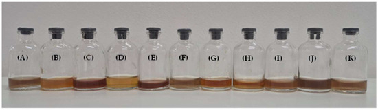
Figure 1.
Mycogenic silver nanoparticles: (A) IBCLP06, (B) IBCLP08, (C) IBCLP11, (D) IBCLP13, (E) IBCLP15, (F) IBCLP16, (G) IBCLP17, (H) IBCLP20, (I) IBCLP22, (J) IBCLP30, and (K) IBCLP40.
The mycogenic colloidal AgNP solutions were stored at room temperature, and their stability was monitored over a six-month period. For this purpose, a portion of each AgNP colloidal solution was centrifuged at 12,000× g for 10 min and washed three times with deionized water [16]. For characterization, the AgNPs were resuspended in distilled water, and the pH was adjusted to 7.0 using a 0.1 M phosphate buffer solution (PBS, Sigma-Aldrich). The morphological characterization of the AgNPs was carried out using X-ray diffraction (XRD) and transmission electron microscopy (TEM). Lyophilized nanoparticles were placed on a glass grid containing silicon substrate for XRD analysis. XRD measurements were performed on a conventional Rigaku diffractometer (MiniFlex II model, Tokyo, Japan) with a CuKα radiation source (λ = 1.54056 Å). The experiments were conducted at a 2θ scanning range from 20° to 90°, with a scan speed of 2° (2θ)/min [20]. The size and shape of the AgNPs were determined using a TEM LEO 906E (Zeiss, Oberkochen, Germany), operated at 200 kV. For sample preparation, colloidal suspensions of AgNPs were dropped onto carbon-coated copper grids (Sigma Aldrich-Merck, São Paulo, Brazil). The particle size was determined by a mean value and standard deviation of at least 50 measured particles, and a distribution histogram was generated by measuring 100 particles per micrograph. Dynamic Light Scattering (DLS) was used to measure the average particle diameter and size distribution of nanoparticles dispersed in water. The charge and aggregation tendency were assessed using zeta potential (Pζ) and the polydispersity index (PDI). All measurements were carried out in triplicate using a Zetasizer Nano ZS90 (Malvern Instruments, Malvern, UK).
2.4. Antimicrobial Activity
2.4.1. Bacteria
The antibacterial activity of the synthesized AgNPs was tested against the Gram-positive bacterium Staphylococcus aureus IPT246 and the Gram-negative bacteria Klebsiella pneumoniae IPT412 and Pseudomonas aeruginosa IPT322. This was determined by agar well diffusion assay [21]. Bacteria were cultured in Mueller–Hinton agar (Himedia), applying 20 μL of an initial inoculum (106 CFU/mL) of each strain to the agar media and spreading this volume uniformly. Subsequently, 20 μL of a colloidal AgNP solution, at the concentrations of 5, 10, 25, 50, 75, and 100 μg/mL, was added into four mm diameter wells, cut out in the center of the Petri dish and incubated at 37 °C, for 24 h. Streptomycin solution (20 μL) was used in the same concentration values as the positive control, and water as the negative control. After incubation, the zones of inhibition (ZOI) were measured in millimeters (mean ± SD). All the assays were performed in quadruplicate.
2.4.2. Fungi
The antifungal activity of the synthesised AgNPs was tested against Aspergillus niger IPT295, Aspergillus fumigatus IPT728, Aspergillus parasiticus IPT729, Trichophyton mentagrophytes IPT311, Penicillium funiculosum IPT423, and Fusarium oxysporum IPT330. This was evaluated using the adapted protocols of the minimal inhibitory concentration (MIC) proposed by Zwar et al. [22] and Ke et al. [23]. Briefly, fungi were grown in Potato Dextrose Agar (PDA, Merck), at 28 °C for seven days. After this period, 10 mL of saline solution with Tween 80 was added to the colonies grown on each Petri dish, spores were scraped, and a spore suspension was obtained. The suspensions were transferred to a sterile flask and homogenized on a shaker platform (Novatecnica, NT715, São Paulo, Brazil) at 30 °C and 100 rpm, for five minutes. The concentrations of the suspensions were adjusted to 1.0 × 10⁵ spores·mL−1, using a counting chamber. Briefly, 100 µL of each AgNP at concentrations of 5, 10, 25, 50, 75, 100, and 125 μg/mL was added in each well of a 96-well plate. Then, 100 μL of spore suspension in Potato Dextrose Broth (PDB, Merck) was added to each respective well. Fluconazole (FLU) was used at the same concentration values as the positive control, and fungal extracts (FEs) were used as the negative control. Firstly, plates were incubated at 30 °C for 24 h. After this period, 10 µL of 0.01% resazurin solution was added to each well and incubated for another 48 h. MICs were defined as the lowest concentration necessary to inhibit 100% fungal growth, shown by the absence of color change after the addition of resazurin. The minimum fungicidal concentration (MFC) test was performed according to the adapted protocol proposed by Fonseca et al. [24]. From each well with no visible growth, 10 µL of suspension was transferred to Petri dishes containing PDA and incubated for a period of seven days. After the incubation period, the AgNP concentration that inhibited 100% of the fungal growth being tested was considered fungicidal. All the assays were performed in triplicate.
3. Results
3.1. Marine-Derived Fungi Screening and Silver Nanoparticles’ Characterization
Twelve different strains of marine-derived fungal strains were grown in batch cultures to evaluate their ability to produce AgNPs. Eleven of them, IBCLP06, IBCLP08, IBCLP11, IBCLP13, IBCLP15, IBCLP16, IBCLP17, IBCLP20, IBCLP22, IBCLP30, and IBCLP40, presented a surface plasmon resonance (SPR) band in the region between 400 and 430 nm. Stability was evaluated using UV–Vis spectroscopy every 30 days (Figure 2) and through visual inspection for precipitation. Only the colloidal solutions of AgNPs produced by strains IBCLP11, IBCLP17, IBCLP20, and IBCLP22 did not exhibit precipitation for up to six months of storage at room temperature.
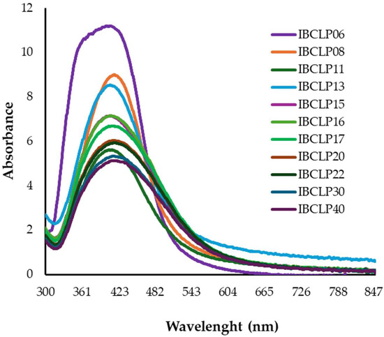
Figure 2.
UV–Vis spectra of different silver nanoparticles: IBCLP06, IBCLP08, IBCLP11, IBCLP13, IBCLP15, IBCLP16, IBCLP17, IBCLP20, IBCLP22, IBCLP30, and IBCLP40.
Table 1 shows the AgNPs, surface plasmon resonance (SPR), hydrodynamic size (DLS), zeta potential (Pζ), polydispersity index (PDI), and mean particle diameter (nm).

Table 1.
Characterization of silver nanoparticles produced by marine-derived fungi.
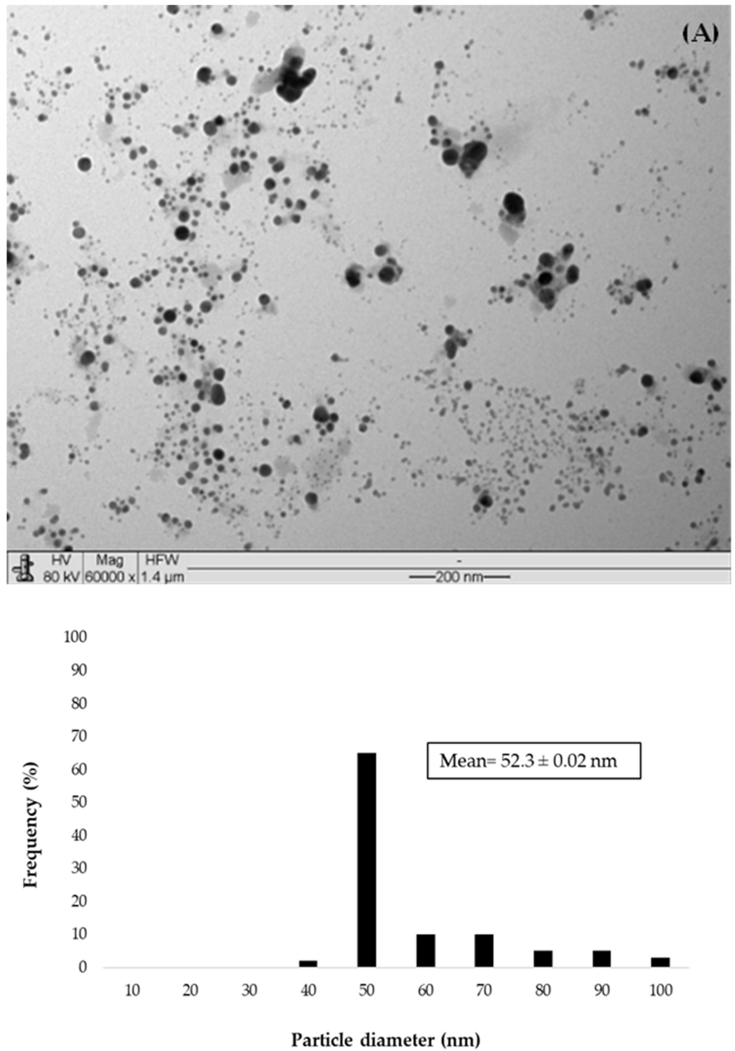
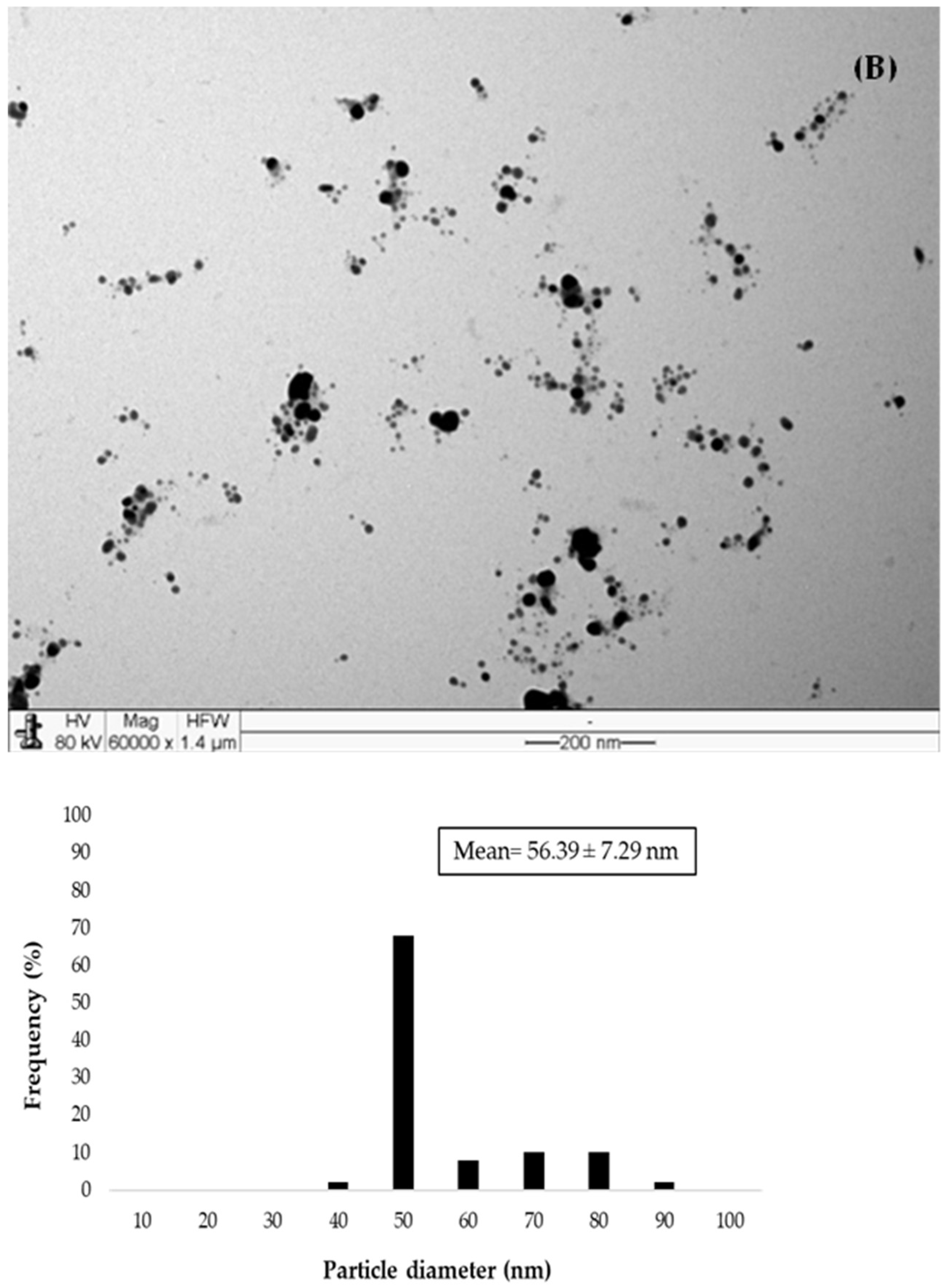
According to the AgNP size determined by DLS and TEM (Figure 3), samples IBCLP 06, IBCLP 16, IBCLP 30, and IBCLP 40 were excluded due to their size being larger than 100 nm. After analysis related to PDI and Pζ, IBCLP13 and IBCLP15 were not considered for the next stage of the study due to the PDI value being higher than 0.3 and the Pζ value in the module being lower than 30 mV. Figure 3A,B show the morphology and size distribution of AgNPIBCLP17 and AgNPIBCLP22. The morphologies of AgNPIBCLP11 and AgNPIBCLP20 were previously described by Aguiar et al. [25] and Assis da Silva et al. [19], respectively. These four AgNPs were selected based on the optimization study of the synthesis parameters. The XRD analysis (Supplementary Matterial, Figure S2.1) revealed a well-defined face-centered cubic (FCC) structure of silver in AgNPIBCLP11, AgNPIBCLP20, AgNPIBCLP17, and AgNPIBCLP22. Characteristic diffraction peaks were observed at 38°, 44°, 64.5°, 77°, and 82°, corresponding to the (111), (200), (220), (311), and (222) crystallographic planes, respectively. The calculated lattice parameter was 0.409 nm, in accordance with the JCPDF #04-783 standard. According to the Debye–Scherrer method [26], the measured average crystallite size of AgNP for each strain was the following: 31 nm for IBCLP11, 32 nm for IBCLP17, 36 nm for IBCLP20, and 37 nm for IBCLP22.


Figure 3.
Transmission electron microscopy (TEM) micrographs and histograms of the mycogenic AgNPs synthesized using (A) Penicillium sclerotigenum IBCLP17 and (B) Penicillium polonicum IBCLP22.
3.2. Antimicrobial Activity of Mycogenic Silver Nanoparticles
Regarding the antibacterial activity of AgNPs against Staphylococcus aureus IPT246, Pseudomonas aeruginosa IPT322, and Klebsiella pneumoniae IPT412, AgNPIBCLP20 presented better results (Figure 4). AgNPIBCLP20 was similarly efficient to Streptomycin at a dose of 25 μg/mL and more effective at concentrations equal to or greater than 50 μg/mL. At a concentration of 100 μg/mL, AgNPIBCLP20 exhibited its highest antibacterial activity against Staphylococcus aureus IPT246 (Figure 4A), as evidenced by the largest ZOI (60.6 mm), followed by Pseudomonas aeruginosa IPT322 (Figure 4B, 53.1 mm) and Klebsiella pneumoniae IPT412 (Figure 4C, 46.9 mm). These results highlight the differential sensitivity of Gram-positive and Gram-negative bacterial strains to mycogenic AgNPIBCLP20.
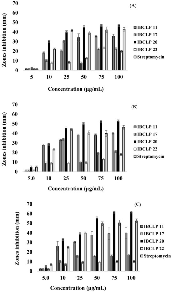
Figure 4.
Antibacterial activity of AgNPs against (A) Klebsiella pneumoniae IPT412, (B) Pseudomonas aeruginosa IPT322, and (C) Staphylococcus aureus IPT246. Zones of inhibition (ZOI) were measured in millimeters (mean ± SD).
The antifungal activity of AgNPs (IBCLP11, IBCLP17, IBCLP20, IBCLP22) was assessed against Aspergillus niger IPT295, Aspergillus fumigatus IPT728, Aspergillus parasiticus IPT729, Trichophyton mentagrophytes IPT311, Penicillium funiculosum IPT423, and Fusarium oxysporum IPT330 by determining their MIC and MFC. Table 2 summarizes the MIC and MFC results, which ranged from 10 to 100 μg/mL and 25 to 125 μg/mL, respectively.
Table 2.
Minimum inhibitory concentrations (MICs) and minimum fungicidal concentrations (MFCs) of AgNPs (IBCLP11, IBCLP17, IBCLP20, IBCLP22), fluconazole (FLU) and fungal extracts (FE) against Aspergillus niger IPT295, Penicillium funiculosum IPT423, Fusarium oxysporum IPT330, Aspergillus fumigatus IPT728, Aspergillus parasiticus IPT729, and Trichophyton mentagrophytes IPT311.
According to Table 2, the MIC and MFC results showed that both AgNPs and FLU exhibited significant fungicidal activity against the evaluated pathogenic fungi at concentrations of 10 µg/mL or higher. The best results were observed for Aspergillus niger IPT295 and Aspergillus parasiticus IPT729 when exposed to AgNPIBCLP20, as well as for Fusarium oxysporum IPT330 and Aspergillus fumigatus IPT728 when exposed to AgNPIBCLP11, with MIC and MFC values of 10 µg/mL and 25 µg/mL, respectively. Based on the characterization and antimicrobial activity results of the AgNPs, AgNPIBCLP11 and AgNPIBCLP20 were selected to investigate the effects of various parameters influencing their synthesis.
3.3. Optimization of Mycogenic Silver Nanoparticles’ Synthesis
The synthesis of AgNPs using fungi is eco-friendly, cost-effective, and is currently considered an important approach that significantly contributes to achieving the 2030 Agenda for Sustainable Development Goals (SDGs). This bionanotechnological method provides a viable pathway toward sustainable innovation and environmental management but must be refined. Some parameters such as AgNO3 and biomass concentration, agitation, temperature, and pH play a critical role in determining the nanoparticles’ size, shape, stability, and other properties.
3.3.1. AgNO3 Concentration
Since Ag+ ions are essential for the synthesis of AgNPs, and a set of experiments was conducted to evaluate the effect of AgNO3 salt concentration. The results showed that higher AgNO3 concentrations directly impacted the size, charge, and aggregation tendency of AgNPs. At concentrations above 1.0 mM, both AgNPs showed a significant increase in size (Table 3). An important consideration when reporting on the synthesis of mycogenic AgNPs is storage stability. This research demonstrates that AgNPs synthesized with higher AgNO3 concentrations have lower stability (3 months) compared to those synthesized with lower AgNO3 concentrations (12 months), as evidenced by visual precipitation.
Table 3.
Effect of AgNO3 concentration on the physicochemical properties of silver nanoparticles produced by extracellular extracts of Penicillium citrinum IBCLP11 and Aspergillus niger IBCLP20 strains.
3.3.2. Fungal Biomass Concentration
Biomass provides reducing agents such as enzymes, proteins, and other metabolites for the reduction in Ag+ ions and stabilization of nanoparticles. According to Table 4, a minimum biomass concentration of 75 g L−1 was required for a successful AgNP synthesis for both fungal strains. Higher biomass concentrations did not disrupt mycogenic synthesis. Although this parameter could potentially lead to nanoparticle aggregation, no such effect was observed under the conditions tested.
Table 4.
Effect of biomass concentration on the physicochemical properties of silver nanoparticles produced by extracellular extracts of Penicillium citrinum IBCLP11 and Aspergillus niger IBCLP20 strains.
3.3.3. Agitation
Table 5 highlights the impact of agitation speed on the synthesis of mycogenic AgNPs (AgNPIBCLP11 and AgNPIBCLP20) and their physicochemical properties. Stirring speed is a crucial parameter influencing both the efficiency of synthesis and the quality of the nanoparticles. Proper agitation promotes the control of NP nucleation and growth, leading to better size distribution. In the present study, 150 rpm was determined to be the optimal stirring speed for both NPs’ formation, as this condition likely increases the interaction between fungal metabolites and Ag⁺ ions, promoting efficient synthesis. In contrast, excessive stirring compromised the size and charge of the NPs, and enhanced their tendency to aggregate.
Table 5.
Effect of agitation speed on the physicochemical properties of silver nanoparticles produced by extracellular extracts of Penicillium citrinum IBCLP11 and Aspergillus niger IBCLP20 strains.
3.3.4. Temperature
Temperature is a crucial abiotic factor influencing the synthesis, yield, size, and stability of AgNPs. In this study, extracellular synthesis using fungal extract from IBCLP11 did not result in AgNP formation at temperatures below 30 °C, while extract from IBCLP20 required at least 25 °C. Conversely, temperatures exceeding 35 °C had a negative impact on AgNPs’ synthesis for both extracts. As shown in Table 6, the optimal temperature range for AgNPs synthesis was 30–35 °C for IBCLP11, whereas IBCLP20 facilitated synthesis within a broader range of 25–35 °C while maintaining a comparable physicochemical profile.
Table 6.
Effect of temperature on the physicochemical properties of silver nanoparticles produced by extracellular extracts of Penicillium citrinum IBCLP11 and Aspergillus niger IBCLP20 strains.
3.3.5. pH
Table 7 indicates that AgNPs synthesized using extracellular extracts from the IBCLP11 strain at pH 6.5 demonstrated superior stability compared to those produced with the IBCLP20 strain, which exhibited optimal stability at pH 6.0. pH values below 6.0 or above 7.5 did not support stable AgNP formation, as the colloidal solution exhibited precipitation after one week of storage. Neutral to slightly alkaline pH values (6.0–8.0) are generally considered optimal for AgNP synthesis. This range of pH facilitates efficient reduction in silver ions and stabilization, resulting in uniform and stable NPs. On the other hand, strongly alkaline conditions may accelerate the reduction process due to the abundance of hydroxide ions, but they often lead to irregular particle shapes and decreased stability over time.
Table 7.
Effect of pH on the physicochemical properties of silver nanoparticles produced by extracellular extracts of Penicillium citrinum IBCLP11 and Aspergillus niger IBCLP20 strains.
4. Discussion
4.1. Marine-Derived Fungi as a New Resource for Silver Nanoparticles’ Synthesis and Their Antimicrobial Activity
The marine ecosystem is recognized as an important resource for biodiversity studies and bioprospecting of enzymes and secondary metabolites. Marine microbial communities are crucial for biogeochemical cycling, and fungi, especially those originating from these environments, are considered promising sources for biotechnological applications [27]. Marine-derived fungi are adapted to more extreme conditions than terrestrial fungi, such as high pressure, low temperature, oligotrophic nutrients, and high salinity. Among the metabolites produced by marine-derived fungi, enzymes stand out, representing enormous potential to be applied in food supplements, medicines, and cosmetics. In addition, these organisms are used in the treatment of aquatic and terrestrial environments contaminated by recalcitrant compounds and xenobiotics [28].
Currently, the synthesis of metallic nanoparticles stands out among compounds produced by terrestrial and marine fungi, mainly due to its environmentally friendly nature and alignment with the 2030 Agenda. Although several studies have investigated the use of mycelium or extracellular extracts of terrestrial fungi for NP synthesis, research focusing on marine fungi remains scarce. Recently, Ameen et al. [29] reported the synthesis of AgNPs using a Cladosporium halotolerans strain isolated from hypersaline environments of Tarout Island, Saudi Arabia. The synthesized AgNPs showed a spherical shape with an average size of 20 nm confirmed by TEM, and a Pζ of −51.8 mV, indicating high stability due to the strong electrostatic repulsion between particles, which minimizes aggregation. Furthermore, AgNPs showed an inhibition of 70% at 1000 ppm of AgNPs against Aspergillus niger.
Basheer et al. [30] evaluated the ability of marine fungi Penicillium simplicissimum, Aspergillus terreus, Aspergillus japonicus, and Aspergillus oryzae isolated from water samples obtained from Lake Qarun in Fayoum, Egypt. All strains produced spherical AgNPs with sizes ranging from 3.8 to 23 nm, with recognized antimicrobial activity against several pathogenic microorganisms. Stable AgNPs ranging in size from 0.72 to 15.21 nm were synthesized using Aspergillus brunneoviolaceus isolated from coastal waters near Diu, India. These NPs exhibited similar antimicrobial activity against Gram-positive bacteria (Bacillus subtilis and Staphylococcus aureus) and Gram-negative bacteria (Pseudomonas aeruginosa, Escherichia coli, and Salmonella spp.) [13]. Compared to our findings, AgNPIBCLP20 was more effective against the Gram-positive bacterium Staphylococcus aureus IPT246 than Gram-negative bacteria (Klebsiella pneumoniae IPT412 and Pseudomonas aeruginosa IPT322). Gram-positive bacteria have a thicker peptidoglycan layer that interacts strongly with AgNPs, leading to enhanced permeability and disruption, whereas Gram-negative bacteria have an additional outer membrane that may act as a barrier, reducing susceptibility [31,32].
Regarding the antifungal activity of AgNPIBCLP11 and AgNPIBCLP20, the results of our study were compared with the AgNPs produced by terrestrial fungi. The scarcity of research on marine fungi in this approach highlights the need for additional studies to investigate these unexplored microorganisms as valuable resources for the development of advanced antifungal agents. According to Ahmad et al. [33], AgNPs biosynthesized from Aspergillus niger showed a maximum zone of inhibition against plant pathogenic fungi P. citrinum (19.33 ± 0.57 mm), Rhizopus stolonifer (17.66 ± 0.57 mm), A. terreus (16.33 ± 1.54 mm), F. oxysporum (14.00 ± 1.00 mm), and Mucor mucedo (13.33 ± 1.15 mm). Liu et al. [34] reported the biosynthesis of AgNPs using Trichoderma longibrachiatum. The study demonstrated that concentrations of 100–200 mg/L effectively inhibited the growth of Fusarium oxysporum. Additionally, a lower concentration of 25 mg/L promoted muskmelon seed germination and reduced the incidence of Fusarium wilt in muskmelon, showcasing the dual potential of AgNPs in agriculture as both a growth enhancer and a disease control agent.
Our data are consistent with the findings of Abd Elghaffar et al. [35], who demonstrated the efficacy of mycogenic AgNPs synthesized by the endophytic Aspergillus luchuensis against pathogenic fungi, including F. oxysporum, Alternaria alternata, Candida albicans, A. brasiliensis, and A. flavus. The AgNPs exhibited significant antimicrobial potential against these pathogenic microorganisms.
4.2. Mycogenic Synthesis Optimization
Saxena et al. [36] observed an increase in the SPR peak at 430 nm with increasing biomass, suggesting that the amount of biomass directly affects the release of reducing agents required for AgNP synthesis. Balakumaran et al. [37] reported that the optimal NP biosynthesis occurred at a 100 g/L fungal biomass concentration. Higher concentrations (200–300 g/L) were less effective due to excess reducing agents, potentially causing NP aggregation. The findings of Balakumaran et al. [38] align with our results, supporting the use of 75–100 g/L of biomass for the stable synthesis of AgNPs. This reinforces the importance of optimizing biomass concentration to ensure nanoparticle stability and efficiency during biosynthesis.
Cui et al. [39] investigated the optimization of AgNP synthesis using Trichoderma longibrachiatum. The study identified the optimal conditions for AgNP production as follows: 2 mmol/L of AgNO3, a temperature of 55 °C, and a neutral pH of 7.0. These parameters facilitated efficient and stable NP synthesis, highlighting the key fine-tuning factors in the biosynthesis process. The parameters, including AgNO3 concentration, cell-free filtrate volume, pH, and reaction time, were applied to optimize the formation of AgNPs by A. oryzae through the central composite design [38]. The results obtained in this study described the synthesis of spherical AgNPs with 50% of the particles smaller than 109.6 nm at the optimized parameters: 3.75 mL of the cell-free filtrate and 2.25 mM of AgNO3 for 108 min at a pH value of 12.25.
The results outlined emphasize distinct findings compared to our study, particularly regarding AgNO3 concentration, temperature, and pH. While higher AgNO3 concentrations were observed to induce AgNP formation, they often resulted in instability. This observation aligns with the findings of Cruz et al. [40] and Guilger-Casagrande and de Lima [14], who reported that high concentrations of metal precursors can result in the formation of large and irregularly shaped NPs. This phenomenon is likely caused by the competition between metal ions and the functional groups in the fungal filtrate, disrupting the stabilization process. Moreover, excessive metal precursor concentrations can lead to significant toxicity, underscoring the critical need to optimize synthesis conditions.
Although studies generally highlight alkaline conditions as more favorable for AgNP synthesis, our results showed that for the IBCLP11 and IBCLP20 strains, pH levels above 6.5 negatively impacted the synthesis of AgNPs. Similarly, Balakumaran et al. [37] reported that a neutral pH (7) produced monodisperse and stable AgNPs, while acidic conditions (pH 1–4) did not induce color changes, and NP aggregation was observed at pH 3–4. In contrast, Velu et al. [41] reinforced that acidic pH conditions were unfavorable for AgNP synthesis, as no SPR peaks were detected and alkaline pH (10.0) promoted smaller diameter nanoparticles with clear SPR peaks, likely due to hydroxide deposition on the AgNPs.
The variations observed across studies regarding the impact of temperature on mycogenic AgNP synthesis can be attributed to several factors. Key considerations include the choice of fungal strains, the synthesis mechanism (extracellular or intracellular), and the potential inactivation or denaturation of enzymes and other metabolites [39,41]. These factors warrant further detailed investigation. Using the Fusarium oxysporum strain, Rajput et al. [18] synthesized AgNPs at 25 °C and 50 °C. The authors reported that higher temperatures led to a narrower size distribution compared to those formed at 25 °C.
5. Conclusions
Coastal and marine ecosystems host diverse microorganisms that are often robust and metabolically versatile, making them ideal candidates for biosynthesis processes. We screened twelve marine-derived fungal strains isolated from mangrove sediments in Peruíbe, Brazil, and found that eleven of them were able to biosynthesize AgNPs. However, after the characterization of these metallic nanomaterials, only the products of the strains IBCLP11, IB-CLP17, IB-CLP20, and IB-CLP22 were selected to evaluate their antimicrobial properties and assess the biosynthesis parameters required for AgNP production. AgNPIBCLP11 and AgNPIBCLP20 demonstrated a greater antimicrobial activity. These strains effectively produced nanoparticles capable of inhibiting the growth of P. aeruginosa IPT322, S. aureus IPT246, and K. pneumoniae IPT412 at concentrations of 50 μg·mL−1 or greater. Additionally, these AgNPs exhibited antifungal activity against A. niger IPT295, A. fumigatus IPT728, A. parasiticus IPT729, T. mentagrophytes IPT311, P. funiculosum IPT423, and F. oxysporum IPT330 at concentrations ranging from 20 to 125 µg·mL−1. These findings underscore the promising antimicrobial potential of AgNPs synthesized by marine fungi.
The biosynthesis of AgNPIBCLP11 and AgNPIBCLP20 was influenced by several key parameters, including metal precursor concentration, biomass concentration, agitation, temperature, and pH, which play a crucial role in determining nanoparticle size, stability, and overall quality. Our results indicate that, at concentrations of a metal precursor above 1.0 mM, AgNPs increased in size, while the minimum biomass concentration required for AgNP formation was 75 g·L−1. Agitation at 150 rpm promoted efficient AgNP synthesis in the tested strains, and temperatures within the ranges of 30–35 °C and 25–30 °C facilitated the synthesis of AgNPIBCLP11 and AgNPIBCLP20, respectively. Higher temperatures resulted in a more narrowly distributed nanoparticle size. Furthermore, while alkaline conditions are generally preferred for AgNPs synthesis, pH levels below 6.0 or above 7.0 negatively impacted nanoparticle formation. These results underscore the importance of optimizing synthesis conditions to achieve stable, size-controlled AgNPs with enhanced properties for specific applications. Future research should focus on further elucidating the biosynthetic mechanisms of fungal-mediated nanoparticle formation and assessing the long-term impacts of these biogenic nanomaterials.
Supplementary Materials
The following supporting information can be downloaded at: https://www.mdpi.com/article/10.3390/suschem6010010/s1, Figure S1.1: Supernatant absorbance using different chemical precursor (AgNO3) concentration for the silver nanoparticles (AgNPs) IBCLP11 (A) and IBCLP20 (B). Figure S1.2: Supernatant absorbance using different fungal biomass concentration for the silver nanoparticles (AgNPs) produced by IBCLP11 (A) and IBCLP20 (B). Figure S1.3: Supernatant absorbance using different agitation values for the silver nanoparticles (AgNPs) produced by IBCLP11 (A) and IBCLP20 (B). Figure S1.4: Supernatant absorbance using different temperature values for the silver nanoparticles (AgNPs) produced by IBCLP11 (A) and IBCLP20 (B). Figure S1.5: Supernatant absorbance using different pH values for the silver nanoparticles (AgNPs) produced by IBCLP11 (A) and IBCLP20 (B). Figure S2.1: Ultraviolet-visible (UV-Vis) spectra of silver nanoparticles synthesized from different fungal strains (IBCLP11, IBCLP17, IBCLP20, and IBCLP22), measured using UV-Vis spectrophotometry (Shimadzu UV-1800, Kyoto, Japan) at room temperature, within a wavelength range of 300 to 850 nanometers.
Author Contributions
Conceptualization, A.L.A., C.A.O., L.d.C., M.F.S., and P.L.; Validation, A.L.A., C.A.O., L.d.C., M.F.S., P.L., and Y.D.; Formal analysis, A.L.A., M.C., C.A.O., L.d.C., Y.D., M.F.S., C.d.S., and C.T.; Investigation, A.L.A., M.C., C.T., C.d.S., and C.T.; Resources, C.A.O. and L.d.C.; Writing—original draft preparation, C.T., C.d.S., C.A.O., and M.F.S.; Writing—review and editing, A.L.A., M.C., C.A.O., L.d.C., M.F.S., P.L., and Y.D.; Supervision, C.A.O. and M.F.S.; Project administration, C.A.O., L.d.C., M.F.S., and Y.D.; Funding acquisition, C.A.O. All authors have read and agreed to the published version of the manuscript.
Funding
The research described in this work was supported by the São Paulo Research Foundation (FAPESP; #2020/12867-2; #19/16023-6) and Conselho Nacional de Desenvolvimento Científico e Tecnológico (CNPq; 421122/2023-4). Marta Filipa Simões is supported by the Science and Technology Development Fund (FDCT), Macau SAR, China (Files No. 002/2024/SKL and FDCT-24-083-SSI). Carolina Assis acknowledges scholarships’ financial support from FAPESP (#23/08202-3; #22/05716-3; #22/03562-9). Caterina do Valle Trotta and Ana Laura Pires de Oliveira acknowledge scholarships’ financial support from the Coordination for the Improvement of Higher Education Personnel (CAPES; #88882.469336/2019-01; #88887.822485/2023-00).
Data Availability Statement
Data will be made available upon request.
Acknowledgments
The authors would also like to extend their gratitude to Jonas Gomes, IPT (Brazil), for his help in completing this research.
Conflicts of Interest
The authors declare no conflicts of interest. The funders had no role in the design of the study; in the collection, analyses, or interpretation of data; in the writing of the manuscript, or in the decision to publish the results.
References
- Fahim, M.; Shahzaib, A.; Nishat, N.; Jahan, A.; Bhat, T.A.; Inam, A. Green synthesis of silver nanoparticles: A comprehensive review of methods, influencing factors, and applications. JCIS Open 2024, 16, 100125. [Google Scholar] [CrossRef]
- Simões, M.F.; Ottoni, C.A.; Antunes, A. Mycogenic metal nanoparticles for the treatment of mycobacterioses. Antibiotics 2020, 9, 569. [Google Scholar] [CrossRef] [PubMed]
- Singh, P.; Kim, Y.J.; Zhang, D.; Yang, D.C. Biological synthesis of nanoparticles from plants and microorganisms. Trends Biotechnol. 2016, 34, 588–599. [Google Scholar] [CrossRef] [PubMed]
- Roy, A.; Bulut, O.; Some, S.; Mandal, A.K.; Yilmaz, M.D. Green synthesis of silver nanoparticles: Biomolecule-nanoparticle organizations targeting antimicrobial activity. RSC Adv. 2019, 9, 2673–2702. [Google Scholar] [CrossRef] [PubMed]
- Bellingeri, A.; Bono, N.; Venditti, I.; Bertelà, F.; Burrati, L.; Faleri, C.; Protano, G.; Paccagnini, E.; Lupetti, P.; Candiani, G.; et al. Capping drives the behavior, dissolution and (eco)toxicity of silver nanoparticles towards microorganisms and mammalian cells. Environ. Sci. Nano 2024, 11, 2049–2060. [Google Scholar] [CrossRef]
- Jafarzadeh, S.; Nooshkam, M.; Zargar, M.; Garavand, F.; Ghosh, S.; Hadidi, M.; Forough, M. Green synthesis of nanomaterials for smart biopolymer packaging: Challenges and outlooks. J. Nanostruct Chem. 2024, 14, 113–136. [Google Scholar] [CrossRef]
- Salem, S.S.; Fouda, A. Green synthesis of metallic nanoparticles and their prospective biotechnological applications: An overview. Biol. Trace Elem. Res. 2021, 199, 344–370. [Google Scholar] [CrossRef]
- Danagoudar, A.; Pratap, G.K.; Shantaram, M.; Ghosh, K.; Kanade, S.R.; Joshi, C.G. Characterization, cytotoxic and antioxidant potential of silver nanoparticles biosynthesised using endophytic fungus (Penicillium citrinum CGJ-C1). Mater. Today Commun. 2020, 25, 101385. [Google Scholar] [CrossRef]
- Feroze, N.; Arshad, B.; Younas, M.; Afridi, M.I.; Saqib, S.; Ayaz, A. Fungal mediated synthesis of silver nanoparticles and evaluation of antibacterial activity. Microsc. Res. Tech. 2020, 83, 72–80. [Google Scholar] [CrossRef]
- Amaral, M.V.M.V.; Carraro, C.B.; Antoniêto, A.C.C.; Costa, M.N.; Fraga-Silva, T.F.C.; Cipriano, U.G.; Abuná, R.P.F.; Rodrigues, T.S.; Martins, R.B.; Luzenti, A.M.; et al. Biogenic silver nanoparticles produced by Trichoderma reesei inhibit SARS-CoV-2 infection, reduce lung viral load and ameliorate acute pulmonary inflammation. Curr. Res. Biotechnol. 2025, 9, 100277. [Google Scholar] [CrossRef]
- Khan, M.; Khan, A.U.; Alam, M.J.; Park, S.; Alam, M. Biosynthesis of silver nanoparticles and its application against phytopathogenic bacterium and fungus. Int. J. Environ. Anal. Chem. 2020, 12, 1390–1401. [Google Scholar] [CrossRef]
- Ribeiro, L.G.; Rezende, K.F.O.; Barbieri, E.; de Souza, A.O. Study of routine metabolism and acute toxicity of mycogenic silver nanoparticles on Palaemon pandaliformis (shrimp). Environ. Sci. Nano 2023, 10, 1715–1729. [Google Scholar] [CrossRef]
- Mistry, H.; Thakor, R.; Patil, C.; Trivedi, J.; Bariya, H. Biogenically proficient synthesis and characterization of silver nanoparticles employing marine procured fungi Aspergillus brunneoviolaceus along with their antibacterial and antioxidative potency. Biotechnol. Lett. 2021, 43, 307–316. [Google Scholar] [CrossRef] [PubMed]
- Guilger-Casagrande, M.; de Lima, R. Synthesis of silver nanoparticles mediated by fungi: A review. Front. Bioeng. Biotechnol. 2019, 7, 287. [Google Scholar] [CrossRef]
- Osorio-Echavarría, J.; Ossa-Orozco, C.P. Synthesis of silver nanoparticles using white-rot fungus anamorphous Bjerkandera sp. R1: Influence of silver nitrate concentration and fungus growth time. Sci. Rep. 2021, 11, 3842. [Google Scholar] [CrossRef]
- Al-Limoun, M.; Qaralleh, H.N.; Khleifat, K.M.; Al-Anber, M.; Al-Tarawneh, A.; Al-sharafa, K.; Kailani, M.H.; Zaitoun, M.A.; Matar, S.A.; Al-soub, T. Culture media composition and reduction potential optimization of mycelia-free filtrate for the biosynthesis of silver nanoparticles using the fungus Tritirachium oryzae W5H. Curr. Nanosci. 2020, 16, 757–769. [Google Scholar] [CrossRef]
- Wang, D.; Xue, B.; Wang, L.; Zhang, Y.; Liu, L.; Zhou, Y. Fungus-mediated green synthesis of nano-silver using Aspergillus sydowii and its antifungal/antiproliferative activities. Sci. Rep. 2021, 11, 10356. [Google Scholar] [CrossRef]
- Rajput, S.; Werezuk, R.; Lange, R.M.; McDermott, M.T. Fungal isolate optimized for biogenesis of silver nanoparticles with enhanced colloidal stability. Langmuir 2016, 32, 8688–8697. [Google Scholar] [CrossRef]
- Assis da Silva, C.; Ribeiro, B.M.; Trotta, C.V.; Perina, F.C.; Martins, R.; Abessa, D.M.S.; Barbieri, E.; Simões, M.F.; Ottoni, C.A. Effects of mycogenic silver nanoparticles on organisms of different trophic levels. Chemosphere 2022, 308, 136540. [Google Scholar] [CrossRef]
- Ottoni, C.A.; Ramos, C.E.D.; de Souza, R.F.B.; da Silva, S.G.; Spinace, E.V.; Neto, A.O. Glycerol and ethanol oxidation in alkaline medium using PtCu/C electrocatalysts. Int. J. Electrochem. Sci. 2018, 13, 1893–1904. [Google Scholar] [CrossRef]
- Chavez-Esquivel, G.; Cervantes-Cuevas, H.; Ybieta-Olvera, L.F.; Castañeda Briones, M.T.; Acosta, D.; Cabello, J. Antimicrobial activity of graphite oxide doped with silver against Bacillus subtilis, Candida albicans, Escherichia coli, and Staphylococcus aureus by agar well diffusion test: Synthesis and characterization. Mater. Sci. Eng. C 2021, 123, 111934. [Google Scholar] [CrossRef] [PubMed]
- Zwar, I.P.; Trotta, C.V.; Ziotti, A.B.S.; Neto, M.L.; Araújo, W.L.; Melo, I.S.; Ottoni, C.A.; Souza, A.O. Biosynthesis of silver nanoparticles using actinomycetes, phytotoxicity on rice seeds, and potential application in the biocontrol of phytopathogens. J. Basic Microbiol. 2023, 63, 64–74. [Google Scholar] [CrossRef] [PubMed]
- Ke, Y.; Ding, B.; Zhang, M.; Dong, T.; Fu, Y.; Lv, Q.; Ding, W.; Wang, X. Study on inhibitory activity and mechanism of chitosan oligosaccharides on Aspergillus flavus and Aspergillus fumigatus. Carbohydr. Polym. 2022, 275, 118673. [Google Scholar] [CrossRef] [PubMed]
- Fonseca, L.M.; Souza, E.J.D.; Radünz, M.; Gandra, E.A.; Zavareze, E.R.; Dias, A.R.G. Suitability of starch/carvacrol nanofibers as biopreservatives for minimizing the fungal spoilage of bread. Carbohydr. Polym. 2021, 252, 117166. [Google Scholar] [CrossRef]
- Aguiar, A.P.; Ottoni, C.A.; Aquaroli, C.L.R.; Mendes, E.C.V.; Araújo, A.L.S.; Simões, M.F.; Barbieri, E. Mycogenic silver nanoparticles from Penicillium citrinum IB-CLP11-their antimicrobial activity and potential toxicity effects on freshwater organisms. Environ. Sci. Nano 2024, 11, 2229–2238. [Google Scholar] [CrossRef]
- Cullity, B.D. Elements of X-Ray Diffraction; Addison-Wesley: Boston, MA, USA, 1967; p. 262. [Google Scholar]
- Jiang, J.-P.; Leng, S.; Liao, Y.-F.; Liu, X.; Li, D.-X.; Chu, C.; Yu, X.-Y.; Liu, C.-H. The potential role of subseafloor fungi in driving the biogeochemical cycle of nitrogen under anaerobic conditions. Sci. Total Environ. 2023, 897, 165374. [Google Scholar] [CrossRef]
- Nogueira, O.M.N.; Bernal, S.P.F.; Peres, C.K.; Boroski, M.; Passarini, M.R.Z. Isolation of marine-derived filamentous fungi and their potential application for bioremediation process. Braz. J. Microbiol. 2024, 55, 3403–3412. [Google Scholar] [CrossRef]
- Ameen, F.; Al-Homaidan, A.A.; Al-Sabri, A.; Almansob, A.; AlNAdhari, S. Anti-oxidant, anti-fungal and cytotoxic effects of silver nanoparticles synthesized using marine fungus Cladosporium halotolerans. Appl. Nanosci. 2023, 13, 623–630. [Google Scholar] [CrossRef]
- Basheer, M.A.; Abutaleb, K.; Abed, N.N.; Mekawey, A.A.I. Mycosynthesis of silver nanoparticles using marine fungi and their antimicrobial activity against pathogenic microorganisms. J. Genet. Eng. Biotechnol. 2023, 21, 127. [Google Scholar] [CrossRef]
- More, P.R.; Pandit, S.; Filippis, A.D.; Franci, G.; Mijakovic, I.; Galdiero, M. Silver nanoparticles: Bactericidal and mechanistic approach against drug resistant pathogens. Microorganisms 2023, 11, 369. [Google Scholar] [CrossRef]
- Saravanan, M.; Arokiyaraj, S.; Lakshmi, T.; Pugazhendhi, A. Synthesis of silver nanoparticles from Phenerochae techrysosporium (MTCC-787) and their antibacterial activity against human pathogenic bacteria. Microb. Pathog. 2018, 117, 68–72. [Google Scholar] [CrossRef] [PubMed]
- Ahmad, N.; Malik, M.A.; Wani, A.H.; Bhat, M.Y. Biogenic silver nanoparticles from fungal sources: Synthesis, characterization, and antifungal potential. Microb. Pathog. 2024, 193, 106742. [Google Scholar] [CrossRef] [PubMed]
- Liu, X.; Li, T.; Cui, X.; Tao, R.; Gao, Z. Antifungal mechanism of nanosilver biosynthesized with Trichoderma longibrachiatum and its potential to control muskmelon Fusarium wilt. Sci. Rep. 2024, 14, 20242. [Google Scholar] [CrossRef] [PubMed]
- Abd Elghaffar, R.Y.; Emam, A.M.; Taher, E.S.; Baz, M.M.; Nayel, H.; Abdeen, A.; El-Nablaway, M.; Alwutayd, K.M.; Mihaela, O.; Ioan, B.-D.; et al. The potential biological activities of Aspergillus luchuensis-aided green synthesis of silver nanoparticles. Front. Microbiol. 2024, 15, 1381302. [Google Scholar] [CrossRef]
- Saxena, J.; Sharma, P.K.; Sharma, M.M.; Sharma, M.M.; Singh, A. Process optimization for green synthesis of silver nanoparticles by Sclerotinia sclerotiorum MTCC 8785 and evaluation of its antibacterial properties. SpringerPlus 2016, 5, 861. [Google Scholar] [CrossRef]
- Balakumaran, M.D.; Ramachandran, R.; Balashanmugam, P.; Mukeshkumar, D.J.; Kalaichelvan, P.T. Mycosynthesis of silver and gold nanoparticles: Optimization, characterization and antimicrobial activity against human pathogens. Microbiol. Res. 2016, 182, 8–20. [Google Scholar] [CrossRef]
- Cui, X.; Zhong, Z.; Xia, R.; Liu, X.; Qin, L. Biosynthesis Optimization of Silver Nanoparticles (AgNPs) Using Trichoderma longibranchiatum and Biosafety Assessment with Silkworm (Bombyx mori). Arab. J. Chem. 2022, 15, 104142. [Google Scholar] [CrossRef]
- Elshafei, A.M.; Othman, A.M.; Elsayed, M.A.; Al-Balakocy, N.G.; Hassan, M.M. Green synthesis of silver nanoparticles using Aspergillus oryzae NRRL447 exogenous proteins: Optimization via central composite design, characterization and biological applications. Environ. Nanotechnol. Monit. Manag. 2021, 16, 100553. [Google Scholar] [CrossRef]
- Cruz, J.N.; Muzammil, S.; Ashraf, A.; Ijaz, M.U.; Siddique, M.H.; Abbas, R.; Sadia, M.; Saba; Hayat, S.; Lima, R.R. A review on mycogenic metallic nanoparticles and their potential role as antioxidant, antibiofilm and quorum quenching agents. Heliyon 2024, 10, e29500. [Google Scholar] [CrossRef]
- Velu, M.; Lee, J.-H.; Chang, W.-S.; Lovanh, N.; Park, Y.-J.; Jayanthi, P.; Palanivel, V.; Oh, B.-T. Fabrication, optimization, and characterization of noble silver nanoparticles from sugarcane leaf (Saccharum officinarum) extract for antifungal application. 3 Biotech. 2017, 7, 147. [Google Scholar] [CrossRef]


Disclaimer/Publisher’s Note: The statements, opinions and data contained in all publications are solely those of the individual author(s) and contributor(s) and not of MDPI and/or the editor(s). MDPI and/or the editor(s) disclaim responsibility for any injury to people or property resulting from any ideas, methods, instructions or products referred to in the content. |
© 2025 by the authors. Licensee MDPI, Basel, Switzerland. This article is an open access article distributed under the terms and conditions of the Creative Commons Attribution (CC BY) license (https://creativecommons.org/licenses/by/4.0/).