Article

Improvement of the Obliteration of Non-Critical Size Defects by Using a Mixture of Bone Dust and Bone Replacement Material (Bioactive Glass S53P4)

Department of Otolaryngology, Head and Neck Surgery, Technische Universität Dresden, Fetscherstrasse 74, 01307 Dresden, Germany
*
Author to whom correspondence should be addressed.
These authors contributed equally to this work.
Osteology 2025, 5(2), 15; https://doi.org/10.3390/osteology5020015
Submission received: 31 January 2025 / Revised: 7 March 2025 / Accepted: 1 May 2025 / Published: 19 May 2025

Abstract

:
Background/Objectives: Obliterates such as autologous bone dust (BD) or the synthetic bioactive glass S53P4 (BA) are frequently used for the obliteration of non-critical size defects (NCSDs), especially in otosurgery. Both obliterates have advantages and disadvantages, so that the combination of both for the obliteration of NCSDs is analysed. Methods: As part of a large animal project with sheep, four NCSDs were created in the calotte of thirteen animals using a drill. These were filled with BD, BD and BA, or BA, and the reference defect remained empty. After three weeks, the explanted calottes were examined with regard to their newly formed bone using digital volume tomography, bone density measurement, fluorochrome sequence labelling, and histological analysis. In addition, human cell culture analyses were carried out on the quality of the BD. Results: BD collected at 7.000 and 15.000 rpm shows a higher activity of new bone formation. In combination with BA, bone is formed centripetally and centrifugally. Defect filling with BA and BD shows a higher bone density and compactness than BD alone. Conclusions: BD should be harvested at a speed of less than 15.000 rpm. Using this BD in combination with BA to obliterate NCSDs enables the defect to be obliterated quickly and completely, with more newly formed bone, creating a bone network with incorporated BA. Further studies are needed to investigate the long-term stability of this obliteration and to determine which other parameters of the extraction can increase the amount of vital BD.

1. Introduction

Although various obliteration techniques and materials have been investigated, there is still no consensus on the optimal approach for closing bone defects, particularly for non-critical size defects (NCSDs). The present study aims to address this gap by systematically evaluating different obliteration materials. In particular, it was investigated whether a combination of bone dust (BD) and bioactive glass S53P4 (BA) can improve bone regeneration and defect closure compared to BD or BA alone. Regardless of the discipline used, the goals of obliteration are generally as follows:
The defect closures should correspond as closely as possible to the original morphology of the local defect bed. The best biomechanical functionality and long-term stability of the closure should be achieved. The main aim here is to minimise the loss of the inserted tissue, e.g., due to wound infection or insufficient healing in the tissue bed. An example of this is the otosurgical closure of the mastoid cavity following reconstructive surgery for chronic mastoiditis. The bone dust (BD) introduced in the process, sometimes in combination with cartilage, can be lost due to debridement caused by local infection [1]. In otosurgery, the use of BD in the obliteration of mastoid cavities or in canal wall down techniques, also in combination with other autologous elements, is a common procedure.
To achieve the sufficiently stable closure of a bony defect, materials must be used that activate bone regeneration and the formation of new bone. Osteostimulative and osteoreparative processes are triggered by pluripotent, undifferentiated, or differentiated, locally fixed osteoprogenitor cells and osteoblasts [2]. Whether the processes mentioned are activated and run permanently also depends on the defect itself. This is because they are also influenced by parameters such as the size of the defect and the content of bone-inductive factors in the surrounding defect margin tissue. Furthermore, the processes depend on the characteristics of the organism (age, metabolic metabolism, and general state of health) [3].
In order to meet these requirements, BD and bioactive glass S53P4 (BA) have been the most commonly used obliteration materials for bony non-critical size defects, despite the many different materials that have been tested [4,5,6]. In a previous study by the authors, it was shown that the sole use of both BD and BA have advantages and disadvantages in the obliteration of defects of non-critical size [7]. Despite the lack of standards in the production and collection method of BD and, thus, qualitative differences in daily practice, this study was able to demonstrate both centrifugal and centripetal new bone formation in the histological evaluation of defects after using BD. When BA was used, no additional bone regeneration was observed compared to the empty hole without obliteration. However, with BA the defects could be filled quickly, consistently, and reliably. In addition, a radiographic density identical to that of the bone bed was found. However, no bone regenerative processes could be detected in the inserted material or obliterated defect.
The findings of the study by Kluge et al. inevitably led to the question of whether the advantages of the materials could be utilised, and the disadvantages eliminated by using a mixture of BD and BA for obliteration [7]. Therefore, in the same large animal project, not only were non-critical size defects filled with either BD or BA examined, but also defects with a combined filling of BD and BA. This leads to the hypothesis to be examined in the present work: NCSDs can be closed quickly, compactly, and completely by the combination of BD with active bone cells and the antibacterial BA. Compared to obliteration with BA alone, the combined defect fillings are more extensively penetrated by active bone.

2. Materials and Methods

Both the materials used and the methods applied in the animal experiment correspond to those already published by Kluge et al. 2019 [7]. The sheep has established itself worldwide as an animal model for research in these areas due to the comparable anatomy of the middle ear and bone metabolism in comparison to humans. Due to the different pneumatisation of the mastoid in sheep, the cranial calvaria was chosen as the defect site in order to be able to set reproducible defined bony defects. Therefore, the sheep model is also used in this study to assess the obliteration of non-critical size defects.

2.1. Materials

2.1.1. Animals

Thirteen 7-year-old female merino sheep were included in the study. These sheep were sourced from a local breeder (Theinert, Canitz (Riesa), Germany) and were housed for a total of 14 days: 7 days before surgery and 7 days post-surgery at the local animal experimentation centre (Tierexperimentelles Zentrum, Dresden, Germany). Throughout this period, the sheep had unrestricted access to water and were fed with standard commercial chow identical to that used at the breeding facility. They were kept together in a field as a herd.
After an acclimatisation period of one week at the centre, the surgical procedure mentioned below was performed. To visualise new bone formation and the healing of osseous defects, fluorochrome labelling was conducted two weeks after the surgical procedure. Three weeks post-surgery, the sheep were euthanised using embutramide (6 mg/50 kg body weight) in combination with tetracainhydrochloride and mebezoniumiodide (T61; Intervet Co., Unterschleißheim, Germany) administered intravenously under general anaesthesia.
The defects and obliteration materials analysed in this study were investigated as part of a large animal study on the osseointegration of middle ear prostheses. In 2019, parts of the results regarding the consideration of obliteration with BD or BA of three NCSDs in the calvaria of a sheep (blank sample, or obliteration with BD or with BA) were already published [7]. This paper now presents the results of further animals operated on as part of this large animal trial, in which the combination was also considered. Thus, this study was conducted as part of an otosurgical animal experiment and received approval from the governmental animal ethics committee (Regierungspräsidium, Dresden, Germany; Reference: 24 (D)-9168.11–1/2013–17).

2.1.2. Harvesting Bone Dust

BD was obtained from the calvaria of the sheep following the previously outlined procedure. The harvesting process involved continuous water irrigation and a consistent drill speed of 15.000 rpm, utilising a BD collection device from DENTSPLY Implants Manufacturing GmbH, Germany. A uniform drill geometry (7.0 mm pear-shaped mill; Figure 1a) and a uniform contact pressure were maintained for all defects at the discretion of the surgeon. The same surgeon performed all operations in the same manner. However, it should be emphasised that the specific parameters depended on the surgeon’s experience. Neither parameters such as the temperature generated during milling nor the contact pressure itself could be objectively measured in this trial. The quality and quantity of the BD could initially only be assessed visually.

2.1.3. Bioactive Glass S53P4

Bioactive glass S53P4, commercially known as BonAlive and produced by BonAlive Biomaterials Ltd. in Turku, Finland, serves as a bone substitute with the capacity to stimulate new bone growth [8]. The osteostimulative properties of BA glass granules actively promote the recruitment and differentiation of osteoblasts, thereby enhancing the process of bone regeneration [9]. Notably, BA granules are fully resorbable over an extended period and can be reconstructed into bone tissue [10]. With a size range of approximately 0.5 to 0.8 mm, the mean volume inserted during application was 0.5 mL.

2.2. Methods

2.2.1. Cell Culture Studies of Human BD

As part of preliminary investigations for the study, human BD was collected from patients during cochlear implant operations at the Department of Otorhinolaryngology at the University Hospital Carl Gustav Carus Dresden at speeds of 7.000 rpm, 15.000 rpm, and 30.000 rpm. The cell culture studies on patients were approved by the local ethics committee (EK 422102015). The BD was obtained during drilling of the implant bed. The cell cultures showed that there were no differences in cell activity between 7.000 rpm and 15.000 rpm.
The BD samples were collected in cryo-tubes, each containing 2 mL of a solution composed of 0.75 mL Dulbeccos Modified Eagle Medium (DMEM high glucose, Thermo Fisher Scientific, Gibco™, Darmstadt, Germany) and 1% Penicillin/Streptomycin (Thermo Fisher Scientific, Gibco™, Darmstadt, Germany). The weight of the individual cryogenic tubes and the DMEM solution were weighed and the data documented.
The BD samples were divided according to speed in a 24-well plate. It was important to disperse the bone dust well in the transport medium. A maximum of half a weighing scoop was filled into each well.
For the cellular experiments, the BD samples are cultured in DMEM (high-glucose) containing 10% Fetal Calf Serum (FCS, Thermo Fisher Scientific, Gibco™), 5 µg/mL ascorbic acid (SIGMA-Aldrich, St-Louis, MO, USA), and Penicillin (100 U/mL)/Streptomycin (100 µg/mL) at 37 °C in a humified atmosphere with 5% CO2 for 42 days. The used medium, as described by Eder in 2011 and Gao in 2018, serves as an osteogenic differentiation medium, promoting the desired cellular responses for the study [11,12].
Each well was subjected to microscopic examination on days 7, 14, 21, 35, and 42 to monitor cell growth. The observations were documented in order to track the progress of cell development. On day 42, the bone dust was carefully removed and the cells in each well were counted. Therefore, after carefully removing the bone dust and medium, the cells were washed with phosphate-buffered saline (PBS, Biochrom, Berlin, Germany), and trypsinised with Trypsin/EDTA Solution (Thermo Fisher Scientific, Gibco™) for 5 min at 37 °C. After centrifugation (900× g, 5 min, room temperature) and discarding the excess liquid, the cells were added to 1 mL DMEM (high-glucose) and counted with the Casy cell counting and analysis system. This comprehensive approach enabled a detailed analysis of cell proliferation in the wells.
The analysis incorporates data from 55 patients ranging in age from 9 months to 87 years, with no differentiation by gender. The patients were divided into three age groups, as it can be assumed that bone regeneration behaviour differs depending on age: Group 1: 0 to 20 years, Group 2: 20 to 60 years, and Group 3: over 60 years. The basis of the categorisation for the first group is the completed bone length growth; for the second group, a stable bone metabolism; and, for the third group, due to the ageing process and the associated change in the hormonal and cochlear metabolism.

2.2.2. Surgery

Surgical intervention on the sheep were performed under general anaesthesia, utilising 50 mg/kg body weight of ketamine hydrochloride (Parker-Davis, Berlin, Germany) administered via subcutaneous injection, along with 15 mg/kg body weight of xylazine hydrochloride (Bayer, Leverkusen, Germany) also administered subcutaneously.
The surgical approach involved detaching and retracting a forehead skin–periosteum flap to expose the calvarian bone. Subsequently, four non-critical size defects, each measuring 10 mm in diameter, were drilled into the calvaria. Drilling in the skull calvaria of the sheep, and, thus, the extraction of the BD, was carried out at a fixed speed of 15.000 rpm using a 7.0 mm pear-shaped burr (Figure 1a) and a commercially available drilling handpiece (BienAir Chiropro 980, Biel, Switzerland). The reamer was held at an angle of approx. 20–30°, ensuring that the wide lateral contact surface engaged the bone at nearly maximum speed. To minimise thermal damage and overheating, continuous irrigation with sterile saline solution was applied during drilling. The rinsing liquid was delivered at a controlled flow rate to ensure adequate cooling. While direct temperature measurements of the drilling site were not performed, previous studies have demonstrated that constant irrigation effectively prevents critical temperature increases that could negatively impact bone viability [13]. The contact pressure and duration of drilling were manually controlled by the surgeon to ensure the collection of bone chips rather than fine-grained bone dust. Additionally, a 10 mm diameter and 4 mm height dummy (Figure 1b) was used to maintain a standardised defect size. This dummy also ensured controlled penetration depth, preventing potential damage to the dura mater (Figure 1e, defect 1).
In each sheep, the four defects were placed in the same arrangement in the frontal calvaria and obliterated. Figure 1e shows an overview of the intraoperative site with the arrangement of the defects with numbering. The calvaria was used for the defects from the front to between the ears. Just as the same healing process cannot be guaranteed at every site in humans, this is only possible to a limited extent in the ovine skull. However, defined defects were placed on the same type of bone so that the conditions for all defects were similar. In some defects there was still a bony boundary to the dura, but there were also defects where the dura was exposed.
The defects were then filled as follows (Figure 1e): defect 1 remained unfilled, serving as a blank sample (BS); defect 2 was filled with BA (Figure 1c); defect 3 was filled with BD at 15.000 rpm (BD) (Figure 1d); and defect 4 was filled with BA and BD 15.000 rpm (BA + BD). Following the procedure, a silicone patch was applied over the defects to prevent periosteal contact. The forehead skin was closed using stitches and a spray-on dressing.

2.2.3. Fluorochrome Sequential Labelling

For the sequential fluorochrome labelling process, the dye Alizarin complexon (obtained from Sigma-Aldrich, Munich, Germany) was subcutaneously injected 2 weeks after the operation. The total amount administered was 30 mg/kg body weight, distributed in 5 mL depots along the backs of the sheep. Fluorochromes, being calcium-binding substances, enter the bloodstream upon administration and specifically bind to newly mineralised tissue in the skeleton and teeth [14]. This binding occurs through chelating calcium ions on the surface of newly formed apatite crystals. The thickness of the histological fluorochrome bands is contingent upon the rate of mineralisation and the duration of retention in the bloodstream [15].

2.2.4. Digital Volume Tomography

To analyse X-ray compact tissues, the defects underwent scanning via Digital Volume Tomography (DVT) using the Accuitomo system from J. Morita Europe GmbH (Dietzenbach, Germany), with a resolution of 125 µm. The acquired image stack files were assessed using Amira software (version number 5.33) from FEI (Hillsboro, OR, USA). Each defect was segmented individually for precise evaluation. The individual segmentation was carried out automatically but was checked manually and corrected if necessary. The bone defect volume and the newly formed radiopaque tissue or compact tissue within the defect were quantified in absolute numbers and expressed as a percentage of the filled defect (radiopaque tissue volume/bone defect volume).

2.2.5. Measurement of Bone Mineral Density

Bone Mineral Density (BMD) assessment was conducted using peripheral quantitative computed tomography (pQCT) (Stratec Medizintechnik GmbH, Pforzheim, Germany), with the surrounding mature bone serving as a reference. pQCT determines the physical density in milligrams per cubic centimeter (mg/cm3) as the ratio of mass to volume for each voxel. The measurements were performed with a voxel size of 70 µm.
To measure the total BMD, three regions of interest (ROI) were defined within each total defect. These ROIs were in 3 slices and filled almost the entire defect (compare to Kluge et al. [7]). The results were derived from the average of the three ROI measurements and represent an approximation of the total defect. Internal quality control measurements were executed using the standard phantom provided by the manufacturer to ensure accuracy and consistency.

2.2.6. Histological Analysis

For histological analysis, the calvaria underwent dehydration using a graded ethanol series and were embedded in a plastic embedding system utilising methylmethacrylate (Technovit 9100; Kulzer GmbH, Wehrheim, Germany). Histological slices were prepared using the saw-grinding method [16], producing slices with a thickness of 80 µm. The specimens were divided in the centre vertically to the defect and processed from there to the border of the defect. The slices were initially analysed for fluorescence and subsequently subjected to Masson–Goldner staining [17].
An Olympus BX 61 microscope (Olympus Europa SE & CO. KG, Hamburg, Germany) equipped with an Olympus Color View II camera and fluorescent light from an Olympus BH2 RFL T3 halogen lamp was used for transmitted light and fluorescence images. All images were digitally captured and further analysed using professional software cell^F (version number 2008) from Olympus Europa SE & CO. KG (Hamburg, Germany). This software facilitated the subclassification of original fluorescence recordings in one phase.
Utilising the phase colour-coding system, the percentage of fluorescence was analysed and calculated. To achieve this, a measurement frame was positioned in the digital slice image, followed by the definition of phase colour thresholds (see Section 3.4). The software then automatically calculated the phase percentage within the measurement frame, employing a method described previously [18].

2.2.7. Statistical Analysis

Statistical analyses were conducted using SPSS 29.0 (SPSS Inc., Chicago, IL, USA) and Microsoft Excel Professional Plus 2019 (Microsoft Corporation, Redmond, WA, USA). It is important to note that not all samples could be evaluated due to the measuring capacities of the participating institutes. Thus, only 10 of 13 animals (40 of 52 specimens) could be analysed using both the DVT and the bone density measurement. To examine the differences between the mean values of the various groups, a one-way analysis of variance (ANOVA) was performed. Prior to conducting the ANOVA, the data were evaluated to ensure compliance with key assumptions, including normality of the residuals and homogeneity of variances. The normality of the distribution was assessed using the Shapiro–Wilk test, while Levene’s test was applied to verify the homogeneity of variances.
In cases where the ANOVA yielded statistically significant results, a Tukey Honestly Significant Difference (HSD) post hoc test was employed to conduct pairwise comparisons among the groups and identify which specific mean differences were statistically significant. The Tukey HSD test was chosen due to its robustness against violations of the normality assumption, its conservative nature, and its ability to reduce the risk of misinterpretation.
For the analysis of categorical data, the chi-square test was used to detect potential associations between categories.
As this is an experimental pilot study, the level of evidence corresponds to an early-stage feasibility study, requiring further validation through larger preclinical and clinical trials.

3. Results

This paper presents the results of the obliteration of NCSDs with a mixture on BA and BD, aiming to achieve a rapid, compact, and complete defect closure. The outcomes are evaluated in comparison to empty holes and obliteration with BD or BA alone, considering a total of four defects. The study specifically investigates whether the combined use of BD and BA enhances bone regeneration and biomechanical stability, while minimising tissue loss due to infection or insufficient healing.

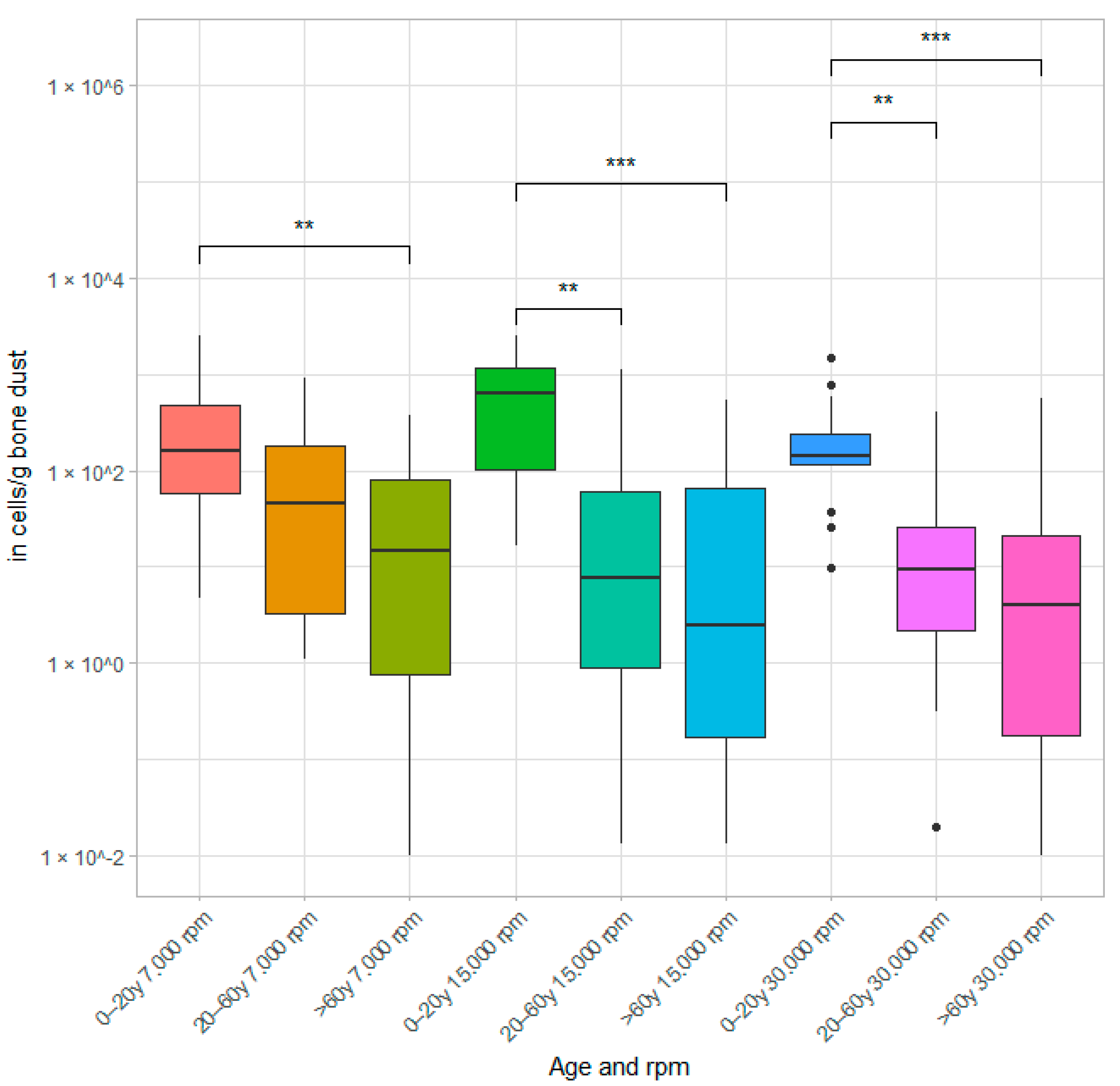
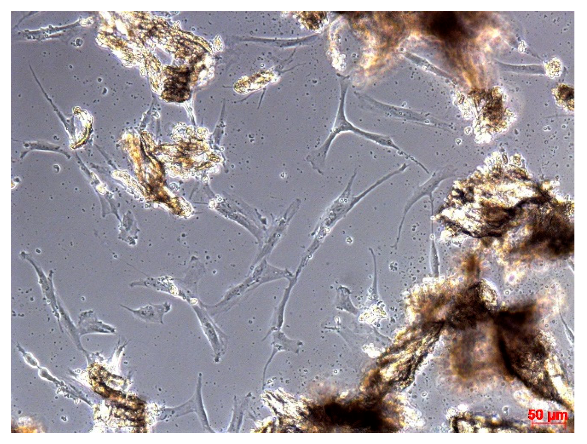
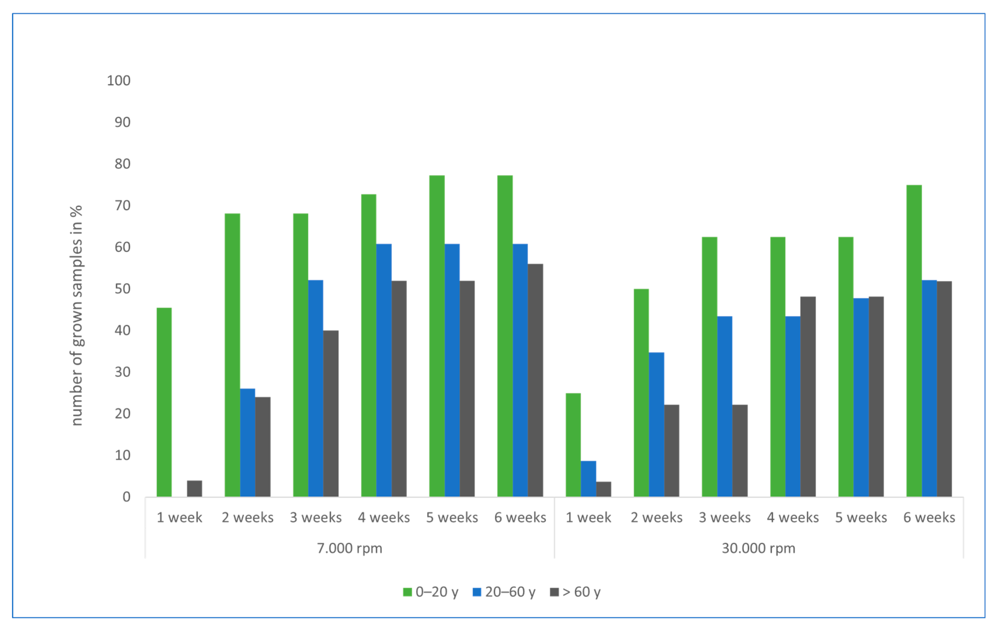
3.1. Results of Cell Culture Studies

The BD obtained at the different rotation speeds of 7.000, 15.000, and 30.000 rpm was distributed over 24-well plates as described in the Section 2.2.1 in order to assess how the rotation speed influences BD particle size, osteogenic activity, and its interaction with BA.
Figure 2 shows the number of osteoblasts (after 6 weeks) per gram of BD. Significant differences were found within the speed groups, in particular, between the age groups (<20 y vs. 20–60 y and <20 y vs. >60 y). No significant difference was found between the 20–60 y and >60 y groups. Due to the large variance, no significant difference was found between speeds within the same age group. However, there is a trend towards more cell outgrowth in the 7.000 and 15.000 rpm samples.
After including 10 patients per age group, no statistically significant difference in osteogenic activity was found between the 7.000 and 15.000 rpm samples.
The analysis examined whether the distribution of cell counts per gram of BD was identical in the two rotation categories for three age groups: 0–20 years, 20–60 years, and over 60 years. The Kruskal-Wallis test, a non-parametric test for independent samples, was used to compare the distributions. The significance level (α) was set at 0.05 and asymptotic significance values (p-values) were reported. For the age group 0–20 years, the p-value was 0.302. As this value is above the significance threshold of 0.05, the null hypothesis—that the distribution of cell counts per gram of BD is identical in the rotation categories—was accepted. Similarly, a p-value of 0.364 was obtained for the 20–60 age group, leading to the same conclusion: the null hypothesis was not rejected. Finally, the p-value for the 60+ age group was 0.642, which also resulted in the null hypothesis being accepted.
In summary, no statistically significant differences in the distribution of cell counts per gram of BD were observed between the rotation categories for any of the age groups, as all p values exceeded the 0.05 significance level. The null hypothesis was, therefore, accepted in all cases. Therefore, we decided to use only 7.000 and 30.000 rpm for the human samples.
Two aspects of the cell culture were assessed: the cell counts after 6 weeks and weekly documentation of outgrowth. Figure 3 shows a microscopic image of a well containing bone dust particles and osteoblasts from a 5-year-old child 3 weeks after bone dust removal.
Figure 4 shows the outgrowth behaviour over a period of 6 weeks. The graph shows the percentage of samples that have outgrown the total number of samples. A chi-square test was performed between age, cell growth, and speed. There was a statistically significant correlation between age and cell growth (χ2(12) = 39.67, p < 0.001, and φ = 0.52). There was no statistically significant correlation between speed and cell growth (χ2(6) = 8.5, p = 0.203, and φ = 0.241). Although there are no significant differences between the speeds, clear trends are recognisable.
In order to calculate the differences in frequency between the age groups and the speed in the absolute outgrowth behaviour, a chi-squared test or a Monte Carlo estimate was carried out, since more than five cells in all groups had an expected frequency of less than five. No significant difference was found between the speeds within the age groups. When the different age groups were analysed separately according to speed, there were sig. differences between the age groups <20 and 21–60 (p < 0.001) and >60 (p = 0.005). The groups 21–60 and >60 did not differ sig. (p = 0.854). At 30.000, the under-20-year-olds differed from the over-60-year-olds (p < 0.001). However, the other two groups did not differ significantly from each other (p = 0.456; p = 0.319).
The microscopic analyses showed that the particles of the 7.000 rpm group have a mean length of 934 µm with a standard deviation (SD) of 370 µm. The particles of the 30.000 rpm group have a mean length of 403 µm ± 262 µm (SD). Due to the non-normal distribution of the data, we decided to use the Wilcoxon rank-sum test to compare two independent metrically scaled groups that do not follow a normal distribution. Hence, there is a significant difference between the two groups. The confidence interval for the mean particle length difference ranges from 364 µm to 610 µm. This significant difference is not only statistically but also practically relevant, given its magnitude. The above results concern exclusively the experiments conducted on human cell cultures. Only the length was considered as an objective parameter; the specific shape or size of the particles was not taken into account. Furthermore, no separate cell culture study of ovine BD was performed. The comparability between human and ovine BD is discussed in the Section 4.
Clinical otosurgical experience showed that BD obtained at 15.000 rpm instead of 7.000 rpm could be better combined with BA and modelled in the defect. In addition, the above-mentioned results for BD harvesting were almost identical for 7.000 and 15.000 rpm, so that it was decided to use 15.000 rpm as the lower speed for the animal experiment.

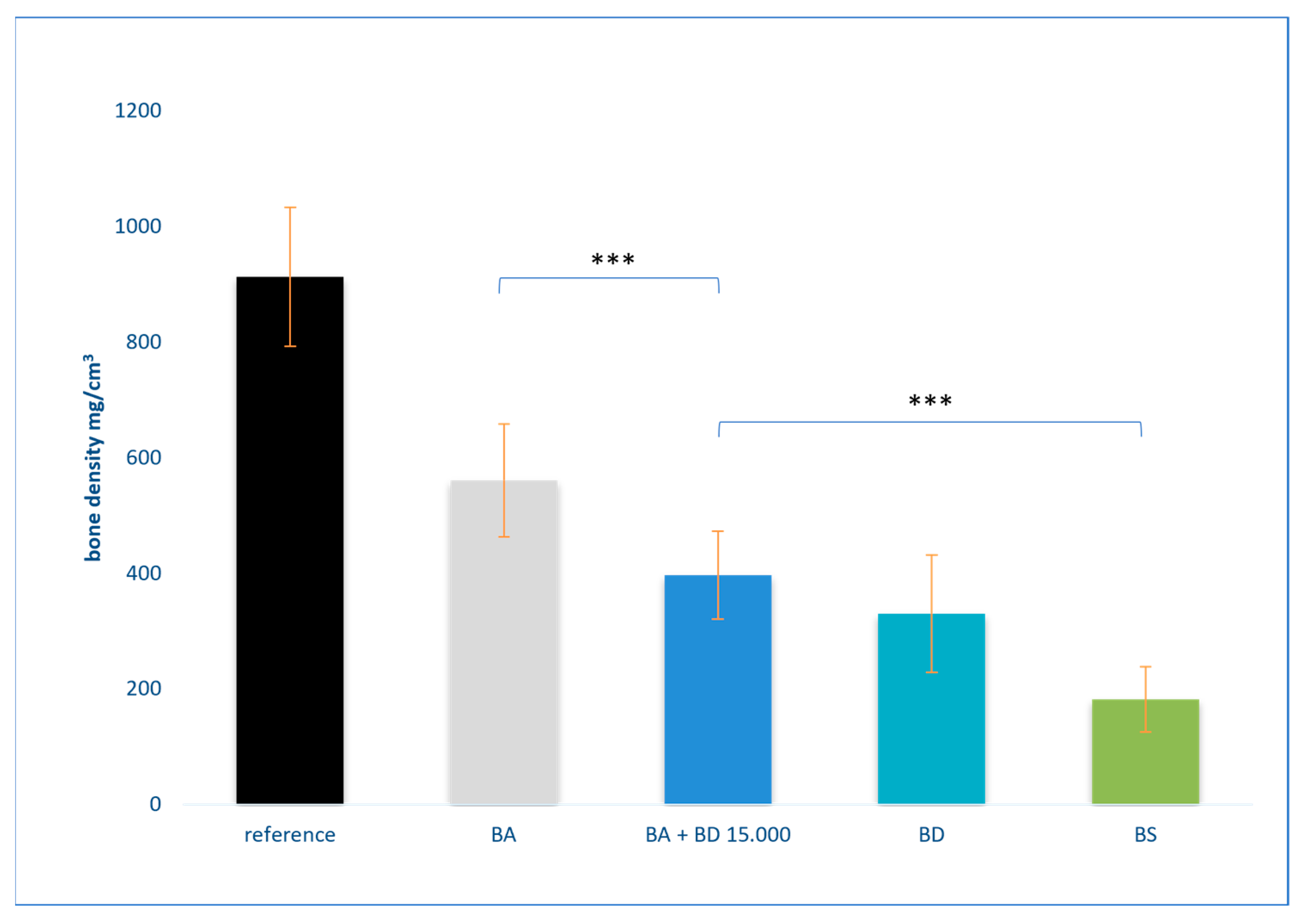
3.2. Results of Bone Density Measurement

In unfilled defect 1, an average bone density of 182.05 mg/cm3 was measured. On the other hand, defect 2, which was filled with BA, had a significantly higher (p < 0.001) mean bone density of 561.03 mg/cm3. Defect 3, which was filled with BD, showed a mean bone density of 330.42 mg/cm3. Finally, the filling of defect 4, which was filled with both BA and BD, had a mean bone density of 387.36 mg/cm3.
The statistical analysis showed significant differences between the individual defects. In particular, defect 4 was significantly denser than defect 1 (p < 0.001). Defect 2, which was filled with BA only, had a significantly higher density than defect 4 (p < 0.001), which contained both BA and BD (Figure 5). The surrounding bone was used as a reference.

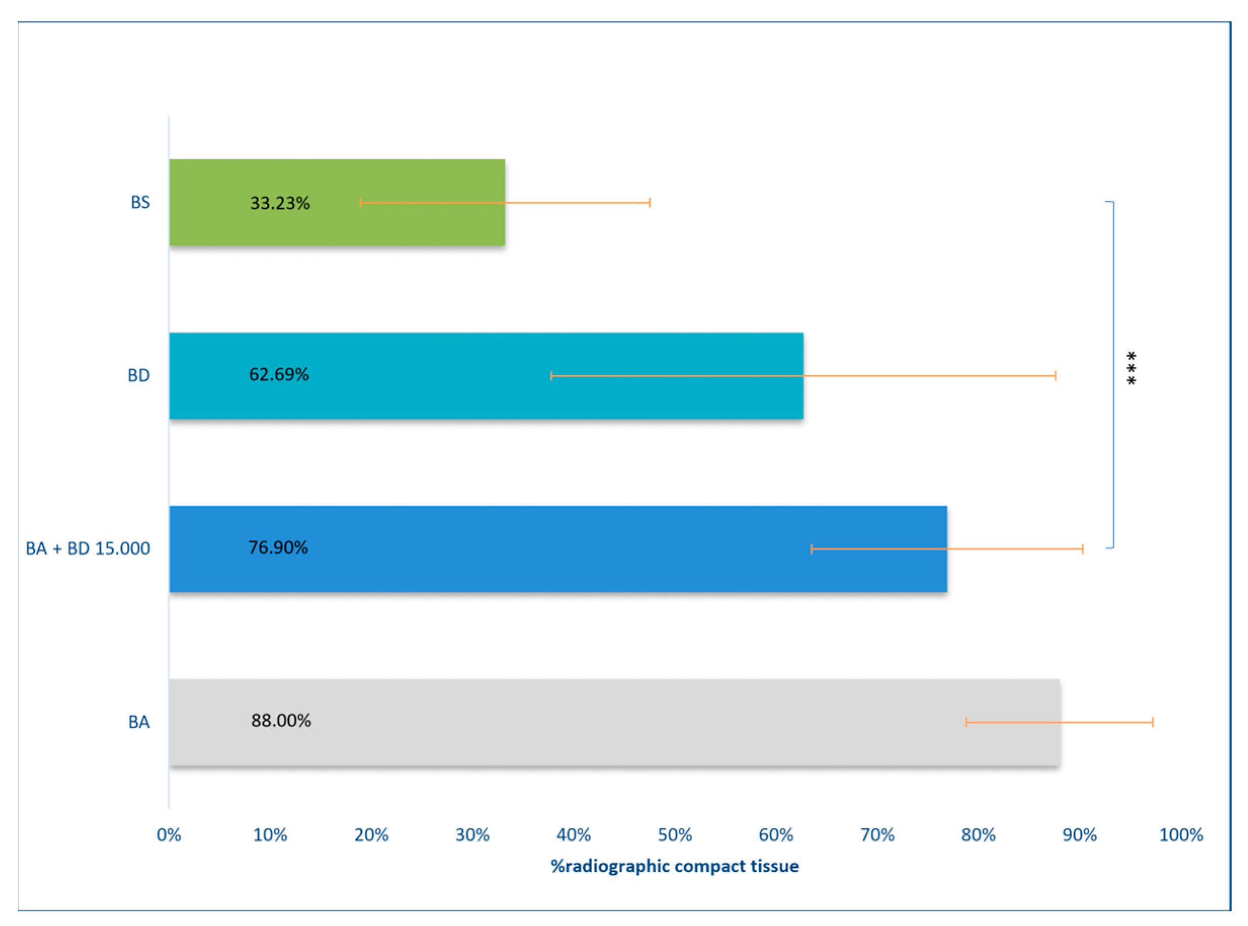
3.3. Results of DVT Measurement

The DVT results are shown in Figure 6 and include data from ten specimens, analysing the total defect volume of each defect. An analysis showed no discrepancies in grey values between the BA and the newly formed bone. The percentage of defective fillings for the different materials are as follows: BA is filled to 88.00%, BA + BD is filled to 76.90%, and BD alone is filled to 62.69%. The blank sample is filled to 33.23%. These values represent the mean values obtained from the analysis. There is no significant difference in the filling between BA + BD and BD alone, or between BA and BA + BD. A trend can be seen, however.

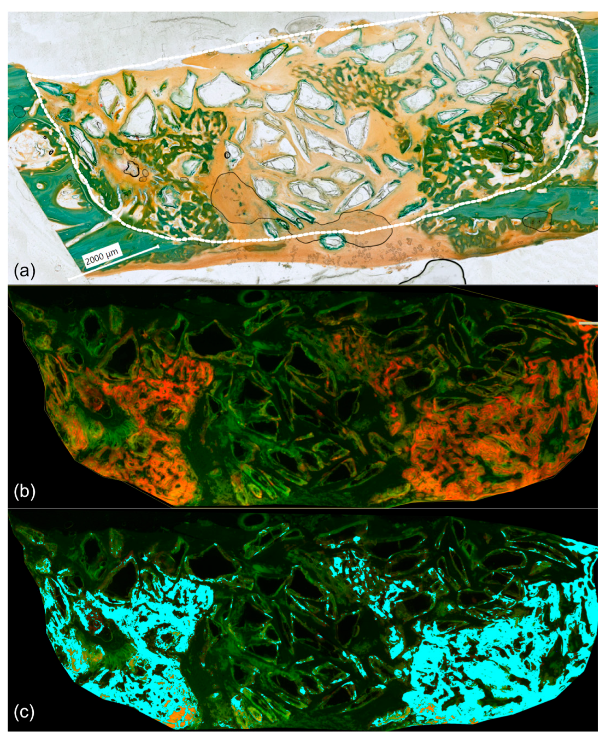
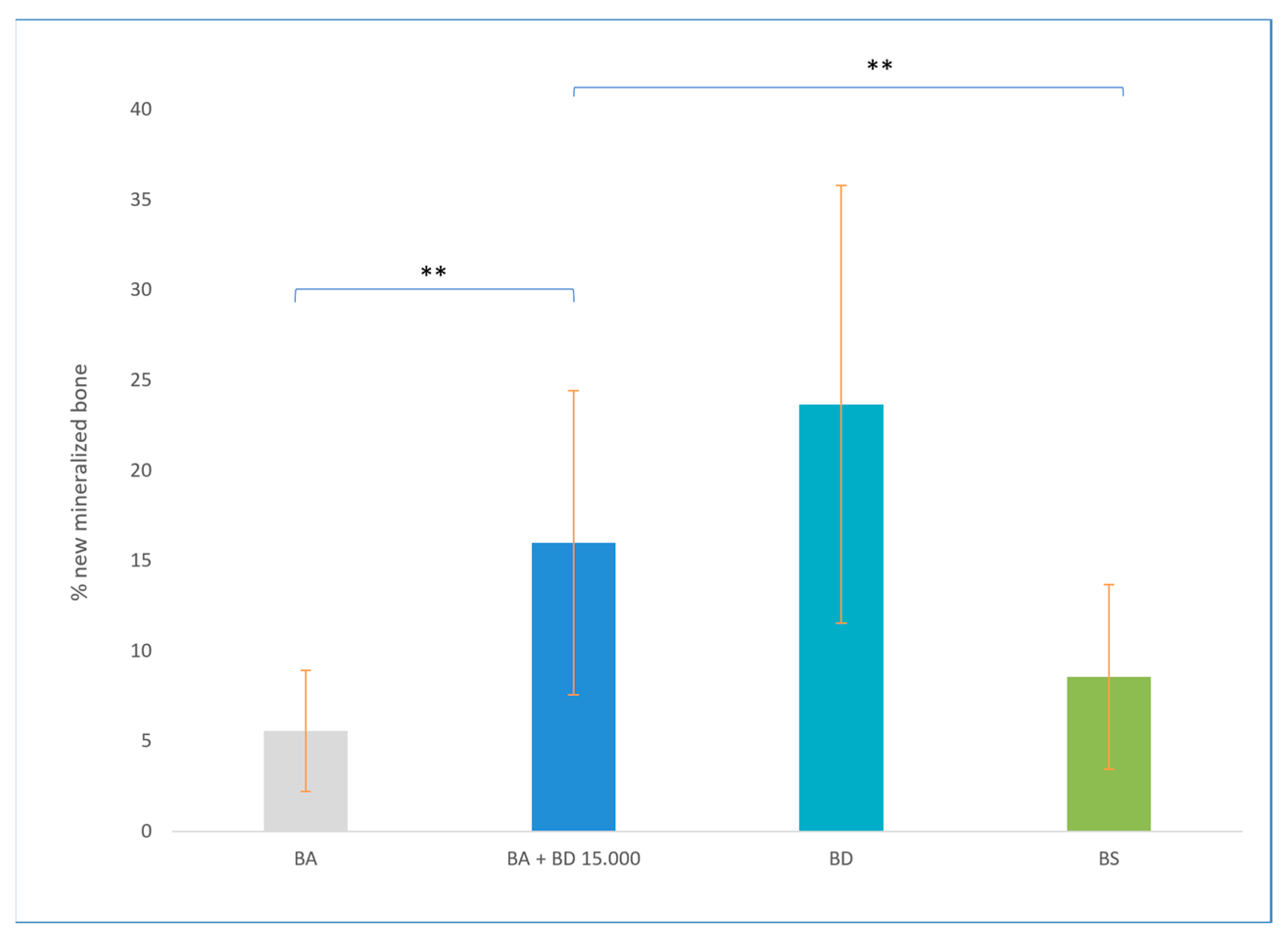
3.4. Results of Phase Colour-Coding System

A phase colour-coding analysis makes it possible for us to visualise the newly formed bone at the time of fluorescence administration. The fluorescent dye alizarin glows red when excited (Figure 7b), marking the newly formed bone within the defect two weeks after surgery. Bone growth normally starts from the edge of the bone (centripetal). Because of the bone dust placed between the glass granules, new bone formation also starts within the defect. (Figure 8).
In the BA defect, the average percentage of new bone formation is 5.57%, which is different from the 8.57% filling of the blank sample (p = 0.11). The Cohen’s d effect size of the difference is medium (d = 0.657), with a 95% confidence interval of −0.14 to 1.44, it but cannot be considered significant. In the BD defect, the percentage of filling is 23.67%. The defect with the mixture of BD and BA has a filling of 15.99% compared to 23.67% with bone dust only (Figure 9). Significant differences can be seen in the filling to BA (p = 0.002) as well as to the BS (p = 0.019). This suggests faster bone formation in the mixed defect compared to BA alone and faster stable healing.

4. Discussion

4.1. BD Harvesting Methods

The quality of the BD depends largely on the collection methods. This is because BD contains vital and avital cells [19], the proportions of which, in turn, depend on the production method. The activity of BD is examined in the literature through individual studies, case descriptions, and case series, and these findings are subsequently compiled cumulatively in a review conducted by Street et al. (2017) [20]. This demonstrates that an increase in the size of the bone fragments is accompanied by an increase in the number of osteoblasts, which, in turn, leads to enhanced mineralisation [11,21]. The use of a high-speed drill has been shown to result in thermal or mechanical damage to cells [22,23]. Consequently, it can be hypothesised that a reduction in temperature would lead to the enhanced activity of the bone dust. If this is indeed the case, the cooling of the drill tip with saline solution and the utilisation of the drill at a reduced speed could prove an effective means of improving the results. In this regard, it has been demonstrated that bone marrow stem cells are more susceptible to thermal stress than bone fragments and bone grafts [24]. In order to determine the quality of the BD and its influencing factors in more detail, the vitality of the BD was first assessed in this study. For this purpose, BD was obtained at 7.000 and 15.000 rpm and 30.000 rpm. There was no significant difference between 7.000 and 15.000 rpm, as almost the same number of new cells was found in both groups. It can, therefore, be stated that, according to these results, both 7.000 and 15.000 rpm can be used for the extraction of BD. However, the comparison of 7.000 vs. 30.000 rpm clearly showed a loss of vitality of the BD. On the other hand, when measuring the bone particles in the BD, statistically significantly larger particles were found in the BD, which was produced at 7.000/15.000 rpm compared to 30.000 rpm. According to this investigation, larger particles and, therefore, a larger surface area from which the osteoblasts can grow out are produced at a lower rotational speed. In addition, the investigations of the outgrowth behaviour show that more cells tend to grow out in each age group at 7.000 rpm. For this reason, we have made this hypothesis. It can, therefore, be concluded that, based on these results, BD extraction should only be carried out at between 7.000 and 15.000 rpm. According to this investigations, larger particles and, therefore, a larger surface area from which the osteoblasts can grow out are produced at a lower rotational speed. This supports the results that bone chips contain more active osteoblasts than BD and would, therefore, be even better suited for the obliteration of NCSDs [21]. In addition, the investigations of the outgrowth behaviour show that more cells tend to grow out in each age group at 7.000 rpm. For this reason, we have made this hypothesis. Our hypothesis is that faster rotational speeds (more than 15.000 rpm) destroy vital cells in the bone dust.
Furthermore, the cell culture examinations showed a clear dependence of the vitality of the BD on age. The BD samples of younger patients showed a higher vitality compared to patients aged 20 years and older. There were no statistically significant differences in the age groups over the age of 20. It can be assumed that the completed longitudinal growth of the body together with the hormonal changes that have taken place can have an influence on bone metabolism and, thus, also on the vitality of the BD from the age of 20 [25,26]. However, the observed vitality of the BD was independent of the number of revolutions tested when the BD was removed.
Even after this study, it is still only possible to speculate about other parameters influencing the BD during harvesting. Thus far, there have only been a few studies on the collection of BD, but these have mainly focused on the time efficiency of the collection [27]. Only a few studies exist on the vitality of BD. For example, the consideration of BD in comparison to bone fragments and perichondrium [28]. It is undisputed that postoperative infections can have a considerable influence on the effectiveness of an obliteration with BD. In principle, autologous transplants are considered sterile. This depends largely on the pre- and intraoperative hygiene measures. The sheep were operated on according to the same high hygiene standards as in human surgery. Irrespective of this, the loss of autonomous grafts is seen in orthopaedic, maxillofacial, otosurgical, and head and neck surgery, so that secondary superinfection and sequestration can be assumed. However, it remains unknown which other factors influence the quality of the BD. It is to be assumed that a higher contact pressure of the drill on the BD will lead to the further destruction of or reduction in the bone chips and, thus, to low vitality. In addition, the temperature increases considerably with speed and contact pressure, which would also have a negative effect on the vitality of the BD. It should, therefore, be noted that the optimum BD should currently be produced with 7.000–15.000 rotations, short and low contact pressure, and intermittent cooling by rinsing. Even though neither the temperature nor the contact pressure could be objectified in this study, these appear to be significant influencing factors. Furthermore, the geometry of the drill and its use for BD production must be taken into account. The drill should be a coarse burr and used as flat as possible in order to obtain coarse-grained bone chips that contain numerous osteoblasts and, thus, the potential for new bone formation. Whether additional influences such as the use of growth factors favour even faster new bone formation remains to be seen.

4.2. Obliteration Techniques

Regardless of the many specialist disciplines involved, the aim of defect obliteration is to achieve closure with the most original morphology possible, optimal biomechanical functionality, sufficient stability, avoidance of infection, and preservation of the surrounding tissue. Numerous autologous and alloplastic materials are used to achieve these goals. In ear surgery, for example, the combination of BD and cartilage slices has become established for the obliteration of the mastoid (canal wall down and up technique) [1]. However, BA is increasingly being used, especially in the absence of sufficient autologous replacement materials. Numerous autologous and alloplastic materials are used to achieve these goals. In ear surgery, for example, the combination of BD and cut cartilage has become established for the obliteration of the mastoid or the radical cavity. However, all autologous materials used are subject to a shrinkage tendency and a risk of infection with a corresponding risk of complications and implant loss [29,30,31]. For this reason, alternative replacement materials such as BA are also used, especially when there are insufficient biological alternatives. BA also has the advantage of having an antibacterial effect, thus reducing the likelihood of wound infection and avoiding further complications [32].
Kluge et al. found that, when non-critical size defects are obliterated with BD, significantly more new bone is formed centrifugally and centripetally than when obliterated with BA alone [7]. However, BA achieved a significantly higher density and compactness of the closure.
In the present study, the obliterates BD and BA were combined with each other. After the observation period, filling the defect with the mixture of BD and BA resulted in similar centripetal and centrifugal new bone formation as the BD alone. A significantly larger amount of newly formed bone was found compared to BA alone. However, the BA in combination with the BD helped to completely fill the defect more quickly and denser. The highest density was found in the non-critical size defects filled with BA. In addition, the defect closure with BA and BD showed a higher density than BD alone.
When considering the radiogenic compactness of the defect filling using DVT, the combination of BA and BD also resulted in a higher compactness than with BD alone. But it was not possible to achieve the same values as with BA alone.
The newly formed bone was visualised using fluorochrome sequence labelling, which can also be used to visualize new bone formation in the middle ear [33]. It was shown that the highest percentage of bone formed after obliteration was only achieved with BD. However, the combination of BA and BD showed significantly greater new bone formation than obliteration with BA alone.
With the present results, no direct interaction between BD and BA can be established. Moreover, there is no indication of such an interaction from the previous literature. According to the manufacturer, only an antibacterial effect is known. It has been shown that the BD leads to new bone formation regardless of the localisation in the defect, e.g., also centrally in the defect. This results in a kind of bone network around the BA. In the present study, no implant loss was observed when BA was used alone or in combination with BD. In clinical practice, the loss of the obliterate BD is frequently observed in postoperative infections [1]. This appears to occur significantly less frequently when BA is used [32]. This advantage of the BA can, therefore, be utilised when using the combination with BD.

4.3. Limitation

This study was a pilot study to test the feasibility of the obliteration of NCSDs with BD or BA or both in combination compared to the blank hole on the calvaria. For this reason, a smaller number of animals was deliberately selected, particularly for ethical reasons. However, the results of the study should lead to a comprehensive study to verify their consistency and transferability to humans. Similarly, only a total of 55 samples could be included in the cell culture tests, resulting in a numerical limitation for both parties to the experiment. Additionally, the study design required the animals to be euthanised after 3 weeks, which likely did not allow for complete bone defect healing. As a preliminary investigation, the level of evidence remains limited, and further large-scale studies are required in order to confirm these findings and strengthen the clinical applicability. As a result, it remains unclear when and in which obliteration technique the defect cavity was ultimately completely closed with bone. The three-week time point provides only a snapshot of the bone regeneration process, limiting the ability to analyse its full dynamics. Consequently, all conclusions drawn from the results pertain only to this specific timeframe. Nevertheless, clear trends in bone formation and obliteration behaviour were observed.
Inflammatory processes that may impair bone formation are expected to begin immediately after surgery and, thus, fall within the study period. This period was chosen based on clinical experience of impaired wound healing after surgical obliteration in middle ear surgery. However, this study provides insufficient information about the long-term stability of the obliteration beyond the three-week observation period. Key aspects such as bone remodelling, BD–BA integration, long-term resorption rates, and potential late-stage inflammatory responses remain unknown. Further research is needed in order to determine whether the obliterated defects achieve stable, mechanically resilient bone formation over extended periods.
Moreover, temperature monitoring and contact pressure control during BD extraction were not objectively measured. While sufficient cooling was ensured through continuous irrigation, the exact thermal effects on bone vitality remain unknown. Similarly, drilling pressure was manually controlled by the surgeon, but no quantitative measurements were recorded. These factors could have influenced BD quality and, consequently, bone regeneration. Future studies should incorporate objective temperature and pressure measurements to better standardise BD collection parameters.
Therefore, further research is needed in order to determine the long-term stability of defect obliteration and to refine the optimal parameters for BD collection, including drill geometry, contact pressure, drill retention angle, and the influence of temperature on BD vitality.

5. Conclusions

This pilot study showed that the obliteration of defects of non-critical size should be performed with BD collected at a speed between 7.000 and 15.000 rpm. This recommendation is based on the observed higher vitality of the BD at these speeds compared to high-speed collection at 30.000 rpm. When a combination of BA and BD is then used for the obliteration of NCSDs, the advantages of both obliteration materials can be combined, leading to improve outcomes.
Through a statistical analysis, including one-way ANOVA with post hoc testing, a significantly larger amount of new bone was confirmed in defects treated with BD and BA in comparison to empty defects and single-material obliterations (BD or BA alone). The results demonstrated statistically significant differences (p < 0.05) in new bone formation after three weeks of healing, as assessed by DVT and bone density measurements. Additionally, the chi-square test revealed significant associations in categorical comparisons of bone growth patterns.
These findings suggest that the combined application of BA and BD not only ensures a faster and more complete defect closure but also promotes enhanced long-term stability and reduces bone loss in the obliterated area.
However, despite these promising results, certain limitations must be addressed in future research. The short observation period of three weeks restricts the ability to assess the long-term stability of BD–BA obliteration. To bridge this gap, future studies should incorporate extended follow-up periods to determine whether obliterated defects develop into stable, mechanically resilient bone over time.
Moreover, further clinical and experimental research is required to refine and standardise the optimal parameters for BD harvesting, including drill geometry, contact pressure, drill retention angle, and the effect of temperature on BD vitality. In addition, long-term stability studies should focus on evaluating the resorption behaviour, structural integrity, and remodelling capacity of BD–BA obliteration, particularly in the context of revision surgeries and broader clinical applications.

Author Contributions

Conceptualisation, M.N.; methodology, M.K., A.K., I.Z.-R. and S.I.G.; validation, I.Z.-R., S.I.G. and M.N.; formal analysis, S.I.G.; investigation, A.K. and I.Z.-R.; data curation, A.K., I.Z.-R. and S.I.G.; writing—original draft preparation, M.K., A.K., I.Z.-R. and S.I.G.; writing—review and editing, M.K., A.K. and S.I.G.; visualisation, A.K.; supervision, M.N.; project administration, M.N., M.K. and A.K.; funding acquisition, M.N. and A.K. All authors have read and agreed to the published version of the manuscript.

Funding

This research received no external funding.

Institutional Review Board Statement

The study was conducted in accordance with the Declaration of Helsinki, and approved by the Ethics Committee Regierungspräsidium Dresden, Germany (Approval Code:24 (D)-9168.11-1/2013-17, Approval Date: 20 June 2013).

Informed Consent Statement

Not applicable.

Data Availability Statement

The original contributions presented in this study are included in the article. Further inquiries can be directed to the corresponding author.

Acknowledgments

The authors thank Roland Jung, Kathrin Spekl, and Elke John for their excellent assistance with the animal experiments. The authors also thank Diana Jünger for the assistance with the histological analysis and Martina Rauner for the assistance with the measurement of bone density.

Conflicts of Interest

The authors declare no conflicts of interest.

Abbreviations

ANOVAanalysis of variance
BAbioactive glass S53P4
BDbone dust
BMDbone mineral density
BSblank sample
CIconfidence interval
DMEMDulbecco Modified Eagle Medium
DVTdigital volume tomography
FCSfetal calf serum
HSDHonestly Significant Difference
NCSDnon-critical size defects
PBSphosphate-buffered saline
ROIregion of interest
Rpmrotation per minute
pQCTperipheral quantitative computed tomography
SDstandard deviation

References

  1. Schimanski, G.; Schimanski, E. Obliteration von Mastoidhöhlen: 30 Jahre Erfahrung mit Empfehlungen zur Operationsstrategie. HNO 2015, 63, 538–545. [Google Scholar] [CrossRef] [PubMed]
  2. Gruber, R.; Baron, M.; Busenlechner, D.; Kandler, B.; Fuerst, G.; Watzek, G. Proliferation and Osteogenic Differentiation of Cells from Cortical Bone Cylinders, Bone Particles from Mill, and Drilling Dust. J. Oral Maxillofac. Surg. 2005, 63, 238–243. [Google Scholar] [CrossRef]
  3. Urist, M.R. Introduction to Update on Osteochondral Allograft Surgery. In Bone Transplantation; Springer: Berlin/Heidelberg, Germany, 1989; pp. 1–6. ISBN 978-3-642-83573-5. [Google Scholar]
  4. Fassone, E.; Fabiano, B.; Caracciolo, A.; Sapino, S.; Ferrero, V. Use of Bonalive in Obliterative Mastoidectomy: Anatomical Results and Clinical Outcome. Eur. Arch. Otorhinolaryngol. 2023, 280, 3577–3583. [Google Scholar] [CrossRef] [PubMed]
  5. Milazzo, M.; Cristofaro, G.; Berrettini, S.; Danti, S. Applications of Bioactive Glasses for Implants in the Ear. In Bioactive Glasses and Glass-Ceramics: Fundamentals and Applications; Wiley: Hoboken, NJ, USA, 2022; pp. 459–477. ISBN 978-1-119-72419-3. [Google Scholar]
  6. Sioshansi, P.C.; Alyono, J.C.; Blevins, N.H. Mastoid Obliteration Using Autologous Bone Dust Following Canal Wall Down Mastoidectomy. Otol. Neurotol. 2021, 42, 68–75. [Google Scholar] [CrossRef] [PubMed]
  7. Kluge, A.; Neudert, M.; Kunert-Keil, C.; Lailach, S.; Zahnert, T.; Kemper, M. The Obliteration of Noncritical Size Bone Defects With Bone Dust or Bone Replacement Material (Bioactive Glass S53P4). Otol. Neurotol. 2019, 40, e415–e423. [Google Scholar] [CrossRef]
  8. Cannio, M.; Bellucci, D.; Roether, J.A.; Boccaccini, D.N.; Cannillo, V. Bioactive Glass Applications: A Literature Review of Human Clinical Trials. Materials 2021, 14, 5440. [Google Scholar] [CrossRef]
  9. Virolainen, P.; Heikkilä, J.; Yli-Urpo, A.; Vuorio, E.; Aro, H.T. Histomorphometric and Molecular Biologic Comparison of Bioactive Glass Granules and Autogenous Bone Grafts in Augmentation of Bone Defect Healing. J. Biomed. Mater. Res. 1997, 35, 9–17. [Google Scholar] [CrossRef]
  10. Pernaa, K.; Koski, I.; Mattila, K.; Gullichsen, E.; Heikkila, J.; Aho, A.J.; Lindfors, N. Bioactive Glass S53P4 and Autograft Bone in Treatment of Depressed Tibial Plateau Fractures—A Prospective Randomized 11-Year Follow-Up. JLT 2011, 21, 139–148. [Google Scholar] [CrossRef]
  11. Eder, C.; Chavanne, A.; Meissner, J.; Bretschneider, W.; Tuschel, A.; Becker, P.; Ogon, M. Autografts for Spinal Fusion: Osteogenic Potential of Laminectomy Bone Chips and Bone Shavings Collected via High Speed Drill. Eur. Spine J. 2011, 20, 1791–1795. [Google Scholar] [CrossRef]
  12. Gao, R.; Street, M.; Tay, M.L.; Callon, K.E.; Naot, D.; Lock, A.; Munro, J.T.; Cornish, J.; Ferguson, J.; Musson, D. Human Spinal Bone Dust as a Potential Local Autograft: In Vitro Potent Anabolic Effect on Human Osteoblasts. Spine 2018, 43, E193–E199. [Google Scholar] [CrossRef]
  13. Pandey, R.K.; Panda, S.S. Drilling of Bone: A Comprehensive Review. J. Clin. Orthop. Trauma 2013, 4, 15–30. [Google Scholar] [CrossRef] [PubMed]
  14. Pautke, C.; Vogt, S.; Tischer, T.; Wexel, G.; Deppe, H.; Milz, S.; Schieker, M.; Kolk, A. Polychrome Labeling of Bone with Seven Different Fluorochromes: Enhancing Fluorochrome Discrimination by Spectral Image Analysis. Bone 2005, 37, 441–445. [Google Scholar] [CrossRef] [PubMed]
  15. An, Y.H.; Martin, K.L. (Eds.) Handbook of Histology Methods for Bone and Cartilage; Humana Press: Totowa, NJ, USA, 2003; ISBN 978-0-89603-960-5. [Google Scholar]
  16. Donath, K.; Breuner, G. A Method for the Study of Undecalcified Bones and Teeth with Attached Soft Tissues. The Säge-Schliff (Sawing and Grinding) Technique. J. Oral Pathol. 1982, 11, 318–326. [Google Scholar] [CrossRef]
  17. Bucher, O.; Wartenberg, H. Cytologie, Histologie und Mikroskopische Anatomie des Menschen, 12th ed.; Hans Huber: Berching, Germany, 1997; ISBN 978-3-456-82785-8. [Google Scholar]
  18. Kunert-Keil, C.; Gredes, T.; Heinemann, F.; Dominiak, M.; Botzenhart, U.; Gedrange, T. Socket Augmentation Using a Commercial Collagen-Based Product—An Animal Study in Pigs. Mater. Sci. Eng. C 2015, 46, 177–183. [Google Scholar] [CrossRef]
  19. Trinkaus, K.; Wenisch, S.; Siemers, C.; Hose, D.; Schnettler, R. Bohrmehl: Eine Quelle vitaler Zellen!: Erste Ergebnisse von humanen Proben. Der Unfallchirurg 2005, 108, 650–656. [Google Scholar] [CrossRef]
  20. Street, M.; Gao, R.; Martis, W.; Munro, J.; Musson, D.; Cornish, J.; Ferguson, J. The Efficacy of Local Autologous Bone Dust: A Systematic Review. Spine Deform 2017, 5, 231–237. [Google Scholar] [CrossRef]
  21. Ye, S.; Seo, K.-B.; Park, B.-H.; Song, K.-J.; Kim, J.-R.; Jang, K.-Y.; Chae, Y.J.; Lee, K.-B. Comparison of the Osteogenic Potential of Bone Dust and Iliac Bone Chip. Spine J. 2013, 13, 1659–1666. [Google Scholar] [CrossRef] [PubMed]
  22. Patel, V.V.; Estes, S.M.; Naar, E.M.; Lindley, E.M.; Burger, E. Histologic Evaluation of High Speed Burr Shavings Collected During Spinal Decompression Surgery. Orthopedics 2009, 32, 1–5. [Google Scholar] [CrossRef]
  23. Hassanein, A.H.; Arany, P.R.; Couto, R.A.; Clune, J.E.; Glowacki, J.; Rogers, G.F.; Mulliken, J.B.; Greene, A.K. Cranial Particulate Bone Graft Ossifies Calvarial Defects by Osteogenesis. Plast. Reconstr. Surg. 2012, 129, 796e–802e. [Google Scholar] [CrossRef]
  24. Karamese, M.; Toksoz, M.R.; Selimoglu, M.N.; Akdağ, O.; Toy, H.; Tosun, Z. Comparison of Bone Dust With Other Types of Bone Grafts for Cranioplasty. J. Craniofacial Surg. 2014, 25, 1155–1158. [Google Scholar] [CrossRef]
  25. Turner, A.S. The Sheep as a Model for Osteoporosis in Humans. Vet. J. 2002, 163, 232–239. [Google Scholar] [CrossRef]
  26. de Pollak, C.; Arnaud, E.; Renier, D.; Marie, P.J. Age-Related Changes in Bone Formation, Osteoblastic Cell Proliferation, and Differentiation during Postnatal Osteogenesis in Human Calvaria. J. Cell. Biochem. 1997, 64, 128–139. [Google Scholar] [CrossRef]
  27. Nagle, S.K.; Hebbar, C.R.; Kumar, R. An Effective Method to Collect Bone Dust for Mastoid Cavity Obliteration. Indian J. Otolaryngol. Head Neck Surg. 2022, 74, 4450–4454. [Google Scholar] [CrossRef] [PubMed]
  28. Gupta, A.; Lobocki, C.; Malhotra, G.; Jackson, I.T. Comparison of Osteogenic Potential of Calvarial Bone Dust, Bone Fragments, and Periosteum. J. Craniofacial Surg. 2009, 20, 1995–1999. [Google Scholar] [CrossRef]
  29. Lindeboom, J.J.; Van Kempen, P.M.W.; Buwalda, J.; Westerlaken, B.O.; Van Zuijlen, D.A.; Bom, S.J.H.; Van Der Beek, F.B. Mastoid Obliteration with Hydroxyapatite vs. Bone Pâté in Mastoidectomy Surgery Performed on Patients with Cholesteatoma and Chronic Suppurative Otitis Media: A Retrospective Analysis. Eur. Arch. Otorhinolaryngol. 2023, 280, 1703–1711. [Google Scholar] [CrossRef] [PubMed]
  30. Beutner, D.; Stumpf, R.; Zahnert, T.; Hüttenbrink, K.-B. Long-Term Results following Mastoid Obliteration in Canal Wall Down Tympanomastoidectomy. Laryngo-Rhino-Otol. 2007, 86, 861–866. [Google Scholar] [CrossRef]
  31. Schimanski, G.; Schimanski, E. Mastoid Cavity Obliteration with Bioactive Glass Granules. In Recent Advances in Otolaryngology Head & Neck Surgery; JP Medical Ltd.: New York, NY, USA, 2016; pp. 249–281. ISBN 978-93-5152-940-8. [Google Scholar]
  32. Fernandes, J.S.; Gentile, P.; Pires, R.A.; Reis, R.L.; Hatton, P.V. Multifunctional Bioactive Glass and Glass-Ceramic Biomaterials with Antibacterial Properties for Repair and Regeneration of Bone Tissue. Acta Biomater. 2017, 59, 2–11. [Google Scholar] [CrossRef]
  33. Kemper, M.; Kluge, A.; Ney, M.; Beleites, T.; Zeidler-Rentzsch, I.; Keil, C.; Zahnert, T.; Neudert, M. Visualization of Bone Formation in Sheep’s Middle Ear by Using Fluorochrome Sequential Labelling (FSL). Sci. Rep. 2024, 14, 7046. [Google Scholar] [CrossRef]
Figure 1. (a) 7 mm Rose burr; (b) drilled defect with dummy; (c) bioactive glass (BA); (d) bone dust (BD) harvested with 15.000 rpm (rotation per minute); and (e) non-critical size defects (NCSDs) in calvarian bone of sheep without filling (blank sample: defect 1) and filled with BA (defect 2), or BD (defect 3) alone and with BA combined with BD (defect 4).
Osteology 05 00015 g001
Figure 2. Logarithmic boxplot representation of the cell count after 6 weeks in relation to age and speed. The proportion of active cells in a gram of bone dust was analysed in three age groups of 0–20, 21–60, and over 60 years (y) and as a function of the speed 7.000, 15.000, or 30.000 rpm (rotation per minutes). The boxplots show the quartiles and the median of the data distribution; the whiskers range from the 1st/3rd quartile to the last data point inside the 1.5 × Interquartile Range (IQR), and outliers outside this range are depicted as dots. Significance levels as pairwise t-test results between boxes connected by brackets are depicted as asterisks with “**” for significance level p < 0.01, and “***” for p < 0.001. Only t-tests inside groups of the same speed and between groups of same speed but different age are performed. Groups: 0–20 y: 7.000 rpm: n = 17, 15.000 rpm: n = 8, 30.000 rpm: n = 13; 20–60 y: 7.000 rpm: n = 18, 15.000 rpm: n = 11, 30.000 rpm: n = 12; >60 y: 7.000 rpm: n = 12; 15.000 rpm: n = 16, 30.000 rpm: n = 15.
Osteology 05 00015 g002
Figure 3. Microscopic view of BD particles and osteoblasts 3 weeks after bone dust removal (patient whose age is 5 years).
Osteology 05 00015 g003
Figure 4. Columns shows the percentage of samples that have outgrown the total number of samples. Visualisation over 6-week cultivation period, different number of rotations, and age of patients (y = years; rpm = routine per minute). Groups: 0–20 y, 7.000 rpm: n = 22; 0–20 y, 30.000 rpm: n = 24; 20–60 y, 7.000 rpm: n = 23; 20–60 y, 30.000 rpm: n = 23; >60 y, 7.000 rpm: n = 25; >60 y, 30.000 rpm: n = 27.
Osteology 05 00015 g004
Figure 5. Results of bone density measurements. Columns show mean bone density (mg/cm3) with standard deviations (BD = bone dust, BA = bioactive glass, and BS = blank sample) (n = 10). p < 0.1% (highly significant, ***).
Osteology 05 00015 g005
Figure 6. Results of DVT. Columns show mean percentages of radiopaque compact tissue with standard deviations (BD = bone dust, and BA = bioactive glass) (n = 10). p < 0.1% (highly significant, ***).
Osteology 05 00015 g006
Figure 7. Histological images of the fluorochrome labelling in the BA + BD defect. (a) Masson–Goldner staining—green coloured areas show new mineralised bone after 3 weeks. The dotted lines mark the NCSDs including BA and BD. (b) Histological image of the fluorochrome labelling—orange coloured areas show new mineralised bone after 2 weeks. (c) Turquoise regions showing the defined areas for the phase (phase colour-coding system).
Osteology 05 00015 g007
Figure 8. Histological image with Masson–Goldner staining. Orange areas show connective tissue, green areas show mineralised bone, and white areas show BA particles (see asterisks). The green glowing edges on the glass particles are not newly formed bone, but probably rather deposits of the dye. These green areas were not included in the analysis.
Osteology 05 00015 g008
Figure 9. Results of the phase colour-coding system. Columns show mean percentage of new mineralisation after 2 weeks (BD = bone dust, BA = bioactive glass) (n = 13). p < 1% (very significant, **).
Osteology 05 00015 g009
Disclaimer/Publisher’s Note: The statements, opinions and data contained in all publications are solely those of the individual author(s) and contributor(s) and not of MDPI and/or the editor(s). MDPI and/or the editor(s) disclaim responsibility for any injury to people or property resulting from any ideas, methods, instructions or products referred to in the content.

