Article

Linear-Chain Nanostructured Carbon with a Silver Film Plated on Metal Components Has a Promising Effect for the Treatment of Periprosthetic Joint Infection

by
Leonid I. Maliuchenko
1,*,
Nikolay S. Nikolaev
1,2,
Nadezhda N. Pchelova
1,
Dmitry Nikolaevich Efimov
1,
Elena V. Preobrazhenskaia
1 and
Vladimir U. Emelianov
1,2
1
Federal Center for Traumatology, Orthopedics and Arthroplasty, 428020 Cheboksary, Russia
2
Faculty of Medicine, Chuvash State University, 428020 Cheboksary, Russia
*
Author to whom correspondence should be addressed.
Osteology 2021, 1(4), 238-246; https://doi.org/10.3390/osteology1040022
Submission received: 30 October 2021 / Revised: 17 November 2021 / Accepted: 6 December 2021 / Published: 8 December 2021

Abstract

:
Background: Due to the aging of the world population, the number of joint diseases, along with the number of arthroplasties, has increased, simultaneously increasing the amount of complications, including periprosthetic joint infection (PPI). In this study, to combat a PPI, we investigated the antimicrobial properties of the new composite cover for titanium implants, silver-doped carbyne-like carbon (S-CLC) film. Methods: The first assay investigated the antimicrobial activity against Pseudomonas aeruginosa and releasing of silver ions from S-CLC films into growth media covered with S-CLC with a thickness of 1, 2, and 4 mm. The second assay determined the direct antibacterial properties of the S-CLC film’s surface against Staphylococcus aureus, Enterococcus faecalis, or P. aeruginosa. The third assay studied the formation of microbial biofilms of S. aureus or P. aeruginosa on the S-CLC coating. Silver-doped carbyne-like carbon (S-CLC)-covered or titanium plates alone were used as controls. Results: S-CLC films, compared to controls, prevented P. aeruginosa growth on 1 mm thickness agar; had direct antimicrobial properties against S. aureus, E. faecalis, and P. aeruginosa; and could prevent P. aeruginosa biofilm formation. Conclusions: S-CLC films on the Ti surface could successfully fight the most common infectious agent in PPI, and prevented biofilm formation.

1. Introduction

Aging of the world population is increasing the number of degenerative diseases of bones and joints [1]. One of the most common age-related joint conditions is osteoarthritis. In the advanced stages of osteoarthritis, only joint-replacement surgery can restore the function of joints. Therefore, there was growth in total arthroplasty procedures in the U.S. from 0.11% in 1980 to 0.83% in 2010 for total hip and 0.13% in 1980 to 1.52% in 2010 total knee replacements, and a shift to younger ages [2].
The most common complications of arthroplasty are bleeding, thrombotic complications, aseptic loosening of prosthetic parts, and periprosthetic joint infection (PPI). PPI, one of the most severe complications despite a minimal incidence rate of 0.5% to 1%, remains a major challenge [3]. The projections of prevalence incidence of revision hip and knee arthroplasty up to 2030 are increases between 43% and 70% for hip joint replacement and between 78% and 182% for knee replacement, particularly due to infection [4]. To reduce the risk of infection on the cement or space between the bone and spacer, vancomycin and gentamicin are added abundantly [5]. From the other side, the articular surface of the spacer can be covered with silver, which has great benefit for patients and healthcare systems [6]. Nevertheless, long-time toxicity of silver coatings for the surrounding tissue has not been established [7]. After extended accumulation in some studies, it was shown that it can be distributed to the blood, brain, heart, and kidney with a toxic effect, or be aggregated and inhibit the immune system [8]. Moreover, insufficient hardness of the coating can lead to rapid wear of the surface.
A composite coating with silver could solve these problems. In this study, we investigated the antibacterial properties of the new nanofilms coated on titanium (Ti), which allows a subsurface coating consisting of linear-chain carbon with an sp-1 connection between atoms, named chain carbyne-like carbon (CLC), with silver atoms incorporated. CLC is one of the hardest materials in the world; it has antibacterial properties, and different atoms, including silver, can be inserted between the carbon chains and on the film surface. This modification allows slowing down the release of silver for a local antibacterial effect.

2. Materials and Methods

2.1. Fabrication of Silver-Doped Carbyne-like Carbon (S-CLC) Films

As a substrate material for applying S-CLC coatings, we used Ti alloy VT6platesof a 50 × 50 × 0.5 mm size. These plates were used without any coating as control samples. The second group of control consisted of CLC-film-coated plates. The CLC coatings were formed by the physical vapor deposition method of ion-stimulated carbon condensation. The process of deposition of coatings was carried out in vacuum coating equipment adjusted to produce linear-chain carbon nanostructures. The scheme of the method is shown in Figure 1. The carbon films were formed by vacuum condensation of a carbon stream while the substrate and the film growing on it were irradiated with ions of an inert gas (argon) [9].
For the third group, silver pins were inserted into the graphite cathode in order to obtain silver-doped carbon S-CLC films. The synthesis of the S-CLC film was carried out with simultaneous ion-plasma evaporation of silver in the same pulsed cathode discharge. A stream of carbon obtained by thermal evaporation or ion sputtering and a stream of Ag+ ions were simultaneously fed to the substrate. Each group consisted of the 3 samples.

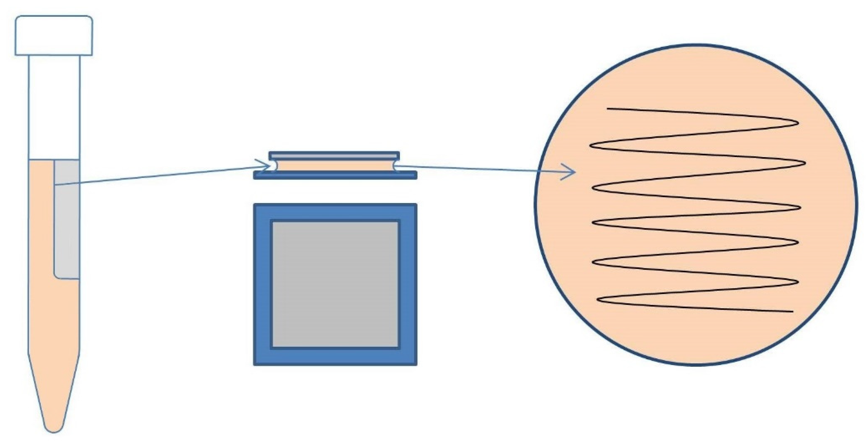
2.2. Evaluation of the Antibacterial Activity of Coatings

All Ti plates were kept in distilled water for 15 min at room temperature in order to remove rapidly soluble components, and then they were sterilized by an air method at 160 °C for 60 min. The antibacterial activity of the S-CLC Ti samples was examined for Gram-positive Staphylococcus aureus (ATCC 25923) and Enterococcus faecalis (ATCC 29212), and Gram-negative Pseudomonas aeruginosa (ATCC 27853orP-142). P. aeruginosa (P-142) isolated from a patient with post-traumatic osteomyelitis had resistance to most antibiotics, with the exception of polymyxins; produced the metallo-beta-lactamase VIM; and had the ability to intensively form microbial biofilms.

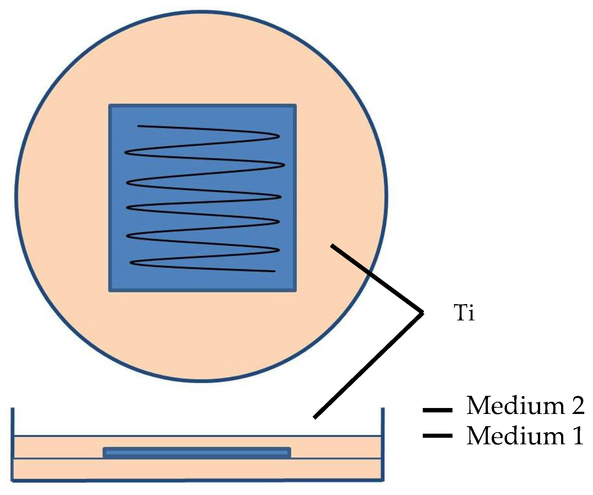
2.2.1. Assessment of the Release of Silver Ions from S-CLC Films

Sterilized samples were placed in the center 90 mm polystyrene Petri dishes with Mueller Hinton II Agar. Following up, the second layer was molten and cooled to 45 °C in Mueller Hinton II Agarin a volume of 8.3 mL, 14.6 mL, and 27.6 mL, and was poured with the second layer on the surface of the plate (Figure 1). The volume was calculated and chosen so as to cover the plates with a height of the agar layer above the Ti plate surface of1, 2, and 4 mm, respectively. The cups were kept on a horizontal surface until the medium completely solidified, then additionally dried in a thermostat for 15 min [10].
The antibacterial activity was evaluated against P. aeruginosa (ATCC 27853). Microbial stock cultures for investigations were prepared on a daily culture grown on nutrient agar (M001, Hi Media, India). Following up, we prepared abacterial suspension in a 0.85% sterile isotonic sodium chloride solution (ISC) with a final optical density of 0.5 McFarland, corresponding to a concentration of microbial cells of ~108 mL−1. The selected bacteria concentration was chosen according to the literature data [11]. The cups were inoculated with a bacterial suspension on agar with the use of a cotton swab and incubated for 18 h at 35 °C. The presence and growth pattern of microorganisms on the Mueller Hinton II Agar surface were evaluated in the area of the control Ti plates and Ti plates coated with S-CLC (Figure 1).

2.2.2. Qualifying the Direct Antibacterial Properties of S-CLC Films

Japan industrial standard Z 2801:2000 was applied for qualification of the surface antibacterial activities of S-CLC films [12] (Figure 2).
Briefly, for this test, sterilized Ti plates were placed in the center of 90 mm polystyrene Petri dishes without culture; then, 400 µL of Staphylococcus aureus (ATCC 25923), Enterococcus faecalis (ATCC 29212) S. aureus, E. faecalis, or P. aeruginosa P-142 bacterial suspensions were applied on the plate’s surface and covered with a polyethylene film (40 × 40 mm) previously sterilized by the ethylene oxide method. The closed cups were carefully transferred to a wet chamber and incubated in a thermostat at 35 °C for 24 h.
Microbial stock cultures were grown on nutrient agar (M001, Hi Media, India). The bacterial suspension was prepared in a sterile ISC to an optical density of 0.5 McFarland; then, for the working solution, the bacterial suspension was mixed at 1:500 in sterile distilled water while adding 1:400 of nutrient broth (M002, Hi Media, India) to a final bacteria concentration of 2.5 × 10⁵ cells/mL, as described in the literature [13].
At the end of incubation, 50 µL of solution was taken under a plastic film with a pipette from the surface of the plate and was transferred to labeled glass tubes with 5 mL of sterile ISC. The contents of the test tubes were mixed in a vortex to distribute the microorganisms evenly in the volume. Using a sterile pipette and a glass rod, 100 mL of the content of the vial was poured into cups with Mueller Hinton Agar (MPZ, Hi Media, Maharashtra, India). The plates were incubated in a thermostat at 35 °C for 24 h, after which the number of colony-forming units per ml culture medium was calculated as described previously [13], according to the following equation:
N = ΣC/[(1 × n1) + (0.1 × n2)]d
where N = number of colonies per ml or per gram of product, ΣC = sum of all colonies on all plates counted, n1 = number of plates in first dilution counted, n2 = number of plates in second dilution counted, and d = dilution from which the first counts were obtained.
The level of antimicrobial activity for a series of experimental samples was calculated as a log reduction in bacterial units:
R = log ( NK NT )
where R is the log reduction in bacterial cells, NK is the average number of microbial cells for a series of control samples, and NT is the average number microbial cells for a series of experimental samples [14].
The bactericidal index was calculated using the following formula:
I   =   NK     NT NK   ×   100 %
where I is bactericidal activity index (Figure 2).

2.2.3. Study of the Formation of Microbial Biofilm on the S-CLC Coating

The Ti plates without the covering or with CLC or S-CLC coating, sized 12.5 × 50 × 0. 5 mm, were sterilized in 15 mL glass centrifuge tubes at 160 °C for 60 min. Then, 10 mL of tryptic soy broth (BD BBL, Sparks, MD, USA) with 50μLof bacterial cell suspension of S. aureus (ATCC 25923) or P. aeruginosa P-142 in ISC with an optical density of 0.5 McFarland were added. Each bacterial culture was tested three times for each bacterial species in each group. All tubes containing samples were incubated for 1 h at 37 °C in a shaker at 100 rpm.
To visualize biofilm formation, the Ti plates were gently removed from the test tubes and washed once with distilled water, followed by drying in a thermostat for 30 min at 35 °C. After that, the samples were stained with 0.1% aqueous lab-grade crystal violet aqueous solution in a thermostat for 30 min at 35 °C, rewashed in water, and dried. The presence of biofilm was assessed by light microscope examination of stained areas in reflected light at 100-fold magnification and 10 fields of vision.
Furthermore, the intensity of biofilm formation was quantified by solubilizing the crystal violet from the surface in 10 mL of 96% ethanol for 24 h at 44 °C and measuring the concentration of crystal violet. After adding the crystal violet extraction solution and standard alcohol solutions to the wells of a flat-bottomed 96-well tablet (Sarstedt, Germany), absorbance was measured at 540 nm using a spectrophotometer (Infinite M200 multifunctional microplate reader, TECAN, Männedorf, Switzerland) with wavelength 540 nm [15].
The biomass of the biofilm formed on the surface of the samples was accepted to be directly proportional to the concentration of crystalline violet in the washing solution. The mass of the biofilm was calculated by multiplying the mass of the dye absorbed by the biofilm during staining by the following formula:
m = V ( C 1 + C 2 + C 3 V 1 V 2 V 3 ) 3
where m is the mass of crystal violet absorbed by the microbial biofilm (in μg); V is the volume of the washing solution for one sample; C1, C2, and C3 denote the concentration of the dye in the washing solutions from the set of samples (numbers 1, 2, and 3) (in mcg/mL); and V1, V2, and V3 denote the concentration of the dye in the washing solutions of a series of samples (numbers 1, 2 and 3) used as a “sterility control” (in mcg/mL).

3. Results

3.1. Antibacterial Activity of CLC or S-CLC Surface

3.1.1. S-CLC Film Had an Influence on P. aeruginosa in Solution Only at a Very Short Distance

The investigation found that the distance of antibacterial action against P. aeruginosa showed inhibition of bacterial growth only in a 1 mm layer of culture solution on the S-CLC covered samples only, as a result of releasing soluble and diffusing Ag+ ions from the film. There was no growth of P. aeruginosa in either the 2 or 4 mm culture solutions or the CLC or S-CLC covered or uncovered Ti plates (Table 1.)

3.1.2. S-CLC Film Could Inhibit S. aureus, E. faecalis, and P. aeruginosa Surface Growth

Ti plates alone had no antibacterial activities against the studied microorganisms. S-CLC had moderate surface activity against the control strains of S. aureus and E. faecalis (bactericidal indices of 51.7 and 61.8%). The S-CLC coating had pronounced surface antibacterial activity against all microorganisms included in the study (bactericidal index against S. aureus and P. aeruginosa—98.7%, E. faecalis—99.9%) (Table 2).

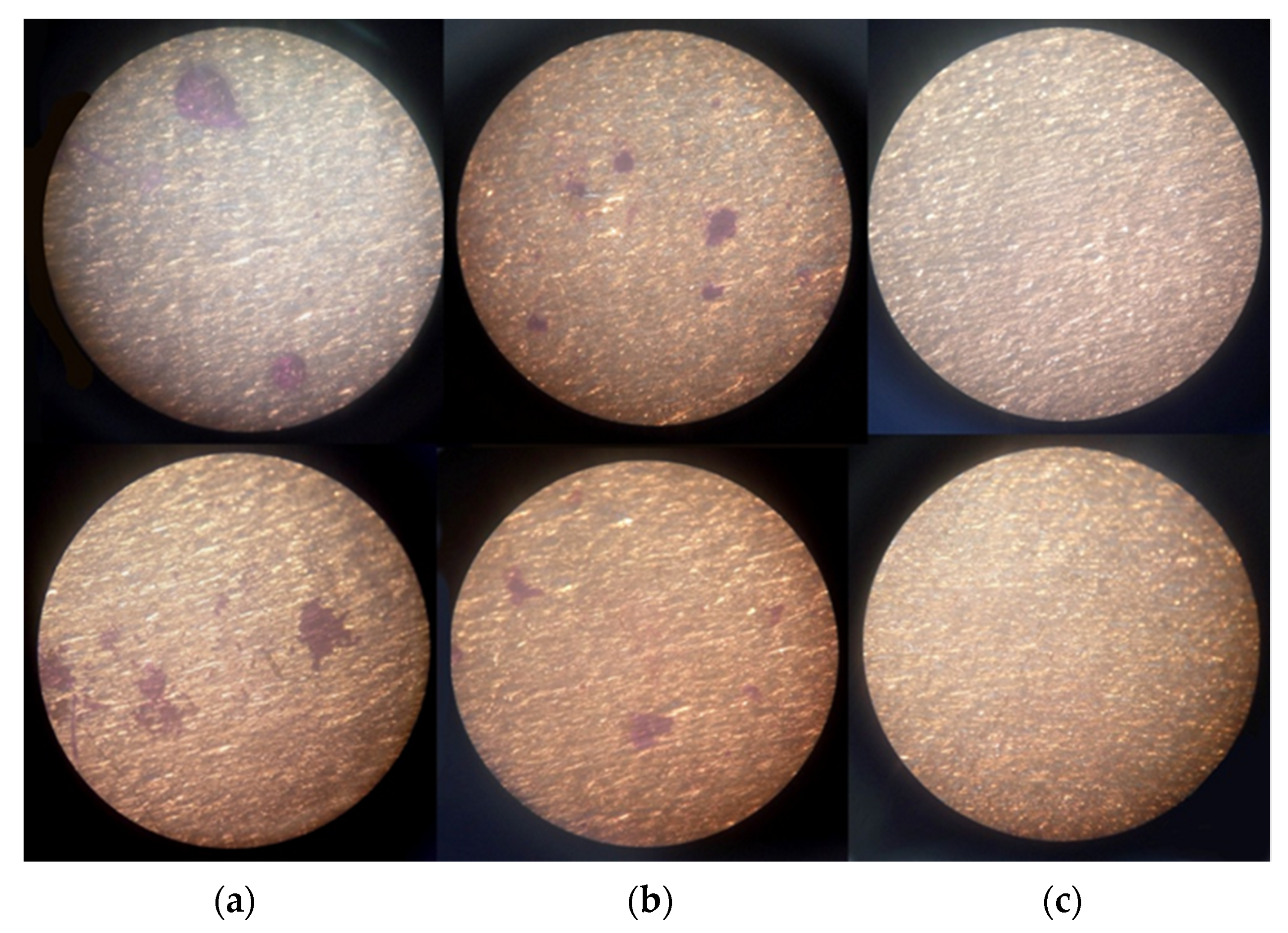
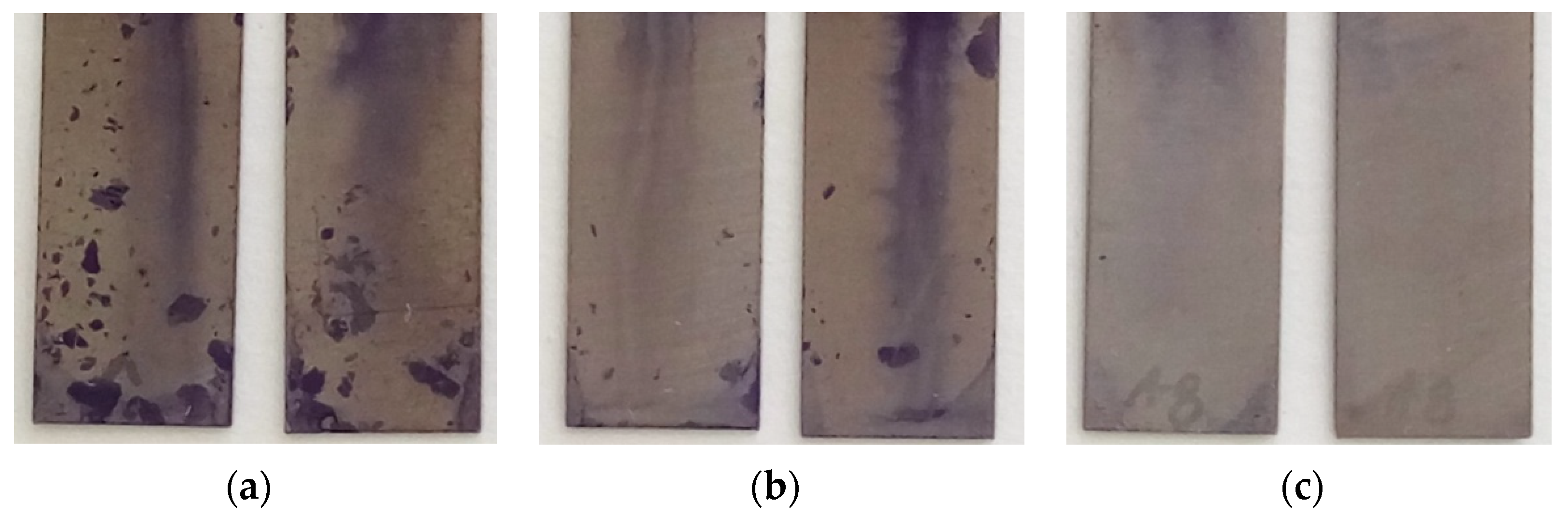
3.1.3. S-CLC Film Can Inhibit the P. aeruginosa Surface Biofilm Formation

The general results of the study are shown in Figure 3. Ti plates alone or with a covering of the CLC or S-CLC coating had no effect on the formation of microbial biofilms in the test cultures of microorganisms. The S-LCU coating almost completely inhibited the formation of biofilms of P. aeruginosa and S. aureus (Figure 3 and Figure 4; Table 3). The intensity of biofilm formation by the P. aeruginosa strain P-142 was significantly greater than that of the S. aureus strain, and the crystal-violet-colored biofilms on the surface of the samples could be seen with the naked eye (Figure 3 and Figure 4), and were confirmed by evaluation of the mass (m) of the crystal violet absorbed by the microbial biofilm (Table 3).

4. Discussion

Different antimicrobial covering materials of the Ti implants with a different efficiency were offered [6]. A Ti surface alone did not have antibacterial activities against S. aureus and E. coli [12]. Silver nanoparticles are one of the most common antimicrobial agents for covering of titanium implants [16]. Silver nanoparticles have broad-spectrum bactericidal activities, as confirmed in a range of studies [17]. The alternative of pure Ag+ nanoparticles can be composite materials. In recent decades, it has been shown that other newly found forms of carbons, including carbon nanotubes, graphene/graphene oxide, and fullerenes, have antimicrobial properties [18]. In this study, we showed antimicrobial and antibiofilm activity of the composite material S-CLC applied on a Ti surface, which could be used for treating a joint PPI.
The first assay showed that the S-CLC had antibacterial action only at a small distance of around 1 mm. The average microbial inhibitory concentration value of Ag nanoparticles against Gram-positive and -negative bacteria is around 2.1–4.27 μg/L [19,20]. According to this data, we could calculate the concentration based on the volume of the 1 mm culture media on our plate of 2.5 mL (0.05 m length × 0.05 m width × 0.001 m height), and the release of silver ions from S-CLC could be calculated at a level of 0.21–0.43 ng/cm2. Ag+ ions are weak acids with a pKa of 11.7. In water solutions, Ag+ ions form poor soluble AgOH, and can begin to precipitate at a pH of about 7–8. Bacteria in periprosthetic wounds can raise the pH, which leads to a decrease in the Ag+ ions’ solubility and their antibacterial properties, as well as the distance of action [21]. Thus, increasing the pH from 6.0 to 9.0 in a PPI wound requires about 2 times more silver ions to reduce growth of P. aeruginosa than that found in our in vitro study [22].
CLC alone showed moderate antimicrobial activities against P. aeruginosa and S. aureus, in contrast to diamondlike carbon nanoparticles, which had no effect on Staphylococcus epidermidis, Staphylococcus aureus, or Pseudomonas aeruginosa. S-CLC, compared to diamondlike carbon doped with Ag on the Ti implants, had similar antibacterial activities and antimicrobial effect to the tested antibacterial. Antimicrobial potency of the diamondlike carbon nanoparticle surfaces with embedded silver ions was positively correlated with embedded Ag concentrations. CLC is one of the most durable materials on the planet, and in a thin film, the stability of the coating will be determined by the underlying Ti material [23]. Using a combination of CLC and Ag+ can add additional mechanical strength to the coating compared to pure silver. Moreover, combination with Ag+ almost fully prevents biofilm formation.
There are no investigations about carbyne antibiofilm activity. It is possible only to compare the antibiofilm activity of S-CLC with other form of carbon similar to diamondlike nanoparticles with Ag+ nanoparticles. They also decrease biofilm formation [24]. This effect was similar to a silver nanoparticle covering alone, which was predicted to prevent biofilm formation. However, there was no antibacterial effect when diamondlike nanoparticles were used alone, as compared to that of CLC, which had low antibacterial activity [25]. Thus, S-CLC would have a similar effect on biofilm formation on titanium implants with Ag+, as compared to pure silver, with much smaller contents, a lower price, and wider application in titanium implants in orthopedics.
To clarify the benefits of the S-CLC covering on Ti implants in traumatology and orthopedic patients, additional clinical investigation will be needed.

5. Conclusions

The S-CLC film covering could successfully combat the most common periprosthetic joint infection agents, such as Staphylococcus aureus, Enterococcus faecalis, and P. aeruginosa. S-CLC released Ag+ ions locally only, and prevented microbial biofilm formation.

6. Patents

The intellectual property of S-CLC films on a Ti surface is licensed under patent RU 2 697 855 C1.

Author Contributions

Conceptualization, N.S.N.; methodology, L.I.M., N.S.N., E.V.P. and V.U.E.; software, V.U.E.; validation, L.I.M. and V.U.E.; formal analysis, L.I.M. and V.U.E.; investigation, L.I.M., N.N.P. and D.N.E.; resources, L.I.M. and N.N.P.; data curation, L.I.M., N.N.P., D.N.E. and V.U.E.; writing—original draft preparation, V.U.E.; writing—review and editing, L.I.M., N.S.N., E.V.P. and V.U.E.; visualization, V.U.E.; supervision, N.S.N.; project administration, N.S.N. and V.U.E.; funding acquisition, N.S.N. All authors have read and agreed to the published version of the manuscript.

Funding

This research received no external funding.

Institutional Review Board Statement

The study was conducted according to the guidelines of the Declaration of Helsinki, and approved by the Institutional Ethics Committee of Chuvash State University (009).

Informed Consent Statement

Informed consent was obtained from all subjects involved in the study.

Data Availability Statement

Data available on request.

Acknowledgments

We thank D.V. Tapalski for performing the experiment.

Conflicts of Interest

The authors declare no conflict of interest.

References

  1. Loeser, R.F.; Collins, J.A.; Diekman, B.O. Ageing and the pathogenesis of osteoarthritis. Nat. Rev. Rheumatol. 2016, 12, 412–420. [Google Scholar] [CrossRef] [Green Version]
  2. Maradit Kremers, H.; Larson, D.R.; Crowson, C.S.; Kremers, W.K.; Washington, R.E.; Steiner, C.A.; Jiranek, W.A.; Berry, D.J. Prevalence of Total Hip and Knee Replacement in the United States. J. Bone Jt. Surg. Am. 2015, 97, 1386–1397. [Google Scholar] [CrossRef] [PubMed] [Green Version]
  3. Sukeik, M.; Haddad, F.S. Periprosthetic joint infections after total hip replacement: An algorithmic approach. SICOT J. 2019, 5, 5. [Google Scholar] [CrossRef] [PubMed] [Green Version]
  4. Schwartz, A.M.; Farley, K.X.; Guild, G.N.; Bradbury, T.L., Jr. Projections and Epidemiology of Revision Hip and Knee Arthroplasty in the United States to 2030. J. Arthroplast. 2020, 35, S79–S85. [Google Scholar] [CrossRef]
  5. Corro, S.; Vicente, M.; Rodriguez-Pardo, D.; Pigrau, C.; Lung, M.; Corona, P.S. Vancomycin-Gentamicin Prefabricated Spacers in 2-Stage Revision Arthroplasty for Chronic Hip and Knee Periprosthetic Joint Infection: Insights Into Reimplantation Microbiology and Outcomes. J. Arthroplast. 2020, 35, 247–254. [Google Scholar] [CrossRef] [PubMed] [Green Version]
  6. Romano, C.L.; Tsuchiya, H.; Morelli, I.; Battaglia, A.G.; Drago, L. Antibacterial coating of implants: Are we missing something? Bone Jt. Res. 2019, 8, 199–206. [Google Scholar] [CrossRef] [PubMed]
  7. Hardes, J.; Ahrens, H.; Gebert, C.; Streitbuerger, A.; Buerger, H.; Erren, M.; Gunsel, A.; Wedemeyer, C.; Saxler, G.; Winkelmann, W.; et al. Lack of toxicological side-effects in silver-coated megaprostheses in humans. Biomaterials 2007, 28, 2869–2875. [Google Scholar] [CrossRef] [PubMed]
  8. Ferdous, Z.; Nemmar, A. Health Impact of Silver Nanoparticles: A Review of the Biodistribution and Toxicity Following Various Routes of Exposure. Int. J. Mol. Sci. 2020, 21, 2375. [Google Scholar] [CrossRef] [PubMed] [Green Version]
  9. Cataldo, F. Polyynes: Synthesis, Properties, and Applications; Taylor & Francis: Boca Raton, FL, USA, 2005; p. xvii. 506p. [Google Scholar]
  10. Green, J.B.; Fulghum, T.; Nordhaus, M.A. Review of immobilized antimicrobial agents and methods for testing. Biointerphases 2011, 6, CL2-43. [Google Scholar] [CrossRef] [PubMed] [Green Version]
  11. Cendra, M.D.M.; Blanco-Cabra, N.; Pedraz, L.; Torrents, E. Optimal environmental and culture conditions allow the in vitro coexistence of Pseudomonas aeruginosa and Staphylococcus aureus in stable biofilms. Sci. Rep. 2019, 9, 16284. [Google Scholar] [CrossRef] [Green Version]
  12. Kawakami, H.; Yoshida, K.; Nishida, Y.; Kikuchi, Y.; Sato, Y. Antibacterial Properties of Metallic Elements for Alloying Evaluated with Application of JIS Z 2801:2000. Isij Int. 2008, 48, 1299–1304. [Google Scholar] [CrossRef] [Green Version]
  13. Food, É.-U.; Administration, D.; Safety, C.F.F.; Nutrition, A. Bacteriological Analytical Manual (BAM); AOAC International: Gaithersburg, MD, USA, 2020. [Google Scholar]
  14. De Vries, T.A.; Hamilton, M.A. Estimating the Antimicrobial Log Reduction: Part 1. Quantitative Assays. Quant. Microbiol. 1999, 1, 29–45. [Google Scholar] [CrossRef]
  15. O’Toole, G.A. Microtiter dish biofilm formation assay. J. Vis. Exp. 2011, 30, 2437. [Google Scholar] [CrossRef] [PubMed]
  16. Gunputh, U.F.; Le, H.; Lawton, K.; Besinis, A.; Tredwin, C.; Handy, R.D. Antibacterial properties of silver nanoparticles grown in situ and anchored to titanium dioxide nanotubes on titanium implant against Staphylococcus aureus. Nanotoxicology 2020, 14, 97–110. [Google Scholar] [CrossRef]
  17. Lara, H.H.; Garza-Treviño, E.N.; Ixtepan-Turrent, L.; Singh, D.K. Silver nanoparticles are broad-spectrum bactericidal and virucidal compounds. J. Nanobiotechnol. 2011, 9, 30. [Google Scholar] [CrossRef] [PubMed] [Green Version]
  18. Azizi-Lalabadi, M.; Hashemi, H.; Feng, J.; Jafari, S.M. Carbon nanomaterials against pathogens; the antimicrobial activity of carbon nanotubes, graphene/graphene oxide, fullerenes, and their nanocomposites. Adv. Colloid Interface Sci. 2020, 284, 102250. [Google Scholar] [CrossRef] [PubMed]
  19. Abalkhil, T.A.; Alharbi, S.A.; Salmen, S.H.; Wainwright, M. Bactericidal activity of biosynthesized silver nanoparticles against human pathogenic bacteria. Biotechnol. Biotechnol.Equip. 2017, 31, 411–417. [Google Scholar] [CrossRef] [Green Version]
  20. Pisarcik, M.; Lukac, M.; Jampilek, J.; Bilka, F.; Bilkova, A.; Paskova, L.; Devinsky, F.; Horakova, R.; Brezina, M.; Opravil, T. Silver Nanoparticles Stabilized with Phosphorus-Containing Heterocyclic Surfactants: Synthesis, Physico-Chemical Properties, and Biological Activity Determination. Nanomaterials 2021, 11, 1883. [Google Scholar] [CrossRef] [PubMed]
  21. Casolaro, M.; Casolaro, I.; Akimoto, J.; Ueda, M.; Ueki, M.; Ito, Y. Antibacterial Properties of Silver Nanoparticles Embedded on Polyelectrolyte Hydrogels Based on alpha-Amino Acid Residues. Gels 2018, 4, 42. [Google Scholar] [CrossRef] [PubMed] [Green Version]
  22. Wiegand, C.; Abel, M.; Ruth, P.; Elsner, P.; Hipler, U.C. pH influence on antibacterial efficacy of common antiseptic substances. Skin Pharmacol. Physiol. 2015, 28, 147–158. [Google Scholar] [CrossRef]
  23. Liu, M.; Artyukhov, V.I.; Lee, H.; Xu, F.; Yakobson, B.I. Carbyne from First Principles: Chain of C Atoms, a Nanorod or a Nanorope. ACS Nano 2013, 7, 10075–10082. [Google Scholar] [CrossRef] [PubMed] [Green Version]
  24. Harrasser, N.; Jussen, S.; Banke, I.J.; Kmeth, R.; von Eisenhart-Rothe, R.; Stritzker, B.; Gollwitzer, H.; Burgkart, R. Antibacterial efficacy of titanium-containing alloy with silver-nanoparticles enriched diamond-like carbon coatings. AMB Express 2015, 5, 77. [Google Scholar] [CrossRef] [PubMed]
  25. Al-Ansari, M.; Alkubaisi, N.; Gopinath, K.; Karthika, V.; Arumugam, A.; Govindarajan, M. Facile and Cost-Effective Ag Nanoparticles Fabricated by Lilium lancifolium Leaf Extract: Antibacterial and Antibiofilm Potential. J. Clust. Sci. 2019, 30, 1081–1089. [Google Scholar] [CrossRef]
Figure 1. Titanium plates with no coating had either a CLC or an S-CLC coating placed on their surface, and coated plates were additionally coated with a second layer of medium so that the obtained thickness of medium on the plates was 1, 2, or 4 mm.
Osteology 01 00022 g001
Figure 2. Bacterial suspension kept on the titanium plates for 24 h, after solutions from the plates were sown on Petri dishes with using a microbiological loop.
Osteology 01 00022 g002
Figure 3. Formation of P. aeruginosa P-142 bacterial biofilms on the surface of Ti plates stained with 0.1% aqueous crystal violet solution: (a) no coating; (b) CLC coating; (c) S-CLC coating.
Osteology 01 00022 g003
Figure 4. Adhesion of S. aureus on the surface of Ti plates stained with 0.1% aqueous crystal violet solution: (a) no coating; (b) CLC coating; (c) S-CLC coating.
Osteology 01 00022 g004
Table 1. Antibacterial activity of various compositions against P. aeruginosa (ATCC 27853) by two-layer agar method.
Thickness of Growth Media Layer above the PlateNo CoverCLCS-CLC
1 mmHeavy growthHeavy growthNo growth
2 mmHeavy growthHeavy growthHeavy growth
4 mmHeavy growthHeavy growthHeavy growth
Table 2. Antibacterial activity of various compositions against P. aeruginosa (ATCC 27853) by two-layer agar method.
No CoverCLCS-CLC
RI%RI%RI%
S. aureus0.00.00.461.81.998.7
E. faecalis0.00.00.451.72.099.9
P. aeruginosa0.00.00.00.01.998.7
Table 3. The mass (m) of crystal violet absorbed by microbial biofilm (in μg) on Ti plates only and Ti plates covered with CLC or S-CLC.
No CoveringCLCS-CLC
Dye mass absorbed by biofilm, μg17.23.90.4
Dye mass absorbed by biofilm, μg2.80.60.1
Publisher’s Note: MDPI stays neutral with regard to jurisdictional claims in published maps and institutional affiliations.

