Article

Unveiling the Relationship between Structure and Anticancer Properties of Permethylated Anigopreissin A: A Study with Thirteen Analogues

1
Department of Science, University of Basilicata, Via dell’Ateneo Lucano, 10, 85100 Potenza, Italy
2
Department of Chemistry, Sapienza University of Rome, p.le Aldo Moro 5, 00185 Roma, Italy
*
Author to whom correspondence should be addressed.
These authors contributed equally to this work.
Organics 2024, 5(3), 237-251; https://doi.org/10.3390/org5030012
Submission received: 18 June 2024 / Revised: 8 July 2024 / Accepted: 29 July 2024 / Published: 1 August 2024

Abstract

:
Permethylated anigopreissin A (PAA), a fully protected form of the natural anigopreissin A, was found in our previous study to be active against several cancer cells, up to IC50 0.24 μM for HepG2 cells. Herein, a total of thirteen PAA analogues with variations in the number, position of substituents and unsaturation were synthesised starting from a common precursor, and their ability to induce cell growth inhibition was tested. By comparing the antiproliferative effect of the analogues with PAA and with the help of computational studies, we have gained valuable insights into both the biological activity and structure of this natural class of compounds. Indeed, we discovered the importance of the C-3 ring in modulating the biological activity of PAA, as well as the crucial role of the trans configuration of the styryl double bond and the significance of substitutions on the other parts of the molecule.

Graphical Abstract

1. Introduction

Stilbenoids, widely distributed in plants such as vegetables, fruits, teas, and some herbs, are recognised as essential dietary components contributing to the management of chronic inflammation [1]. Resveratrol and its oligomers, which belong to the stilbenoid class, are renowned for their diverse biological activities, encompassing anti-inflammatory, antioxidative, antimicrobial, and anticancer properties [2,3]. Several resveratrol dimers feature a benzofuran or dihydrobenzofuran core, along with a styryl moiety exhibiting either a trans configuration (e.g., anigopreissin A) or a cis configuration (e.g., ε-viniferin) [4], or an arylethyl substituent (e.g., gneafricanin D) [5]. Additionally, other dimers arise from the dimerisation of oxyresveratrol, piceatanol, or isorhapontigenin (e.g., bisisorhapontigenin B) [6] (Figure 1).
The significant interest in discovering new anticancer compounds has spurred us to advance our investigation of anigopreissin A and its analogues. Anigopreissin A, a dimer of resveratrol naturally occurring in Anigozanthos preissi, Musa Cavendish [7], and Macropidia fuliginosa, exhibits modest antimicrobial activity against S. aureus and S. pyogenes [8]. Furthermore, permethylated anigopreissin A [9] (PAA), representing a fully protected version of anigopreissin A, demonstrates compelling cytotoxicity against a wide spectrum of human tumour cell lines, including HepG2, MCF7, U937, and SHSY5Y.
PAA triggers cancer cell death by inducing cell cycle arrest in the G1 phase, likely mediated through the cyclin E/CDK2 complex. Notably, exposure to PAA leads to an up-regulation of the CDKN1B gene, a specific inhibitor of the cyclin E/CDK2 complex [10]. The molecular mechanisms underlying the death of PAA-treated cancer cells involve the intrinsic apoptotic pathway.
Our previous studies have shown that PAA exerts the most potent antiproliferative effect on hepatocellular carcinoma cell lines, specifically HepG2 and Alexander [10]. Importantly, its cytotoxic activity is also selective towards cancer cells, as non-tumorigenic cells such as HEK293 and human fibroblasts remain unaffected by PAA. A recent study suggests that PAA induces its effect in HCC cells via GDH1 inhibition, although other molecular mechanisms could be involved [11].
Moreover, PAA demonstrated higher activity compared to trimethylated resveratrol, as well as higher activity compared to simpler structures or synthetic precursors, which exhibited either no activity or lower activity than PAA [10].
To deepen our comprehension of the structure–activity relationship of PAA and to develop more potent cytotoxic compounds, it is crucial to construct various analogues. For this objective, our primary focus was on varying the number of methoxy groups on the aryl moieties and the degree of unsaturation of the ethylene bridge within the PAA scaffold. We opted to employ the same synthetic strategy used for PAA, with modifications limited to the sequence and adoption of common precursors as advised by the diversity-oriented synthesis (DOS) methodology [12].
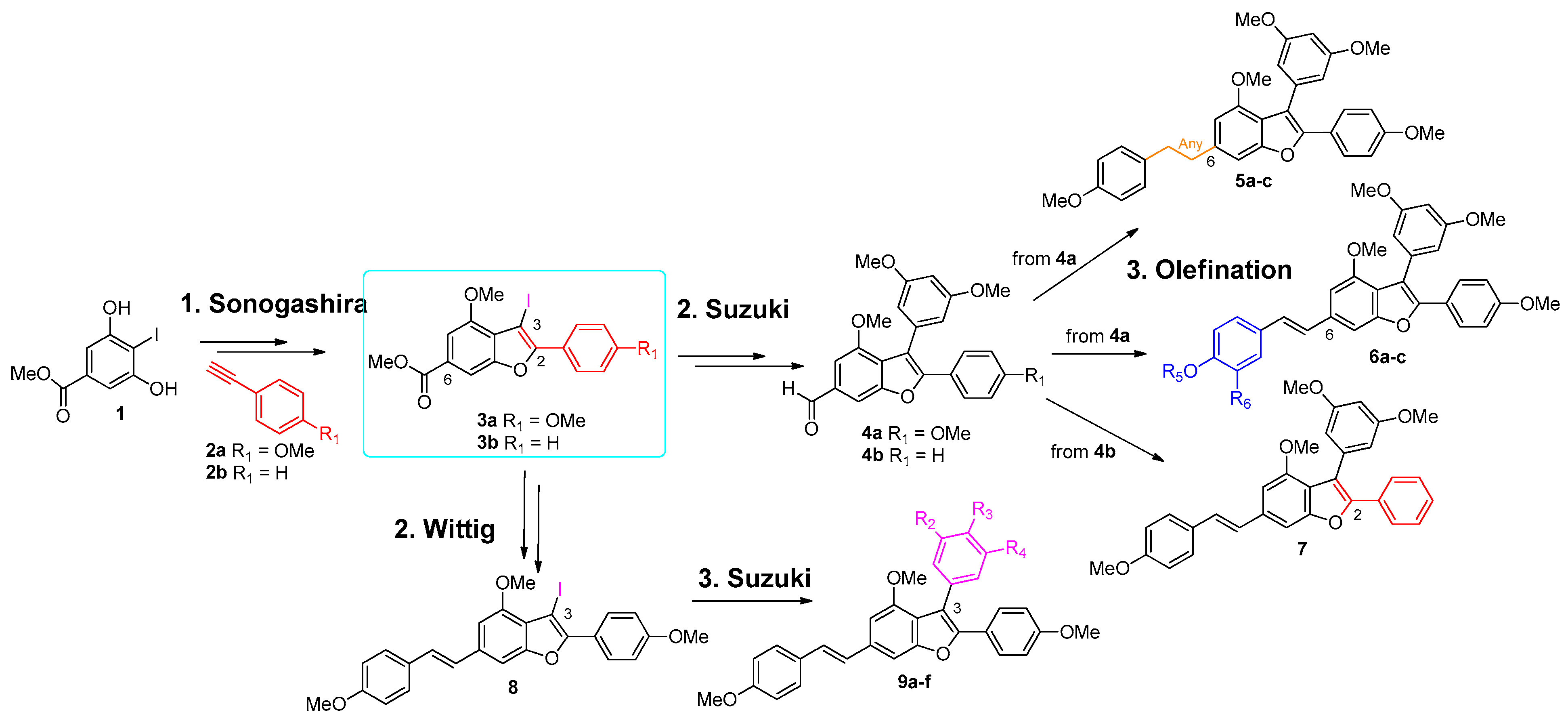
In this work, thirteen PAA analogues were efficiently synthesised using domino Sonogashira-heteroannulation reactions [13], olefination reactions (Wittig or Julia–Kocienski reactions), chemoselective reductions and Suzuki cross-couplings (Scheme 1). We decided to engage nearly fully permethylated derivatives since it is known that methylated resveratrol derivatives have shown a higher antiproliferative effect on cancer cells compared to their hydroxylated counterparts. This is likely due to their increased lipophilic properties that enhance uptake through the cell membrane [14] and due to the inhibition of metabolic conjugation reactions. To elucidate the structure–activity relationship of PAA, cell viability assays of all synthetised analogues were performed using PAA as the reference compound and computational studies were conducted on a selected number of analogues.

2. Materials and Methods

2.1. General Procedures

All reagents were supplied by Sigma-Aldrich, TCI, and AlfaAesar companies and were used without further purification unless otherwise stated.. All reactions were carried out in oven-dried glassware under an argon atmosphere unless otherwise noted. Flash chromatography was performed using 60–200 mesh silica gel. 1H NMR spectra were recorded on Varian 400 MHz and Varian 500 MHz spectrometers at room temperature with CDCl3 as the solvent unless otherwise noted. 13C NMR spectra were recorded on Varian 100 MHz and Varian 125 MHz spectrometers, all at room ambient temperature. Chemical shifts are reported in parts per million relative and referenced internally to the residual solvent resonances: 1H NMR spectra to CDCl3 at δ 7.26 and 13C NMR spectra to CDCl3 at δ 77.0. Data for 1H NMR are reported as follows: chemical shift, multiplicity (s = singlet, d = doublet, t = triplet, q = quartet, and m = multiplet), coupling constants (in Hertz), and integration. Mass spectra were obtained using FT-ICR/MS by using ESI in the positive ion mode.

2.1.1. General Procedure of Wittig Olefination for the Preparation of Compound 5a, 6b, 6c, 7, and 8

To a stirred solution of triphenyl arylphosphonium bromide 10 ((4-methoxybenzyl)triphenylphosphonium bromide) or 16 ((3,4-dimethoxybenzyl)triphenylphosphonium bromide) or 17 ((4-((tert-butyldimethylsilyl)oxy)-3-methoxybenzyl)triphenylphosphonium bromide) (0.85 mmol, 1.1 equiv.) in i-PrOH (20 mL), LiBr (0.224 g, 2.61 mmol, 3.4 equiv) and LiOH∙H2O (0.058g, 1.39 mmol, l.8 equiv) were added at room temperature. After 15 min, a solution of 4a (3-(3,5-dimethoxyphenyl)-4-methoxy-2-(4-methoxyphenyl)benzofuran-6-carbaldehyde) or 4b (3-(3,5-dimethoxyphenyl)-4-methoxy-2-phenylbenzofuran-6-carbaldehyde) or 34 (3-iodo-4-methoxy-2-(4-methoxyphenyl)benzofuran-6-carbaldehyde) (0.76 mmol, 1 equiv.) in i-PrOH (10 mL) was added to the mixture and heated to reflux. After 3 h, the mixture was quenched with H2O and extracted with EtOAc (3 × 40 mL). The combined organic layers were dried over Na2SO4, filtered, and concentrated under reduced pressure. The cis/trans crude mixture of product 5a was purified by column chromatography (petroleum ether/EtOAc 9:1), avoiding light, while the crude compounds 6b, 6c, 7, and 8 were firstly isomerized with catalytic amounts of I2 in heptane/DCM (7:3) at room temperature in the dark for one night and then purified by column chromatography.

2.1.2. Reduction of the Styryl Double Bond for Preparation of Compound 5b

To a stirred solution of PAA (21.1 mg, 0.0404 mmol) in dry THF (0.400 mL), 2-nitrobenzensulfonyl chloride (20.3 mg, 0.0916 mmol) was added. The reaction mixture was cooled to 0 °C, and then hydrazine (16 μL, 0.326 mmol) was added. After stirring for 5 days at room temperature, the reaction mixture was quenched by dilution with AcOEt. The organic phase was washed with a saturated aqueous solution of NH4Cl (2 × 5 mL), dried over Na2SO4 and concentrated under reduced pressure. The crude was purified by column chromatography on silica gel (EP/AcOEt, 9:1). Compound 5b was isolated (11.6 mg, 55% yield) as a pale yellow solid.

2.1.3. Julia–Kocienski Reaction for Preparation of Compound 6a

To a solution of 4-((benzo[d]thiazol-2-ylsulfonyl)methyl)phenyl tert-butyl carbonate 14 (0.121 g, 0.3 mmol) in dry THF (0.1M) at −78 °C, a solution of KHMDS in toluene (1M) (0.3 mmol) was added and the mixture was stirred for 30 min. A solution of aldehyde 4a (0.084 g, 0.2 mmol) in THF (0.3M) was cannulated into the reaction mixture. The reaction mixture was allowed to warm to room temperature. After 8 h, it was quenched by adding a saturated aqueous solution of NaHCO3 and extracted with Et2O. The organic phases were washed with brine, dried over Na2SO4, and concentrated under reduced pressure. The crude mixture cis/trans of the reaction was isomerized by using a catalytic amount of I2 in heptane/DCM 7:3 for a night. The crude mixture was then purified by column chromatography on silica gel (petroleum ether/Et2O 8:2), affording 15 (72%, 0.088 g, 0.14 mmol) as a pale yellow solid.

2.1.4. General Procedure for the Suzuki Reaction for the Preparation of Compounds 33, 9a9f

To a solution of iodide 3b or 8 (1.14 mmol, 1.0 equiv.) in DMF/H2O (4:1, 22.8 mL), aryl boronic acid (1.6 mmol, 1.4 equiv.), NaHCO3 (0.133 g, 1.6 mmol), and PdCl2(PPh3)2 (0.040 g, 0.057 mmol) were added. The solution was stirred for 10 min at room temperature and then heated at 80 °C for 7 h. After cooling, the mixture was diluted with AcOEt and washed with a saturated aqueous solution of NH4Cl, brine, and water. The organic layer was dried over Na2SO4, filtered, and concentrated under reduced pressure. The crude was purified (silica gel; petroleum ether/AcOEt, 9:1) to afford the desired product 33, or 9a9f.

2.2. Cell Culture, Treatments, and Viability Assay

HepG2 cells were cultured as previously described [11]. Alexander cells were grown in Roswell Park Memorial Institute (RPMI) 1640 Medium (Thermo Fisher Scientific, San Jose, CA, USA) supplemented with 10% foetal bovine serum, 2mM L-glutamine, 100 U/mL of penicillin, and 100 µg/mL of streptomycin at 37 °C in a humidified atmosphere containing 5% CO2. Cells were screened periodically for mycoplasma contamination.
PAA and the thirteen analogues were dissolved in 100% dimethyl sulfoxide (DMSO) (Sigma-Aldrich, St Louis, MO, USA) and diluted in a complete culture medium to obtain a final concentration of 0.1% DMSO in the cells.

2.2.1. Cell Viability Assay

Cell viability was determined by colourimetric 3-(4,5-dimethylthiazol-2-yl)-2,5-diphenyltetrazolium bromide (MTT) assay. The cells (5 × 10⁴) were seeded in a 96-well plate and, 24 h later, were treated with PAA or PAA analogues at increasing concentrations (10 nM, 100 nM, 1 μM, 10 μM, and 100 μM) for 72 h. The control groups received DMSO equivalent to the maximum percentage of <0.1% solvent used in the experimental settings. At the end of the treatments, an MTT assay was performed as described in [15]. The optical density was determined at 570 nm with a GloMax® Discover Microplate Reader (Promega, Madison, WI, USA).

2.2.2. Statistical Analysis

The statistical analysis was performed by GraphPad Prism software (La Jolla, CA, USA). The results are presented as mean ± standard deviation (SD) from three independent experiments with four replicates in each. Data were analysed using one-way ANOVA followed by Dunnett’s multiple comparison test. The asterisks in the figures denote statistical significance (* p < 0.05, ** p < 0.01, and *** p < 0.001).

2.3. Computational Details

All the computations were performed with the Gaussian 09 software, version C01 [16] (G09 in the following). The M06 xc functional [17] was used in all the reported computations. The integration grid was set to the “ultrafine” level (as implemented in G09). SCF procedures and structure optimizations were performed with the default convergence thresholds as set in G09.
For the research of the most stable conformers of PAA—9a, 9b, and 9c—the 6-31G(d) basis set [18] (as implemented in G09) was used for a quicker investigation of the potential energy surface. The more accurate cc-pVTZ basis set [19] (as implemented in G09) was used for refining the structures and their electronic structures. All the data reported in Table S1 and Figures S4 and S5 were extracted from the cc-pVTZ-based computations.
The atomic charges in Table S1 (see Section S9 in Supporting Information) are Mulliken’s well-known atomic charges and the natural charges obtained by the NBO-based electron density analysis, known as natural population analysis (NPA) [20]. In the last case, the NPA implementation inside G09 was used with the default options (“population npa” option).
Graphical representations of computational studies were prepared through the MolDen 5.7 program [21].

3. Results and Discussion

3.1. Synthesis of the Thirteen Analogues

As depicted in Scheme 1, the thirteen analogues were prepared using two key building blocks, 4 and 8, synthetised alternatively by Suzuki arylation or by Wittig olefination of 3 derivatives. On these compounds, the olefination or the Suzuki reaction was performed in order to insert the desired structural motif.

3.1.1. Synthesis of Analogues 5ac and 6ac

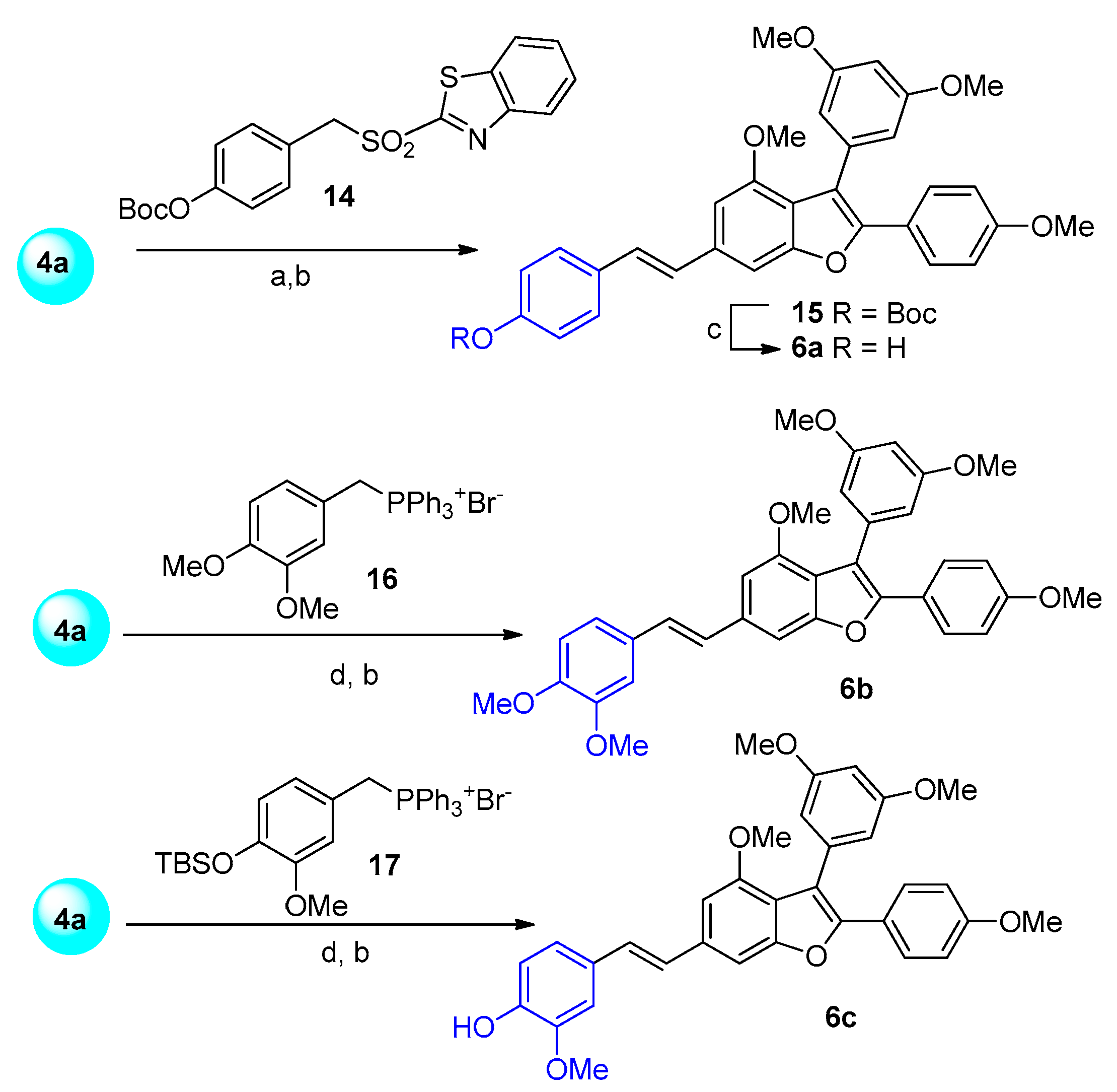
To decipher the significance of the trans double bond in the PAA biological activity, we constructed the first series of analogues containing various linkages between the benzofuran portion and the p-methoxybenzene: a cis double bond (5a), a fully saturated bridge (5b), or a triple bond (5c). The synthesis of these new derivatives was carried out following the procedure outlined in Scheme 2. The key intermediate 4a was prepared according to our previously reported strategy [9]. Then, it was involved in the Wittig olefination reaction with the phosphonium salt 10, LiOH, and LiBr in i-PrOH to afford PAA as a mixture of cis/trans isomers in a 1:1 ratio [22]. Compound 5a, representing the cis isomer of PAA, was isolated from the mixture via column chromatography. Subsequently, the trans isomer, PAA, was chemoselectively reduced to compound 5b using o-NO2PhCl and hydrazine hydrate in THF [23].
The derivative 5c, featuring a triple bond, was synthesised starting from the same aldehyde 4a, which was converted into the corresponding dibromovinyl compound 11 using PPh3 and CBr4 in DCM [24]. Subsequently, compound 11 was transformed into the alkyne 12 with Cs2CO3 in DMSO [25] at 110 °C, yielding compound 12 (50%, two steps). Compound 12 was then subjected to a Sonogashira reaction with p-methoxy-phenyl iodide 13, leading to compound 5c (40%).
The second class of compounds (6a6c) was designed to assess the impact of the substituents on the styrene ring, and again, compound 4a was the key intermediate. Among them, compound 6a features a hydroxyl group on the aryl of the styryl moiety instead of the methoxy group found in PAA, while compounds 6b and 6c contain an additional methoxy group in the ortho position to the hydroxyl or methoxyl group of 6a or PAA, respectively. The analogue 6a, bearing a free hydroxyl group, was prepared via the Julia–Kocienski olefination using the heteroaryl-sulfone 14 (Scheme 3).
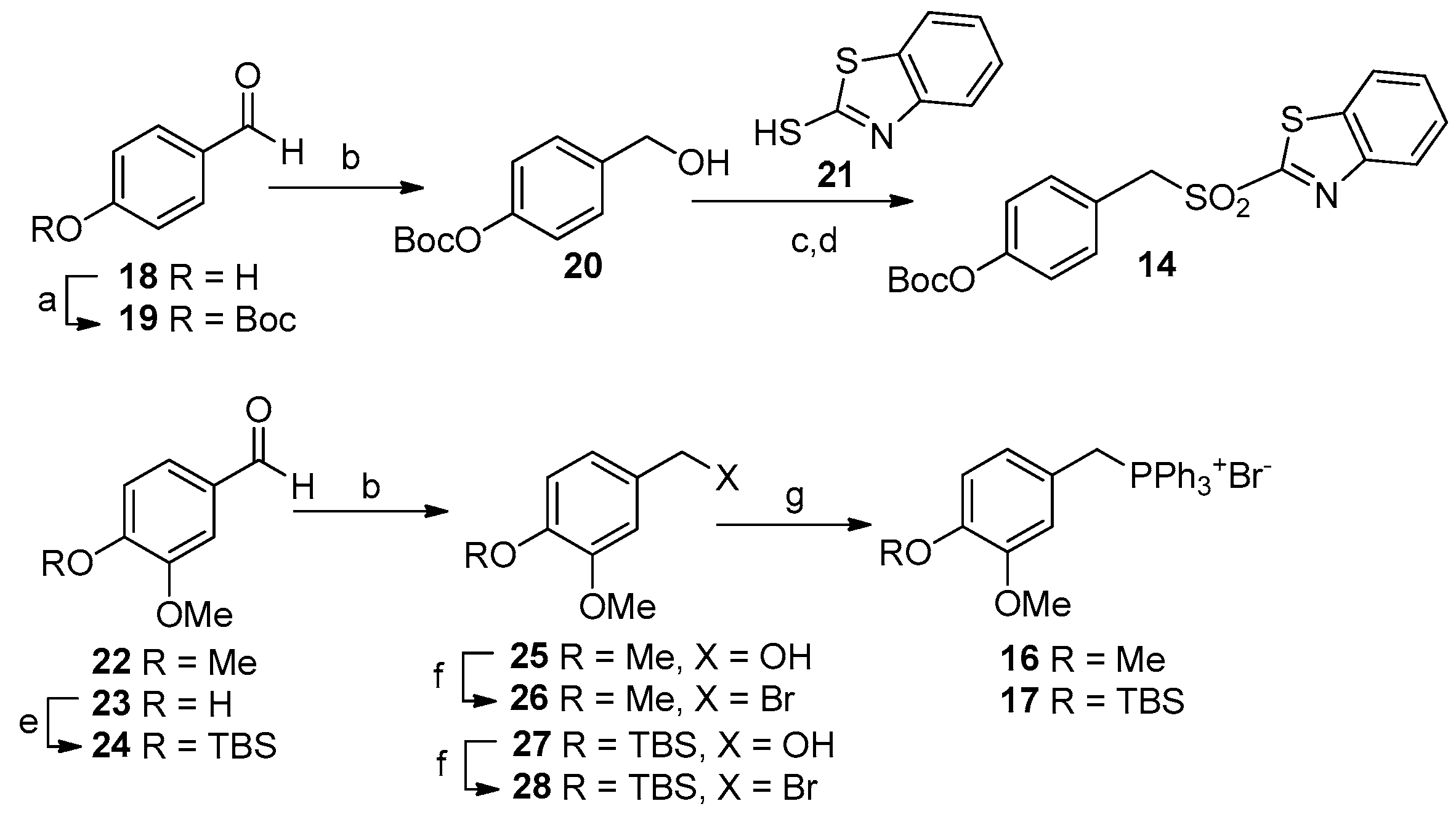
Attempts to carry out the Wittig olefination with phosphonium salts bearing a TBS or a Boc [26] protecting group resulted in very low yields. Consequently, the Julia–Kocienski reaction was selected as the preferred olefination method. The synthesis of heteroaryl-sulfone 14 started from the commercially available aldehyde 18, which was first protected as the Boc carbonate 19 [27] and then reduced to alcohol 20 using NaBH4 [26] (as depicted in Scheme 4). The benzylic alcohol, by the corresponding tosylated [25], was converted into the sulphide during the nucleophilic substitution with mercaptobenzothiazol 21. The obtained BT-sulphide was then oxidised quantitatively to sulfone 14 with mCPBA [28]. The Julia–Kocienski olefination employing KHMDS as the base furnished olefin 15 [29] as a mixture of cis/trans isomers (approximately 1:1 ratio), which was then isomerized by iodine to the trans isomer 15 in 72% yield. Finally, the removal of the Boc group with CBr4 and PPh3 in MeOH afforded compound 6a in a quantitative yield (Scheme 3) [30].
The preparation of compounds 6b and 6c was performed employing the classical Wittig protocol, where phosphonium salts 16 and 17 were prepared starting from the commercially available aldehydes 22 and 23 (Scheme 4).
Aldehyde 22 was directly reduced with NaBH4 to benzylic alcohol 25 [31] in a 73% yield, which was then transformed into bromide 26 using NBS and PPh3 in DCM [32] in a 47% yield. Vanillin 23 was first protected by a TBS group and subsequently reduced to benzylic alcohol 27 [33] (a 79% yield over two steps). Alcohol 27 was then converted into bromide 28 [32] with a 49% yield. Both bromides were treated with PPh3 in toluene to form the phosphonium salts 16 and 17 in 96% and 88% yields, respectively. The final Wittig olefination and isomerization furnished exclusively trans isomers of compounds 6b (in a 44% yield over two steps) and 6c (in a 34% yield over two steps with the simultaneous removal of the protecting group) (Scheme 3).

3.1.2. Synthesis of Analogues 7 and 9af

The third class of designed analogues features modifications at the C2 position.
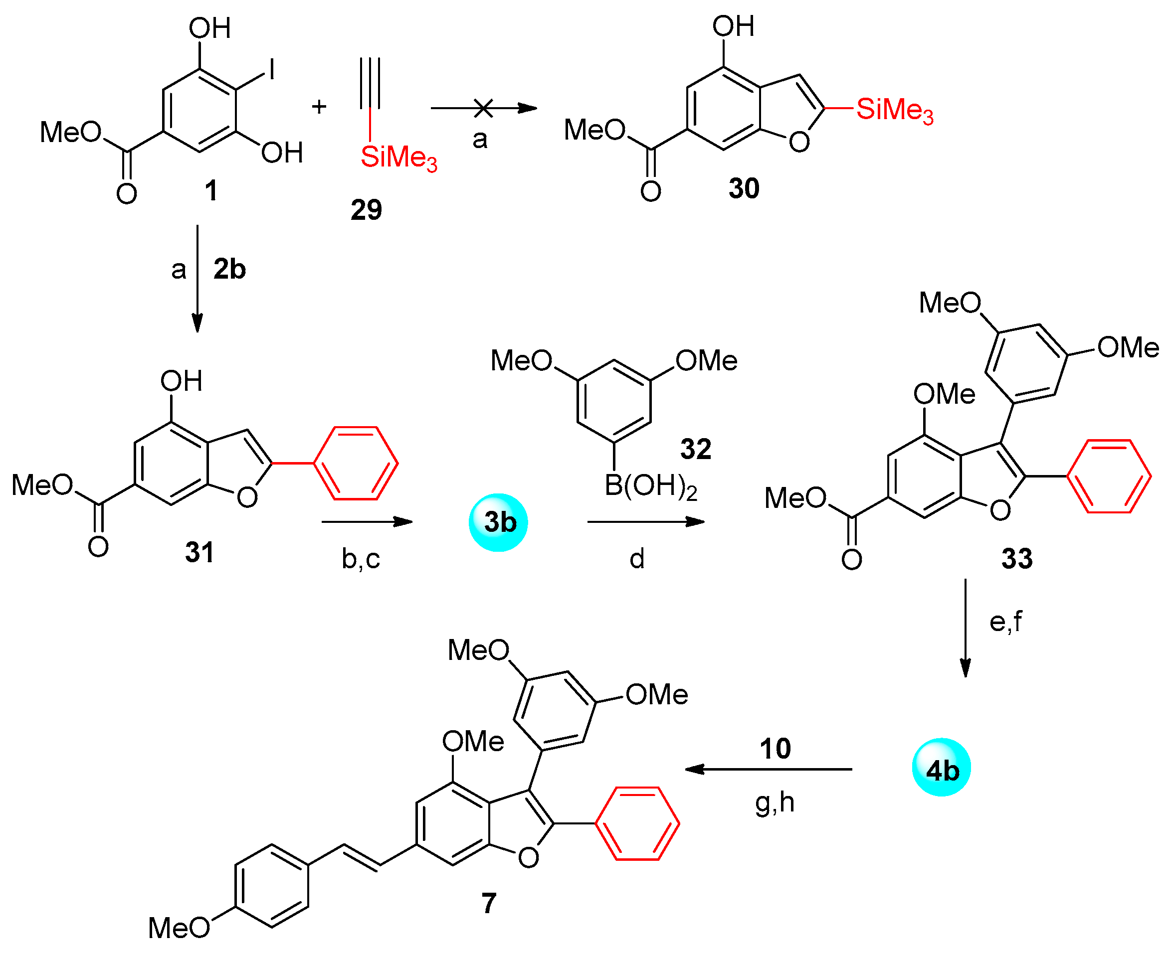
Our initial synthetic strategy for these compounds involved performing a direct arylation or Suzuki coupling on an advanced precursor utilising iodide 1 and trimethylsylyl acetylene 29 in the Larock cyclisation [13,34] (Scheme 5). However, this transformation did not yield the desired benzofuran derivative 30 but instead produced its open and irrelevant structural isomer.
Nevertheless, to understand the influence of the para-methoxy group of the C2 aryl ring on PAA’s biological activity, we decided to follow the complete sequence of reactions used for PAA’s synthesis to prepare the analogue 7 bearing a simple phenyl ring. Indeed, iodide 1 [9] was used in the domino Sonogashira-hetero-annulation reaction with phenylacetylene 2b, affording the desired benzofuran 31 in a good yield (85%).
Quantitative methylation followed by iodination produced 3b (Scheme 5), which was then used in the Suzuki reaction with boronic acid 32 to furnish the cross-coupled product 33 in a 75% yield. This product, reduced to aldehyde 4b, was used in the Wittig olefination and isomerisation with the standard conditions to afford compound 7 in an overall good yield (Scheme 5).
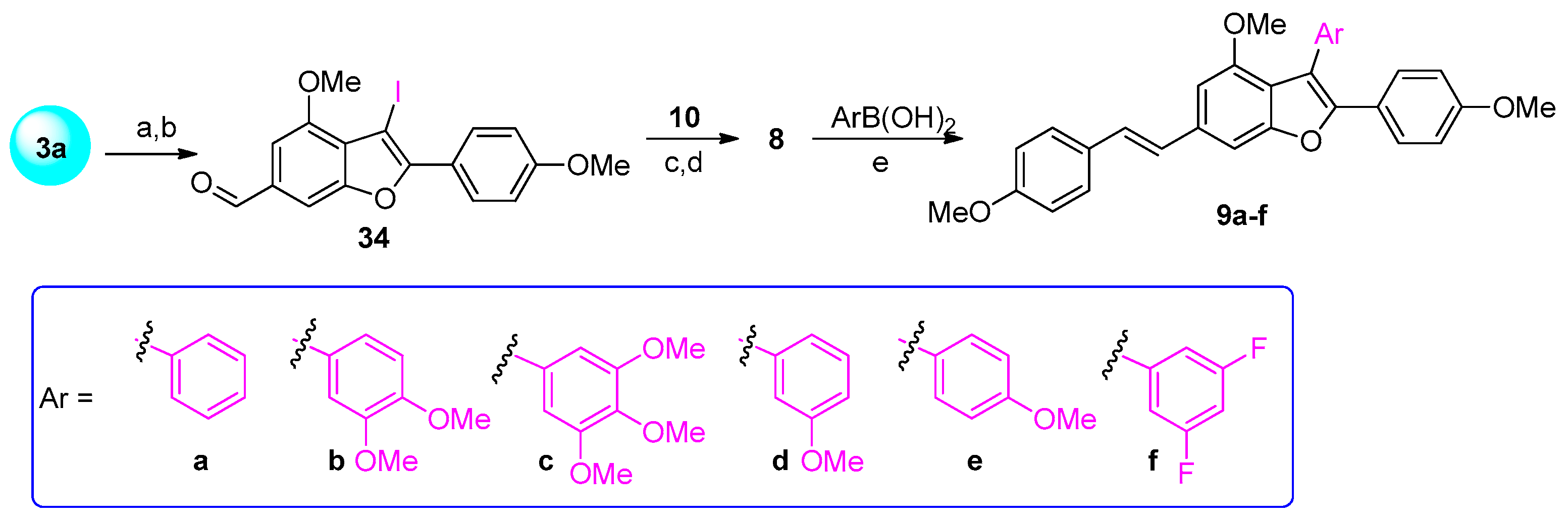
Finally, to investigate the effect of the substituents at the C-3 position, we prepared analogues with different aryl moieties at C-3 (compounds 9af). The synthesis of these analogues was performed using the intermediate iodide 8 in Suzuki cross-coupling reactions with variously substituted aryl boronic acids (Scheme 6).
Intermediate 8 was prepared starting from iodide 3a [9], which was chemoselectively reduced to the benzylic alcohol by DIBAL-H and then oxidised by DMP to the corresponding aldehyde 34 (90% yield over two steps). The usual Wittig reaction with phosphonium salt 10 furnished the trans isomer 8 (66% yield) after the isomerisation of the resulting cis/trans mixture. In the final step, compounds 9af were obtained through Suzuki reactions, introducing either a phenyl (9a), a 3,4-dimethoxybenzene (9b), a 3,4,5-trimethoxybenzene (9c), a 3-methoxybenzene (9d), a 4-methoxybenzene (9e), or a 3,5-difluorobenzene (9f) group at the C3-position of compound 8 in 56-86% yields.

3.2. Antiproliferative Effect of the 13 Analogues Compared to PAA

3.2.1. Antiproliferative Effect on HepG2 Cells

After their synthesis and purity determination by NMR spectroscopy (>99%), the antiproliferative effect of the PAA derivatives was evaluated on the hepatocellular carcinoma HepG2 cell line using the MTT assay. To this end, HepG2 cells were exposed to a broad spectrum of concentrations, ranging from 10 nM to 100 μM, of PAA or PAA analogues for 72 h. Among the thirteen new compounds tested, only compounds 9a and 9f exhibited significant cytotoxic activity against HepG2 cells (Figure 2). Specifically, analogue 9a, which features a phenyl ring at the C3 position, reduced cell viability by approximately 50% at concentrations of 10 μM and 100 μM, although this effect was less pronounced than that of PAA (Figure 2). In contrast, compound 9f, which has fluorine atoms on the C3 aryl ring, showed a variable response, with a slightly proliferative effect at a 10 nM concentration and a cytotoxic activity of about 30% at the highest concentration tested. The other PAA analogues did not demonstrate significant antiproliferative effects up to 100 μM (Figure 2). Since all these new compounds were less active or completely inactive than PAA, assays on healthy cells were not performed.

3.2.2. Antiproliferative Effect on Alexander Cells

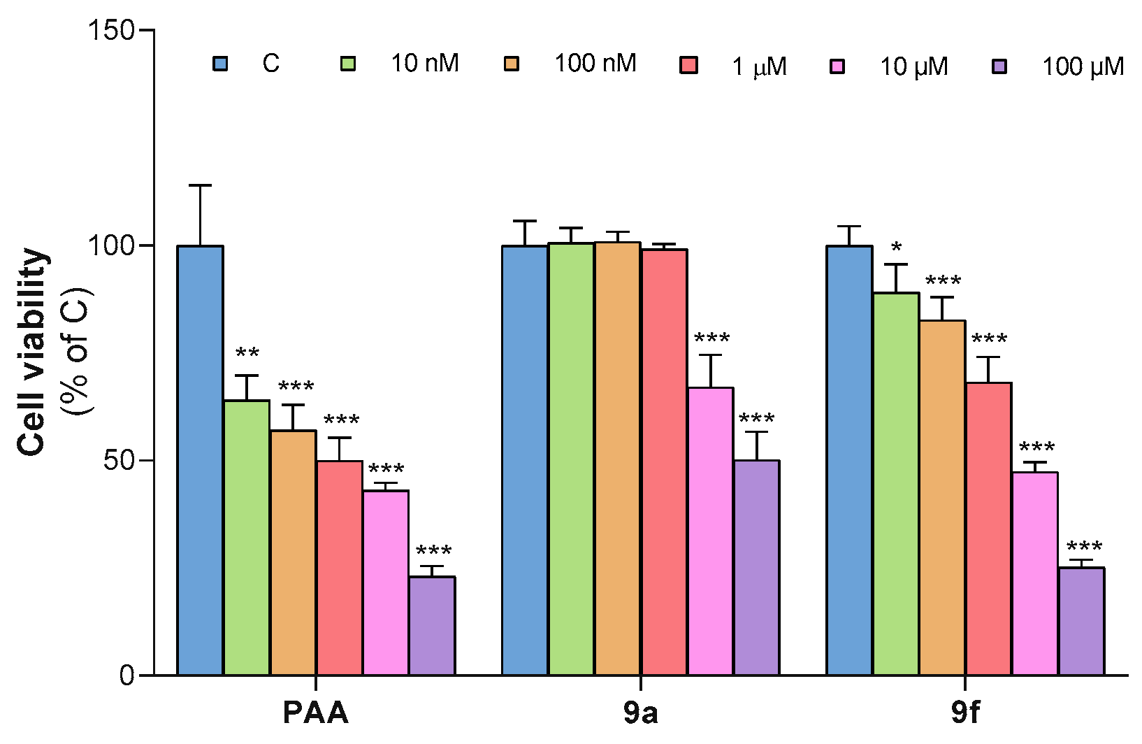
We also tested the most active analogues, 9a and 9f, against the Alexander cell line, which consists of epithelial cells isolated from the liver of a donor with hepatoma. The cytotoxic activity of 9a and 9f, along with PAA used as a reference, was assessed using the MTT assay at different concentrations (ranging from 10 nM to 100 μM), as illustrated in Figure 3.
Interestingly, both 9a and 9f exhibited cytotoxic activity against the Alexander cells but in different ways. Specifically, 9a was active only at the higher concentrations of 10 and 100 μM, where cell viability was reduced by about 30% and 50%, respectively. In contrast, analogue 9f was more active than 9a, with a 12% reduction in cell viability, starting at a concentration of 10 nM and reaching 50% and 75% reduction at 10 μM and 100 μM, respectively. However, both analogues were less active than PAA, which achieved a 40% reduction in cell viability just at the low concentration of 10 nM.

3.3. Computational Studies

3.3.1. Structure Optimisations

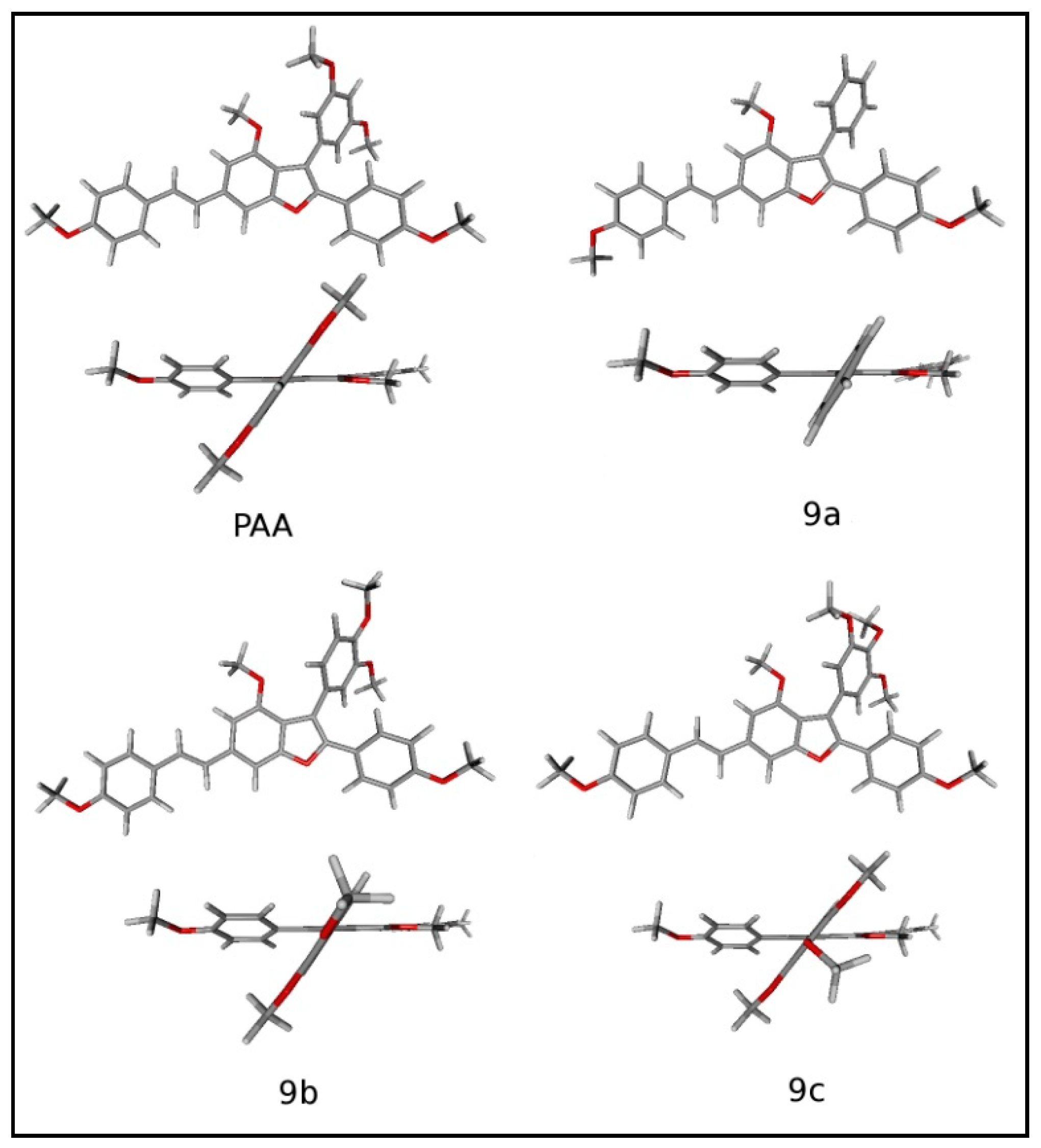
Computational studies were aimed at understanding the 3D structure of the PAA analogues synthesised in this article and looking for the interaction between the structure and relative electron density on the one hand and the biological activity on the other (see “Computational details” in Materials and Methods, Section 2.3, for a description of the used methods). In this regard, we decided to limit such studies to PAA, 9a (with an unsubstituted phenyl at C-3), 9b, and 9c (both bearing a para-methoxy group). PAA was chosen as a reference compound due to its high antiproliferative effect. Additionally, 9a, 9b, and 9c were chosen because of their different C-3 substituents and their related different biological activities. Indeed, based on our empirical results, the functionalisation of C-3 is essential to distinguish between active compounds (e.g., 9a and 9f) and inactive ones (9b and 9c).
A graphical view of the obtained structures is shown in Figure 4. All the compounds present the benzofuran core, the styryl substituent at C-6, and the para-methoxyphenyl group at C-2 in a substantially coplanar arrangement. In contrast, the phenyl group at C-3 is significantly rotated relative to the rest of the molecular structure. The dihedral angle between the average planes of the benzofuran core and the C-3 phenyl group is computed to be approximately 60 degrees in all four studied compounds.

3.3.2. Conformational Study and Electronic Properties

A conformational study was performed for PAA, 9a, 9b, and 9c to research alternative structures in which the phenyl group at C-3 is coplanar with the benzofuran core. These attempts did not lead to new stable conformers, suggesting a strong energetic preference for the not-planar orientation of the phenyl group at C-3. Such a preference is unaffected by the presence of the methoxy groups on this phenyl ring. The inability to arrange both the phenyl groups at C-2 and C-3 in a coplanar manner is likely due to steric strain occurring between the two rings. Hence, from our studies, we can conclude that only one phenyl group can be coplanar with the benzofuran core, and evidently, the ring at C-2 is energetically favoured.
The fact that the phenyl group at C-3 is neatly rotated in the studied compounds suggests that no significant differences should be expected in terms of conjugative effects between the benzofuran moiety and the phenyl ring when comparing PAA to 9ac.
In this respect, atomic charges for the oxygen and carbon atoms of the benzofuran core were computed in the series of studied compounds. The obtained Mulliken and natural charges (based on the NBO analysis) are reported in Table S1 of the Supporting Information. From the data, it is evident that no significant changes were observed when comparing PAA with 9ac. Indeed, it is reasonable to state that the different substitutions on the phenyl ring at C-3 do not induce significant changes in the electronic density of the remaining molecular structure.
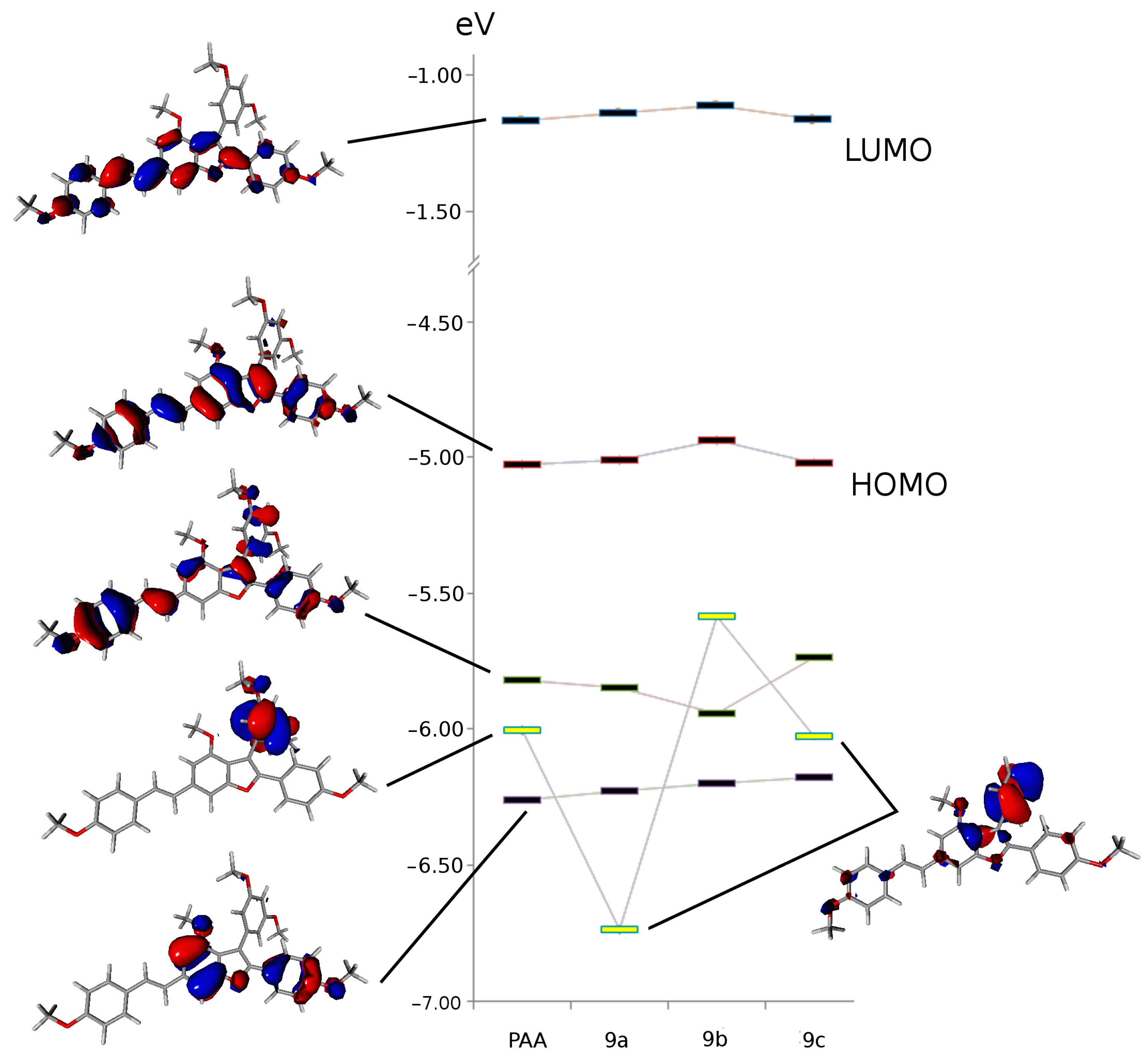
A substantial invariance was also observed in the molecular orbitals (MOs) close to the HOMO and LUMO in terms of MO shapes and energies. Figure 5 shows the MOs calculated in PAA and 9ac. Interestingly, all the reported MOs are mainly localised on the coplanar part of the molecule, and their energy levels are in black in the figure, with the exception of one MO, which is primarily localised on the phenyl group at C-3 (the yellow level). The MO shapes are shown for PAA as an example in Figure 4.
It is evident that the first type of MO (black levels) exhibits substantial invariance in terms of orbital energy in the series of compounds. This finding is mirrored by a significant invariance in the MO shape across the series of the four compounds. In contrast, and as expected, the MO primarily localised on the phenyl ring at C-3 (yellow levels) shows big changes in both shape and energy.

3.4. Structure–Properties Relationship Conclusion

Based on the biological assays and computational studies, we can gain valuable information about the PAA biological activity. Indeed, we can assess the following:
  • The trans configuration of the double bond is essential for the cytotoxicity of PAA, as the cis isomer (5a) and the analogues with the ethylene bridge (5b) or a triple bond (5c) were inactive.
  • The presence of only one methoxy group in the para position on the styryl ring is important since additional methoxy substituents (as in 6b and 6c) or a hydroxyl group instead of the methoxy group all resulted in inactive compounds.
  • The para-methoxy substituent on the C-2 ring is fundamental for the biological activity of PAA since its absence (as in analogue 7) resulted in an inactive species.
  • The C-3 ring is the only portion of the molecule that can be modulated to produce other active species, such as compound 9c, with a plain unsubstituted phenyl ring, or analogue 9f, with two fluorine atoms, even if both analogues were less active than PAA.
Moreover, through computational studies, we have learned that PAA has a rigid and planar structure, with the C-3 phenyl ring being the only exception. This ring is out of the plane with a 60° dihedral angle relative to the rest of the molecule. We also computed a conformational invariance between PAA and 9ac analogues, all of which have a planar structure except for the C-3 ring, which is about 60° out of the plane, regardless of its substitution.
Due to the lack of electronic conjugation between the C-3 phenyl ring and benzofuran core, no significant changes were computed in terms of electronic density and, in general, the electronic properties of the coplanar core of the studied molecules. However, the electronic structure of the C-3 phenyl ring changes significantly from one compound to another. Therefore, the measured changes in the antiproliferative activities in the four studied compounds should be associated with the direct effects of the C-3 phenyl rather than an indirect perturbation on the rest of the molecule. In this respect, and on an empirical basis, we can highlight that the presence of the -OMe substituent in the para position (as in 9b and 9c) is completely deleterious for the desired biological activity of the PAA derivatives. The presence of two OMe groups in the meta positions improves the antiproliferative effect (PAA compared to 9a) but does not seem to be strictly necessary.
Moreover, compound 9f showed higher cytotoxic activity than 9a, especially against the Alexander cell line. This can be explained by the similar size of hydrogen and fluorine atoms, making 9a and 9f structurally similar. However, the fluorine atoms, unlike hydrogen atoms, exert both inductive and mesomeric effects on the phenyl C-3 ring, resulting in a greater influence on the overall structure [35]. Due to its high electronegativity, F can affect the lipophilicity/hydrophilicity balance of the compound. Moreover, likewise to the methoxy moiety, F is a moderate hydrogen acceptor, so the two fluorines in compound 9f interact with the biological target in a similar manner to the two methoxy groups of the C-3 ring of PAA. In the end, it can be affirmed that the phenyl C-3 ring seems to be fundamental for the antiproliferative activity [10], as well as the nature and the position of the substituents on the ring.

4. Conclusions

In summary, thirteen new PAA derivatives were synthesised by using different synthetic strategies. In vitro MTT assays were performed to evaluate the cytotoxic activity of the synthetised compounds. The biological results clearly demonstrated that the overall structure of PAA plays a critical role in cell viability. The trans stereochemistry of the double bond is essential for activity, as the cis isomer (5a) was inactive. Neither the extreme rigidity of the linear compound 5c nor the flexibility of compound 5b increased cytotoxicity. A single methoxy group on the styryl portion and one on the aryl ring at the C-2 position are essential; additional methoxy groups in compounds 6b and 6c or a hydroxyl group instead of methoxy in 6a or 6c are ineffective. Moreover, the C-3 group proved to be the only portion of the molecule that can be modulated to obtain other active compounds. In fact, only the analogues 9a, with an unsubstituted phenyl ring at the C-3 position, and 9f, with fluorine atoms on the same ring, showed antiproliferative activity, although this was less than PAA. In contrast, the presence of a para OMe group on the C-3 ring, as in analogues 9b, 9c, and 9e, dropped the biological activity. The importance of the C-3 ring was also confirmed by computational studies on PAA and analogues 9ac. These studies showed that the C-3 ring is the only part of the molecule that deviates from the planar structure formed by the benzofuran and the styryl core. Furthermore, the C-3 ring exhibited electronic variance among the studied analogues, highlighting its significant role in the biological activity of PAA and PAA’s analogues. Studies aimed at developing more potent and selective derivatives based on these new findings are currently ongoing in our laboratory. Different positions of fluorine, as well as different halogens or substituents (EWG as CF3, NO2, etc.) on the ring, will be introduced, and the results will be presented in due course.

Supplementary Materials

The following supporting information can be downloaded at: https://www.mdpi.com/article/10.3390/org5030012/s1. Procedures for the preparation of synthetical precursors and final compounds, cell culture and cell viability assays, and computational details. Copies of 1H and 13C NMR spectra of selected compounds were reported.

Author Contributions

Conceptualisation, L.C.; methodology, I.C., A.S. (Alessandro Santarsiere), and A.S. (Anna Santarsiero); software, M.A.; validation, P.C. and A.S. (Anna Santarsiero); investigation, I.C., A.S. (Alessandro Santarsiere), and A.S. (Anna Santarsiero); data curation, A.S. (Alessandro Santarsiere), P.C., and I.C.; writing—original draft preparation, L.C.; writing—review and editing, M.A., M.F., and P.L.; supervision, L.C.; funding acquisition, L.C. All authors have read and agreed to the published version of the manuscript.

Funding

This research received no external funding.

Conflicts of Interest

The authors declare no conflicts of interest. The funders had no role in the design of the study, in the collection, analyses, or interpretation of data, in the writing of the manuscript, or in the decision to publish the results.

References

  1. Kotecha, R.; Takami, A.; Espinoza, J.L. Dietary Phytochemicals and Cancer Chemoprevention: A Review of the Clinical Evidence. Oncotarget 2016, 7, 52517–52529. [Google Scholar] [CrossRef] [PubMed]
  2. Keylor, M.H.; Matsuura, B.S.; Stephenson, C.R.J. Chemistry and Biology of Resveratrol-Derived Natural Products. Chem. Rev. 2015, 115, 8976–9027. [Google Scholar] [CrossRef] [PubMed]
  3. Vang, O. Resveratrol: Challenges in Analyzing Its Biological Effects. Ann. N. Y. Acad. Sci. 2015, 1348, 161–170. [Google Scholar] [CrossRef] [PubMed]
  4. Bala, A.E.A.; Kollmann, A.; Ducrot, P.-H.; Majira, A.; Kerhoas, L.; Leroux, P.; Delorme, R.; Einhorn, J. Cis Ε-viniferin: A New Antifungal Resveratrol Dehydrodimer from Cyphostemma crotalarioides Roots. J. Phytopathol. 2000, 148, 29–32. [Google Scholar] [CrossRef]
  5. Lin, M.; Yao, C.-S. Natural Oligostilbenes. In Studies in Natural Products Chemistry, 1st ed.; Atta-ur-Rahman, Ed.; Elsevier: Amsterdam, The Netherlands, 2006; Volume 33, pp. 601–644. [Google Scholar] [CrossRef]
  6. Kloypan, C.; Jeenapongsa, R.; Sri-in, P.; Chanta, S.; Dokpuang, D.; Tip-pyang, S.; Surapinit, N. Stilbenoids from Gnetum macrostachyum Attenuate Human Platelet Aggregation and Adhesion. Phytother. Res. 2012, 26, 1564–1568. [Google Scholar] [CrossRef] [PubMed]
  7. Hölscher, D.; Schneider, B. A Resveratrol Dimer from Anigozanthos preissii and Musa cavendish. Phytochemistry 1996, 43, 471–473. [Google Scholar] [CrossRef]
  8. Brkljača, R.; White, J.M.; Urban, S. Phytochemical Investigation of the Constituents Derived from the Australian Plant Macropidia fuliginosa. J. Nat. Prod. 2015, 78, 1600–1608. [Google Scholar] [CrossRef] [PubMed]
  9. Chiummiento, L.; Funicello, M.; Lopardo, M.T.; Lupattelli, P.; Choppin, S.; Colobert, F. Concise Total Synthesis of Permethylated Anigopreissin A, a New Benzofuryl Resveratrol Dimer. Eur. J. Org. Chem. 2012, 2012, 188–192. [Google Scholar] [CrossRef]
  10. Convertini, P.; Tramutola, F.; Iacobazzi, V.; Lupattelli, P.; Chiummiento, L.; Infantino, V. Permethylated Anigopreissin A Inhibits Human Hepatoma Cell Proliferation by Mitochondria-Induced Apoptosis. Chem.-Biol. Interact. 2015, 237, 1–8. [Google Scholar] [CrossRef]
  11. Marsico, M.; Santarsiero, A.; Pappalardo, I.; Convertini, P.; Chiummiento, L.; Sardone, A.; Di Noia, M.A.; Infantino, V.; Todisco, S. Mitochondria-Mediated Apoptosis of HCC Cells Triggered by Knockdown of Glutamate Dehydrogenase 1: Perspective for Its Inhibition through Quercetin and Permethylated Anigopreissin A. Biomedicines 2021, 9, 1664. [Google Scholar] [CrossRef]
  12. Schreiber, S.L. Target-Oriented and Diversity-Oriented Organic Synthesis in Drug Discovery. Science 2000, 287, 1964–1969. [Google Scholar] [CrossRef] [PubMed]
  13. Herraiz-Cobo, J.; Albericio, F.; Álvarez, M. The Larock Reaction in the Synthesis of Heterocyclic Compounds. In Advances in Heterocyclic Chemistry; Elsevier: Amsterdam, The Netherlands, 2015; Volume 116, pp. 1–35. [Google Scholar] [CrossRef]
  14. Chao, J.; Li, H.; Cheng, K.-W.; Yu, M.-S.; Chang, R.C.-C.; Wang, M. Protective Effects of Pinostilbene, a Resveratrol Methylated Derivative, against 6-Hydroxydopamine-Induced Neurotoxicity in SH-SY5Y Cells. J. Nutr. Biochem. 2010, 21, 482–489. [Google Scholar] [CrossRef]
  15. Chimento, A.; Santarsiero, A.; Iacopetta, D.; Ceramella, J.; De Luca, A.; Infantino, V.; Parisi, O.I.; Avena, P.; Bonomo, M.G.; Saturnino, C.; et al. A Phenylacetamide Resveratrol Derivative Exerts Inhibitory Effects on Breast Cancer Cell Growth. Int. J. Mol. Sci. 2021, 22, 5255. [Google Scholar] [CrossRef] [PubMed]
  16. Frisch, M.J.; Trucks, G.W.; Schlegel, H.B.; Scuseria, G.E.; Robb, M.A.; Cheeseman, J.R.; Scalmani, G.; Barone, V.; Mennucci, B.; Petersson, G.A.; et al. Gaussian 09, Revision C.01; Gaussian, Inc.: Wallingford, CT, USA, 2010. [Google Scholar]
  17. Zhao, Y.; Truhlar, D.G. The M06 Suite of Density Functionals for Main Group Thermochemistry, Thermochemical Kinetics, Noncovalent Interactions, Excited States, and Transition Elements: Two New Functionals and Systematic Testing of Four M06-Class Functionals and 12 Other Functionals. Theor. Chem. Acc. 2008, 120, 215–241. [Google Scholar] [CrossRef]
  18. Ditchfield, R.; Hehre, W.J.; Pople, J.A. Self-Consistent Molecular-Orbital Methods. IX. An Extended Gaussian-Type Basis for Molecular-Orbital Studies of Organic Molecules. J. Chem. Phys. 1971, 54, 724–728. [Google Scholar] [CrossRef]
  19. Dunning, T.H. Gaussian Basis Sets for Use in Correlated Molecular Calculations. I. The Atoms Boron through Neon and Hydrogen. J. Chem. Phys. 1989, 90, 1007–1023. [Google Scholar] [CrossRef]
  20. Reed, A.E.; Weinstock, R.B.; Weinhold, F. Natural Population Analysis. J. Chem. Phys. 1985, 83, 735–746. [Google Scholar] [CrossRef]
  21. Schaftenaar, G.; Noordik, J.H.J. Molden: A pre- and post-processing program for molecular and electronic structures. Comput.-Aided Mol. Design 2000, 14, 123–134. [Google Scholar] [CrossRef] [PubMed]
  22. Antonioletti, R.; Bonadies, F.; Ciammaichella, A.; Viglianti, A. Lithium Hydroxide as Base in the Wittig Reaction. A Simple Method for Olefin Synthesis. Tetrahedron 2008, 64, 4644–4648. [Google Scholar] [CrossRef]
  23. Marsh, B.J.; Carbery, D.R. One-Pot o-Nitrobenzenesulfonylhydrazide (NBSH) Formation−Diimide Alkene Reduction Protocol. J. Org. Chem. 2009, 74, 3186–3188. [Google Scholar] [CrossRef]
  24. Fang, Z.; Song, Y.; Sarkar, T.; Hamel, E.; Fogler, W.E.; Agoston, G.E.; Fanwick, P.E.; Cushman, M. Stereoselective Synthesis of 3,3-Diarylacrylonitriles as Tubulin Polymerization Inhibitors. J. Org. Chem. 2008, 73, 4241–4244. [Google Scholar] [CrossRef] [PubMed]
  25. Zhao, M.; Kuang, C.; Yang, Q.; Cheng, X. Cs2CO3-Mediated Synthesis of Terminal Alkynes from 1,1-Dibromo-1-Alkenes. Tetrahedron Lett. 2011, 52, 992–994. [Google Scholar] [CrossRef]
  26. Nicolaou, K.C.; Lister, T.; Denton, R.M.; Gelin, C.F. Total Synthesis of Artochamins F, H, I, and J through Cascade Reactions. Tetrahedron 2008, 64, 4736–4757. [Google Scholar] [CrossRef] [PubMed]
  27. Schechter, A.; Goldrich, D.; Chapman, J.R.; Uberheide, B.M.; Lim, D. MgBr2·OEt2: A Lewis Acid Catalyst for the O- and N-Boc Protection of Phenols and Amines. Synth. Commun. 2015, 45, 643–650. [Google Scholar] [CrossRef]
  28. Bonini, C.; Chiummiento, L.; Videtta, V. One-Pot Practical Preparation of Novel Propargylic Aryl and Heteroaryl Sulfides and Sulfones. Synlett 2005, 20, 3067–3070. [Google Scholar] [CrossRef]
  29. Bonini, C.; Chiummiento, L.; Videtta, V. Direct Preparation of Z-1,3-Enyne Systems with a TMS-Propargylic Sulfone: Application of a One-Pot Julia Olefination. Synlett 2006, 2006, 2079–2082. [Google Scholar] [CrossRef]
  30. Chankeshwara, S.V.; Chebolu, R.; Chakraborti, A.K. Organocatalytic Methods for Chemoselective O-Tert-Butoxycarbonylation of Phenols and Their Regeneration from the O-t-Boc Derivatives. J. Org. Chem. 2008, 73, 8615–8618. [Google Scholar] [CrossRef] [PubMed]
  31. Rachakonda, V.; Kotapalli, S.S.; Ummanni, R.; Alla, M. Ring Functionalization and Molecular Hybridization of Quinolinyl Pyrazole: Design, Synthesis and Antimycobacterial Activity. ChemistrySelect 2017, 2, 6529–6534. [Google Scholar] [CrossRef]
  32. Firouzabadi, H.; Iranpoor, N.; Ebrahimzadeh, F. Facile Conversion of Alcohols into Their Bromides and Iodides by N-Bromo and N-Iodosaccharins/Triphenylphosphine under Neutral Conditions. Tetrahedron Lett. 2006, 47, 1771–1775. [Google Scholar] [CrossRef]
  33. Singh, S.B.; Pettit, G.R. Synthesis of (±)-Isocombretastatins A–C. Synth. Commun. 1987, 17, 877–892. [Google Scholar] [CrossRef]
  34. Chiummiento, L.; D’Orsi, R.; Funicello, M.; Lupattelli, P. Last Decade of Unconventional Methodologies for the Synthesis of Substituted Benzofurans. Molecules 2020, 25, 2327. [Google Scholar] [CrossRef] [PubMed]
  35. Shabir, G.; Saeed, A.; Zahid, W.; Naseer, F.; Riaz, Z.; Khalil, N.; Muneeba; Albericio, F. Chemistry and Pharmacology of Fluorinated Drugs Approved by the FDA (2016–2022). Pharmaceuticals 2023, 16, 1162. [Google Scholar] [CrossRef] [PubMed]
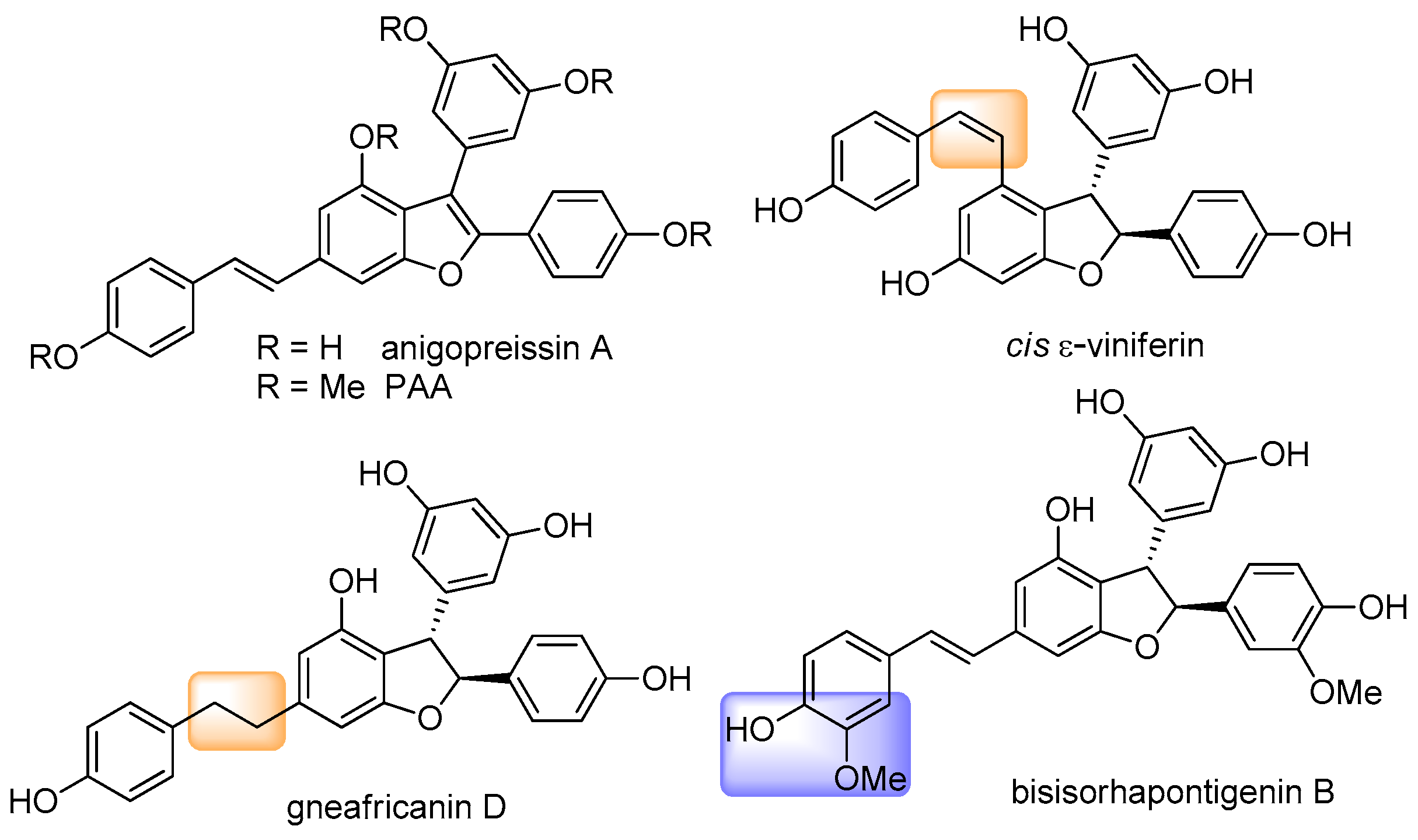
Figure 1. Examples of resveratrol dimer derivatives.
Organics 05 00012 g001
Scheme 1. General strategy to prepare derivatives 5ac, 6ac, 7 and 9af.
Organics 05 00012 sch001
Scheme 2. (a) 10, LiOH, LiBr, i-PrOH, reflux, 80%; (b) o-NO2PhCl, NH2NH2.H20, THF, rt, 55%; (c) PPh3, CBr4, DCM, rt; (d) Cs2CO3, DMSO, 110 °C, 50% in two steps; (e) 13, PdCl2(PPh3)2, CuI, Et3N, reflux, 40%.
Organics 05 00012 sch002
Scheme 3. (a) 14, KHMDS, THF, −78 °C; (b) I2 cat., n-heptane: DCM (7:3), 72%, 44%, and 34% in two steps for compounds 15, 6b, and 6c, respectively; (c) CBr4, PPh3, MeOH, quant.; (d) LiOH, LiBr, i-PrOH.
Organics 05 00012 sch003
Scheme 4. (a) (Boc)2O, DIPEA, DMAP, THF, 2h, rt, quant.; (b) NaBH4, EtOH, 10 min, quant. for 20, 73% for 25 and 79% in two steps for 27; (c) TsCl, Bu4NI, NaOH, toluene, 4h, rt, then, 21, 2h, rt 56% in two steps; (d) mCPBA, DCM, 8h, rt, quant.; (e) only for aldehyde 23, TBSCl, DMAP, DCM, 1h, rt; (f) NBS, PPh3, DCM, 8h, 0 °C, 47% and 49% for compounds 26 and 28, respectively; (g) PPh3, toluene, reflux, 96% and 88% for compounds 16 and 17.
Organics 05 00012 sch004
Scheme 5. (a) 2b or 29, PdCl2(PPh3)2, CuI, Et3N, 80 °C, 85% for 31; (b) MeI, K2CO3, acetone, quant.; (c) NIS, TFA, CH3CN, 45%; (d) 32, PdCl2(PPh3)2, NaHCO3, DMF/H2O (4:1), 75%; (e) DIBAL-H, DCM; (f) DMP, DCM; 80% in two steps; (g) 10, LiOH, LiBr, i-PrOH, reflux; (h) I2 cat., n-heptane: DCM (7:3), 64% in two steps.
Organics 05 00012 sch005
Scheme 6. (a) DIBAL-H, DCM, 0 °C, 24 h; (b) DMP, DCM, rt, 90% in two steps; (c) 10, LiOH, LiBr, i-PrOH, reflux; (d) I2 cat., n-heptane: DCM (7:3), 66% in two steps; (e) ArB(OH)2, PdCl2(PPh3)2, NaHCO3, DMF/H2O (4:1), 80 °C, 56–86% for 9af.
Organics 05 00012 sch006
Figure 2. Antiproliferative effects of PAA and derivatives 5ac, 6ac, 7, and 9af. HepG2 cells were treated with DMSO (C), PAA, or PAA derivatives at the indicated concentrations (10 nM, 100 nM, 1 μM, 10 μM, and 100 μM). Cell viability was assessed after 72 h exposure by MTT assay. Data are expressed as mean ± SD of three independent experiments with four replicates. Where indicated, differences were significant according to one-way ANOVA followed by Dunnett’s multiple comparison test (* p < 0.05, ** p < 0.001, *** p < 0.001).
Organics 05 00012 g002
Figure 3. Antiproliferative effects of PAA and derivatives 9a and 9f. Alexander cells were treated with DMSO (C), PAA, or PAA derivatives at the indicated concentrations (10 nM, 100 nM, 1 μM, 10 μM, and 100 μM). Cell viability was assessed after 72 h exposure by MTT assay. Data are expressed as mean ± SD of three independent experiments with four replicates in each. Where indicated, differences were significant according to one-way ANOVA followed by Dunnett’s multiple comparison test (* p < 0.05, ** p < 0.001, *** p < 0.001).
Organics 05 00012 g003
Figure 4. Computed stable conformers of PAA, 9a, 9b, and 9c.
Organics 05 00012 g004
Figure 5. Selected MOs of PAA, 9a, 9b, and 9c, which are close in energy to the HOMO and LUMO. The yellow levels are associated with MOs mainly localised on the phenyl group at the benzofuran C-3. The black levels refer to MOs localised on the benzofuran core and the remaining nearly coplanar groups.
Organics 05 00012 g005
Disclaimer/Publisher’s Note: The statements, opinions and data contained in all publications are solely those of the individual author(s) and contributor(s) and not of MDPI and/or the editor(s). MDPI and/or the editor(s) disclaim responsibility for any injury to people or property resulting from any ideas, methods, instructions or products referred to in the content.