Review

Exploring Vitamin D as a Modifiable Risk Factor in Cognitive Decline and Dementia


1
FCS-UBI, Faculty of Health Sciences, University of Beira Interior, 6200-506 Covilha, Portugal
2
RISE-Health, Department of Medical Sciences, Faculty of Health Sciences, University of Beira Interior, Av. Infante D. Henrique, 6200-506 Covilha, Portugal
*
Author to whom correspondence should be addressed.
Endocrines 2026, 7(3), 35; https://doi.org/10.3390/endocrines7030035 (registering DOI)
Submission received: 15 April 2026 / Revised: 8 June 2026 / Accepted: 29 June 2026 / Published: 6 July 2026
(This article belongs to the Section Neuroendocrinology and Pituitary Disorders)

Abstract

Background/Objectives: Dementia is a progressive, multifactorial neurodegenerative syndrome that poses a major public health challenge, with an increasing number of cases due to an aging population. Vitamin D (vitD) is crucial not only for bone and calcium homeostasis, but also as a neuroactive steroid that influences brain functions such as neurotransmission, neuroprotection and immunomodulation. Emerging evidence suggests that vitD deficiency may be a modifiable risk factor for the development of dementia. Thus, the aim of this review is to understand the possible role of vitD as a modifiable risk factor in the prevention of dementia. Methods: Research was conducted in the PubMed and Scopus databases using combinations of the MeSH terms ‘calcitriol’, ‘dementia’, ‘vitamin D receptors’, and ‘brain function’ for articles from 2014 until 2026. Results: The analysis of the 30 articles retrieved exhibited a significant association between vitD deficiency and an increased risk of dementia. Longitudinal studies and meta-analyses have indicated an increased risk of dementia and Alzheimer’s disease proportional to the severity of the deficit. A significant association with vascular dementia was also highlighted, with the risk increasing synergistically in the presence of hypertension. Conclusions: The evidence reviewed suggests that both vitD deficiency and insufficiency are strongly associated with increased risk of dementia, particularly Alzheimer’s disease and vascular dementia. Addressing vitD deficiency by maintaining adequate levels is proposed as a potentially effective preventive strategy against cognitive decline and dementia.

1. Introduction

Vitamin D (vitD) is a fat-soluble steroid hormone derived from cholesterol. It plays a crucial role in maintaining bone and calcium homeostasis. VitD may be produced endogenously in the human body, when the ultraviolet B (UVB) radiation stimulates its synthesis in the skin, or it can be obtained through diet. In the diet the two main forms are vitamin D2 (vitD2), or ergocalciferol, produced in some types of mushrooms and yeasts, and vitamin D3 (vitD3), or cholecalciferol, whose main diet sources are from animal origin [1,2,3]. Upon absorption, these two forms of vitD obtained through diet or supplementation can either be incorporated into micelles or absorbed by enterocytes [3]. In the skin, vitD3 is produced from 7-dehydrocholesterol (7DHC), and exposure to ultraviolet B (UVB) radiation triggers a rearrangement that creates the pre-vitamin D3 compound, that is converted in cholecalciferol, accounting for approximately 80% of total production of vitamin D. Hydroxylation of cholecalciferol (obtained from the diet and synthetized in the skin) and ergocalciferol occurs in the liver, producing 25-hydroxyvitamin D (25(OH)D) or calcidiol, and finally, hydroxylation in the kidneys transforms this into 1α,25-dihydroxyvitamin D (1α,25(OH)2D) or calcitriol, the active form of the molecule [1,2]. Calcidiol is easily detected in urine, whole blood, serum, plasma or blood spots, and is extremely stable under various pre-analytical laboratory conditions and during long-term storage. Conversely, calcitriol concentrations are much lower and are largely regulated peripherally via autocrine and paracrine mechanisms, which escape systemic endocrine control and detection [1,3].
VitD has been clearly recognized as a molecule with endocrine, paracrine and autocrine effects in multiple tissues and organs, in addition to maintaining skeletal homeostasis [1]. It is well known that the binding of vitD, which is a neuroactive steroid, to the vitamin D receptor (VDR), widely expressed in the brain, affects a variety of brain functions, being crucial for neurodevelopment, neurotransmission, neuroprotection, immunomodulation and synaptic plasticity through both genomic and non-genomic mechanisms [2,4,5,6]. Additionally, calcitriol can be locally synthesized by neurons and microglia, influencing cognitive, emotional, and motor functions playing a key role in controlling neuroinflammation with neuroprotective effects, particularly in the clearance of amyloid plaques [2,4,7].
VitD deficiency is highly prevalent worldwide, and although supplementation practices are increasing, optimal levels remain debated. Either way, it is recommended to maintain vitD levels above 20 ng/mL, as deficiency is associated with adverse health outcomes and may be linked to neurological disorders such as dementia [2,3,8,9].
Dementia is a multifactorial neurodegenerative syndrome characterized by progressive cognitive decline severe enough to impair daily functioning, affecting millions worldwide and influenced by various lifelong risk factors [10,11,12]. It is the loss of intellectual abilities severe enough to interfere with social or occupational functioning [10]. Its origins and mechanisms are complex and, despite the progress made over the last three decades, it remains poorly understood [13]. Some common causes include Alzheimer’s disease (AD), vascular dementia (VaD), dementia with Lewy bodies (DLB), frontotemporal dementia (FTD), brain infections, severe thyroid deficiency, or severe brain injury [10]. Specifically, AD is a degenerative dementia, as brain cells deteriorate and die, disrupting the production and distribution of neurotransmitters [10]. VaD is a neurological disorder caused by damage to the blood vessels in the brain, whether due to large strokes, micro-strokes, small-vessel disease or cerebral atherosclerosis, leading to significant cognitive decline [14,15]. It is the second leading cause of dementia, accounting for 20–30% of cases worldwide [10,14,15]. DLB is suspected to be caused by a variety of factors (genetic, lifestyle and environmental factors) being characterized by attention deficits, fluctuations in cognitive function, recurrent visual hallucinations, and certain features of parkinsonism, including repeated unprovoked falls, which can be dangerous and lead to hip or spinal fractures [10,12,16,17]. Lastly, FTD emerged as part of a group of clinically, pathologically, and genetically heterogeneous diseases in which degeneration is relatively selective for the frontal and temporal lobes of the brain [10,18,19].
Dementia diagnosis is complex and often delayed, requiring a multidisciplinary evaluation based on clinical history, neuropsychological assessment, biomarkers, and imaging, as no single test can confirm the condition. Early and accurate diagnosis is crucial for intervention, management, and care planning [10,20]. Treatment varies according to the type of dementia, but in general it focuses on managing cognitive and neuropsychiatric symptoms, controlling risk factors, and addressing comorbidities, although effective disease-modifying therapies are still lacking [10,14,16,18,21].
Due to an aging population, the number of people with dementia is rising, posing a major challenge to the health system. In fact, a third of the cases can be attributed to potential modifiable risk factors, including nutritional habits or deficiencies [22]. It is known that vitD deficiency most commonly affects older people and that VDRs are found in the central nervous system (CNS), particularly in the pyramidal regions of the hippocampus, playing a crucial role in memory function [13,22]. Taking this into consideration, emerging evidence suggests that vitD deficiency may be a risk factor for the development of dementia [13,22]. Thus, the aim of this review is to evaluate the current literature to understand the possible role of vitD as a modifiable risk factor in the prevention of dementia.

2. Materials and Methods

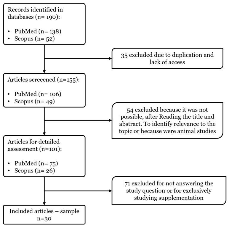
A search was conducted in the MEDLINE database, using the PubMed and Scopus search engines, with associations between the MeSH terms “calcitriol”, “dementia”, “vitamin d receptors”, “brain function” and using the Boolean operators “AND” and “OR” in the various associations. A total of 190 articles from 2014 to May 2026 were obtained. The search was limited to articles available in Portuguese, Spanish, and English, and took place between April 2024 and May 2026. Thirty original articles addressing calcitriol deficiency and/or dementia and the relationship between them were selected (Figure 1). The inclusion and exclusion criteria are expressed in Table 1. Regarding the exclusion criteria, since the review focuses on human epidemiological studies, animal studies were excluded as well as secondary publications (reviews, etc.) to avoid duplication and to base conclusions on primary evidence. Considering the predisposed population to dementia and to limit heterogeneity, only studies on adult populations living in the community or the general population were included, excluding specific contexts and conditions (pregnancy, institutional care) that could introduce confounding factors into the association between vitamin D and dementia. Confounding factors may also be attributed to pandemic context (lockdowns, disruptions to healthcare) that cannot be generalized to the overall relationship.

3. Results and Discussion

Thirty original articles were analyzed, which presented a total population of 1,728,602 individuals. The study by Navale et al. (2022) [23] had the largest sample population, involving a total of 427,690 individuals. On the other hand, Richter et al. (2024) [24] studied the smallest sample with only 25 individuals. In total, 19 countries were involved, including Germany, Belgium, China, South Korea, Denmark, the United States of America, Finland, France, India, Iceland, Japan, Mexico, Norway, the Netherlands, Poland, the United Kingdom, Sweden, Thailand, and Turkey. The studies analyzed were published between 2014 and 2026, when the research ended.
The results of these studies were analyzed according to calcitriol concentration, the definition of dementia, and the relationship between vitD and mild cognitive impairment, dementias, Alzheimer’s disease, vascular dementia, dementia with Lewy bodies, and mixed dementia. Therefore, all these parameters will be summarized below in Table 2 and discussed in detail in Table S1.

3.1. Calcitriol Parameters

The analysis of the studies included in this review reveals considerable variability in the criteria adopted for classifying serum vitD levels, which represents a significant methodological challenge in comparing the results.
In general, the most commonly used cutoff values for vitD deficiency are below 25–30 nmol/L or <10–12 ng/mL, this threshold being adopted in various studies [23,24,25,28,29,30,31,34,35,36,44]. VitD insufficiency was generally defined between 25 and 50 nmol/L (or 10–20 ng/mL), while sufficiency was considered from values above 50 nmol/L (20 ng/mL) [25,30,31,34,35,36,44,47,48,51], with some variations classifying levels between 75 and 100 nmol/L (30–40 ng/mL) as “optimal state” [30,32]. Other authors classified as deficient ≤ 30 nmol/L, insufficient > 30 and ≤75 nmol/L, and sufficient > 75 nmol/L [52] or vitD deficiency less than 30 ng/mL and an adequacy above 30 ng/mL [50].
Some publications have adopted more refined classifications, including categories such as severe deficiency (<25 nmol/L), moderate deficiency (25–50 nmol/L), insufficiency (50–75 nmol/L), and optimal state (>75 nmol/L) [24,30]. Other subdivisions, such as the use of quartiles or quintiles, on the other hand, reflect specific statistical approaches to the analysis of dose–response associations [26,27,49].
This heterogeneity limits direct comparison between studies and highlights the lack of universal consensus on ideal vitD values for cognitive function and dementia prevention. Even so, almost all articles established that levels below 50 nmol/L indicated vitD insufficiency or deficiency [23,24,25,26,28,29,31,33,34,35,36,37,38,39,41,43,44,45,46,47,48,51,52].
This methodological variation reinforces the importance of future investigations standardizing the criteria for classifying vitD, aiming for greater homogeneity and robustness of the results.

3.2. Definition of Dementia

The articles analyzed reveal considerable diversity in the diagnostic criteria and instruments used to assess parameters of dementia and cognitive impairment. This heterogeneity reflects not only chronological and geographical differences between the studies, but also scientific progress in the field of neuropsychiatry and the adaptation of criteria to clinical contexts.
The most frequently used diagnostic criteria were those of the Diagnostic and Statistical Manual of Mental Disorders (DSM-IV and DSM-5), widely employed for the identification of dementia and MCI [29,30,32,34,35,38,41,42,43]. Additionally, for the classification of AD, the National Institute of Neurological and Communicative Disorders and Stroke–Alzheimer’s Disease and Related Disorders Association (NINCDS-ADRDA) criteria [25,35,43] as well as the International Classification of Diseases—ICD-8 (290.0 and 290.1), ICD-9 and ICD-10 (F00, F03 and G30) codes [28,47,49,50,51]—were used. For the classification of VaD, the National Institute of Neurological and Communicative Disorders and Stroke–Association Internationale pour la Recherché et l’Enseignement en Neurosciences (NINDS-AIREN) criteria [29] and the ICD-8 (293.0 and 293.1) and ICD-10 (F01) codes [28] stand out.
In a recent multicenter study, specific criteria for dementia subtypes were also applied, such as the Rascovsky criteria for FTD and the diagnostic criteria for DLB [43].
Regarding neurocognitive assessment, most studies used standardized instruments such as the Mini-mental State Examination (MMSE) [24,26,33,35,37,38,41,46], frequently associated with other batteries such as the Trail Making Test (TMT), Free and Cued Selective Reminding Test (FCSRT), Montreal Cognitive Assessment (MoCA), and specific scales such as the Cornell Scale for Depression in Dementia (CSDD) or the Middelheim Frontality Score (MFS) [24,35,42]. Sakuma et al. (2019) [39], who performed a study with a Japanese population, used the version of the MMSE characteristic of the country, the MMSE-Japan (MMSE-J). Behavioral assessments were also included, especially when addressing dementia in moderate or advanced stages [24].
Population-based studies have sometimes used diagnostic algorithms based on medical records, interviews with informants or family members, and results of laboratory and imaging tests, with the diagnosis being confirmed by multidisciplinary panels or committees of experts (geriatricians, neurologists, and neuropsychologists) [23,25,27,34,37,40,41,45,48,52].
This diversity of diagnostic criteria and assessment methodologies influences the heterogeneity of results, making direct comparison between studies difficult. The standardization of diagnostic criteria for dementia and cognitive assessment methods therefore emerges as an essential recommendation for future research aiming to clarify the role of vitD in cognitive health.

3.3. Relationship Between Mild Cognitive Impairment and Vitamin D

The relationship between vitD levels and cognitive decline shows mixed results, with evidence of both significant association and neutrality.
The studies performed by Miller et al. (2015) [30] and Moon et al. (2015) [31] demonstrated that participants with severe vitD deficiency (<25 nmol/L) had a higher risk of developing MCI when compared with participants in the higher or sufficient vitD categories, as well as of accelerated decline in episodic memory and executive function. Aguilar-Navarro et al. (2019) [38] reinforced this association, reporting a Hazard Ratio (HR) of 25.02 (95% Confidence Interval) for MCI in cases of vitamin deficiency. Additionally, Yeşil et al. (2015) [32] observed significantly lower mean vitD levels in groups with MCI (20.68 ng/mL) compared to controls (23.74 ng/mL), while Chei et al. (2014) [26] reported a doubled risk of cognitive impairment (adjusted Odds Ratio (OR) 2.15) in individuals with lower levels (31.9 nmol/L vs. 45.6 nmol/L). In addition, Sakuma et al. (2019) [39] also observed that low levels of vitD were independently associated with a higher prevalence of cognitive impairment (MMSE-J ≤ 23). The lowest quartile of vitD had an adjusted OR of 2.70 (95% CI, 1.38–5.28) for cognitive impairment compared to the highest quartile. Furthermore, there was a positive correlation between vitD levels and MMSE-J scores in both sexes.
Cochar-Soares et al. identified vitD deficiency as an independent risk factor for cognitive decline among adults over 50 years of age, thus providing further supporting longitudinal evidence that insufficient vitD levels may contribute to age-related cognitive decline [52]. Consistent with the previous studies, the meta-analysis by Goodwill et al. (2017) [53] that analyzed 26 observational studies, concluded that individuals with low vitD levels (25 and 50 nmol/L) had worse cognitive performance (OD = 1.24 with 95% CI) and a higher risk of cognitive decline (OR = 1.26 with 95% CI) compared with participants with higher or sufficient vitD concentrations. However, recent studies such as those from Asante et al. (2023) [42], Janse et al. (2023) [43] and Imerbesin et al. (2025) [46] did not find significant associations between vitD deficiency and risk of neurocognitive disorders or mild cognitive impairment, even in longitudinal analyses with a 10-year interval [42].
Regarding the study conducted by Xie et al. (2023) [44], the authors concluded that older adults with vitD deficiency had a higher prevalence of cognitive impairment and that sufficiency was associated with a 33 to 55% reduced risk of cognitive impairment. In this study, vitD deficiency was defined as below 25 nmol/L, insufficiency as 25 to 50 nmol/L, and sufficiency as above 50 nmol/L or higher; therefore, the suggested protective association refers to the sufficient category compared with the deficient or insufficient categories. The authors went even further by being able to associate the additional benefit of sufficiency when combined with good quality and normal duration of sleep, and the opposite was also observed, since vitD deficiency combined with poor sleep quality or inadequate duration resulted in an increased risk of cognitive deficit [44]. Similarly, both Zhou et al. [47] and Ren et al. [49] explored the influence of sleep characteristics in the association between vitamin D and dementia risk. The authors found that lower vitD levels were associated with an increased risk of developing dementia, being more pronounced when combined with unfavorable sleep characteristics, including duration [47] and daytime sleepiness [49]. These findings suggest that there is an interaction between vitD status and sleep characteristics, reinforcing the importance of lifestyle factors in cognitive aging and dementia development. This divergence can be attributed to methodological differences, such as varying criteria for disability (e.g., <25 nmol/L vs. <30 nmol/L) or distinct study designs (cross-sectional vs. longitudinal). Some studies highlight specific impacts on cognitive domains (episodic memory and executive function) [30,33], while another study does not identify relationships with decline in semantic memory or visuospatial skills [30].
Overall, most evidence suggests that adequate vitD levels may have a protective role, while severe deficiency appears to increase the risk of MCI. Furthermore, the link between vitamin D and dementia may be influenced by sleep behavior. Thus, preventive strategies should consider both nutritional and lifestyle factors simultaneously. However, causality remains uncertain, and confounding factors (sun exposure, mobility, or comorbidities) may influence the results. Future studies should standardize diagnostic criteria and include therapeutic interventions to elucidate underlying mechanisms.

3.4. Relationship Between Dementia and Vitamin D

Regarding the relationship between vitD and dementia, Chen et al. (2024) [45] highlighted that both insufficiency and deficiency of vitD are associated with an increased risk of all types of dementia, with HR ranging between 1.11 and 1.25, respectively. Meta-analyses by Zhang et al. (2024) [22] and Kalra et al. (2020) [54] reinforce these results, with the former pointing to a 42% increase in the risk of dementia in individuals with deficient vitD levels compared with individuals with sufficient or higher vitD concentrations.
The other studies analyzed in this review corroborate these meta-analyses; however, they quantify the risk according to the severity of the disability and also provide additional evidence. For example, Littlejohns et al. (2014) [25] showed that the risk of dementia was 1.53 times higher among the disabled and 2.25 times higher among the severely deficient in vitD. Similar results were found by Licher et al. (2017) [36], with an increase in the adjusted risk for each standard deviation decrease in vitD levels (HR: 1.11; 95% CI). Consistent with these studies, the meta-analysis conducted by Chai et al. (2019) [55] observed a 32% increase in risk with general disability, 48% with moderate deficiency, and even a high risk in severe disability.
Additionally, the studies from Knekt et al. (2014) [27] and Fashanu et al. (2019) [40], analyzed the risk differences in specific populations, such as women and middle-aged individuals. Knekt et al. showed a significant association between higher vitD levels and a lower risk of dementia among women, but not among men, suggesting possible gender differences [27], while Fashanu et al. demonstrated that deficient vitD levels in middle-aged were linked to an increased risk of developing dementia in subsequent decades (HR: 1.26) compared with participants with non-deficient or sufficient vitD levels [40]. In a different perspective, Cheng et al. [50] and Hung et al. [51] conducted two studies using the same cohort (TriNetX) to evaluate the association between vitD and dementia risk among adults with sensory impairment, specifically vision or hearing impairment. Cheng et al. identified a significant increase in the risk of new-onset dementia among patients with glaucoma and vitD deficiency, also suggesting that treating vitD deficiency may have a positive impact on the development of neurodegenerative diseases [50]. Considering that sensory impairment has been recognized as a risk factor for cognitive decline and dementia, the study conducted by Hung et al. also found that low vitD levels were associated with a significantly increased risk of incident dementia over the follow-up period [51].
Lu et al. (2025) [48] presented the results of a case–cohort study integrated into the Japan Public Health Centre-based Prospective (JPHC) Study. The authors observed that participants with higher plasma vitD levels had a lower risk of developing disabling dementia compared with those in the lowest categories. These results support the hypothesis that adequate levels of vitD may contribute to the prevention of dementia in older adults [48].
Conversely, two other studies point to less conclusive results. Asante et al. (2023) [42], for example, found no significant association between vitD deficiency, assessed at two time points 10 years apart, and the risk of neurocognitive disorders when deficient or insufficient vitD levels were compared with the respective references. Similarly, Nagel et al. (2015) [33] observed a non-significant association after statistical adjustments. These results indicate that the relationship between vitD and dementia may depend on additional factors, such as the timing of the assessment, duration of follow-up, and the presence of confounders, including age, sex, BMI, smoking status, alcohol consumption, physical activity, diabetes, blood pressure, serum cholesterol, depression, and serum creatinine. However, considering that the Nagel et al. study has a cross-sectional design, it is not possible to rule out reverse causality, as the temporal relationship is unclear.
Regarding the duration of follow-up, in 2024, Grant et al. [56] published a study on the implications of follow-up periods for the effect of vitD deficiency on the risk of dementia, Alzheimer’s disease and cognitive impairment. The authors found that the duration of follow-up significantly altered the association between low serum vitD levels and an increased risk of the three neurological conditions, with the link being stronger in shorter follow-up periods (less than 5 years), and weaker or null in very long follow-up periods (10–20 years). The authors stated that the decline in vitD levels in the years immediately preceding diagnosis is likely a consequence of reduced outdoor activity and an inadequate diet during the preliminary phase of dementia, rather than evidence of a long-term causal relationship [56].
Despite the contradictions, there is more evidence suggesting a positive causal relationship. In addition to the studies cited above, Navale et al. (2022) [23], through Mendelian randomization analysis, identified a threshold effect below 25 nmol/L, in which the risk of dementia increased sharply, supporting a causal link between severe vitD deficiency and the development of dementia. Łukaszyk et al. (2018) [37], Moon et al. (2015) [31], Arnljots et al. (2017) [34], Lu et al. [48], Cheng et al. [50] and Hung et al. [51] also point to vitD deficiency as a modifiable factor associated with cognitive dysfunction and dementia, especially among the elderly and sensory-impaired adults. These results suggest that maintaining adequate vitD levels may represent an important preventive strategy in the context of aging and cognitive health. In a different perspective, Eymundsdottir et al. 2020 [41] stated that individuals with dementia have a higher risk of vitD deficiency, regardless of other lifestyle factors; however, the authors did not make the inverse correlation.
In summary, these studies point to a significant proportional relationship between vitD deficiency and an increased risk of dementia. Studies with different methodological designs indicate that both vitD deficiency and insufficiency are associated with an increased risk of developing dementia, with these being especially notable in subgroups such as women and middle-aged individuals. Although some studies yield less conclusive results, most research, including Mendelian randomization analyses, reinforces the hypothesis of a causal role of vitD in cognitive processes and in the prevention of dementia.

3.5. Relationship Between Alzheimer’s Disease and Vitamin D

Regarding studies on the relationship between AD and vitD, several studies have shown a significant association between low serum vitD levels, and an increased risk of AD. Chen et al. (2024) [45] concluded that both vitamin D insufficiency and deficiency are associated with a higher risk, with an HR of 1.10. Concordantly, but more expressively, the meta-analysis by Kalra et al. (2020) [54] reported that individuals with vitD deficiency had a 1.87 times greater risk of developing AD, while those with insufficiency had a 1.25 times greater risk, compared to those with normal levels. Another meta-analysis developed by Zhang et al. (2024) [22] also corroborated these data, estimating a 57% increase in the risk of AD in individuals with vitamin deficiency.
In this sense, the studies analyzed in this work corroborate this association, namely Feart et al. (2017) [35], after following a cohort with 125 cases of AD, concluded that vitD deficiency (<25 nmol/L compared with >50 nmol/L) was associated with almost three times the risk of developing the disease (HR: 2.85). Similarly, Littlejohns et al. (2014) [25] observed that deficient individuals had a 1.69 times increased risk of developing AD, while severely deficient individuals presented a 2.22 times greater risk, compared with participants with sufficient concentrations of 50 nmol/L or higher. Licher et al. (2017) [36] identified that each standard deviation decrease in vitD levels was associated with a 13% increase in the risk of AD (HR: 1.13). Finally, Afzal et al. (2014) [28] after multivariate adjustment, defined that participants with vitD levels < 25 nmol/L had a higher risk for AD (HR 1.25) when compared to those with ≥50 nmol/L.
The severity of the deficiency appears to directly influence the risk level, as demonstrated in the meta-analysis by Chai et al. (2019) [55], showing a 34% increased risk of developing AD in individuals with vitD deficiency (<20 ng/mL) compared with individuals with sufficient vitD levels, a risk that was even higher in moderate deficiency—HR: 1.51—and significant in severe deficiency—HR: 1.36.
A particularly surprising finding comes from Aguilar-Navarro et al. (2019) [38], who reported an extreme increase in the risk of AD among individuals with vitD deficiency (≤20 ng/mL, compared with individuals with sufficient vitamin D levels > 30 ng/mL; HR: 41.7). Although this value should be interpreted with caution, possibly reflecting specific sample characteristics or methodology, the authors suggest that the poorer performance in executive function, attention, evocation, orientation domains found in subjects with VitD deficiency may be due to damage to the frontal-subcortical circuits.
Observational studies reinforce the relationship between low vitD levels and the presence of AD. Yeşil et al. (2015) [32] found significantly lower mean vitD levels in patients with AD (20.29 ng/mL) compared to the control group (23.74 ng/mL). Janse et al. (2023) [43] presented similar results, in which patients with AD had significantly lower adjusted mean levels (7.77) compared to individuals without dementia (8.27). These data indicate that vitD deficiency may be present even in the early stages of the disease.
Additionally, there is evidence of biological mechanisms that may explain this association. Richter et al. (2024) [24] observed an inverse correlation between vitD levels and Aβ1-42 levels in cerebrospinal fluid, suggesting that vitamin deficiency may contribute to the deposition of amyloid plaques, a central pathological feature of AD. The authors also suggested that vitD deficiency is associated with serotonergic alterations, influencing cognitive and behavioral symptoms of the disease. These results reinforce the hypothesis that vitD may have a relevant neuroprotective role, with implications for both the prevention and the clinical course of AD [24].
In conclusion, current evidence indicates a robust association between low serum vitD levels and an increased risk of developing AD, with this risk being proportional to the severity of the deficiency. Different types of studies, including cohort and observational ones, corroborate this relationship, revealing that individuals with vitD deficiency or insufficiency are more prone to developing the disease, even in its early stages. Furthermore, the data suggested that vitD may exert important neuroprotective functions, influencing mechanisms such as amyloid plaque deposition and the modulation of serotonergic pathways. Despite certain variations in the results, possibly attributed to methodological differences, the body of research reinforces the relevance of maintaining adequate vitD levels as a potential strategy for preventing and controlling AD progression.

3.6. Relationship Between Vascular Dementia and Vitamin D

The association between low vitD levels and risk of VaD has been evidenced by some recent studies. Chen et al. (2024) [45] found an association between reduced serum levels and vitD insufficiency (<50 nmol/L compared with ≥50 nmol/L) with increased risk of VaD (HR of 1.15). These results indicate that even levels not necessarily classified as severe deficiency may be implicated in increased vulnerability to this subtype of dementia [45]. Afzal et al. (2014) [28] found that the risk for VaD was elevated among the participants with vitD levels < 25 nmol/L (HR 1.22; 95% CI 0.77–1.91) compared with participants with adequate serum vitD levels (≥50 nmol/L), although it did not obtain statistical significance.
Corroborating this evidence, the study conducted by Janse et al. (2023) [43] showed that patients diagnosed with VaD presented significantly lower levels of vitD compared to individuals without dementia. This difference reinforces the hypothesis that vitamin deficiency may be related to the development of VaD, even when comparing between clinical groups.
A previous study by Prabhakar et al. (2015) [29] adds a more detailed perspective. Although the difference in mean vitD levels between the VaD cases (15.9 ng/mL) and controls (16.6 ng/mL) was not statistically significant, the prevalence of severe deficiency (≤12 ng/mL) was slightly higher (53% vs. 50%). Importantly, after adjusting for demographic and biochemical covariates, vitD deficiency (≤12 ng/mL compared with vitD sufficiency > 20 ng/mL) was associated with a 2.2 times higher risk of VaD (adjusted OR: 2.19). Notably, the combination of hypertension and vitD deficiency increased the risk for VaD sharply, reaching an increase of up to 31.6-fold, suggesting a possible synergistic effect between these risk factors [29].
In conclusion, the data analyzed throughout all studies suggest a relevant association between low vitD levels and an increased risk of VaD, although the evidence is more limited compared to other dementia subtypes. Recent studies indicate that even levels considered merely insufficient may be implicated in the development of VaD, reinforcing the importance of vitD as a possible modifiable risk factor. Furthermore, clinical data demonstrate that individuals with VaD have significantly lower levels of vitD compared to controls, and adjusted analyses revealed a substantially higher risk among those deficient. The potential interaction between vitD deficiency and other vascular factors, such as hypertension, is particularly noteworthy, as it can considerably amplify the risk of VaD.

3.7. Relationship Between Dementia with Lewy Bodies, Mixed Dementia and Vitamin D

The only study that refers to these two types of dementia is the one performed by Janse et al. (2023) [43], where 1758 patients over the age of 60 with cognitive diagnoses were analyzed. All participants underwent a standardized cognitive assessment performed by a multidisciplinary team, which included a geriatrician, neurologist, specialist nurse and, if necessary, neuropsychologist. Clinical diagnoses followed current international criteria, and vitD status was measured at the first appointment. However, the authors found no significant differences in vitD levels between patients with MCI, DLB and mixed dementia [43].
Thus, this work suggests that the evidence on the association of vitD with all types of dementia analyzed is currently scarce and inconclusive. It is therefore essential that future investigations and longitudinal clinical trials include these patient groups to determine whether vitD deficiency is a relevant risk factor as observed for other etiologies of dementia.

4. Conclusions

The evidence gathered throughout this review confirms the role of vitD in brain function and in the risk of developing dementia, especially AD and VaD.
Recent large-scale demographic studies analyzed throughout this work, such as the Navale et al. (2022) [23] and Chen et al. (2024) [45] studies, which evaluated hundreds of thousands of individuals, demonstrate that vitD deficiency or even insufficiency is associated with a significant increase in the risk of dementia in general and AD in particular, with risks proportional to the severity of the deficit. Furthermore, several epidemiological studies, both cross-sectional and longitudinal, point to a significant relationship between low vitD levels and MCI, especially in domains such as memory and executive function. However, although they are useful methods for identifying associations and estimating prevalence, cross-sectional studies lack a timeframe and therefore cannot establish causality. In this case, longitudinal studies are preferrable since a temporal analysis allows us to understand how the disease state may affect serum vitD concentrations.
The methodological heterogeneity among the studies was notable, related to the diagnostic criteria for dementia and the cutoff values for vitD deficiency. Although this variability limits the direct comparability of the results, there is consensus that concentrations below 50 nmol/L correspond to significant deficits and that maintaining adequate levels can be a fundamental prevention strategy.
Although some investigations have not found significant associations after adjusting for confounding factors (e.g., age, sex, season, ethnicity, cardiovascular factors, calcium, renal function, depression, outdoor activity, and APOE4), the preponderance of evidence reinforces the hypothesis of a causal and modifiable role of vitD in dementia. Finally, the synergistic interaction between vitD deficiency and classic vascular risk factors, notably hypertension, in increasing the risk of vascular disease is highlighted.
An additional methodological consideration is the type of evidence available for evaluating vitD with respect to cognitive decline and dementia. Although randomized controlled trials are generally considered the highest level of evidence for interventions, vitD trials present specific challenges that may hinder their interpretability. Nutrient trials differ from drug trials because the biological effect depends not only on the assigned dose, but also on baseline nutrient status, achieved serum vitD concentrations, dose–response relationships, adherence, background intake, and the duration of exposure. Trials that include participants with sufficient vitD status at baseline, use inadequate doses, fail to measure achieved vitD concentrations, or have insufficient follow-up may underestimate potential benefits in deficient individuals. These issues have been highlighted in methodological guidelines for nutrient studies and in critical appraisals of large randomized controlled trials of vitD [57,58]. Therefore, well-designed observational studies, particularly prospective cohort studies with baseline vitD assessment and long-term follow-up, remain highly relevant for evaluating the epidemiological association between vitD status and cognitive decline or dementia. However, such studies are still susceptible to residual confounding and reverse causation, and their findings should be interpreted as supportive rather than definitive evidence of causality.
Thus, this review suggests that combating vitD deficiency, especially in the elderly, could represent an effective preventive approach to cognitive decline and dementia, particularly AD and VaD. However, defining ideal levels and confirming the clinical impact of supplementation requires further standardized and, eventually, interventional research.

Supplementary Materials

The following supporting information can be downloaded at: https://www.mdpi.com/article/10.3390/endocrines7030035/s1, Table S1: Detailed overview of the thirty studies analysed regarding vitamin D status and different dementia outcomes.

Author Contributions

Conceptualization, I.S. and E.C.; methodology, I.S. and E.C.; validation, E.C.; investigation, I.S. and E.C.; writing—original draft preparation, I.S.; writing—review and editing, I.S., M.M. and E.C.; visualization, I.S., M.M. and E.C.; supervision, E.C.; project administration, E.C.; funding acquisition, E.C. All authors have read and agreed to the published version of the manuscript.

Funding

This article was supported by National Funds through FCT—Fundação para a Ciência e a Tecnologia, I.P., within the project RISE-Health—UID/06397/2025.

Data Availability Statement

No new data were created or analyzed in this study.

Conflicts of Interest

The authors declare no conflicts of interest.

Abbreviations

The following abbreviations are used in this manuscript:
1α,25(OH)2D1α,25-dihydroxyvitamin D or calcitriol
7DHC7-dehydrocholesterol
25(OH)D25-hydroxyvitamin D or calcidiol
ADAlzheimer’s disease
ADLActivities of daily living
BMIBody mass index
BNTBoston Naming Test
BPBlood pressure
CDRClinical dementia rating scale
CERADConsortium to Establish a Registry for Alzheimer’s Disease
CMAICohen-Mansfield agitation inventory
CNSCentral nervous system
CSDDCornell Scale for Depression in Dementia
CVDCerebrovascular disease
DLBDementia with Lewy bodies
DSMDiagnostic and Statistical Manual of Mental Disorders
FCSRTFree Selective Recall Test and Cued
FTDFrontotemporal dementia
HDLHigh-density lipoprotein
HRHazzard ratio
IADLInstrumental activities of daily living
ICDInternational Classification of Diseases
MCIMild cognitive impairment
MFSMiddelheim frontality score
MINIMini international neuropsychiatric interview
MMSEMini-mental state examination
MNA-SFMini-form nutrition assessment
MoCAMontreal Cognitive Assessment
MRIMagnetic resonance imaging
NIA-AANational Institute on Aging and Alzheimer’s Association
NINCDS-ADRDANational Institute of Neurological and Communicative Disorders and Stroke-Alzheimer’s Disease and Related Disorders Association
NINCDS-AIRENNational Institute of Neurological Disorders and Stroke (NINDS) and the Association Internationale pour la Recherche et l’Enseignement en Neurosciences (AIREN)
ODOdds ratio
TMTTrail Making Test
UVBUltraviolet B
VaDVascular dementia
VDRVitamin D receptor
VitDVitamin D
VitD2Vitamin D2
VitD3Vitamin D3
VFTVerbal fluency test

References

  1. Saponaro, F.; Saba, A.; Zucchi, R. An Update on Vitamin D Metabolism. Int. J. Mol. Sci. 2020, 21, 6573. [Google Scholar] [CrossRef] [PubMed]
  2. Wassif, G.A.; Alrehely, M.S.; Alharbi, D.M.; Aljohani, A.A. The Impact of Vitamin D on Neuropsychiatric Disorders. Cureus 2023, 15, e47716. [Google Scholar] [CrossRef] [PubMed]
  3. Sorrenti, V.; Buriani, A.; Davinelli, S.; Scapagnini, G.; Fortinguerra, S. Vitamin D Physiology, Deficiency, Genetic Influence, and the Effects of Daily vs. Bolus Doses of Vitamin D on Overall Health: A Clinical Approach. Nutraceuticals 2023, 3, 403–420. [Google Scholar] [CrossRef]
  4. Mirarchi, A.; Albi, E.; Beccari, T.; Arcuri, C. Microglia and Brain Disorders: The Role of Vitamin D and Its Receptor. Int. J. Mol. Sci. 2023, 24, 11892. [Google Scholar] [CrossRef] [PubMed]
  5. Hii, C.S.; Ferrante, A. The Non-Genomic Actions of Vitamin D. Nutrients 2016, 8, 135. [Google Scholar] [CrossRef] [PubMed]
  6. Carlberg, C. Genomic signaling of vitamin D. Steroids 2023, 198, 109271. [Google Scholar] [CrossRef] [PubMed]
  7. Lang, F.; Ma, K.; Leibrock, C.B. 1,25(OH)(2)D(3) in Brain Function and Neuropsychiatric Disease. Neurosignals 2019, 27, 40–49. [Google Scholar] [CrossRef] [PubMed]
  8. Amrein, K.; Scherkl, M.; Hoffmann, M.; Neuwersch-Sommeregger, S.; Kostenberger, M.; Tmava Berisha, A.; Martucci, G.; Pilz, S.; Malle, O. Vitamin D deficiency 2.0: An update on the current status worldwide. Eur. J. Clin. Nutr. 2020, 74, 1498–1513. [Google Scholar] [CrossRef] [PubMed]
  9. Vieth, R. Vitamin D supplementation: Cholecalciferol, calcifediol, and calcitriol. Eur. J. Clin. Nutr. 2020, 74, 1493–1497. [Google Scholar] [CrossRef] [PubMed]
  10. World Health Organization. Ageing Gracefully: Diversity of Dementia; WHO: Geneva, Switzerland, 2017. [Google Scholar]
  11. Dresden, S.M.; Dickens, J.C.; Lo, A.X. Emergency Care for Persons Living with Dementia. Emerg. Med. Clin. N. Am. 2025, 43, 235–248. [Google Scholar] [CrossRef] [PubMed]
  12. Goddard, T.R.; Brookes, K.J.; Sharma, R.; Moemeni, A.; Rajkumar, A.P. Dementia with Lewy Bodies: Genomics, Transcriptomics, and Its Future with Data Science. Cells 2024, 13, 223. [Google Scholar] [CrossRef] [PubMed]
  13. Annweiler, C. Vitamin D in dementia prevention. Ann. N. Y Acad. Sci. 2016, 1367, 57–63. [Google Scholar] [CrossRef] [PubMed]
  14. Altahrawi, A.Y.; James, A.W.; Shah, Z.A. The Role of Oxidative Stress and Inflammation in the Pathogenesis and Treatment of Vascular Dementia. Cells 2025, 14, 609. [Google Scholar] [CrossRef] [PubMed]
  15. Agrawal, N.; Afzal, M.; Khan, N.H.; Ganesan, S.; Kumari, M.; Sunitha, S.; Dash, A.; Goyal, K.; Kushwaha, B.; Rekha, A.; et al. The role of VEGF in vascular dementia: Impact of aging and cellular senescence. Biogerontology 2025, 26, 77. [Google Scholar] [CrossRef] [PubMed]
  16. An, D.; Xu, Y. Environmental risk factors provoke new thinking for prevention and treatment of dementia with Lewy bodies. Heliyon 2024, 10, e30175. [Google Scholar] [CrossRef] [PubMed]
  17. Laurell, A.A.S.; Mak, E.; O’Brien, J.T. A systematic review of diffusion tensor imaging and tractography in dementia with Lewy bodies and Parkinson’s disease dementia. Neurosci. Biobehav. Rev. 2025, 169, 106007. [Google Scholar] [CrossRef] [PubMed]
  18. Buccellato, F.R.; D’Anca, M.; Tartaglia, G.M.; Del Fabbro, M.; Galimberti, D. Frontotemporal dementia: From genetics to therapeutic approaches. Expert Opin. Investig. Drugs 2024, 33, 561–573. [Google Scholar] [CrossRef] [PubMed]
  19. Antonioni, A.; Raho, E.M.; Lopriore, P.; Pace, A.P.; Latino, R.R.; Assogna, M.; Mancuso, M.; Gragnaniello, D.; Granieri, E.; Pugliatti, M.; et al. Frontotemporal Dementia, Where Do We Stand? A Narrative Review. Int. J. Mol. Sci. 2023, 24, 11732. [Google Scholar] [CrossRef] [PubMed]
  20. Arvanitakis, Z.; Shah, R.C.; Bennett, D.A. Diagnosis and Management of Dementia: Review. JAMA 2019, 322, 1589–1599. [Google Scholar] [CrossRef] [PubMed]
  21. Khan, B.; Iqbal, M.K.; Khan, M.A.; Khan, H.; Kiyani, M.M.; Bashir, S.; Li, S. Unraveling the Complexity of Alzheimer’s Disease: Insights into Etiology and Advancements in Treatment Strategies. J. Mol. Neurosci. 2025, 75, 57. [Google Scholar] [CrossRef] [PubMed]
  22. Zhang, X.X.; Wang, H.R.; Meng, W.; Hu, Y.Z.; Sun, H.M.; Feng, Y.X.; Jia, J.J. Association of Vitamin D Levels with Risk of Cognitive Impairment and Dementia: A Systematic Review and Meta-Analysis of Prospective Studies. J. Alzheimers Dis. 2024, 98, 373–385. [Google Scholar] [CrossRef] [PubMed]
  23. Navale, S.S.; Mulugeta, A.; Zhou, A.; Llewellyn, D.J.; Hypponen, E. Vitamin D and brain health: An observational and Mendelian randomization study. Am. J. Clin. Nutr. 2022, 116, 531–540. [Google Scholar] [CrossRef] [PubMed]
  24. Richter, A.L.; Diepeveen-de Bruin, M.; Balvers, M.G.J.; De Groot, L.; De Deyn, P.P.; Engelborghs, S.; Witkamp, R.F.; Vermeiren, Y. Association between Low Vitamin D Status, Serotonin, and Clinico-Biobehavioral Parameters in Alzheimer’s Disease. Dement. Geriatr. Cogn. Disord. 2023, 52, 318–326. [Google Scholar] [CrossRef] [PubMed]
  25. Littlejohns, T.J.; Henley, W.E.; Lang, I.A.; Annweiler, C.; Beauchet, O.; Chaves, P.H.; Fried, L.; Kestenbaum, B.R.; Kuller, L.H.; Langa, K.M.; et al. Vitamin D and the risk of dementia and Alzheimer disease. Neurology 2014, 83, 920–928. [Google Scholar] [CrossRef] [PubMed]
  26. Chei, C.L.; Raman, P.; Yin, Z.X.; Shi, X.M.; Zeng, Y.; Matchar, D.B. Vitamin D levels and cognition in elderly adults in China. J. Am. Geriatr. Soc. 2014, 62, 2125–2129. [Google Scholar] [CrossRef] [PubMed]
  27. Knekt, P.; Saaksjarvi, K.; Jarvinen, R.; Marniemi, J.; Mannisto, S.; Kanerva, N.; Heliovaara, M. Serum 25-hydroxyvitamin d concentration and risk of dementia. Epidemiology 2014, 25, 799–804. [Google Scholar] [CrossRef] [PubMed]
  28. Afzal, S.; Bojesen, S.E.; Nordestgaard, B.G. Reduced 25-hydroxyvitamin D and risk of Alzheimer’s disease and vascular dementia. Alzheimers Dement. 2014, 10, 296–302. [Google Scholar] [CrossRef] [PubMed]
  29. Prabhakar, P.; Chandra, S.R.; Supriya, M.; Issac, T.G.; Prasad, C.; Christopher, R. Vitamin D status and vascular dementia due to cerebral small vessel disease in the elderly Asian Indian population. J. Neurol. Sci. 2015, 359, 108–111. [Google Scholar] [CrossRef] [PubMed]
  30. Miller, J.W.; Harvey, D.J.; Beckett, L.A.; Green, R.; Farias, S.T.; Reed, B.R.; Olichney, J.M.; Mungas, D.M.; DeCarli, C. Vitamin D Status and Rates of Cognitive Decline in a Multiethnic Cohort of Older Adults. JAMA Neurol. 2015, 72, 1295–1303. [Google Scholar] [CrossRef] [PubMed]
  31. Moon, J.H.; Lim, S.; Han, J.W.; Kim, K.M.; Choi, S.H.; Kim, K.W.; Jang, H.C. Serum 25-hydroxyvitamin D level and the risk of mild cognitive impairment and dementia: The Korean Longitudinal Study on Health and Aging (KLoSHA). Clin. Endocrinol. 2015, 83, 36–42. [Google Scholar] [CrossRef] [PubMed]
  32. Yesil, Y.; Kuyumcu, M.E.; Kara, O.; Halacli, B.; Etgul, S.; Kizilarslanoglu, M.C.; Yavuz, B.B.; Ozcan, M.; Halil, M.G.; Sahin Cankurtaran, E.; et al. Vitamin D status and its association with gradual decline in cognitive function. Turk. J. Med. Sci. 2015, 45, 1051–1057. [Google Scholar] [CrossRef] [PubMed]
  33. Nagel, G.; Herbolsheimer, F.; Riepe, M.; Nikolaus, T.; Denkinger, M.D.; Peter, R.; Weinmayr, G.; Rothenbacher, D.; Koenig, W.; Ludolph, A.C.; et al. Serum Vitamin D Concentrations and Cognitive Function in a Population-Based Study among Older Adults in South Germany. J. Alzheimers Dis. 2015, 45, 1119–1126. [Google Scholar] [CrossRef] [PubMed]
  34. Arnljots, R.; Thorn, J.; Elm, M.; Moore, M.; Sundvall, P.D. Vitamin D deficiency was common among nursing home residents and associated with dementia: A cross sectional study of 545 Swedish nursing home residents. BMC Geriatr. 2017, 17, 229. [Google Scholar] [CrossRef] [PubMed]
  35. Feart, C.; Helmer, C.; Merle, B.; Herrmann, F.R.; Annweiler, C.; Dartigues, J.F.; Delcourt, C.; Samieri, C. Associations of lower vitamin D concentrations with cognitive decline and long-term risk of dementia and Alzheimer’s disease in older adults. Alzheimers Dement. 2017, 13, 1207–1216. [Google Scholar] [CrossRef] [PubMed]
  36. Licher, S.; de Bruijn, R.; Wolters, F.J.; Zillikens, M.C.; Ikram, M.A.; Ikram, M.K. Vitamin D and the Risk of Dementia: The Rotterdam Study. J. Alzheimers Dis. 2017, 60, 989–997. [Google Scholar] [CrossRef] [PubMed]
  37. Lukaszyk, E.; Bien-Barkowska, K.; Bien, B. Cognitive Functioning of Geriatric Patients: Is Hypovitaminosis D the Next Marker of Cognitive Dysfunction and Dementia? Nutrients 2018, 10, 1104. [Google Scholar] [CrossRef] [PubMed]
  38. Aguilar-Navarro, S.G.; Mimenza-Alvarado, A.J.; Jimenez-Castillo, G.A.; Bracho-Vela, L.A.; Yeverino-Castro, S.G.; Avila-Funes, J.A. Association of Vitamin D with Mild Cognitive Impairment and Alzheimer’s Dementia in Older Mexican Adults. Rev. Investig. Clin. 2019, 71, 381–386. [Google Scholar] [CrossRef] [PubMed]
  39. Sakuma, M.; Kitamura, K.; Endo, N.; Ikeuchi, T.; Yokoseki, A.; Onodera, O.; Oinuma, T.; Momotsu, T.; Sato, K.; Nakamura, K.; et al. Low serum 25-hydroxyvitamin D increases cognitive impairment in elderly people. J. Bone Miner. Metab. 2019, 37, 368–375. [Google Scholar] [CrossRef] [PubMed]
  40. Fashanu, O.E.; Zhao, D.; Schneider, A.L.C.; Rawlings, A.M.; Sharrett, A.R.; Lutsey, P.L.; Gottesman, R.F.; Gross, A.L.; Guallar, E.; Alonso, A.; et al. Mid-life serum Vitamin D concentrations were associated with incident dementia but not late-life neuropsychological performance in the Atherosclerosis Risk in Communities (ARIC) Study. BMC Neurol. 2019, 19, 244. [Google Scholar] [CrossRef] [PubMed]
  41. Eymundsdottir, H.; Chang, M.; Geirsdottir, O.G.; Gudmundsson, L.S.; Jonsson, P.V.; Gudnason, V.; Launer, L.; Jonsdottir, M.K.; Ramel, A. Lifestyle and 25-hydroxy-vitamin D among community-dwelling old adults with dementia, mild cognitive impairment, or normal cognitive function. Aging Clin. Exp. Res. 2020, 32, 2649–2656. [Google Scholar] [CrossRef] [PubMed]
  42. Asante, E.O.; Mai, X.M.; Eldholm, R.S.; Skjellegrind, H.K.; Kolberg, M.; Brumpton, B.M.; Selbaek, G.; Chen, Y.; Sun, Y.Q. Vitamin D Status Over Time and Cognitive Function in Norwegian Older Adults: A Prospective Cohort of the HUNT Study. J. Nutr. Health Aging 2023, 27, 30–37. [Google Scholar] [CrossRef] [PubMed]
  43. Janse, A.; van de Rest, O.; de Groot, L.; Witkamp, R.F. The Association of Vitamin D Status with Mild Cognitive Impairment and Dementia Subtypes: A Cross-Sectional Analysis in Dutch Geriatric Outpatients. J. Alzheimers Dis. 2023, 91, 1359–1369. [Google Scholar] [CrossRef] [PubMed]
  44. Xie, Y.; Bai, C.; Feng, Q.; Gu, D. Serum Vitamin D(3) Concentration, Sleep, and Cognitive Impairment among Older Adults in China. Nutrients 2023, 15, 4192. [Google Scholar] [CrossRef] [PubMed]
  45. Chen, L.J.; Sha, S.; Stocker, H.; Brenner, H.; Schottker, B. The associations of serum vitamin D status and vitamin D supplements use with all-cause dementia, Alzheimer’s disease, and vascular dementia: A UK Biobank based prospective cohort study. Am. J. Clin. Nutr. 2024, 119, 1052–1064. [Google Scholar] [CrossRef] [PubMed]
  46. Imerbsin, N.; Shantavasinkul, P.C.; Witoonpanich, P.; Sirivarasai, J.; Taonam, N.; Phanachet, P.; Warodomwichit, D.; Jayanama, K.; Boonyawat, K.; Somlaw, N.; et al. Vitamin D and Cognitive Impairment. Nutrients 2025, 17, 1301. [Google Scholar] [CrossRef] [PubMed]
  47. Zhou, J.; Huang, N.; Huang, T.; Ma, J. Investigating the longitudinal relationship between Vitamin D levels, sleep duration, and dementia risk: Insights from the UK Biobank. Psychiatry Res. 2025, 353, 116759. [Google Scholar] [CrossRef] [PubMed]
  48. Lu, Y.; Yamaji, T.; Yasuda, N.; Kuchiba, A.; Shimizu, Y.; Nakano, S.; Yamagishi, K.; Inoue, M.; Tsugane, S.; Sawada, N.; et al. Association between plasma 25-hydroxyvitamin D concentration and incident disabling dementia risk in Japan: A case-cohort study. J. Neurol. Sci. 2025, 478, 123720. [Google Scholar] [CrossRef] [PubMed]
  49. Ren, J.J.; Li, Z.H.; Zhong, W.F.; Gao, J.; Chen, P.L.; Wang, X.M.; You, F.F.; Mao, C. Serum 25-hydroxyvitamin D concentrations, sleep characteristics and the risk of incident dementia. Maturitas 2026, 206, 108864. [Google Scholar] [CrossRef] [PubMed]
  50. Cheng, Y.C.; Lu, C.L.; Wang, J.; Tsai, M.L.; Lu, K.C. Increased risk of incident dementia associated with vitamin D deficiency in glaucoma patients: A TriNetX cohort study. Front. Nutr. 2026, 13, 1760959. [Google Scholar] [CrossRef] [PubMed]
  51. Hung, K.C.; Weng, H.L.; Lai, Y.C.; Chen, I.W. Low vitamin D status and 10-year dementia risk in sensory-impaired adults: A propensity score-matched cohort study. Front. Nutr. 2026, 13, 1822195. [Google Scholar] [CrossRef] [PubMed]
  52. Cochar-Soares, N.; da Silva, T.B.P.; Luiz, M.M.; Tofani, P.S.; Delinocente, M.L.B.; de Oliveira Maximo, R.; Steptoe, A.; de Oliveira, C.; da Silva Alexandre, T. Vitamin D deficiency as a risk factor for cognitive decline in individuals aged 50 or older. Geroscience 2026, 48, 2491–2503. [Google Scholar] [CrossRef] [PubMed]
  53. Goodwill, A.M.; Szoeke, C. A Systematic Review and Meta-Analysis of The Effect of Low Vitamin D on Cognition. J. Am. Geriatr. Soc. 2017, 65, 2161–2168. [Google Scholar] [CrossRef] [PubMed]
  54. Kalra, A.; Teixeira, A.L.; Diniz, B.S. Association of Vitamin D Levels with Incident All-Cause Dementia in Longitudinal Observational Studies: A Systematic Review and Meta-analysis. J. Prev. Alzheimers Dis. 2020, 7, 14–20. [Google Scholar] [CrossRef] [PubMed]
  55. Chai, B.; Gao, F.; Wu, R.; Dong, T.; Gu, C.; Lin, Q.; Zhang, Y. Vitamin D deficiency as a risk factor for dementia and Alzheimer’s disease: An updated meta-analysis. BMC Neurol. 2019, 19, 284. [Google Scholar] [CrossRef] [PubMed]
  56. Grant, W.B. Follow-Up Period Affects the Association between Serum 25-Hydroxyvitamin D Concentration and Incidence of Dementia, Alzheimer’s Disease, and Cognitive Impairment. Nutrients 2024, 16, 3211. [Google Scholar] [CrossRef] [PubMed]
  57. Heaney, R.P. Guidelines for optimizing design and analysis of clinical studies of nutrient effects. Nutr. Rev. 2014, 72, 48–54. [Google Scholar] [CrossRef] [PubMed]
  58. Pilz, S.; Trummer, C.; Theiler-Schwetz, V.; Grubler, M.R.; Verheyen, N.D.; Odler, B.; Karras, S.N.; Zittermann, A.; Marz, W. Critical Appraisal of Large Vitamin D Randomized Controlled Trials. Nutrients 2022, 14, 303. [Google Scholar] [CrossRef] [PubMed]
Figure 1. Flowchart for selecting articles for systematic review, based on the PRISMA model.
Endocrines 07 00035 g001
Table 1. Inclusion and exclusion criteria for article selection.
Inclusion CriteriaExclusion Criteria
Articles written in English, Spanish, and Portuguese.
Original scientific articles that have been published in the last 10 years.
Studies in adult human populations, over 18 years of age.
Original articles involving animal research.
Reviews, systematic reviews and meta-analyses, notes, letters, editorials.
Articles related to pediatrics and obstetrics.
Articles that study the association between vitamin D and dementia only in the context of the COVID-19 pandemic.
Articles concerning populations residing in nursing homes and bedridden individuals.
Duplicate, unrelated, and inaccessible items.
Table 2. Summary comparison of vitD status and cognitive outcomes of the reviewed articles. Legend: Risk signal refers to the association between lower vitD status and MCI, cognitive impairment, dementia, AD, or VaD, compared with the highest or sufficient vitamin D category within each study. +++ strong positive association; ++ moderate positive association; + weak, domain-specific, or marginal association; +/− mixed or attenuated after adjustment; Ø no significant association; Ref/↓ reference group or lower-risk group; — not reported or not applicable. Feature codes: L: longitudinal/prospective; X: cross-sectional; CC: case–control; R: registry-based diagnosis; I: neuroimaging; MR: Mendelian randomization/genetic analysis; S: supplement-related analysis; SL: sleep-related analysis; V: vascular dementia or vascular cognitive impairment; B: biomarker/CSF analysis; SP: special population; N: null-adjusted finding; D: domain-specific cognitive decline. Studies using ng/mL were harmonized approximately to nmol/L.
Author
Year
Sample
Number
Main
Outcome
Vitamin D LevelsCritical FindingCodes
<25 nmol/L25–50 nmol/L≥50 nmol/L
Littlejohns et al.
2014
[25]
1658Dementia, AD+++++Ref/Severe deficiency and deficiency were associated with higher dementia and AD risk; risk increased below 50 nmol/L.L
Chei et al.
2014
[26]
2004Cognitive impairment+++++Ref/Lowest vitD quartile was associated with about twice the odds of cognitive impairment.X
Knekt et al.
2014
[27]
5010Dementia+++Ref/↓Inverse association, strongest and statistically significant among women.L
Afzal et al.
2014
[28]
10,186AD, VaD++Ref/↓Reduced 25(OH)D was associated with higher long-term risk, strongest for combined AD/VaD.L, R, V
Prabhakar et al.
2015
[29]
272Vascular cognitive disorder+++/−Ref/↓Deficiency was associated with higher vascular cognitive disorder risk, especially with hypertension.CC, V
Miller et al.
2015
[30]
382Cognitive decline++++Ref/↓Deficiency/insufficiency predicted faster decline in episodic memory and executive function.L, D
Moon et al.
2015
[31]
405MCI, dementia++++/−Ref/↓Severe deficiency independently predicted incident MCI or dementia.L
Yeşil et al.
2015
[32]
989MCI, AD++Ref/↓MCI and AD groups had lower mean vitD than cognitively normal controls.X
Nagel et al.
2015
[33]
1506Dementia, cognitive domains+/−Ref/↓Dementia association was attenuated after adjustment; low vitD related to poorer domain-specific performance.X, D
Arnljots et al.
2017
[34]
545Dementia+++Ref/↓VitD deficiency was more frequent among nursing-home residents with dementia.X, SP
Feart et al.
2017
[35]
916Cognitive decline, dementia, AD++++Ref/↓Deficiency was associated with faster cognitive decline and higher AD risk.L
Licher et al.
2017
[36]
6220Dementia, AD++Ref/↓Lower vitD was associated with modestly increased incident dementia and AD risk.L
Łukaszyk et al.
2018
[37]
357Cognitive dysfunction, dementia++Ref/↓Higher vitD was independently associated with better cognition and lower dementia risk.X, SP
Aguilar-Navarro et al.
2019
[38]
208MCI, AD+++Ref/↓VitD deficiency was strongly associated with MCI and AD after adjustment.X
Sakuma et al.
2019
[39]
740Cognitive impairment+++Ref/↓Low vitD was independently associated with cognitive impairment.X
Fashanu et al.
2019
[40]
13,039Dementia++Ref/↓Midlife vitD deficiency predicted higher later-life dementia risk.L
Eymundsdottir et al.
2020
[41]
5162MCI, dementia++Ref/↓Dementia group had lower vitD and higher deficiency prevalence; supplementation increased levels.X, S
Navale et al.
2022
[23]
427,690Dementia, stroke, brain MRI++++/−Ref/↓Deficiency was associated with higher dementia risk; genetic analysis supported a threshold effect below 25 nmol/L.L, I, MR, V
Asante et al.
2023
[42]
717MCI, dementiaØRefRepeated vitD insufficiency measurements were not associated with MCI, dementia, or MoCA score.L, N
Janse et al.
2023
[43]
1758Dementia subtypes, MCI++Ref/↓AD and VaD patients had lower vitD than non-dementia patients; no clear association for MCI.X, V
Xie et al.
2023
[44]
3412Cognitive impairment+++++Ref/↓Sufficiency was associated with lower cognitive impairment risk, including among participants with poor sleep.X, SL
Richter et al.
2023
[24]
25AD biomarkers, cognition, behavior+/−+/−Deficiency was common in AD and related to amyloid/serotonergic markers, but sample was small.X, B, SP
Chen et al.
2024
[45]
269,229Dementia, AD, VaD+++Ref/↓Deficiency and insufficiency predicted higher dementia, AD, and VaD risk; supplements were associated with lower AD/VaD risk.L, R, S, V
Imerbsin et al.
2025
[46]
718MCI, cognitionØRefNo significant association between vitD status and MCI, MMSE, or MoCA.X, N
Zhou et al. 2025
[47]
411,966Dementia, AD, VaD+++++Ref/↓Lower vitD predicted dementia risk, especially with unfavorable sleep duration.L, R, SL
Lu et al.
2025
[48]
4081Disabling dementia++Ref/↓Higher plasma vitD was associated with lower incident disabling dementia risk.L
Ren et al. 2026
[49]
366,160Dementia+++++Ref/↓Lower vitD and unfavorable sleep characteristics showed the highest dementia risk.L, R, SL
Cheng et al., 2026
[50]
32,240Dementia in glaucoma+++VitD deficiency, defined using a higher study-specific threshold, was associated with dementia risk in glaucoma patients.L, R, SP
Hung et al. 2026
[51]
158,382Dementia in sensory impairment+++Low vitD predicted dementia among adults with sensory impairment after matching and adjustment.L, R, SP
Cochar-Soares et al.
2026
[52]
2625Cognitive decline+++++/RefDeficiency and insufficiency were associated with cognitive decline; sufficiency was defined above 75 nmol/L.L, D
Disclaimer/Publisher’s Note: The statements, opinions and data contained in all publications are solely those of the individual author(s) and contributor(s) and not of MDPI and/or the editor(s). MDPI and/or the editor(s) disclaim responsibility for any injury to people or property resulting from any ideas, methods, instructions or products referred to in the content.