Review

Diagnosis of Congenital and Acquired Generalized Lipodystrophies—Similarities and Differences

by
Josivan Gomes Lima
1,2,*,
Lucas Nobrega Lima
1,
Vitor Yan Bezerra Araujo
1,
Lucia Helena Coelho Nobrega
1,2 and
Julliane Tamara Araújo de Melo Campos
2,3
1
Departamento de Medicina Clínica, Hospital Universitário Onofre Lopes, UFRN, Natal 59012-300, RN, Brazil
2
Brazilian Group for the Study of Inherited and Acquired Lipodystrophies (BRAZLIPO), Fortaleza 60430-370, CE, Brazil
3
Departamento de Morfologia, Centro de Biociências, UFRN, Natal 59064-741, RN, Brazil
*
Author to whom correspondence should be addressed.
Endocrines 2025, 6(4), 55; https://doi.org/10.3390/endocrines6040055 (registering DOI)
Submission received: 21 July 2025 / Revised: 2 November 2025 / Accepted: 12 November 2025 / Published: 17 November 2025
(This article belongs to the Section Obesity, Diabetes Mellitus and Metabolic Syndrome)

Abstract

Generalized lipodystrophies (GLs) are rare diseases characterized by a lack of body fat. When patients with a GL phenotype are referred with a presumptive diagnosis of congenital generalized lipodystrophy (CGL) but genetic testing for known pathogenic variants is negative, the diagnosis of acquired generalized lipodystrophy (AGL) becomes a more likely diagnosis. No single test confirms such a diagnosis, and it is crucial to recognize the similarities and differences between these diseases. We review the literature and report four GL cases from our lipodystrophy outpatient clinic, highlighting the main points for an accurate diagnosis. Similarities: phlebomegaly, umbilical scar protrusion, loss of Bichat’s fat pad, muscle hypertrophy, and hepatomegaly can occur in both. Cirrhosis can also arise, but in AGL, it occurs as a consequence of hepatic steatosis and also due to autoimmune hepatitis. Insulin resistance is frequent, and patients present acanthosis nigricans and acrochordons and may develop difficult-to-control diabetes and its complications, despite very high daily doses of insulin. Low HDL and hypertriglyceridemia are frequent and may progress to acute pancreatitis. Serum leptin levels are typically low and contribute to hyperphagia. Differences: AGL patients’ body fat loss occurs gradually in childhood or adolescence, whereas CGL patients are born with the characteristic phenotype. Evaluating photographs of AGL patients in the first years of life can provide evidence of this selective and gradual fat loss. Some AGL patients may have panniculitis (inflamed and painful subcutaneous nodules), with or without autoimmune diseases. In conclusion, recognizing both similarities and differences is crucial for making an accurate diagnosis and ensuring the most appropriate treatment.

1. Introduction

Generalized lipodystrophies (GL) are rare diseases characterized by a lack of body fat. Unlike partial lipodystrophies, where fat loss is limited to parts of the body, patients with GL have very little body fat, usually below 7% [1,2]. The congenital type (CGL), also known as Berardinelli–Seip syndrome [3,4], is more frequent than the acquired type (AGL or Lawrence syndrome) [5,6]. When patients with a GL phenotype are referred with a presumptive diagnosis of Berardinelli–Seip syndrome, but genetic testing for known pathogenic variants causing CGL (AGPAT2, BSCL2, CAV1, and CAVIN1) [7] is negative, a diagnosis of Lawrence syndrome becomes more likely. Unfortunately, no single test confirms such a diagnosis, and patients experience long delays before receiving the correct diagnosis [8].
This article aims to describe the similarities and differences between these two syndromes. We review the literature and report four GL cases from our lipodystrophy outpatient clinic, highlighting key aspects relevant to an accurate diagnosis. Written informed consent was obtained from the patients’ legal guardians for publication, and it was approved by the Ethics Committee (CAAE 14070213.3.0000.5537).

2. Cases Description

Case 1. An 11-year-old boy was referred to us who had had difficult-to-control diabetes since he was nine years old. He had been using NPH and regular insulins for a year, at a total daily dose of 12 U/kg. He was also diagnosed with hemolytic anemia and juvenile rheumatoid arthritis and denied a family history of lipodystrophy. On physical examination, he presented with joint pain and deformities in his hands, Acanthosis nigricans on the neck, elbows, and armpits (Figure 1A), umbilical scar protrusion, abdominal distension, and hepatomegaly. Laboratory tests revealed a glycated hemoglobin of 12%, elevated liver enzymes, hypertriglyceridemia, and a low HDL.
Case 2. A 3-year-old boy was referred to us for GL follow-up. The mother had already noticed the child’s different physical appearance, as she had a relative who had been diagnosed with CGL. There were no other associated diseases, except for elevated serum triglycerides and liver enzymes found in previous blood tests. On physical examination, he presented with protrusion of the umbilical scar, hepatomegaly, and low body fat, as well as loss of Bichat’s fat pad, Acanthosis nigricans, and hypertrichosis on the arms and back (Figure 1B).
Case 3. A 3-year-old girl presented with a lesion on her right thigh, with redness, followed by gradual loss of fat in that location (Figure 1C) and later throughout the body. She had recently received the rotavirus vaccine. She had a family history of autoimmune hepatitis and lupus. On physical examination, she had very low body fat, protrusion of the umbilical scar, loss of Bichat’s fat pad, and muscular hypertrophy. She did not have Acanthosis nigricans or acrochordons. There was a scar on the front surface of the right thigh (Figure 1D). Blood tests, including glycemia, lipids, insulin, and liver enzymes, were normal.
Case 4. A 9-month-old boy (Figure 1E) was brought by his mother for consultation due to elevated liver enzymes, hypertriglyceridemia, and low HDL. He was born via vaginal delivery, without complications. He was breastfed until six months of age, was using commercially available milk recommended by the nutritionist for his age, and had hypertriglyceridemia. He has a brother with a GL, and his parents are consanguineous and healthy. On physical examination, he presented with an appearance of muscular hypertrophy, lack of body fat, reduction of Bichat’s fat pad, and mild Acanthosis nigricans on the neck and armpits.

3. Similar Findings in Congenital and Acquired Generalized Lipodystrophies

CGL and AGL, as seen in the four reported cases, exhibit common clinical and laboratory features (Table 1). The shared findings do not help to differentiate whether the cause is congenital or acquired, but they support the diagnosis of GL.

3.1. Clinical History

The lack of body fat is the main finding that usually leads patients to seek medical help and characterizes generalized forms of lipodystrophies. In adults, the reason for medical consultation may sometimes be muscular hypertrophy disproportionate to the intensity of physical activity [9]. Often, patients are sedentary or perform minimal exercise and still have increased muscle mass [10]. Patients may also report difficult-to-control diabetes and hypertriglyceridemia with or without episodes of acute pancreatitis [11,12,13]. Reduced body fat decreases leptin production, leading to hyperphagia [10,14,15], which compromises dietary adherence and worsens both hypertriglyceridemia and diabetes control [14].

3.2. Physical Examination

Physical examination typically confirms the almost complete lack of body fat, which can occur throughout the body and is evidenced by the characteristic facies, including loss of Bichat’s fat pad [16]. Measuring skinfold thickness with a caliper is generally unnecessary because the almost total absence of subcutaneous fat is easily noticeable [17]. Veins in arms and legs are often prominently visible (phlebomegaly) [17,18,19,20]. Limb musculature becomes more evident, giving a muscular appearance [14,18,19,20,21]. Specifically in children, the abdomen is enlarged due to hepatomegaly, and the scarcity of periumbilical fat evidences the protrusion of the umbilical scar [22]. Acanthosis nigricans on the neck and armpits can be mild, but it is often intense (Figure 1A,B) and may also affect elbows and hands [14,15].

3.3. Laboratory

Given the history and physical examination suggestive of GL, blood tests are requested and are usually abnormal [23], except during the early phase of AGL. The presence of acanthosis nigricans is sufficient clinical evidence of insulin resistance, and measuring serum insulin is usually not necessary. If measured, it will generally be two to three times above the upper limit of normal. Elevated serum insulin levels in patients with a normal or generally low body mass index may suggest the diagnosis of lipodystrophy or other syndromes with severe insulin resistance [24]. Hyperinsulinemia is compensatory and sometimes sufficient to maintain euglycemia. Diabetes only begins after a few years of disease and is generally hard to control despite high doses of insulin [25,26].
Hypertriglyceridemia and low HDL are characteristic findings of any disease with insulin resistance [7]. Due to the severe insulin resistance of GL, these findings are amplified. HDL cholesterol is usually below 30 mg/dL [2], and triglyceridemia may reach levels high enough to cause acute pancreatitis [17,27,28]. Hyperphagia (due to low leptin) impairs dietary adherence and exacerbates hypertriglyceridemia.
Hepatic fat deposition elevates liver enzymes, and even children already show some increase in aspartate aminotransferase (AST) and alanine aminotransferase (ALT) [23,25]. Liver functional impairment usually occurs later, when the patient progresses to cirrhosis [15]. At this stage, liver enzymes typically decrease, giving the false impression of clinical improvement; however, platelets also decrease, suggesting the presence of liver fibrosis. Serum albumin may also decrease. The Fibrosis-4 (FIB-4) index can be used to screen for liver fibrosis. Because patients are usually young and age is a variable in the calculator, the results may not represent the true state of the liver disease. In cases of AGL, clinicians should be aware that autoimmune thrombocytopenia can develop even in the absence of liver involvement [29].

3.4. Imaging Exams

Ultrasonography, tomography (CT), or abdominal magnetic resonance imaging can confirm the presence of hepatic steatosis and determine whether hepatosplenomegaly is present. Sometimes hepatomegaly is so severe that the liver passes behind the stomach and touches the spleen (“kissing sign”—Figure 1F) [30]. Elastography can identify patients who are progressing to liver cirrhosis. GL children, regardless of type, may present with accelerated growth and advanced bone age [1,10,31].
Assessment of body fat using Dual X-ray Absorptiometry (DXA) can help confirm the diagnosis of GL. The body fat values are as low as 4 to 5% [2], but may be higher in patients with AGL who have not yet completely lost body fat [17,20]. Assessment of the fat shadow on DXA will visually show where this fat is located and may even suggest the type of CGL, once type 1 preserves mechanical fat in some locations [32].

3.5. Metreleptin Treatment

Metreleptin therapy has demonstrated efficacy in both CGL and AGL [33,34]. Although most clinical studies include both subtypes, the number of patients with AGL is typically smaller [35]. Despite this, pooled analyses consistently show beneficial metabolic effects across the generalized forms [35]. AGL case reports also confirm that metreleptin remains effective, even in the presence of autoimmune disease [36].

4. Differences Between Congenital and Acquired Generalized Lipodystrophies

4.1. Clinical History

The main difference in clinical history is the onset of body fat loss [9,22]. Fat scarcity is already noticeable at birth in patients with CGL. In contrast, AGL patients are born with normal fat distribution [7]. They lose fat in limited locations during childhood or adolescence, progressing to the entire body over weeks or even years [7,12,14,15,22,25]. Body fat loss rarely begins in adulthood [13,17,20,29,37]. Only in the late stage of the disease does the AGL patient become a phenocopy of CGL. Evaluating photographs of AGL patients from early childhood can reveal this selective and gradual fat loss [24,37,38].
Hyperphagia due to hypoleptinemia may not be present in the initial phase of AGL, when body fat loss has not yet occurred entirely. In contrast, most patients with CGL have a voracious appetite early on [7,9].
The presence of other cases of GL in the family and a history of consanguinity between parents increase the chance of CGL [2,9,38]. Similarly, intellectual disability or some degree of cognitive deficit makes CGL (type 2) more likely [2]. Some patients with BSCL2 pathogenic variants may even develop encephalopathy [39].
AGL patients may have panniculitis (inflamed and painful subcutaneous nodules). In 80% to 85% of cases [17,21], they can also have a personal or family history of autoimmune diseases [22,38,40,41], such as juvenile rheumatoid arthritis, chronic urticaria [10,36], autoimmune hepatitis [14,17,21,36,37,42], dermatomyositis [36,43], severe hemolytic anemia [10,37], type 1 diabetes [17,21,44], Graves’ disease [18,36], Hashimoto thyroiditis [17,37,42], Addison’s disease [18], autoimmune polyglandular syndrome type 1 [45], and others [36,37,43,46]. A case series reported that 13% of patients with juvenile dermatomyositis also had AGL [47,48]. Lymphoproliferative disease and lymphomas can also occur [49,50].
Some AGL cases may occur after vaccination [17] or infections, such as chickenpox, measles, otitis, and pneumonia [6,17,24]. Drugs, such as immune checkpoint inhibitors, can trigger immune-mediated adipose tissue loss [51]. Hypergammaglobulinemia can occur in some patients [21]. More recently, IgG and IgM antibodies against perilipin 1 (PLIN1) have been described in 37 to 50% of patients with AGL [21,52,53]. PLIN1 inhibits lipid droplet lipolysis, and pathogenic variants in this gene have been associated with type 4 partial lipodystrophy [54]. Further studies are needed to elucidate the role of autoantibodies against PLIN1 in AGL. Genetic counseling is not indicated in AGL as no consistent genetic abnormalities have been identified.
Usually, the loss of body fat is irreversible. However, there is a report of an AGL patient with spontaneous remission in the postpartum period [55]. There are no reports of lipodystrophy reversibility in CGL patients.
Although late-onset GL may suggest AGL, a differential diagnosis should still be considered for other causes of significant weight loss [38], such as anorexia, cancer, adrenal insufficiency, uncontrolled diabetes, hyperthyroidism, HIV infection, and malnutrition [12,16,17].
Due to insulin resistance, difficult-to-control diabetes can occur in both types of GL [12,38]. Patients with CGL usually develop diabetes around puberty, although the onset can occur earlier [2,26]. It is important to note that patients with AGL may develop type 1 diabetes [44], as autoimmune diseases are common. Since the treatment of these diseases may include high doses of corticosteroids, this can potentially trigger diabetes.

4.2. Physical Examination

Findings on physical examination are very similar in both types of GL, but some features may suggest a specific type. Hypertrichosis primarily occurs in children with CGL (Figure 1B), while panniculitis [56] (Figure 1C) and hand deformities suggestive of rheumatoid arthritis (Figure 1A) suggest AGL [12,16].

4.3. Laboratory

Measuring serum leptin and adiponectin is not routinely indicated [16,38,57]. Since CGL patients already lack body fat from birth, serum leptin is generally reduced (<2.0 ng/mL) at the first clinical visit [2]. On the other hand, in cases of AGL, the drop in serum leptin is proportional to the loss of body fat and can be undetectable, low or normal [18,21,42,45,46]. Before significant fat loss and the onset of insulin resistance, these patients may exhibit normal blood glucose, lipids, and liver enzymes. In contrast, patients with CGL may already present hypertriglyceridemia and elevated liver enzymes from the first months of life. Eruptive xanthomas may be present early (Figure 1G) [14]. Low or undetectable C-peptide values may indicate type 1 diabetes in patients with AGL [21].
Although liver disease can occur in both types of GL due to ectopic fat deposition, patients with AGL may also develop autoimmune-related cirrhosis [14,15,42]. Activation of the complement system via the classical pathway may lead to reductions in C4 levels in patients with AGL [12,17,21,38,58], especially if it is associated with autoimmune hepatitis [59,60] or type 1 diabetes [47]. This low C4 can be considered an indirect marker of immune activity. Some patients may also have a reduction in C3 [46], although this is more common in patients with partial lipodystrophy [47]. CK enzyme may be elevated in patients with type 4 CGL [14].
If screening for usual CGL gene mutations is negative, AGL is the more likely diagnosis [9,18,40,42].

4.4. Imaging Exams

Patients with CGL may present with bone pain, and imaging tests, such as plain radiography and CT, can reveal bone cysts [14,61,62], which are not usually found in AGL.
As previously mentioned, DXA usually shows a very low body fat percentage in patients with GL. However, depending on the amount of fat loss, patients with AGL may have higher values than those with CGL [17,46,63]. It depends on the stage of the disease, but it is usually <15% [40].
Cardiovascular disease can be detected early in patients with CGL [64,65,66], and is not frequent in AGL [17]. Hypertrophic cardiomyopathy with left ventricular hypertrophy and electrocardiographic changes can be detected on transthoracic echocardiography and ECG [57]. Cardiac arrhythmias are the leading cause of death in patients with CGL type 4 [67].

5. Back to Cases—Clinical Practice Algorithm

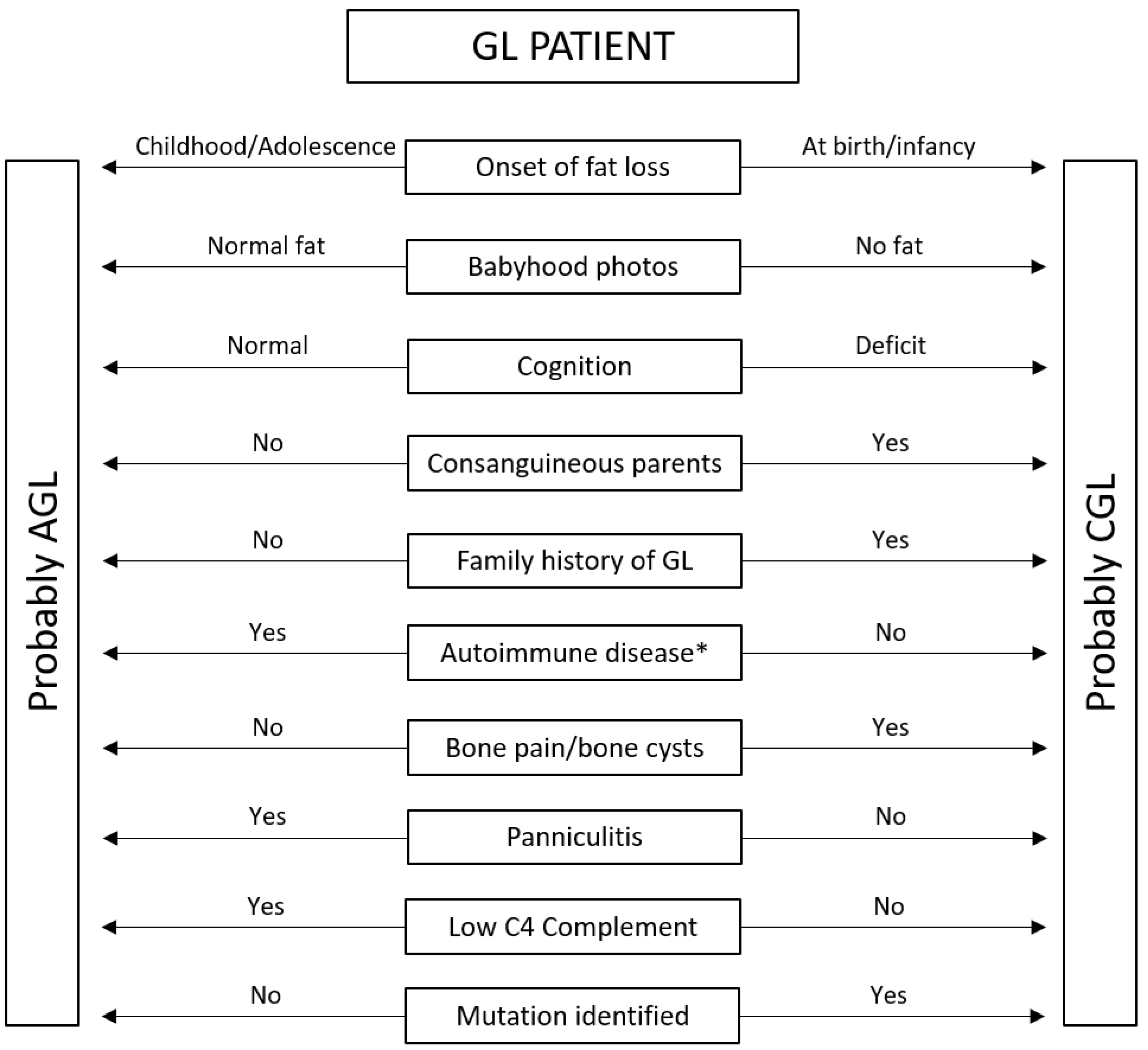
First, it is essential to confirm that fat scarcity is generalized. Next, we must identify when fat loss began. Babyhood photos can help determine whether lipodystrophy was present at birth [38]. A family history of lipodystrophies suggests a diagnosis of CGL [17,38], while the presence of autoimmune diseases in the patient or in their family members indicates a greater likelihood of AGL. Bone pain and cysts are common in CGL [61], whereas panniculitis occurs in approximately one-quarter of patients with AGL [37]. Figure 2 presents a clinical algorithm that helps with differential diagnosis.
After highlighting the differences and similarities in the clinical and laboratory findings of patients with CGL and AGL, we can return to the clinical cases and define their diagnoses.
A classification based on a large cohort of AGL cases categorized the disease into three subtypes: Type 1, associated with panniculitis; Type 2, linked to autoimmune disorders; and Type 3, considered idiopathic [37]. Nevertheless, this classification may require revision, as autoimmunity is much more frequent than previously reported [17,37], and some patients with panniculitis also exhibit concurrent autoimmune or other systemic conditions, challenging the exclusivity of these categories [40].
Patient 1 was referred to us with a diagnosis of CGL, as at that time, the loss of body fat was already significant. However, the association with hemolytic anemia and juvenile rheumatoid arthritis suggested this was an AGL case. Genetic testing for the classic genes associated with CGL (AGPAT2, BSCL2, CAV1, CAVIN) was negative, confirming the diagnosis of AGL.
In patient 2, the family history of CGL supports the diagnosis, making further mutation research unnecessary. The absence of autoimmune diseases in the patient and his family strengthens the diagnosis of CGL.
Patient 3 had a lesion on her right thigh suggestive of panniculitis (Figure 1C). It preceded the onset of body fat loss. Together with a family history of autoimmune disease, this suggests the diagnosis of AGL.
Patient 4, like patient 2, has a family history of CGL and was diagnosed with GL very early, in the first months of life (Figure 1E), suggesting the diagnosis of CGL. This patient was breastfed and was using milk according to his age. As milk is generally rich in fat, it can exacerbate hypertriglyceridemia, resulting in triglyceride values exceeding 1000 mg/dL. The characteristic phenotype of lipodystrophy in a child with severe hypertriglyceridemia confirms CGL, making other causes, such as familial chylomicronemia syndrome, unlikely.
Photos from the first year of life showed that patients 1 and 3 had a normal body fat distribution, suggesting they had AGL. Genetic study of patients 2 and 4 confirmed mutation in BSCL2.

6. Conclusions

In conclusion, we found several differences and similarities between Lawrence and Berardinelli–Seip syndromes. Recognizing them is essential for accurate diagnosis and appropriate treatment. Family history of GL, consanguinity between parents, associated panniculitis or autoimmune diseases, and the moment body fat loss begins are key points that help make the differential diagnosis. Failure to identify classic mutations increases the chance of AGL. Rare diseases are often underdiagnosed, particularly when classical signs are overlooked.

Author Contributions

Conceptualization, J.G.L.; methodology, J.G.L., L.N.L.; writing—original draft preparation, J.G.L., L.N.L.; writing—review and editing, V.Y.B.A., L.H.C.N., J.T.A.d.M.C. All authors have read and agreed to the published version of the manuscript.

Funding

This research received no external funding.

Institutional Review Board Statement

This research was approved by the Ethics Committee (CAAE 14070213.3.0000.5537, 10 October 2019).

Informed Consent Statement

Informed consent was obtained from all subjects involved in the study.

Data Availability Statement

The original contributions presented in the study are included in the article, further inquiries can be directed to the corresponding author.

Conflicts of Interest

The authors declare no conflicts of interest.

Abbreviations

The following abbreviations are used in this manuscript:
CGLCongenital Generalized Lipodystrophy
AGLAcquired Generalized Lipodystrophy
GLGeneralized Lipodystrophy

References

  1. Patni, N.; Garg, A. Lipodystrophy for the Diabetologist-What to Look For. Curr. Diab. Rep. 2022, 22, 461–470. [Google Scholar] [CrossRef] [PubMed]
  2. Lima, J.G.; Nobrega, L.H.C.; De Lima, N.N.; Do Nascimento Santos, M.G.; Baracho, M.F.P.; Jeronimo, S.M.B. Clinical and Laboratory Data of a Large Series of Patients with Congenital Generalized Lipodystrophy. Diabetol. Metab. Syndr. 2016, 8, 23. [Google Scholar] [CrossRef]
  3. Berardinelli, W. An Undiagnosed Endocrinometabolic Syndrome: Report of 2 Cases. J. Clin. Endocrinol. Metab. 1954, 14, 193–204. [Google Scholar] [CrossRef] [PubMed]
  4. Seip, M. Lipodystrophy and Gigantism with Associated Endocrine Manifestations. A New Diencephalic Syndrome? Acta Paediatr. 1959, 48, 555–574. [Google Scholar] [CrossRef]
  5. Lawrence, R.D. Lipodystrophy and Hepatomegaly, with Diabetes, Lipaemia, and Other Metabolic Disturbances; a Case Throwing New Light on the Action of Insulin. Lancet 1946, 247, 724–731. [Google Scholar] [CrossRef]
  6. Ziegler, L.H. Lipodystrophies: Report of seven cases. Brain 1928, 51, 147–167. [Google Scholar] [CrossRef]
  7. Hussain, I.; Garg, A. Lipodystrophy Syndromes. Endocrinol. Metab. Clin. N. Am. 2016, 45, 783–797. [Google Scholar] [CrossRef]
  8. Patni, N.; Chard, C.; Araújo-Vilar, D.; Phillips, H.; Magee, D.A.; Akinci, B. Diagnosis, Treatment and Management of Lipodystrophy: The Physician Perspective on the Patient Journey. Orphanet. J. Rare Dis. 2024, 19, 263. [Google Scholar] [CrossRef]
  9. Yildirim Simsir, I.; Tuysuz, B.; Ozbek, M.N.; Tanrikulu, S.; Celik Guler, M.; Karhan, A.N.; Denkboy Ongen, Y.; Gunes, N.; Soyaltin, U.E.; Altay, C.; et al. Clinical Features of Generalized Lipodystrophy in Turkey: A Cohort Analysis. Diabetes Obes. Metab. 2023, 25, 1950–1963. [Google Scholar] [CrossRef]
  10. Seip, M.; Trygstad, O. Generalized Lipodystrophy, Congenital and Acquired (Lipoatrophy). Acta Paediatr. 1996, 413, 2–28. [Google Scholar] [CrossRef]
  11. Akinci, B.; Onay, H.; Demir, T.; Ozen, S.; Kayserili, H.; Akinci, G.; Nur, B.; Tuysuz, B.; Nuri Ozbek, M.; Gungor, A.; et al. Natural History of Congenital Generalized Lipodystrophy: A Nationwide Study from Turkey. J. Clin. Endocrinol. Metab. 2016, 101, 2759–2767. [Google Scholar] [CrossRef]
  12. Sorkina, E.; Chichkova, V. Generalized Lipoatrophy Syndromes. Presse Medicale 2021, 50, 104075. [Google Scholar] [CrossRef] [PubMed]
  13. Handelsman, Y.; Oral, E.A.; Bloomgarden, Z.T.; Brown, R.J.; Chan, J.L.; Einhorn, D.; Garber, A.J.; Garg, A.; Timothy Garvey, W.; Grunberger, G.; et al. The Clinical Approach to the Detection of Lipodystrophy-An Aace Consensus Statement. Off. J. Am. Coll. Endocrinol. Am. Assoc. Clin. Endocrinol. 2013, 19, 107. [Google Scholar] [CrossRef] [PubMed]
  14. Brown, R.J.; Akinci, B.; Al Yaarubi, S.; Bismuth, E.; Cappa, M.; Deeb, A.; Kamrath, C.; Musso, C.; Patni, N.; Prodam, F.; et al. The Clinical Approach to Child and Adolescent Patients with Lipodystrophy: A Series of International Case Discussions. Front. Endocrinol. 2025, 16, 1597053. [Google Scholar] [CrossRef] [PubMed]
  15. Garg, A. Acquired and Inherited Lipodystrophies. N. Engl. J. Med. 2004, 350, 1220–1234. [Google Scholar] [CrossRef]
  16. Araújo-Vilar, D.; Santini, F. Diagnosis and Treatment of Lipodystrophy: A Step-by-Step Approach. J. Endocrinol. Investig. 2019, 42, 61–73. [Google Scholar] [CrossRef]
  17. Fernandez-Pombo, A.; Prado-Moraña, T.; Diaz-Lopez, E.J.; Sanchez-Iglesias, S.; Castro, A.I.; Cobelo-Gomez, S.; Araujo-Vilar, D. Clinical Characterisation and Comorbidities of Acquired Generalised Lipodystrophy: A 14-Year Follow-Up Study. J. Clin. Med. 2023, 12, 7344. [Google Scholar] [CrossRef]
  18. Deeb, A.; Hassan Beck, R.; Fatima, U.; Yoosuf, H. Acquired Generalized Lipodystrophy with Extensive Autoimmune Involvement: A Case Report and Review of the Literature. Horm. Res. Paediatr. 2025, 1–19. [Google Scholar] [CrossRef]
  19. Vatier, C.; Vigouroux, C.; Mosbah, H. Primary Disease of Adipose Tissue: When to Think about and How to Evaluate It in Clinical Practice? Ann. Endocrinol. 2024, 85, 190–194. [Google Scholar] [CrossRef]
  20. Llamas-Velasco, M.; Daudén, E.; Martínez-Peñas, G.; García-Diez, A. Late-Onset Acquired Generalized Lipodystrophy with Muscle Involvement. Actas Dermo-Sifiliogr. 2012, 103, 729–732. [Google Scholar] [CrossRef]
  21. Triaille, C.; Corvillo, F.; Mertens, A.; Hoffman, I.; Roskams, T.; López-Trascasa, M.; De Somer, L.; Casteels, K. Perilipin-1 Autoantibodies Are a Robust Marker of Acquired Lipodystrophy and May Precede Clinical Detection. Pediatr. Allergy Immunol. 2025, 36, e70026. [Google Scholar] [CrossRef] [PubMed]
  22. Mainieri, F.; Chiarelli, F. Lipodystrophies in Children. Horm. Res. Paediatr. 2022, 95, 305–320. [Google Scholar] [CrossRef] [PubMed]
  23. Fernández-Pombo, A.; Sánchez-Iglesias, S.; Castro-Pais, A.I.; Ginzo-Villamayor, M.J.; Cobelo-Gómez, S.; Prado-Moraña, T.; Díaz-López, E.J.; Casanueva, F.F.; Loidi, L.; Araújo-Vilar, D. Natural History and Comorbidities of Generalised and Partial Lipodystrophy Syndromes in Spain. Front. Endocrinol. 2023, 14, 1250203. [Google Scholar] [CrossRef] [PubMed]
  24. Pliszka, M.; Szablewski, L. Severe Insulin Resistance Syndromes: Clinical Spectrum and Management. Int. J. Mol. Sci. 2025, 26, 5669. [Google Scholar] [CrossRef] [PubMed]
  25. Özen, S.; Akıncı, B.; Oral, E.A. Current Diagnosis, Treatment and Clinical Challenges in the Management of Lipodystrophy Syndromes in Children and Young People. J. Clin. Res. Pediatr. Endocrinol. 2020, 12, 17–28. [Google Scholar] [CrossRef]
  26. Cortés, V.A.; Fernández-Galilea, M. Lipodystrophies: Adipose Tissue Disorders with Severe Metabolic Implications. J. Physiol. Biochem. 2015, 71, 471–478. [Google Scholar] [CrossRef]
  27. İşlek, A.; Sayar, E.; Yılmaz, A.; Duman, Ö.; Artan, R. A Very Rare Cause of Acute Pancreatitis: Berardinelli-Seip Congenital Lipodystrophy. Turk J. Gastroenterol. 2014, 25, 216–219. [Google Scholar] [CrossRef]
  28. Brahmanandam, L.; Vivekanand, B.; Mythili, A.; Subrahmanyam, K.A.V. Congenital Generalized Lipodystrophy 3 & 4 Presenting as Pancreatitis. J. Evid. Based Med. Healthc. 2019, 6, 2679–2683. [Google Scholar] [CrossRef]
  29. Cunningham, J.; Nadal, R.; Broome, C. Acquired Generalized Lipodystrophy Following Immune Thrombocytopenia. Am. J. Med. 2017, 130, e445–e446. [Google Scholar] [CrossRef]
  30. Giambelluca, D.; Di Martino, E.; Salvaggio, G. The “Kissing Sign” of Liver and Spleen. Abdom. Radiol. 2019, 44, 2323–2324. [Google Scholar] [CrossRef]
  31. Sahhar, N.N.; Ahmad, A.T.; Ammari, F.L.; Ajlouni, K.M. Dysregulation of Growth Hormone in Acquired Generalized Lipodystrophy. Saudi. Med. J. 2004, 25, 1766–1767. [Google Scholar]
  32. Meral, R.; Ryan, B.J.; Malandrino, N.; Jalal, A.; Neidert, A.H.; Muniyappa, R.; Akıncı, B.; Horowitz, J.F.; Brown, R.J.; Oral, E.A. “Fat Shadows” From DXA for the Qualitative Assessment of Lipodystrophy: When a Picture Is Worth a Thousand Numbers. Diabetes Care 2018, 41, 2255–2258. [Google Scholar] [CrossRef]
  33. Brush, M.; Auh, S.; Cochran, E.; Tuska, R.; Koh, C.; Kleiner, D.E.; Lightbourne, M.; Brown, R.J. Effects of Metreleptin in Patients with Generalized Lipodystrophy Before vs After the Onset of Severe Metabolic Disease. J. Clin. Endocrinol. Metab. 2025, 110, e1051–e1061. [Google Scholar] [CrossRef] [PubMed]
  34. Oral, E.A.; Simha, V.; Ruiz, E.; Andewelt, A.; Premkumar, A.; Snell, P.; Wagner, A.J.; DePaoli, A.M.; Reitman, M.L.; Taylor, S.I.; et al. Leptin-Replacement Therapy for Lipodystrophy. N. Engl. J. Med. 2002, 346, 570–578. [Google Scholar] [CrossRef] [PubMed]
  35. Diker-Cohen, T.; Cochran, E.; Gorden, P.; Brown, R.J. Partial and Generalized Lipodystrophy: Comparison of Baseline Characteristics and Response to Metreleptin. J. Clin. Endocrinol. Metab. 2015, 100, 1802–1810. [Google Scholar] [CrossRef] [PubMed]
  36. Lebastchi, J.; Ajluni, N.; Neidert, A.; Oral, E.A. A Report of Three Cases with Acquired Generalized Lipodystrophy with Distinct Autoimmune Conditions Treated with Metreleptin. J. Clin. Endocrinol. Metab. 2015, 100, 3967–3970. [Google Scholar] [CrossRef]
  37. Misra, A.; Garg, A. Clinical Features and Metabolic Derangements in Acquired Generalized Lipodystrophy: Case Reports and Review of the Literature. Medicine 2003, 82, 129–146. [Google Scholar] [CrossRef]
  38. Brown, R.J.; Araujo-Vilar, D.; Cheung, P.T.; Dunger, D.; Garg, A.; Jack, M.; Mungai, L.; Oral, E.A.; Patni, N.; Rother, K.I.; et al. The Diagnosis and Management of Lipodystrophy Syndromes: A Multi-Society Practice Guideline. J. Clin. Endocrinol. Metab. 2016, 101, 4500–4511. [Google Scholar] [CrossRef]
  39. Sánchez-Iglesias, S.; Fernández-Pombo, A.; Cobelo-Gómez, S.; Hermida-Ameijeiras, Á.; Alarcón-Martínez, H.; Domingo-Jiménez, R.; Riquelme, A.I.R.; Requena, J.R.; Araújo-Vilar, D. Celia’s Encephalopathy (Bscl2-Gene-Related): Current Understanding. J. Clin. Med. 2021, 10, 1435. [Google Scholar] [CrossRef]
  40. Ceccarini, G.; Magno, S.; Gilio, D.; Pelosini, C.; Santini, F. Autoimmunity in Lipodystrophy Syndromes. Presse Medicale 2021, 50, 104073. [Google Scholar] [CrossRef]
  41. Knebel, B.; Müller-Wieland, D.; Kotzka, J. Lipodystrophies—Disorders of the Fatty Tissue. Int. J. Mol. Sci. 2020, 21, 8778. [Google Scholar] [CrossRef]
  42. Ribeiro, A.; Brandão, J.R.; Cleto, E.; Santos, M.; Borges, T.; Santos Silva, E. Fatty Liver and Autoimmune Hepatitis: Two Forms of Liver Involvement in Lipodystrophies. GE Port J. Gastroenterol. 2019, 26, 362–369. [Google Scholar] [CrossRef]
  43. Liu, Q.; Pan, M.; Cao, H.; Zheng, J.; Ruan, Y.; Zhao, X. Acquired Generalized Lipodystrophy in a Juvenile Dermatomyositis Patient. Int. J. Rheum. Dis. 2024, 27, 15101. [Google Scholar] [CrossRef]
  44. Kumar, R.; Pilania, R.K.; Bhatia, A.; Dayal, D. Acquired Generalised Lipodystrophy and Type 1 Diabetes Mellitus in a Child: A Rare and Implacable Association. BMJ Case Rep. 2018, 2018, bcr-2018-225553. [Google Scholar] [CrossRef] [PubMed]
  45. Sorkina, E.; Frolova, E.; Rusinova, D.; Polyakova, S.; Roslavtseva, E.; Vasilyev, E.; Petrov, V.; Tiulpakov, A. Progressive Generalized Lipodystrophy as a Manifestation of Autoimmune Polyglandular Syndrome Type 1. J. Clin. Endocrinol. Metab. 2016, 101, 1344–1347. [Google Scholar] [CrossRef] [PubMed]
  46. Cavdar, U.; Akinci, B.; Foss de Freitas, M.C.; Conjeevaram, H.; Fox, D.; Fontana, R.; Tayeh, M.; Frame, D.; Innis, J.; Walkovich, K.; et al. Untangling the Heterogeneity of Acquired Generalized Lipodystrophy. J. Endocr. Soc. 2021, 5, A39–A40. [Google Scholar] [CrossRef]
  47. Foss-Freitas, M.C.; Akinci, B.; Luo, Y.; Stratton, A.; Oral, E.A. Diagnostic Strategies and Clinical Management of Lipodystrophy. Expert Rev. Endocrinol. Metab. 2020, 15, 95–114. [Google Scholar] [CrossRef]
  48. Mathiesen, P.; Hegaard, H.; Herlin, T.; Zak, M.; Pedersen, F.K.; Nielsen, S. Long-Term Outcome in Patients with Juvenile Dermatomyositis: A Cross-Sectional Follow-up Study. Scand. J. Rheumatol. 2012, 41, 50–58. [Google Scholar] [CrossRef]
  49. Esfandiari, N.H.; Rubenfire, M.; Neidert, A.H.; Hench, R.; Eldin, A.J.; Meral, R.; Oral, E.A. Diagnosis of Acquired Generalized Lipodystrophy in a Single Patient with T-Cell Lymphoma and No Exposure to Metreleptin. Clin. Diabetes Endocrinol. 2019, 5, 4. [Google Scholar] [CrossRef]
  50. Brown, R.J.; Chan, J.L.; Jaffe, E.S.; Cochran, E.; Depaoli, A.M.; Gautier, J.F.; Goujard, C.; Vigouroux, C.; Gorden, P. Lymphoma in Acquired Generalized Lipodystrophy. Leuk. Lymphoma 2016, 57, 45–50. [Google Scholar] [CrossRef]
  51. Kennedy, R.; Macrohon, C.R.L.; David, M.L.G.V.; Lee, M.; Salama, A.K.S.; Shariff, A. Acquired Generalized Lipodystrophy as an Adverse Event of Combined Immune Checkpoint Inhibitor Therapy. JCEM Case Rep. 2025, 3, luaf023. [Google Scholar] [CrossRef]
  52. Corvillo, F.; Aparicio, V.; López-Lera, A.; Garrido, S.; Araújo-Vilar, D.; de Miguel, M.P.; López-Trascasa, M. Autoantibodies Against Perilipin 1 as a Cause of Acquired Generalized Lipodystrophy. Front. Immunol. 2018, 9, 2142. [Google Scholar] [CrossRef] [PubMed]
  53. Mandel-Brehm, C.; Vazquez, S.E.; Liverman, C.; Cheng, M.; Quandt, Z.; Kung, A.F.; Parent, A.; Miao, B.; Disse, E.; Cugnet-Anceau, C.; et al. Autoantibodies to Perilipin-1 Define a Subset of Acquired Generalized Lipodystrophy. Diabetes 2023, 72, 59–70. [Google Scholar] [CrossRef] [PubMed]
  54. Kozusko, K.; Tsang, V.H.M.; Bottomley, W.; Cho, Y.H.; Gandotra, S.; Mimmack, M.; Lim, K.; Isaac, I.; Patel, S.; Saudek, V.; et al. Clinical and Molecular Characterization of a Novel PLIN1 Frameshift Mutation Identified in Patients with Familial Partial Lipodystrophy. Diabetes 2015, 64, 299–310. [Google Scholar] [CrossRef]
  55. Bhatia, R.; Chennupathi, P.; Rosenstein, E.D.; Advani, S. Spontaneous Remission of Acquired Generalized Lipodystrophy Presenting in the Postpartum Period. JCEM Case Rep. 2024, 2, luae009. [Google Scholar] [CrossRef]
  56. Hill, M.; Weissman, A.S.; Hirshburg, J.; McBride, J.D.; Lawrence, H. An Unusual Case of Acquired Generalized Lipodystrophy (Panniculitis Variety). Pediatr. Dermatol. 2024, 41, 1152–1155. [Google Scholar] [CrossRef]
  57. Fourman, L.T.; Lima, J.G.; Simha, V.; Cappa, M.; Alyaarubi, S.; Montenegro, R.; Akinci, B.; Santini, F. A Rapid Action Plan to Improve Diagnosis and Management of Lipodystrophy Syndromes. Front. Endocrinol. 2024, 15, 1383318. [Google Scholar] [CrossRef]
  58. Garg, A. Lipodystrophies: Genetic and Acquired Body Fat Disorders. J. Clin. Endocrinol. Metab. 2011, 96, 3313–3325. [Google Scholar] [CrossRef]
  59. Savage, D.B.; Semple, R.K.; Clatworthy, M.R.; Lyons, P.A.; Morgan, B.P.; Cochran, E.K.; Gorden, P.; Raymond-Barker, P.; Murgatroyd, P.R.; Adams, C.; et al. Complement Abnormalities in Acquired Lipodystrophy Revisited. J. Clin. Endocrinol. Metab. 2009, 94, 10–16. [Google Scholar] [CrossRef]
  60. Eren, E.; Özkan, T.B.; Papatya Çakir, E.D.; Saǧlam, H.; Tarim, Ö. Acquired Generalized Lipodystrophy Associated with Autoimmune Hepatitis and Low Serum C4 Level. JCRPE J. Clin. Res. Pediatr. Endocrinol. 2010, 2, 39–42. [Google Scholar] [CrossRef]
  61. Gregory, J.M.; Arkader, A.; Bothari, A.; Dormans, J.P. Case Report: Unicameral Bone Cysts in a Young Patient with Acquired Generalized Lipodystrophy. Clin. Orthop. Relat. Res. 2010, 468, 1440–1446. [Google Scholar] [CrossRef]
  62. Westvik, J. Radiological Features in Generalized Lipodystrophy. Acta Paediatr. 1996, 85, 44–51. [Google Scholar] [CrossRef] [PubMed]
  63. Haque, W.A.; Shimomura, I.; Matsuzawa, Y.; Garg, A. Serum Adiponectin and Leptin Levels in Patients with Lipodystrophies. J. Clin. Endocrinol. Metab. 2002, 87, 2395. [Google Scholar] [CrossRef] [PubMed]
  64. Ponte, C.M.M.; Fernandes, V.O.; Gurgel, M.H.C.; Vasconcelos, I.; Karbage, L.; Liberato, C.B.R.; Negrato, C.A.; Gomes, M.B.; Montenegro, A.; Montenegro Junior, R.M. Early Commitment of Cardiovascular Autonomic Modulation in Brazilian Patients with Congenital Generalized Lipodystrophy. BMC Cardiovasc. Disord. 2018, 18, 6. [Google Scholar] [CrossRef]
  65. Hussain, I.; Patni, N.; Garg, A. Lipodystrophies, Dyslipidaemias and Atherosclerotic Cardiovascular Disease. Pathology 2019, 51, 202–212. [Google Scholar] [CrossRef]
  66. Feijó, B.M.X.C.R.R.; Mendonça, R.M.; Egito, E.S.T.; Lima, D.N.; Campos, J.T.A.D.M.; Lima, J.G. Coronary Arterial Calcification in Patients with Congenital Generalised Lipodystrophy: A Case Series. Clin. Endocrinol. 2022, 97, 863–866. [Google Scholar] [CrossRef] [PubMed]
  67. Akinci, G.; Alyaarubi, S.; Patni, N.; Alhashmi, N.; Al-Shidhani, A.; Prodam, F.; Gagne, N.; Babalola, F.; Al Senani, A.; Muniraj, K.; et al. Metabolic and Other Morbid Complications in Congenital Generalized Lipodystrophy Type 4. Am. J. Med. Genet. A 2024, 194, e63533. [Google Scholar] [CrossRef]
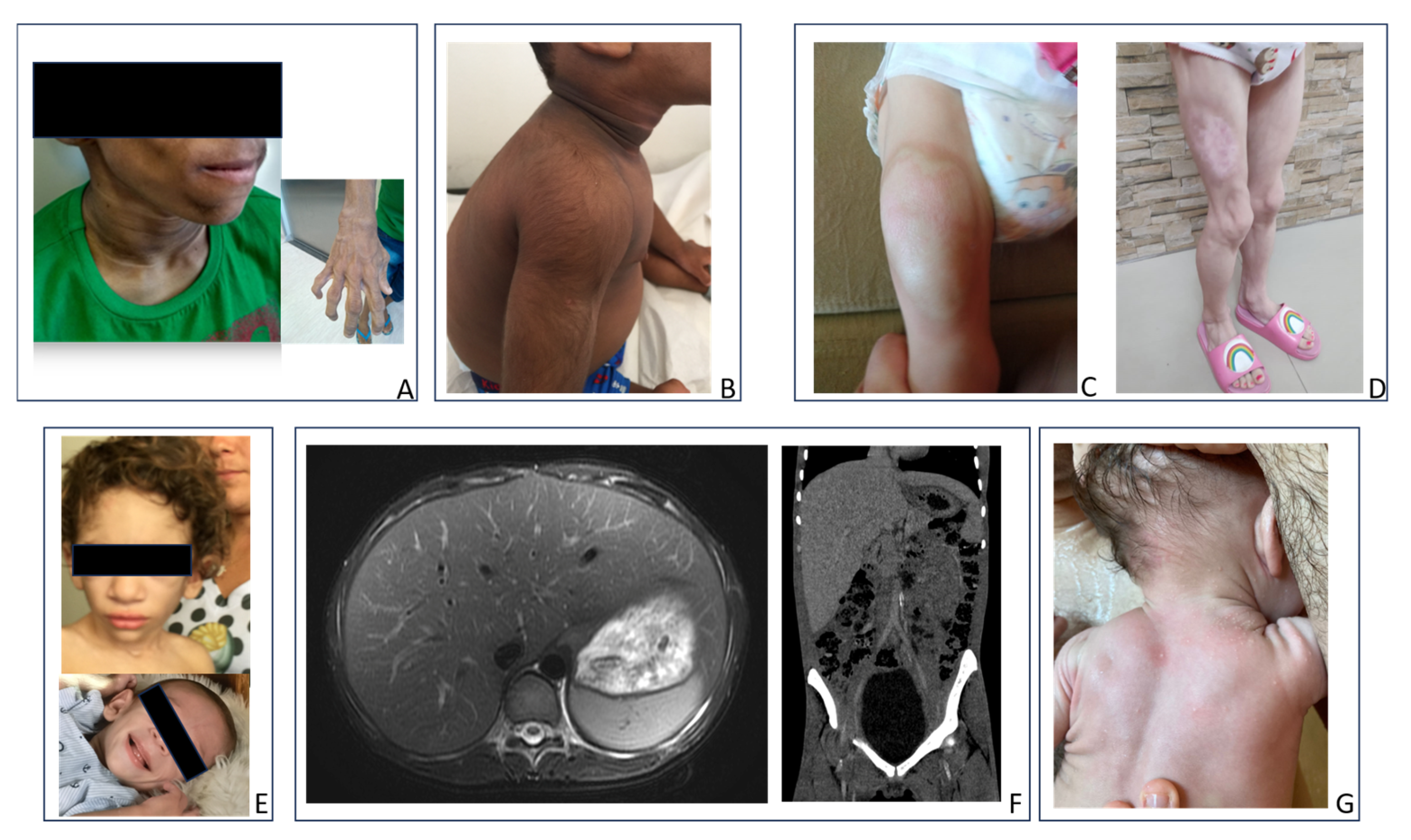
Figure 1. Clinical and imaging data of patients with generalized lipodystrophy (GL). (A) Acanthosis nigricans, loss of the fat pad of Bichat, and hand deformity (Case 1). (B)—Hypertrichosis and Acanthosis nigricans. (C) Panniculitis (acute phase). (D) Panniculitis (late phase) and muscular hypertrophy in the lower limbs. (E) Patient with GL at birth and during the first consultation. (F) Severe hepatomegaly (“kissing sign”) in a patient with type 1 CGL. (G) Eruptive xanthomas in an infant with CGL and severe hypertriglyceridemia.
Endocrines 06 00055 g001
Figure 2. Clinical algorithm to differentiate between congenital (CGL) and acquired (AGL) generalized lipodystrophies (GL). * Personal or family history of autoimmune disease.
Endocrines 06 00055 g002
Table 1. Differences and similarities between patients with congenital and acquired generalized lipodystrophies.
CGL
(Berardinelli–Seip Syndrome)
AGL
(Lawrence Syndrome)
DIFFERENCES
Onset of body fat lossAt birth or infancyChildhood or adolescence
Body fat at birth *Low or very lowNormal
Voracious appetiteEarly childhoodLater (if present)
CognitionIntellectual disability can occur in CGL type 2Normal cognition
Physical examHypertrichosisPanniculitis, joint deformities (if rheumatoid arthritis)
Associated diseasesBony cysts, cardiovascular diseasePanniculitis, autoimmune diseases **
Liver diseaseInduced by ectopic fatEctopic fat or autoimmune
Serum leptinAlways lowIt may be normal if the loss of body fat is not yet general
C4 complementNormalCan be low
Body fat (DXA)Always lowIt may not be low if the loss of body fat is not yet general
Insulin resistanceAppears earlyAppears later, depending on the loss of body fat
SIMILARITIES
Total body fatLow or very low
Physical examLoss of Bichat’s fat pad, Acanthosis nigricans, acrochordons, phlebomegaly, umbilical scar protrusion, hepatomegaly, muscle hypertrophy
Blood testsHyperinsulinemia, hyperglycemia, low HDL, hypertriglyceridemia
CGL—congenital generalized lipodystrophy; AGL—acquired generalized lipodystrophy. * Best assessed using photographs from the first year of life. ** Personal or family history of autoimmune disease.
Disclaimer/Publisher’s Note: The statements, opinions and data contained in all publications are solely those of the individual author(s) and contributor(s) and not of MDPI and/or the editor(s). MDPI and/or the editor(s) disclaim responsibility for any injury to people or property resulting from any ideas, methods, instructions or products referred to in the content.