Article

Euglycemic Hyperinsulinemia Lowers Blood Pressure and Impedes Microvascular Perfusion More Effectively in Persons with Cardio-Metabolic Disease

by
Zhenqi Liu
1,
Linda A. Jahn
1 and
Eugene J. Barrett
1,2,*
1
Division of Endocrinology and Metabolism, Department of Medicine, University of Virginia School of Medicine, Charlottesville, VA 51540, USA
2
Department of Pharmacology, University of Virginia School of Medicine, Charlottesville, VA 51540, USA
*
Author to whom correspondence should be addressed.
Endocrines 2025, 6(3), 36; https://doi.org/10.3390/endocrines6030036
Submission received: 12 May 2025 / Revised: 29 June 2025 / Accepted: 16 July 2025 / Published: 24 July 2025
(This article belongs to the Special Issue Feature Papers in Endocrines 2025)

Abstract

In healthy humans, insulin at physiological concentrations exerts acute vasodilatory actions on both resistance and terminal arterioles, leading, respectively, to increased total blood flow and the microvascular network volume being perfused. The process of increasing capillary network volume is frequently referred to as “capillary recruitment”. Together these two vascular actions of insulin enhance the delivery of oxygen, nutrients, and insulin itself to tissues. Both processes are diminished by insulin resistance. Here we examined interactions between insulin’s acute (within 2 h) actions on blood pressure (both central and peripheral) and on capillary recruitment in healthy controls and in four distinct groups of people with heightened cardio-metabolic disease (CMD) risk: individuals with obesity, metabolic syndrome, and type 1 or type 2 diabetes. Insulin increased microvascular blood volume (MBV) more effectively in controls than in each of the four CMD risk groups (p < 0.001). Conversely, insulin lowered both central and peripheral systolic pressure (p < 0.05 or less) in each of the CMD risk groups but not in the controls. The insulin-induced blood pressure decrements were greater in the metabolic syndrome, type 2 diabetes, and obesity groups (p < 0.05 or less) than in the controls. The greater blood pressure declines likely reflect decreased sympathetic baroreceptor reflex tone. These effects on blood pressure combined with the diminished dilation of terminal arterioles due to microvascular insulin resistance in the CMD risk subjects led to decreased distal microvascular perfusion as evidenced by changes in MBV. These findings highlight the complex interplay between insulin’s actions on resistance and terminal arterioles in individuals with a high CMD risk, underscoring the importance of addressing microvascular dysfunction in these conditions.

1. Introduction

Insulin relaxes blood vessels both in vivo and in vitro via direct action on the vascular endothelial cell insulin receptor to activate the phosphotidylinositol-3-kinase signaling pathway and increase nitric oxide (NO) production [1,2,3]. This action is currently thought to be responsible for most, if not all of insulin’s vasodilatory actions. Insulin infusion under euglycemic conditions also raises plasma norepinephrine concentrations but does not elevate blood pressure [4,5], suggesting a balance between insulin’s direct vasodilatory effect and adrenergic vasoconstriction [6,7]. Surprisingly, insulin’s acute effect on either peripheral [8] or central blood pressure [9] has not been extensively studied in subjects with cardio-metabolic disease (CMD) [8,9]. We recently reported that euglycemic hyperinsulinemia lowered both central and peripheral pressures in humans with type 2 diabetes (T2DM) [10] and this effect was enhanced by treatment with empagliflozin.
Using contrast-enhanced ultrasound (CEU) coupled with the euglycemic insulin clamp, we tested in animals [11] and humans [12] the hypothesis that insulin’s microvascular action in muscle is secondary to the relaxation of terminal arterioles with the consequent increase in capillary network perfused volume. We and others have shown that physiologic hyperinsulinemia promptly (within 15 min) increases muscle microvascular blood volume (MBV) [11,13]. This increase facilitates insulin’s metabolic action in skeletal muscle [14], adipose tissue [15] and myocardium [16] by accelerating both insulin and nutrient delivery to myocytes and adipocytes. Inhibition of NO synthase abolishes insulin-mediated muscle microvascular perfusion and blunts insulin-stimulated muscle glucose uptake [14].
While we observed that acute insulin infusion (euglycemic clamp) increased muscle MBV in most healthy humans, in settings of CMD it frequently either failed to increase or paradoxically diminished the MBV perfused [10,17,18,19]. Here we compared the frequency of occurrence of decreased MBV in healthy young controls and in each of four CMD risk groups including metabolic syndrome, T2DM, type 1 diabetes (T1DM), and obesity and provide the first report of the frequency of this dysfunctional vasoconstrictive response to insulin. In these same subjects, we also measured brachial artery pressure and used radial artery tonometry to examine heart rate and central aortic blood pressure changes in response to euglycemic hyperinsulinemia.
We were intrigued to find that in these studies both peripheral and central aortic pressures declined significantly during two hours of euglycemic hyperinsulinemia in the T1DM, T2DM, metabolic syndrome, and obesity groups but not in the healthy controls. We suggest that the contrasting responses of insulin-induced blood pressure and microvascular perfused volume changes seen in the insulin-resistant subjects compared to the controls can be explained by a diminished systemic baroreceptor reflex-mediated sympathetic vascular constriction of resistance arterioles simultaneous with lessened terminal arteriolar relaxation in skeletal muscle thereby decreasing muscle microvascular perfused volume in the insulin-resistant subjects.

2. Methods

2.1. Study Participants

Study participants were recruited either through newspaper advertisements, posters, or letters mailed to patients followed at University of Virginia outpatient clinics. Respondents were contacted by telephone to review inclusion/exclusion criteria. Anyone taking sodium glucose transporter-2 inhibitors, glucagon-like peptide receptor agonists, or having a body mass index (BMI) ≤ 19 or ≥43 kg/m2, or with active cardiac, pulmonary, renal, hepatic, or central nervous system illnesses were excluded. Subsequently, each participant completed a written informed consent and underwent a screening visit that included a medical history and physical examination, and measurements of serum electrolytes, liver, and renal function, a complete blood count, fasting glucose, C-peptide, hemoglobin A1c, a lipid panel, and a pregnancy test (if applicable). Women with positive pregnancy tests were excluded from the study. In total, 125 respondents passed the screening and were studied. This included 22 participants with at least a 1-year history of T2DM, 25 subjects with T1DM, 22 subjects with metabolic syndrome, 21 subjects with obesity (BMI ≥ 30 kg/m2), and 35 healthy controls. Diabetes was diagnosed based on the American Diabetes Association (ADA) diagnostic criteria. Metabolic syndrome diagnosis was based on the criteria defined by the American Heart Association–National Heart Lung and Blood Institute (AHA-NHLBI). Participants who were on stable doses of antihypertensive or lipid-lowering medications (≥6 months) at the time of recruitment were included in the study. All study protocols were reviewed and approved by the University of Virginia Human Investigation Committee (IRB protocols # 15792, # 18237, # 18895 and # 21403). All infusions, vascular and imaging studies were performed in the University of Virginia Clinical Research Unit (CRU).

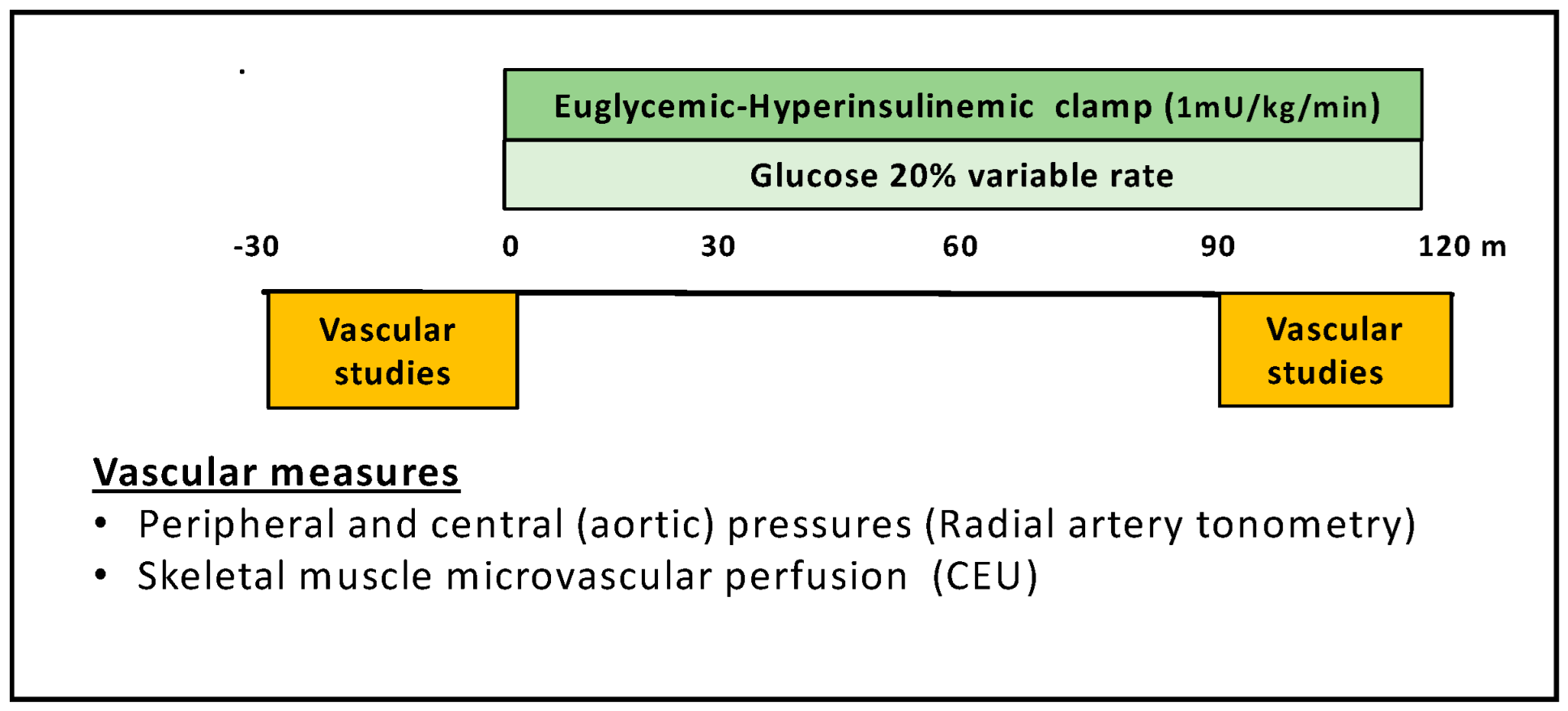
2.2. Experimental Protocol

Figure 1 illustrates the study design for measuring insulin’s vascular and metabolic actions. Participants were admitted to the CRU at 7 am after an overnight fast. An intravenous catheter was placed in the right antecubital vein for infusion of insulin, glucose, and ultrasound contrast agent (Definity microbubbles, Lantheus Medical Imaging, N. Billerica, MA, USA) and a second catheter was placed at the right wrist for blood sampling. For participants with diabetes, fasting plasma glucose was measured and if outside a target range of 100–140 mg/dL, a slow infusion of either glucose (1 mg/kg/min) or insulin (0.15 mU/kg/min) was begun and continued until plasma glucose was within the desired target range. We then obtained baseline (time 0) measurements of metabolic parameters including plasma glucose, insulin, C-peptide, and hemoglobin A1c, and vascular functions including peripheral and central blood pressure and microvascular parameters. Peripheral blood pressure was measured at the brachial artery, while central aortic blood pressure and pulse pressures were measured by radial artery tonometry. Measures of skeletal muscle MBV, microvascular flow velocity (MFV), and microvascular blood flow (MBF) were obtained using CEU. After these measurements were completed, a hyperinsulinemic–euglycemic clamp (2 mU/kg/min × 10 min followed by 1 mU/kg/min × 110 min) was begun and vascular measurements were repeated between 90 and 120 min of the clamp. Glucose was measured every 5 min and insulin every 30 min during the clamp.

2.3. Determination of Microvascular Perfusion by CEU

Forearm flexor muscle microvascular perfusion was assessed with a Phillips Epiq 7 ultrasound system during steady-state infusion of Definity microbubbles (Lantheus Medical Imaging; North Billerica, MA, USA) using low mechanical index (MI = 0.10) continuous imaging of forearm flexor muscles for 30 s at a frame rate of 15 s−1 with a flash at 0.88 MI to initiate a replenishment curve, as described previously [10]. Four replenishment curves were acquired immediately before starting and again at the end of the insulin clamp (Figure 1). Replenishment curves were analyzed using Q-Lab software (Philips Research; Cambridge, MA, USA) yielding measures of MBV (in video intensity units) and MFV (in s−1).

2.4. Recording of Peripheral and Central Blood Pressure

Systemic blood pressure was measured at the brachial artery by automated sphygmomanometry and central blood pressures were quantified at the radial artery using applanation tonometry (SphygmoCor System, AtCor Medical, Itasca, IL, USA) before, and between 90 and 120 min of insulin infusion. Subjects were supine with the head elevated approximately 30° for all blood pressure measurements.

2.5. Euglycemic Hyperinsulinemic Clamp

After obtaining baseline plasma samples and vascular measurements, a 2 mU/kg/min insulin infusion was started and the rate decreased to 1 mU/kg/min at 10 min and maintained until 120 min. Plasma glucose was measured every 5 min and maintained at baseline through a variable rate glucose infusion. We chose two hours of insulin infusion for vascular and metabolic measurements as prior studies have shown that insulin’s actions reach a near-steady state within this interval. During the insulin clamp, to avoid the confounding systemic vasodilation due to limb heating, we did not use a “heated hand” to arterialize venous blood [20].

2.6. Biochemical Analyses

Serum electrolytes, creatinine, liver function, C-peptide, hemoglobin A1c, complete blood counts, lipid profiles, and pregnancy tests were assayed at the University of Virginia Clinical Chemistry Laboratory. Plasma glucose was measured using a YSI glucose analyzer (Yellow Spring Instruments, Inc.; Yellow Springs, OH, USA). Plasma insulin was determined by ELISA (Alpco Corp., Salem, NH, USA).

2.7. Statistics

The primary endpoint for microvascular insulin responsiveness was the change (increase or decrease) in forearm muscle MBV from baseline to end-of-clamp. The secondary endpoint for vascular insulin responsiveness was central and peripheral blood pressure changes. For assessing metabolic insulin sensitivity, we used the glucose infusion rate (GIR) needed to maintain euglycemia during the clamp. Data are presented as the mean ± standard error of the mean (SE). Comparisons were made between measurements made before beginning (time −30–0 min, baseline) and at the end (time 90–120 min) of the insulin clamp using paired Student’s t-test, one- or two-way ANOVA, and Chi-squared test as appropriate. All statistical analyses were conducted with GraphPad Prism Software (Version 8, Dotmatics, Boston, MA, USA). Statistical significance was defined as p ≤ 0.05 for primary and secondary endpoints.

3. Results

Both skeletal muscle microvascular perfusion indices, and peripheral and central blood pressures before and after insulin were available on all 125 subjects. This included 35 healthy young controls and 21 to 25 subjects with T2DM, or T1DM, or metabolic syndrome or obesity (Table 1). The control subjects were significantly younger and had lower BMIs than the other four groups.
Microvascular and Glucose Metabolic Responses to Insulin: Thirty-one of the 35 control subjects (89%) responded to insulin infusion with an increase in MBV, i.e., a microvascular vasodilatory response, while 4 of the 35 control subjects (11%) responded with a decline in MBV; i.e., a terminal arteriolar vasoconstrictive response (Table 2) that decreased the perfused capillary volume. A significantly greater fraction of subjects with increased CMD risk responded to insulin infusion with a decline in MBV ranging from 46% in the T1DM subjects up to 73% in the metabolic syndrome group. The fraction with a vasoconstrictive response in the control group was significantly less (p < 0.01, Chi-square test) than each of the other groups. The glucose metabolic response to insulin infusion was greatest in the control subjects (Table 2). At steady-state over the last 30 min of the clamp the controls required glucose infusions of 6.2 mg/kg/min on average to maintain euglycemia. This was modestly greater than the GIR for T1DM and significantly greater than the metabolic syndrome, T2DM, or obesity groups, indicating metabolic insulin resistance in each CMD group.
Brachial and Central Aortic Blood Pressure Responses to Insulin: The effects of two hours of euglycemic hyperinsulinemia on brachial systolic and central aortic systolic blood pressures are shown in Table 3. As reported by others [5,21], acute insulin infusion had no significant effect on peripheral (brachial) systolic blood pressure in healthy controls. However, it did provoke a modest but significant (~3 mmHg) decline in the aortic systolic pressure in the control subjects. Surprisingly, in each of the CMD groups, systolic blood pressure declined significantly in both the systemic and central aortic circulation. Heart rate rose significantly in the control participants as well as in those with obesity and T2DM, but not in the metabolic syndrome or T1DM subjects, consistent with a more variable sympathetic baroreceptor response in the latter two groups. Table 4 provides the comparison of the changes in peripheral and aortic systolic and diastolic pressures between CMD groups and controls. The insulin infusion yielded significantly greater decrements of both peripheral and aortic systolic pressures in the subjects with metabolic syndrome, obesity, or T2DM compared to the controls. In T1DM subjects both peripheral and central systolic pressures trended towards a greater decline than in controls but this was not statistically significant. Both peripheral and central diastolic pressure declined modestly during the insulin infusion in all groups, with no statistically significant differences between groups.

4. Discussion

It has been more than 40 years since the first report that euglycemic hyperinsulinemia raised circulating catecholamines unaccompanied by changes in blood pressure in healthy young subjects [21]. Subsequent studies demonstrated that euglycemic hyperinsulinemia increases muscle blood flow by decreasing systemic vascular resistance [22], indicating a primary vasodilatory effect of insulin. Thus, insulin, independent of changes in blood glucose, directly affects vascular tone. This subsequently was shown secondary to insulin increasing production of NO in vascular tissue [2,23,24]. These findings suggested a dynamic balance between adrenergic vasoconstriction and insulin-induced vasodilation resulting in minimal or no changes in systemic blood pressure [4,5]. Recent studies support a role for baroreceptor-mediated increases in α1 adrenergic action in muscle microvasculature balancing the vasodilator effect of insulin [6,7].
Here we confirm that insulin infusion has no direct effect on peripheral systolic or diastolic pressures in healthy young adults, and demonstrate for the first time that it has a highly significant effect to lower peripheral systolic pressures in subjects with insulin resistance of diverse origins. We provide a further novel finding that central aortic pressure, derived using radial artery tonometry, responds to euglycemic hyperinsulinemia by decreasing systolic pressure even in young healthy subjects and these decreases are significantly exaggerated in insulin-resistant subjects. These findings suggest a previously unrecognized dysfunction of the balancing process in subjects with increased CMD risk. This was particularly evident in the metabolic syndrome, T2DM and obesity groups in whom both systemic and aortic systolic pressure fell most markedly with insulin infusion. In the same groups, during the two hours of insulin infusion there was a higher prevalence of decreased muscle microvascular perfusion as measured by CEU. Taken together, this pattern of vascular response in the subjects with heightened CMD risk is most consistent with the fall in blood pressure during insulin infusion diminishing microvascular perfusion distally perhaps facilitated by the absence of a vasodilatory effect on the precapillary arterioles.
Our observation in controls that insulin induces a significant decrease in central aortic blood pressure alongside an increase in heart rate, without affecting systemic blood pressure, aligns with the previously established inverse relationship between heart rate and central aortic blood pressure. An elevated heart rate increases the reflection of pressure waves back towards the heart, leading to lower central aortic pressure, and multiple regression adjusted for brachial arterial blood pressure showed heart rate to be a major determinant of central aortic pressure [25]. Whether the increases in heart rate in the healthy control were secondary to insulin-mediated vasodilation in the peripheral arteries and the microvasculature remains unclear.
Another novel aspect of insulin’s vascular actions reported here is the first evaluation of the prevalence of a paradoxical vasoconstrictive microvascular response to euglycemic hyperinsulinemia. This response occurred at a low frequency (approximately 10%) in healthy young individuals but was far more prevalent (50–70% or greater) in individuals with increased CMD risk. Even this may be an underestimate as we have taken care to study individuals with increased CMD risk who are without clinical cardiovascular disease. The prevalence of paradoxical microvascular vasoconstrictive responses was greatest in the groups with the most marked metabolic insulin resistance as indicated by the GIR (metabolic syndrome and T2DM). This would be consistent with previously published data that diminishing insulin’s microvascular vasodilatory action contributes to metabolic insulin resistance [17,26,27]. Collectively, the diminished sympathetic vasoconstrictive effect on resistance arterioles and lack of precapillary arteriolar relaxation (indicated by the decline of MBV with insulin) appear to critically affect perfusion of muscle capillary networks. It is noteworthy that while declines in systolic pressure in the CMD subjects were significant, there were also non-significant downward trends of both brachial and central aortic diastolic pressures. The better preservation of diastolic pressure may be related to the fluid volume replacement which was occurring with the glucose and insulin infusions.
We note that both men and women were included, though not in equal numbers, in each of the five study groups. We did not see any sex-based differences in either the microvascular or blood pressure responses. However, we recognize that the study was not powered for such a subgroup analysis. To truly resolve the question, sex-based differences in insulin’s action on blood pressure or microvascular perfusion will require larger prospectively designed studies.
These findings carry significant clinical implications. The microvasculature plays a pivotal role in delivering oxygen, nutrients, and trophic hormones to tissues, while also facilitating the removal of metabolic wastes from tissues. Additionally, microvasculature, particularly the small arterioles within it, crucially determines the peripheral vascular resistance. Thus, vasoconstrictive responses to insulin in conditions such as obesity, metabolic syndrome and diabetes may help explain the propensity for hypertension, easy fatigability, and poor wound healing commonly associated with insulin resistance.
In conclusion, insulin effectively increased MBV, along with increased heart rate and decreased aortic blood pressure without affecting peripheral arterial blood pressure in young and healthy humans. In contrast, insulin lowered both central aortic and peripheral arterial systolic blood pressure and frequently induced microvascular vasoconstriction in individuals with obesity, metabolic syndrome, T1DM or T2DM, all conditions linked to elevated CMD risk. These effects on blood pressure, likely due to diminished sympathetic baroreceptor reflex tone, combined with reduced dilation of terminal arterioles from microvascular insulin resistance, lead to decreased distal microvascular flow. The findings underscore the importance of addressing vascular dysfunction to mitigate the cardiometabolic risks associated with insulin resistance.

Author Contributions

Conceptualization, E.J.B.; data curation, E.J.B. and L.A.J.; formal analysis, E.J.B.; funding acquisition, Z.L.; investigation, L.A.J. and Z.L.; methodology, L.A.J. and Z.L.; project administration, E.J.B.; writing—original draft, E.J.B. and Z.L. All authors have read and agreed to the published version of the manuscript.

Funding

This work was supported by National Institutes of Health grants R01DK124344 and R01DK125330 (to Z.L.) and HL142250 (to E.B.).

Institutional Review Board Statement

All study protocols were reviewed and approved by the University of Virginia Human Investigation Committee (IRB protocols # 15792, # 18237, # 18895 and # 21403). All infusions, vascular and imaging studies were performed in the University of Virginia Clinical Research Unit (CRU).

Informed Consent Statement

Informed consent was obtained from all subjects involved in the study.

Data Availability Statement

The data that support the findings of this study are available on reasonable request from the corresponding author.

Acknowledgments

The authors extend their thanks to the clinical research coordinators and technicians staffing the UVA clinical research unit, and to the subjects who participated as volunteers in these studies.

Conflicts of Interest

The authors declare no conflicts of interest.

Abbreviations

CMD—cardio-metabolic disease; MBV—microvascular blood volume; T2DM—type 2 diabetes mellitus; T1DM—type 1 diabetes mellitus; CEU—contrast-enhanced ultrasound; NO—nitric oxide; BMI—body mass index; CRU—clinical research unit.

References

  1. Zeng, G.; Nystrom, F.H.; Ravichandran, L.V.; Cong, L.-N.; Kirby, M.; Mostowski, H.; Quon, M.J. Roles for insulin receptor, PI3-kinase, and Akt in insulin-signaling pathways related to production of nitric oxide in human vascular endothelial cells. Circulation 2000, 101, 1539–1545. [Google Scholar] [CrossRef] [PubMed]
  2. Zeng, G.; Quon, M.J. Insulin-stimulated production of nitric oxide is inhibited by wortmannin. Direct measurement in vascular endothelial cells. J. Clin. Investig. 1996, 98, 894–898. [Google Scholar] [CrossRef] [PubMed]
  3. Jiang, Z.Y.; Lin, Y.-W.; Clemont, A.; Feener, E.P.; Hein, K.D.; Igarashi, M.; Yamauchi, T.; White, M.F.; King, G.L. Characterization of selective resistance to insulin signaling in the vasculature of obese Zucker (fa/fa) rats. J. Clin. Investig. 1999, 104, 447–457. [Google Scholar] [CrossRef] [PubMed]
  4. Anderson, E.A.; Hoffman, R.P.; Balon, T.W.; Sinkey, C.A.; Mark, A.L. Hyperinsulinemia produces both sympathetic neural activation and vasodilation in normal humans. J. Clin. Investig. 1991, 87, 2246–2252. [Google Scholar] [CrossRef] [PubMed]
  5. Anderson, E.A.; Balon, T.W.; Hoffman, R.P.; Sinkey, C.A.; Mark, A.L. Insulin increases sympathetic activity but not blood pressure in borderline hypertensive humans. Hypertension 1992, 19, 621–627. [Google Scholar] [CrossRef] [PubMed]
  6. Limberg, J.K.; Smith, J.A.; Soares, R.N.; Harper, J.L.; Houghton, K.N.; Jacob, D.W.; Mozer, M.T.; Grunewald, Z.I.; Johnson, B.D.; Curry, T.B.; et al. Sympathetically mediated increases in cardiac output, not restraint of peripheral vasodilation, contribute to blood pressure maintenance during hyperinsulinemia. Am. J. Physiol.-Heart Circ. Physiol. 2020, 319, H162–H170. [Google Scholar] [CrossRef] [PubMed]
  7. McMillan, N.J.; Soares, R.N.; Harper, J.L.; Shariffi, B.; Moreno-Cabanas, A.; Curry, T.B.; Manrique-Acevedo, C.; Padilla, J.; Limberg, J.K. Increased Muscle Sympathetic Nerve Activity with Acute Hyperinsulinemia: Role of Insulin-stimulated Peripheral Vasodilation and the Response of the Arterial Baroreflex. FASEB J. 2022, 36, R4106. [Google Scholar] [CrossRef]
  8. Heise, T.; Magnusson, K.; Heinemann, L.; Sawicki, P.T. Insulin resistance and the effect of insulin on blood pressure in essential hypertension. Hypertension 1998, 32, 243–248. [Google Scholar] [CrossRef] [PubMed]
  9. Tamminen, M.; Westerbacka, J.; Vehkavaara, S.; Yki-Järvinen, H. Insulin-induced decreases in aortic wave reflection and central systolic pressure are impaired in type 2 diabetes. Diabetes Care 2002, 25, 2314–2319. [Google Scholar] [CrossRef] [PubMed]
  10. Jahn, L.A.; Hartline, L.M.; Nguyen, T.; Aylor, K.; Horton, W.B.; Liu, Z.; Barrett, E.J. Empagliflozin improves vascular insulin sensitivity and muscle perfusion in persons with type 2 diabetes. Am. J. Physiol. Endocrinol. Metab. 2024, 326, E258–E267. [Google Scholar] [CrossRef] [PubMed]
  11. Vincent, M.; Dawson, D.; Clark, A.; Lindner, J.; Rattigan, S.; Clark, M.; Barrett, E.J. Skeletal muscle microvascular recruitment by physiological hyperinsulinemia precedes increases in total blood flow. Diabetes 2002, 51, 42–48. [Google Scholar] [CrossRef] [PubMed]
  12. Coggins, M.; Lindner, J.; Rattigan, S.; Jahn, L.; Fasy, E.; Kaul, S.; Barrett, E. Physiologic hyperinsulinemia enhances human skeletal muscle perfusion by capillary recruitment. Diabetes 2001, 50, 2682–2690. [Google Scholar] [CrossRef] [PubMed]
  13. Eggleston, E.M.; Jahn, L.A.; Barrett, E.J. Early microvascular recruitment modulates subsequent insulin-mediated skeletal muscle glucose metabolism during lipid infusion. Diabetes Care 2013, 36, 104–110. [Google Scholar] [CrossRef] [PubMed]
  14. Vincent, M.; Barrett, E.; Lindner, J.; Clark, M.; Rattigan, S. Inhibiting NOS blocks microvascular recruitment and blunts muscle glucose uptake in response to insulin. Am. J. Physiol. Endocrinol. Metab. 2003, 285, E123–E129. [Google Scholar] [CrossRef] [PubMed]
  15. Hu, D.; Remash, D.; Russell, R.D.; Greenaway, T.; Rattigan, S.; Squibb, K.A.; Jones, G.; Premilovac, D.; Richards, S.M.; Keske, M.A. Impairments in adipose tissue microcirculation in type 2 diabetes mellitus assessed by real-time contrast-enhanced ultrasound. Circ. Cardiovasc. Imaging 2018, 11, e007074. [Google Scholar] [CrossRef] [PubMed]
  16. Liu, Z. Insulin at physiological concentrations increases microvascular perfusion in human myocardium. Am. J. Physiol. Endocrinol. Metab. 2007, 293, E1250–E1255. [Google Scholar] [CrossRef] [PubMed]
  17. Clerk, L.H.; Vincent, M.A.; Jahn, L.A.; Liu, Z.; Lindner, J.R.; Barrett, E.J. Obesity blunts insulin-mediated microvascular recruitment in human forearm muscle. Diabetes 2006, 55, 1436–1442. [Google Scholar] [CrossRef] [PubMed]
  18. Jahn, L.A.; Hartline, L.; Rao, N.; Logan, B.; Kim, J.J.; Aylor, K.; Gan, L.-M.; Westergren, H.U.; Barrett, E.J. Insulin enhances endothelial function throughout the arterial tree in healthy but not metabolic syndrome subjects. J. Clin. Endocrinol. Metab. 2016, 101, 1198–1206. [Google Scholar] [CrossRef] [PubMed]
  19. Jahn, L.A.; Logan, B.; Love, K.M.; Horton, W.B.; Eichner, N.Z.; Hartline, L.M.; Weltman, A.L.; Barrett, E.J. Nitric oxide-dependent micro-and macrovascular dysfunction occurs early in adolescents with type 1 diabetes. Am. J. Physiol. Endocrinol. Metab. 2022, 322, E101–E108. [Google Scholar] [CrossRef] [PubMed]
  20. Astrup, A.; Simonsen, L.; Bulow, J.; Christensen, N.J. Measurement of forearm oxygen consumption: Role of heating the contralateral hand. Am. J. Physiol. 1988, 255, E572–E578. [Google Scholar] [CrossRef] [PubMed]
  21. Rowe, J.W.; Young, J.B.; Minaker, K.L.; Stevens, A.L.; Pallotta, J.; Landsberg, L. Effect of insulin and glucose infusions on sympathetic nervous system activity in normal man. Diabetes 1981, 30, 219–225. [Google Scholar] [CrossRef] [PubMed]
  22. Baron, A.D. Hemodynamic actions of insulin. Am. J. Physiol. 1994, 267, E187–E202. [Google Scholar] [CrossRef] [PubMed]
  23. Steinberg, H.O.; Brechtel, G.; Johnson, A.; Fineberg, N.; Baron, A.D. Insulin-mediated skeletal muscle vasodilation is nitric oxide dependent. A novel action of insulin to increase nitric oxide release. J. Clin. Investig. 1994, 94, 1172–1179. [Google Scholar] [PubMed]
  24. Scherrer, U.; Randin, D.; Vollenweider, P.; Vollenweider, L.; Nicod, P. Nitric oxide release accounts for insulin’s vascular effects in humans. J. Clin. Investig. 1994, 94, 2511–2515. [Google Scholar] [CrossRef] [PubMed]
  25. Williams, B.; Lacy, P.S.; CAFE and the ASCOT (Anglo-Scandinavian Cardiac Outcomes Trial) Investigators. Impact of heart rate on central aortic pressures and hemodynamics: Analysis from the CAFE (Conduit Artery Function Evaluation) study: CAFE-Heart Rate. J. Am. Coll. Cardiol. 2009, 54, 705–713. [Google Scholar] [CrossRef] [PubMed]
  26. Barrett, E.J.; Wang, H.; Upchurch, C.T.; Liu, Z. Insulin regulates its own delivery to skeletal muscle by feed-forward actions on the vasculature. Am. J. Physiol. Endocrinol. Metab. 2011, 301, E252–E263. [Google Scholar] [CrossRef] [PubMed]
  27. Liu, Z.; Liu, J.; Jahn, L.A.; Fowler, D.E.; Barrett, E.J. Infusing lipid raises plasma free fatty acids and induces insulin resistance in muscle microvasculature. J. Clin. Endocrinol. Metab. 2009, 94, 3543–3549. [Google Scholar] [CrossRef] [PubMed]
Figure 1. Study design.
Endocrines 06 00036 g001
Table 1. Study population demographics.
N (M/F)Age (Years)BMI (kg/m2)
Metabolic Syndrome22 (11/11)46 ± 236 ± 1
T2DM22 (9/13)52 ± 132 ± 1
T1DM25 (16/9)28 ± 226 ± 1
Obesity21 (5/16)32 ± 333 ± 2
Control35 (17/18)23 ± 122 ± 1
Table 2. Fraction of subjects in each group with a diminished MBV in response to insulin and the corresponding GIR in each group.
Fraction Affectedp-Value
(Chi-Squared)
GIR (mg/min/kg)p-Value (ANOVA)
Metabolic Syndrome16/22<0.012.5 ± 0.2<0.001
T2DM15/22<0.012.5 ± 0.3<0.001
T1DM12/25<0.015.2 ± 0.4NS
Obesity14/21<0.013.6 ± 0.4<0.001
Control4/35-----6.2 ± 0.4------
p-values vs. control by Chi-square test or one-way ANOVA.
Table 3. Effect of 120 min insulin infusion on brachial and aortic systolic blood pressure and heart rate.
Brachial Systolic Blood Pressure (mmHg)Aortic Systolic Blood Pressure (mmHg)Heart Rate
(BPM)
Baseline120 minBaseline120 minBaseline120 min
Metabolic Syndrome130 ± 2122 ± 2 *118 ± 2108 ± 3 **72 ± 375 ± 3
T2DM128 ± 3121 ± 3 *119 ± 3112 ± 3 **67 ± 270 ± 2 #
T1DM123 ± 3118 ± 3 #108 ± 2102 ± 2 **70 ± 371 ± 2
Obesity122 ± 3116 ± 3 *108 ± 399 ± 3 **61 ± 265 ± 2 *
Control109 ± 3109 ± 295 ± 292 ± 2 **55 ± 159 ± 1 *
p-values * < 0.001, ** < 0.01, # < 0.05 vs. baseline within each group.
Table 4. Changes in brachial and aortic systolic and diastolic pressures and heart rate in response to euglycemic hyperinsulinemia.
Systolic Blood Pressure (mmHg)Diastolic Blood Pressure (mmHg)Heart Rate
(BPM)
BrachialAorticBrachialAortic
Metabolic Syndrome−7.2 ± 2 *−9.1 ± 1.9 *−4.7 ± 1.7−4.7 ± 1.63.8 ± 1.5
T2DM−6.2 ± 2.1 #−7.0 ± 1.3 #−3.8 ± 2.1−4.0 ± 2.02.1 ± 1.3
T1DM−4.1 ± 1.7−5.6 ± 1.6−3.3 ± 1.4−3.0 ± 3.11.0 ± 2.4
Obesity−6.6 ± 1.8 #−8.4 ± 1.8 *−3.6 ± 1.2−2.8 ± 1.24.7 ± 0.9
Control−0.4 ± 1.9−3.1 ± 1.1−1.9 ± 1.0−2.0 ± 1.04.1 ± 0.8
* p < 0.01, # p < 0.05, vs. controls.
Disclaimer/Publisher’s Note: The statements, opinions and data contained in all publications are solely those of the individual author(s) and contributor(s) and not of MDPI and/or the editor(s). MDPI and/or the editor(s) disclaim responsibility for any injury to people or property resulting from any ideas, methods, instructions or products referred to in the content.

