Article

Induction of Mandibular Cortical Bone Defects to Study Bone Regeneration

1
Department of Orthopedic Surgery, University of Minnesota, Minneapolis, MN 55455, USA
2
Medical School, University of Minnesota, Minneapolis, MN 55455, USA
3
College of Veterinary Medicine, University of Minnesota, St. Paul, MN 55108, USA
4
School of Dentistry, University of Minnesota, Minneapolis, MN 55455, USA
5
Stem Cell Institute, University of Minnesota, Minneapolis, MN 55455, USA
*
Author to whom correspondence should be addressed.
Endocrines 2025, 6(1), 9; https://doi.org/10.3390/endocrines6010009
Submission received: 27 November 2024 / Revised: 10 January 2025 / Accepted: 10 February 2025 / Published: 14 February 2025

Abstract

Background/Objectives: In contrast to endochondral bone healing, the process of intramembranous bone regeneration is poorly understood. This limits our ability to repair and regenerate the craniofacial skeleton to either correct deformity or optimally heal tissues following injury. While there are several preclinical models of intramembranous regeneration within the craniofacial skeleton, some are not load bearing and others are technically challenging. The goal of this pilot study is therefore to describe a simple method for induction of cortical defects within the mandible that does not involve compounding injury to the surrounding tissues. Methods: Single cortex defects were generated in the mandible body of 8-week-old male and female mice. The extent of bone regeneration within the defect was characterized at days 0, 3, 14, and 28 following defect generation via micro-computed tomography and histology. Conclusions: Observed healing was predictable and reproducible and resulted in intramembranous bone formation. This model will help aid the understanding of intramembranous bone healing in load bearing bones (e.g., mandible) within the craniofacial skeleton

1. Introduction

Generation of the craniofacial skeleton involves a complex process that includes several cell lineages of varying developmental origins that contribute to bone formation through both intramembranous and endochondral ossification. In contrast to endochondral bone regeneration, intramembranous bone healing is poorly understood. Despite this, intramembranous ossification is an important modality of bone regeneration. During this process, skeletal stem cells within the periosteum migrate to the area of injury [1,2]. Ensuing osteoblastogenesis mediates the formation of de novo osteoid and subsequent mineralization to create new bone tissue [1,2]. Intramembranous ossification is critical for healing fractures within the appendicular skeleton (e.g., bridging of the cortex) and the skull, as many craniofacial bones form via intramembranous ossification during development [1,2]. Despite similar processes, intramembranous bone regeneration may be accomplished via different cellular and molecular mechanisms at various skeletal sites, as supported by recent research suggesting diversity of skeletal stem cell types [3,4,5]. Hence, further understanding of intramembranous ossification is deeply pertinent to craniofacial research.
The developmental process of intramembranous bone formation is recapitulated during healing after injury or as part of regenerative strategies. The most common conditions requiring regeneration of the craniofacial skeleton include congenital conditions such as cleft palate, trauma, and surgical resection of tumors or infected tissues [6]. Currently, craniofacial deformities are treated with bone grafting, particularly autografts from flat bone (e.g., the iliac crest) and long bone (e.g., the tibia) [7,8]. Weaknesses in this approach are substantiated by limitations in the availability of graft material (particularly in critical-size deformities), contouring, donor site morbidity, and capricious bone resorption [9,10]. Moreover, use of allografts or xenografts pose a risk of immune reactions and infection. As such, additional models to improve our understanding of intramembranous bone regeneration would help address current limitations within the field.
There are several preclinical models used to simulate intramembranous bone regeneration. These models include distraction osteogenesis models, as well as induction of stabilized cortical bone and calvarial defects. Each model has advantages and limitations. Distraction osteogenesis is an excellent model of intramembranous bone regeneration in which a fibrous callus at an osteotomy site is extended through gradual stretching of the injury site prior to immobilization and subsequent ossification. This method is used clinically to lengthen craniofacial bones including the mandible, midface, and cranial vault [11,12]. Limitations of the model include technical complexity of the procedure, including generation of the osteotomy, placement of the distraction devices, as well as maintenance of device stability, alignment, and tension during the distraction phase.
Induction of cortical defects represents another model of intramembranous ossification that is less technically challenging. This model involves generating a small defect with a powered drill. Common sites for cortical bone defects include the tibia, femur, and mandible. When performed correctly, the defect should regenerate via intramembranous ossification, but improper induction where the periosteum is injured can result in cartilage formation. While defects generated using this model are highly reproducible and predictable, the small defect size may limit potential applications. Calvarial defects are also utilized to simulate intramembranous ossification within the craniofacial skeleton. Advantages of this model include the induction of standardized, reproducible defects, technical feasibility, and lack of mechanical loading. In addition, the model also can be adapted for generation of spontaneous healing defects as well as critical sized defects. These models give researchers options to study intramembranous bone regeneration, but development of additional models will allow us to tailor our choice to specific questions.
In this manuscript we describe and characterize a model of intramembranous bone regeneration that involves induction of a cortical bone defect within the mandible. This model generates a defect within the craniofacial skeleton that is clinically relevant, but less technically challenging than prior reports generating mandibular cortical defects.

2. Materials and Methods

2.1. Use of Animals for Study

Male and female 8-week-old C57BL/6J mice were used for this study. Mice were housed in an accredited facility under a 12-h light/dark cycle and provided water and food ad libitum. All animal research was conducted according to guidelines provided by the National Institute of Health and the Institute of Laboratory Animal Resources, National Research Council. The University of Minnesota Institutional Animal Care and Use Committee approved all animal studies (#2402-41820A).

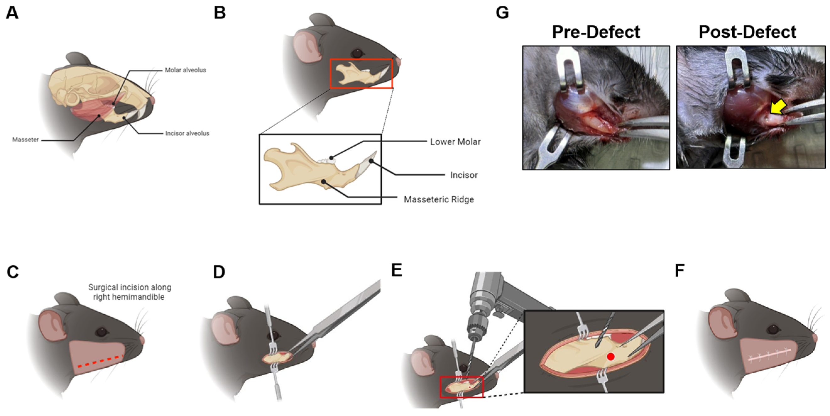
2.2. Mandible Defect Generation

Mice in all experimental groups were given 0.5 mg/kg Buprenorphine-ER preoperatively 2–4 h prior to surgery via subcutaneous injection. Mice were then anesthetized with isoflurane and prepared for aseptic surgery. A small incision on the right inferior hemi-mandible was made, extending to the incisor alveolus. Using forceps, skin and underlying tissue was pushed aside to expose the proximal portion of the incisor. Tissue retractors were used to assist in holding excess skin when needed. Once exposed, the incisor was held using a forceps and a 0.7 mm diameter steel burr drill bit (#19007-07, Fine Science Tools, Foster City, CA, USA) and an electric drill was used to induce a single-cortex defect in the mid portion of the mandible, adjacent to the superficial masseter muscle and directly below the lower molar alveolus (Figure 1). Defects were immediately irrigated with 0.5 mL sterile saline followed by incision closure. Warm saline (1 mL) was administered subcutaneously immediately after surgery. Mice were given moistened food post-operation for three days. In addition, carprofen (10 mg/kg) and buprenorphine SR (0.5 mg/kg) were given to mice post-operatively for 72 h following defect generation. The incision site was monitored for signs of infection daily for 72 h, weekly thereafter, as well as following euthanasia. Mild swelling of the face was observed 24 h post-defect generation that resolved by 48 h. No signs of infection were observed in any animals and we did not have any attrition within the study. Animals we weighed on days 0, 3, 14, and 28 post-defect generation. In this pilot study, male and female mice were sacrificed via carbon dioxide inhalation at postoperative day 0 (n = 2 males, n = 2 females), day 3 (n = 3 males, n = 3 females), day 14 (n = 5 males, n = 5 females), and day 28 (n = 5 males, n = 5 females). Taking into account the variability of our data, five mice within each group provides 80% power to detect a 20% change in BV/TV within the defect site (α = 0.05). All animals were included within the study.

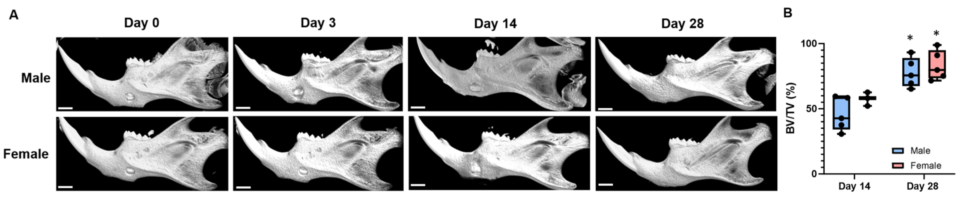
2.3. Micro-Computed Tomography of the Bone Defect Site

Primary outcomes for this study were increases in BV/TV within the defect site over time as assessed by micro-computed tomography (μCT). Skulls were collected on either day 0, 3, 14 or 28 and fixed in 10% neutral buffered formalin for 24 h. Skulls were then stored in 70% ethanol prior to unblended scanning using the XT H 225 micro-computed tomography machine (Nikon Metrology Inc., Brighton, MI, USA). Scans were performed at 108 kV, 54 μA, 720 frames per second. Scans were performed at an isometric voxel size of 21 μm, 35 minutes per scan. Each scan volume was reconstructed using CT Pro 3D (Nikon Metrology Inc.). Reconstructions were converted to bitmap data sets using VGStudio MAX 3.2 (Volume Graphics GmbH, Heidelberg, Germany). Scans were reoriented via DataViewer (SkyScan, Bruker microCT, Kontich, Belgium) to create a new bitmap data set for consistent analysis. From this data, representative transverse slices were selected for qualitative analysis. Morphometric analysis was performed using SkyScan CT-Analyzer (CTAn, Bruker micro-CT, Belgium). Bruker’s instructions and guidelines for analysis within the field were followed throughout analysis. 3D analysis of the defect site was performed in the right mandibular region. Lower and upper bounds of the defect were found. The midpoint was then identified and an ROI was set extending 30 slices or 0.7mm bilaterally. ROI was manually contoured to fit the parameters within the defect. Whole bone 3D analysis of this ROI was conducted following subsequent automated contouring. Binary selection of all samples resulted in a global threshold used to separate bone from surrounding tissue within the ROI.

2.4. Image Reconstruction of the Mandible

3D reconstructions of the mandible were created using the CTVOX program (Bruker, Kontich, Belgium). Tissue was manually delineated by adjusting the transfer function scale. Skulls were oriented laterally, where the nasal region faced left and the occipital region faced right. The right mandible region was manually isolated by adjusting the view in both the sagittal and the coronal plane. Lighting was adjusted and images were saved accordingly.

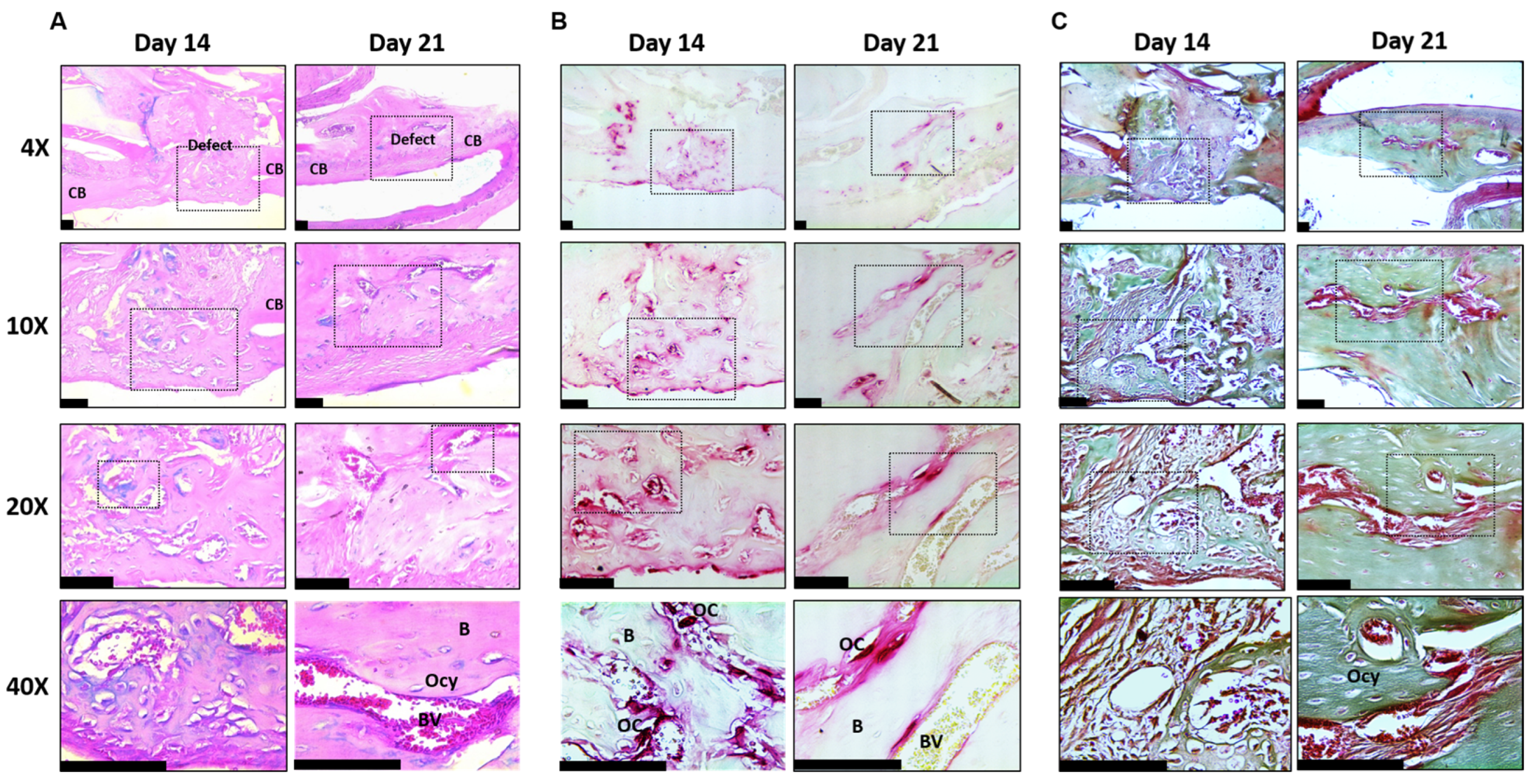
2.5. Mandible Defect Histology

Mandibles from male and female mice were isolated at 0, 3, 14, and 28 days following defect generation. Mandibles were then fixed 10% neutral buffered formalin for 48 h, decalcified in 15%EDTA for 14 days, and then stored in 70% ethanol. Tissues were paraffin embedded and 7 micron sections were collected. Serial sections were stained with TRAP/Fast green, Alcian blue/eosin, or Masson’s trichrome as previously described [13,14,15].

2.6. Statistics

Statistics were performed in GraphPad Prism (Version 9) using Student’s t-test or one-way ANOVA as appropriate and post-hoc tests for multiple comparisons when necessary. Specific p values under 0.1 are shown within figures. Data are shown as box plots from the 25th to 75th percentiles, with whiskers extending to the minimum and maximum value and means shown by horizontal lines or means ± standard deviation.

3. Results

Mandible Defects Heal via Intramembranous Ossification

We generated single cortex defects within the mandible body. At day 0, minimal variation in defect size was observed (Figure 2), demonstrating reproducibility of this model. Moreover, no mandibular fractures occurred following defect generation in either males or females. At day 3 post-defect generation, minimal bone was observed within the defect, consistent with prior reports [16,17]. In contrast, micro-computed tomography within the defect at day 14 revealed partial healing in both males and females, with BV/TV approximating 50% (Figure 2A,B). By day 28 following generation of the defect, BV/TV observed in both males and females was 78 ± 11% in males and 82 ± 14% in females, representing nearly complete regeneration of the defect (Figure 2A,B). Animals did not lose weight post-defect generation. We did not observe a difference in healing between males and females, but we may not be sufficiently powered within this pilot study to detect altered healing between sexes. These data demonstrate the robust, reproducible, and predictable model of cortical bone regeneration achieved via this model.
When performed correctly, single cortex defects heal through intramembranous ossification. To demonstrate that this was true in our mandible defect model, we performed a histological assessment on tissues collected at day 0, 3, 14, and 28 post-defect generation. On day 0 and day 3, we did not observe formation of bone or cartilage within the defect (Figure 3A,B). In contrast, bone formation was evident by day 14 with no cartilage tissue (e.g., Alcian blue staining) present within the defect (Figure 3A,B). Trichrome staining revealed a replacement of woven bone observed at day 14, whereas mineralized bone was observed on day 28 following defect generation (Figure 3B). Remodeling of the defect was evident with the presence of TRAP-positive cells within the defect at both day 14 and 28 post-injury (Figure 3C).

4. Discussion

In this manuscript we describe generation of a mandible defect to study spontaneous intramembranous bone healing that can be used to study normal mechanisms of bone regeneration within the mandible body. We generated a 0.7 mm defect that nearly heals within 28 days, similar to healing of single cortex femoral defects [16]. The underlying incisor alveolus can also be observed in our micro-CT reconstructions, so this model could also be applied to study how bony mandible injuries affect underlying dentition in children. One key aspect of this method is to limit injury of the adjacent periosteal tissue (e.g., do not let the bit slip along the bone surface) as this may induce bone formation along the periosteal surface. We did not observe a difference in healing between males and females within our study, but further time points could help to determine if the rate of healing differed. Overall, the method we describe represents a robust, reproducible, and predictable model of intramembranous regeneration of the mandible body.
Development of the mandible is complex, and includes portions that form via endochondral and intramembranous ossification along the proximal-distal axis [18,19]. The mandibular body forms via intramembranous ossification around the Meckel’s cartilage, whereas endochondral ossification forms the mandibular condyle and symphysis [18,19]. Due to these differences in developmental formation, we chose to place a defect within the mandibular body adjacent to the massenteric ridge, the site where Meckel’s cartilage was present during embryogenesis.
Stabilized cortical defects have been used to study bone regeneration within different anatomical locations. This includes the long bones (e.g., femur, tibia [17,20,21,22,23,24]), calvarium, as well as the mandible [25,26]. To date, a method for generation of defects within the mandible body in the absence of endochondral bone formation has not been described. In prior work, an osteotomy was used to model regeneration of the mandible that healed primarily through intramembranous regeneration, but evidence of cartilage formation was also observed [25]. Other groups reported induction of a 2.3 mm defect that represented a critical sized defect that did not spontaneously heal after 8 weeks, but this model also required injury of the masseter and was placed within the angular process of the mandible [26]. Although calvarial defects are commonly used to study intramembranous regeneration in the craniofacial skeleton, development of the model described within this manuscript will aid in studying site-specific healing. Unlike the calvaria, the mandible is subjected to cyclic mechanical loading resulting from mastication. Moreover, site-specific mechanisms of skeletal healing, including location specific progenitors, may greatly enhance our understanding of musculoskeletal regeneration [3,27,28,29].
We did not generate a critical sized defect within this pilot study, as our goal was to establish a model of bone healing within the mandible that is amenable to studying how interventions (e.g., cellular, genetic, therapeutics) may modulate healing within the mandible. It may be possible to scale the defect size using the method described within this manuscript to achieve a critical sized defect that does not spontaneously heal.
While single cortex defect models are useful for understanding the basic cellular processes in bone repair, these models have limitations. The rate of skeletal healing is likely accelerated as compared to a fracture. Although little bone formation is expected three days post-defect generation, we were not sufficiently powered to observe differences in healing between days 0 and 3 versus later time points within this pilot study. While the defects generated using this method are reproducible, they do not reflect a true fracture of the mandible; thus, reduced inflammatory and immune responses and lack of endosteal marrow components are limitations of this model. Likewise, the effects of mechanical loading of the mandible may be minimized within this fully stabilized model. Nonetheless, advantages of the model make it amenable to screen potential therapies and to understand mechanisms of craniofacial bone regeneration in a manner that is relatively simple to create and standardize.

5. Conclusions

In this manuscript we describe and characterize a model of intramembranous bone regeneration that involves induction of a cortical bone defect within the mandible. This model generates a defect within the craniofacial skeleton that is clinically relevant and subject to mechanical loading but is less technically challenging than prior reports generating mandibular cortical defects.

Author Contributions

Conceptualization, R.B.C. and K.C.M.; methodology, E.K.V., G.K., M.J.S., I.Y.K., J.K., E.C., S.M. and E.W.B.; formal analysis, E.K.V., G.K., M.J.S., E.C., S.M. and E.W.B.; investigation, E.K.V., G.K., M.J.S., I.Y.K., J.K., E.C. and S.M.; resources, K.C.M. and E.W.B.; data curation, E.W.B., G.K., M.J.S., I.Y.K., J.K., E.C. and S.M.; writing—original draft preparation, E.K.V., G.K., M.J.S. and E.W.B.; writing—review and editing, E.K.V., G.K., M.J.S., I.Y.K., J.K., E.C., S.M., R.B.C., K.C.M. and E.W.B.; supervision, K.C.M. and E.W.B.; project administration, K.C.M. and E.W.B.; funding acquisition, K.C.M. and E.W.B. All authors have read and agreed to the published version of the manuscript.

Funding

This research was funded by the Osteoscience Foundations and the University of Minnesota.

Institutional Review Board Statement

The study was conducted in accordance with the Declaration of Helsinki, and approved by the University of Minnesota Institutional Animal Care and Use Committee (protocol code #2402-41820A and was approved on 28 April 2024).

Informed Consent Statement

Not applicable.

Data Availability Statement

All data are contained within this manuscript and are available from the corresponding author upon reasonable request.

Acknowledgments

The authors would like to thank the support from the Comparative Pathology Shared Resource and the Minnesota Dental Research Center for Biomaterials and Biomechanics.

Conflicts of Interest

The authors declare no conflicts of interest.

References

  1. Ghimire, S.; Miramini, S.; Edwards, G.; Rotne, R.; Xu, J.; Ebeling, P.; Zhang, L. The investigation of bone fracture healing under intramembranous and endochondral ossification. Bone Rep. 2021, 14, 100740. [Google Scholar] [CrossRef] [PubMed] [PubMed Central]
  2. Ko, F.C.; Sumner, D.R. How faithfully does intramembranous bone regeneration recapitulate embryonic skeletal development? Dev. Dyn. 2021, 250, 377–392. [Google Scholar] [CrossRef] [PubMed]
  3. Ambrosi, T.H.; Longaker, M.T.; Chan, C.K.F. A Revised Perspective of Skeletal Stem Cell Biology. Front. Cell Dev. Biol. 2019, 7, 189. [Google Scholar] [CrossRef] [PubMed] [PubMed Central]
  4. Bianco, P.; Robey, P.G. Skeletal stem cells. Development 2015, 142, 1023–1027. [Google Scholar] [CrossRef] [PubMed] [PubMed Central]
  5. Bok, S.; Sun, J.; Greenblatt, M.B. Are osteoblasts multiple cell types? A new diversity in skeletal stem cells and their derivatives. J. Bone Miner. Res. 2024, 39, 1386–1392. [Google Scholar] [CrossRef] [PubMed] [PubMed Central]
  6. Aghali, A. Craniofacial Bone Tissue Engineering: Current Approaches and Potential Therapy. Cells. 2021, 10, 2993. [Google Scholar] [CrossRef] [PubMed] [PubMed Central]
  7. Akbay, E.; Aydogan, F. Reconstruction of isolated mandibular bone defects with non-vascularized corticocancellous bone autograft and graft viability. Auris Nasus Larynx 2014, 41, 56–62. [Google Scholar] [CrossRef] [PubMed]
  8. Barone, A.; Covani, U. Maxillary alveolar ridge reconstruction with nonvascularized autogenous block bone: Clinical results. J. Oral Maxillofac. Surg. 2007, 65, 2039–2046. [Google Scholar] [CrossRef] [PubMed]
  9. Chamieh, F.; Collignon, A.M.; Coyac, B.R.; Lesieur, J.; Ribes, S.; Sadoine, J.; Llorens, A.; Nicoletti, A.; Letourneur, D.; Colombier, M.L.; et al. Accelerated craniofacial bone regeneration through dense collagen gel scaffolds seeded with dental pulp stem cells. Sci. Rep. 2016, 6, 38814. [Google Scholar] [CrossRef] [PubMed] [PubMed Central]
  10. Dumanian, Z.P.; Tollemar, V.; Ye, J.; Lu, M.; Zhu, Y.; Liao, J.; Ameer, G.A.; He, T.C.; Reid, R.R. Repair of critical sized cranial defects with BMP9-transduced calvarial cells delivered in a thermoresponsive scaffold. PLoS ONE 2017, 12, e0172327. [Google Scholar] [CrossRef] [PubMed] [PubMed Central]
  11. Klement, K.A.; Black, J.S.; Denny, A.D. Versatility of Distraction Osteogenesis for the Craniofacial Skeleton. J. Craniofacial Surg. 2016, 27, 565–570. [Google Scholar] [CrossRef] [PubMed]
  12. Winters, R.; Tatum, S.A. Craniofacial distraction osteogenesis. Facial Plast. Surg. Clin. N. Am. 2014, 22, 653–664. [Google Scholar] [CrossRef] [PubMed]
  13. Bradley, E.W.; Carpio, L.R.; McGee-Lawrence, M.E.; Castillejo Becerra, C.; Amanatullah, D.F.; Ta, L.E.; Otero, M.; Goldring, M.B.; Kakar, S.; Westendorf, J.J. Phlpp1 facilitates post-traumatic osteoarthritis and is induced by inflammation and promoter demethylation in human osteoarthritis. Osteoarthr. Cartil. 2016, 24, 1021–1028. [Google Scholar] [CrossRef] [PubMed] [PubMed Central]
  14. Karkache, I.Y.; Molstad, D.H.; Vu, E.; Jensen, E.D.; Bradley, E.W. Phlpp1 Expression in Osteoblasts Plays a Modest Role in Bone Homeostasis. JBMR Plus 2023, 7, e10806. [Google Scholar] [CrossRef] [PubMed] [PubMed Central]
  15. Mattson, A.M.; Begun, D.L.; Molstad, D.H.H.; Meyer, M.A.; Oursler, M.J.; Westendorf, J.J.; Bradley, E.W. Deficiency in the phosphatase PHLPP1 suppresses osteoclast-mediated bone resorption and enhances bone formation in mice. J. Biol. Chem. 2019, 294, 11772–11784. [Google Scholar] [CrossRef] [PubMed] [PubMed Central]
  16. Molstad, D.H.H.; Zars, E.; Norton, A.; Mansky, K.C.; Westendorf, J.J.; Bradley, E.W. Hdac3 deletion in myeloid progenitor cells enhances bone healing in females and limits osteoclast fusion via Pmepa1. Sci. Rep. 2020, 10, 21804. [Google Scholar] [CrossRef] [PubMed]
  17. McGee-Lawrence, M.E.; Razidlo, D.F. Induction of fully stabilized cortical bone defects to study intramembranous bone regeneration. Methods Mol. Biol. 2015, 1226, 183–192. [Google Scholar] [CrossRef] [PubMed] [PubMed Central]
  18. Lee, S.K.; Kim, Y.S.; Oh, H.S.; Yang, K.H.; Kim, E.C.; Chi, J.G. Prenatal development of the human mandible. Anat. Rec. 2001, 263, 314–325. [Google Scholar] [CrossRef] [PubMed]
  19. Parada, C.; Chai, Y. Mandible and Tongue Development. Curr. Top. Dev. Biol. 2015, 115, 31–58. [Google Scholar] [CrossRef] [PubMed] [PubMed Central]
  20. Behr, B.; Leucht, P.; Longaker, M.T.; Quarto, N. Fgf-9 is required for angiogenesis and osteogenesis in long bone repair. Proc. Natl. Acad. Sci. USA 2010, 107, 11853–11858. [Google Scholar] [CrossRef] [PubMed] [PubMed Central]
  21. He, Y.X.; Zhang, G.; Pan, X.H.; Liu, Z.; Zheng, L.Z.; Chan, C.W.; Lee, K.M.; Cao, Y.P.; Li, G.; Wei, L.; et al. Impaired bone healing pattern in mice with ovariectomy-induced osteoporosis: A drill-hole defect model. Bone 2011, 48, 1388–1400. [Google Scholar] [CrossRef] [PubMed]
  22. Katae, Y.; Tanaka, S.; Sakai, A.; Nagashima, M.; Hirasawa, H.; Nakamura, T. Elcatonin injections suppress systemic bone resorption without affecting cortical bone regeneration after drill-hole injuries in mice. J. Orthop. Res. 2009, 27, 1652–1658. [Google Scholar] [CrossRef] [PubMed]
  23. Monfoulet, L.; Rabier, B.; Chassande, O.; Fricain, J.C. Drilled hole defects in mouse femur as models of intramembranous cortical and cancellous bone regeneration. Calcif. Tissue Int. 2010, 86, 72–81. [Google Scholar] [CrossRef] [PubMed]
  24. Tanaka, K.; Tanaka, S.; Sakai, A.; Ninomiya, T.; Arai, Y.; Nakamura, T. Deficiency of vitamin A delays bone healing process in association with reduced BMP2 expression after drill-hole injury in mice. Bone 2010, 47, 1006–1012. [Google Scholar] [CrossRef] [PubMed]
  25. Paccione, M.F.; Warren, S.M.; Spector, J.A.; Greenwald, J.A.; Bouletreau, P.J.; Longaker, M.T. A mouse model of mandibular osteotomy healing. J. Craniofacial Surg. 2001, 12, 444–450. [Google Scholar] [CrossRef] [PubMed]
  26. Yu, F.; Liu, L.; Xia, L.; Fang, B. Establishment of a C57BL/6 Mandibular Critical-Size Bone Defect Model. J. Craniofacial Surg. 2021, 32, 2562–2565. [Google Scholar] [CrossRef] [PubMed]
  27. Clark, R.; Park, S.Y.; Bradley, E.W.; Mansky, K.; Tasca, A. Mouse mandibular-derived osteoclast progenitors have differences in intrinsic properties compared with femoral-derived progenitors. JBMR Plus 2024, 8, ziae029. [Google Scholar] [CrossRef] [PubMed] [PubMed Central]
  28. Kingsmill, V.J.; McKay, I.J.; Ryan, P.; Ogden, M.R.; Rawlinson, S.C. Gene expression profiles of mandible reveal features of both calvarial and ulnar bones in the adult rat. J. Dent. 2013, 41, 258–264. [Google Scholar] [CrossRef] [PubMed]
  29. Yahara, Y.; Nguyen, T.; Ishikawa, K.; Kamei, K.; Alman, B.A. The origins and roles of osteoclasts in bone development, homeostasis and repair. Development 2022, 149, dev199908. [Google Scholar] [CrossRef] [PubMed] [PubMed Central]
Figure 1. Depiction of surgical procedure to generate mandible cortical defects. (A) Anatomy of the mouse skull. (B) Anatomy of the mouse mandible indicating position of lower molar and masseteric ridge used as landmarks for defect placement. (CF) Sequential steps of surgical procedure. (G) Images taken pre- and post-defect generation.
Endocrines 06 00009 g001
Figure 2. Mandible cortical bone defect healing time course. (A) Micro-CT reconstructions 0, 3, 14, and 28 days following defect generation. (B) BV/TV within the defect site. Scale bars are 1 mm. * p < 0.05 versus sex-matched D14 samples.
Endocrines 06 00009 g002
Figure 3. Histology of mandible cortical bone defects. (AC) Histology of the mandible defect site 14 and 21 days post-defect generation. The upper row of panels were imaged at 4×, the second row of panels were taken at 10×, the third row at 20×, and lower panels were imaged at 40×. Insets within each image indicate areas selected for higher magnification images. (A) Alcian blue and eosin staining at day 14 and 21 post-defect generation. (B) TRAP/fast green staining of the defect site. (C) Goldner’s trichrome staining. Scale bars are 100 microns. B: Bone, BV: Blood vessel, OC: Osteoclast, Ocy: Osteocyte.
Endocrines 06 00009 g003
Disclaimer/Publisher’s Note: The statements, opinions and data contained in all publications are solely those of the individual author(s) and contributor(s) and not of MDPI and/or the editor(s). MDPI and/or the editor(s) disclaim responsibility for any injury to people or property resulting from any ideas, methods, instructions or products referred to in the content.

