Review

Neuroendocrine Blockade of the Reproductive Axis in Female Athletes

by
Winfried G. Rossmanith
Department of Obstetrics-Gynecology, Klinikum Mittelbaden, Academic Teaching Hospital, University of Heidelberg, D-76532 Baden-Baden, Germany
Endocrines 2022, 3(4), 765-774; https://doi.org/10.3390/endocrines3040063
Submission received: 6 September 2022 / Revised: 21 October 2022 / Accepted: 28 November 2022 / Published: 5 December 2022
(This article belongs to the Section Neuroendocrinology and Pituitary Disorders)

Abstract

:
This review aims at defining the neuroendocrine mechanisms underlying the sport-induced restrictions of the reproductive axis in female athletes. Episodic gonadotropin release was found to be compromised, presumably a result of impaired hypothalamic pulsatile GnRH release. Any deviation from optimal gonadotropin release may result in a suboptimal function of the ovaries, leading to disorders of the menstrual cycle and ovulation. A whole spectrum of menstrual dysfunctions ranging from ovulatory eumenorrhea to luteal phase defects and amenorrhea has been reported in sportive women. As essential neuroendocrine factors underlying these observations, activation of the adrenal axis and altered central nervous neurotransmitter activity have been identified to transfer metabolic, nutritional, and stress signals into the hypothalamic GnRH release. The degree by which the neuroendocrine axis governing reproduction is impaired critically depends on the intensity and duration of exercise and the state of training. Other decisive factors may be energy expenditure and availability, nutritional components, and the maturity of the hypothalamic-pituitary-ovarian (HPO) axis when sport activity was initiated. In conclusion, the gradual cessation of reproductive function observed in female athletes may be interpreted as an adaptive mechanism in response to physical and psychological endurance during sport. This sport-induced restriction of reproductive capacity may serve as protection (endogenous contraception) to preserve a woman’s health.

1. Introduction

Abundant evidence suggests that the reproductive axis is affected during sports in women. Menstrual dysfunctions ranging from anovulatory eumenorrhea, luteal phase defects, and anovulatory oligomenorrhea to long-lasting amenorrhea have been reported in athletic women [1,2,3]. These findings support the notion of close temporal and functional links between sportive activity and the hypothalamic-pituitary-ovarian (HPO) axis governing reproduction [4,5]. This short review aims at defining the principal changes observed in the neuroendocrine control of reproduction in women during sports. In particular, the determinants underly the neuroendocrine restriction of the reproductive axis in athletic women should be defined.

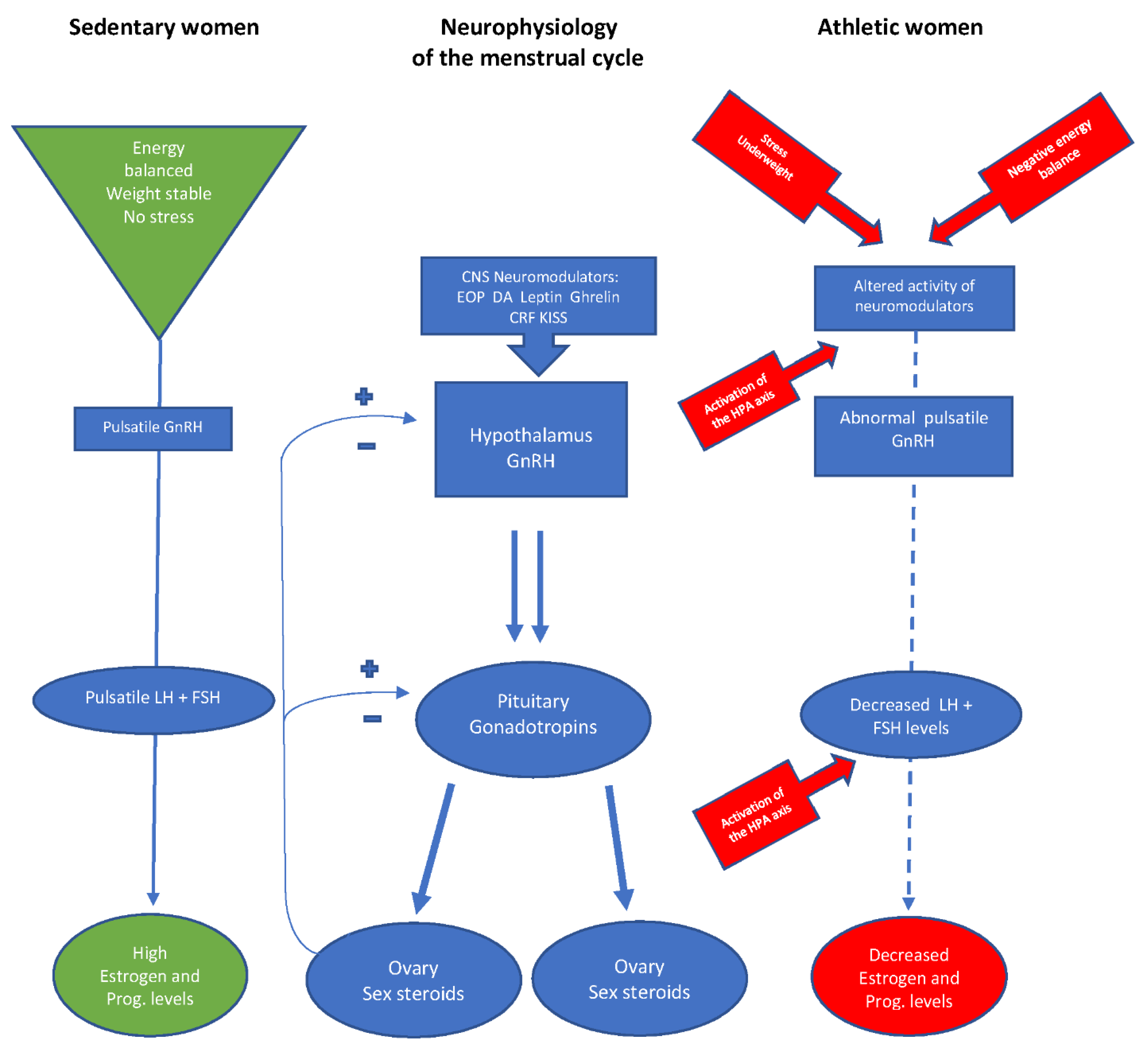
1.1. Neuroendocrinology of Reproduction

For a better understanding of the neuroendocrine control of female reproduction, the mechanisms involved in the neuroendocrine control of normal reproductive function are briefly outlined. When the HPO axis governing reproductive hormone release is fully operative, it ensures regular menstrual cyclicity and uncompromised reproductive capacity [4,6]. Hypothalamic gonadotropin-releasing hormone (GnRH) containing neurons display intrinsic activity so that GnRH is released from the hypothalamus in an intermittent or pulsatile fashion [4,6,7]. Consequently, the episodic release of gonadotropins (luteinizing hormone LH and follicle-stimulating hormone FSH) is observed from the pituitary, reflecting intermittent hypothalamic GnRH stimulation of the pituitary and the effects of factors from within and outside the hypothalamus modulating this secretion [7,8]. Cellular, genetic, and metabolic cues, such as the effects of insulin, leptin, and Ghrelin have been identified as important regulators for differential GnRH secretion [6,8]. In addition, a whole series of neurotransmitters and hormones have also been found to be involved in modulating the activity of GnRH-containing neurons, amongst them ovarian sex steroids, endogenous opioid peptides (EOP), dopamine, kisspeptin, and numerous others [6,8]. Gonadotropin release varies by the stages of the menstrual cycle: LH pulse frequencies increase during the follicular phase of the cycle, while a slowing of the LH pulse frequencies with a concomitant increase in LH pulse amplitudes is noted during the luteal phase of the cycle [9]. It is important to note that a marked slowing of the LH pulse frequencies with a concomitant increase in LH pulse amplitudes is exclusively observed during sleep in the early follicular phase, and increased EOP activity may be accounted for this sleep-entrained manifestation [8,10]. Thus, pulsatile LH and by inference, hypothalamic GnRH release is the result of sex steroid feedback on the central regulatory units during the menstrual cycle, and these effects of sex steroids are translated into neurotransmitter signals impinging on hypothalamic GnRH neurons [4,6,8]. Pulsatile release of the gonadotropins ensures appropriate stimulation of the ovaries, thus promoting sex steroid secretion and follicular growth. In summary, the orchestrated release of gonadotropins with appropriate cycle-dependent feedback by sex steroids is essential for maintaining normal menstrual cycles and optimal reproductive function. Any deviation from this perfectly coordinated GnRH-gonadotropin release may result in suboptimal ovarian function and follicular demise. Menstrual cycle disorders and impaired reproductive capacity observed during physical activity and exercising are therefore clinical manifestations of restrictions in the neuroendocrine drive of reproduction [6,8].

1.2. The Hypothalamic-Pituitary-Ovarian (HPO) Axis in Athletic Women

The increasing popularity of recreational physical exercise during the last decades led to the effects of strenuous exercising on reproductive function. Exercise-associated menstrual dysfunction has been reported at various degrees in women with athletic activities, such as in tennis players, gymnasts, and group sports [1,2,3,11]. High rates of menstrual dysfunctions such as oligo-amenorrhea are found in ballet dancers and long-distance runners (60% to 70%), whereas swimmers, divers, and gymnasts have much lower rates (20%) [3,12]. These incidences of menstrual cycle disturbances are higher than those found in a sedentary population. A cause-effect relationship is difficult to ascertain, as various responses have been observed in different forms of exercises [1,2,3,13]. The spectrum of abnormalities in menstrual cyclicity in athletic women may reflect a pathophysiological continuum of changes in GnRH activity that occur while women exercise over time [8,10]. The degree by which the neuroendocrine axis governing reproduction is critically affected depends on factors such as intensity and duration of exercise, body composition, and stress factors during training programs [1,3,12]. Weight loss has frequently been claimed to play an essential role in female athletes because of their perception of extreme leanness as the perfect body composition for endurance and strenuous exercise [2,14]. Additionally, training intensity, an adaptation of the stress axis, energy availability, and the composition of nutritional components may also be important [1,2,15,16].
Analysis of the LH pulsatility in groups of sedentary and athletic women revealed a slowing of the LH pulse frequencies with a concomitant increase of LH pulse amplitudes, and this finding can be extended to most athletic women [17]. Amenorrheic athletic women showed low LH pulse frequencies and relatively high LH pulse amplitudes, so the mean LH levels were low. These data indicate that even when athletic women experienced normal menstrual bleedings, they had a reduction in LH pulse frequencies (up to 30%) compared with regularly cycling sedentary women. Furthermore, amenorrheic athletes had an additional reduction in LH pulse frequencies of around 20% [17]. The lowest LH pulse frequencies were found in amenorrheic athletes, intermediate pulse frequencies in eumenorrheic athletes, and highest pulse frequencies in non-athletes [15,17]. The 24 h mean LH concentrations were lowest in amenorrheic athletes, compared with eumenorrheic athletes and non-athletic women [17]. Interestingly, a slowing of LH pulse frequencies with a concomitant increase in LH pulse amplitudes was noted during sleep in sedentary [9,17] and cyclic athletes [15,18], but this sleep-associated slowing was absent in amenorrheic athletic women [17]. Interestingly, no differences were found in mean follicle-stimulating hormone (FSH) levels in sedentary and athletic women in most studies [15,17]. Administration of exogenous GnRH to women with exercise-induced amenorrhea elicited exaggerated LH responses in athletic women compared with sedentary control and cyclic athletic women [17,19]. Thus, sport-induced changes in gonadotropin pulsatility may presumably be attributed to a reduced hypothalamic GnRH secretion rather than to impaired pituitary responsiveness [1,17]. These data, although not unequivocal [1,2,3,6,8], indicate that exercise-induced menstrual disorders are causally related to a restriction in the LH pulsatility in athletic women.
Moreover, ovarian steroid secretion was found to be reduced in sportswomen, presumably the result of inappropriate gonadotropin stimulation of the ovaries. This notion is supported by the reduced excretion of urinary estrogen metabolites in amenorrheic athletes [17]. Additionally, a reduction of progesterone metabolites was found in athletic women, as an indicator of luteal phase deficiency (LPD) and anovulation [15,17,20]. These findings characterize approximately half of the menstrual cycles in regularly cycling athletic women and could explain their relative infertility [1,14]. Initially, the changes in the neuroendocrine axis of reproduction may be so subtle that menstruations still appear to be regular. Such subtle defects in the pulsatile GnRH-LH release are likely to contribute to the high rates of anovulation seen even in recreational athletes [1,2]. Almost two-thirds of athletic women were found to have LPD or anovulation in their menstrual cycles [12]. Therefore, the length of the menstrual cycle is not a good parameter for ovarian function in athletic women, as regular menstrual cycles may be either normal or abnormal by undetected anovulation or LPD [1,2]. Taken together, the menstrual abnormalities observed in athletic women represent a pathophysiological continuum from fully coordinated to disordered GnRH-gonadotropin release [1,8,17]. Therefore, changes in the orchestrated 24 h gonadotropin pulsatility could explain, at least in part, why the reproductive capacity is restricted in athletic women [1,8,13].

1.3. Impact of Stress on Reproductive Function

Exercise may impair the hypothalamic–pituitary–ovarian axis (HPO) in some women by acting as a chronic stressor and activator of the hypothalamic-pituitary-adrenal axis (HPA) [21]. Work on the importance of kisspeptin has demonstrated this peptide to be a link between these two axes [22]. Lower plasma cortisol levels are found in women with regular menstrual cycles compared with women suffering from functional hypothalamic amenorrhea, amongst them female athletes [11,17,23]. However, a circadian rhythm in cortisol is preserved in both regularly menstruating women and amenorrheic subjects [23]. Cortisol pulse amplitudes remain high over the course of the day in amenorrheic athletes, while pulse amplitudes decrease in eumenorrheic athletes [17]. These data, although conflicting [2,15,24], suggest higher cortisol levels in amenorrheic than cyclic athletes, presumably indicating hyperactivity of the HPA axis. As for the pituitary responsiveness to corticotropin-releasing hormone (CRH), it appears that the magnitude of ACTH and cortisol response is highest in sedentary compared with athletic women [17]. Furthermore, the adrenal sensitivity to CRH challenges is increased in athlete women with and without amenorrhea, compared with sedentary women [17]. This suggests an exaggerated cortisol response to physiological stimuli such as sports and may also explain the supraphysiological circulating levels of cortisol in most athletic women [15,17].
Furthermore, no differences were observed in ACTH secretory patterns and cortisol secretory pulse frequencies in amenorrheic athletes compared with eumenorrheic athletes and nonathletes [17]. Overnight cortisol levels were found to be higher in both groups of female athletes. Since cortisol stimulates an increase in glucose levels in humans [2], activation of the HPA axis may be interpreted as a mobilizer of energy stores to maintain metabolic homeostasis. In turn, the observed activation of the HPA axis in athletic women may impair the HPO axis, through changes induced by adrenal corticosteroids [25] or altered neurotransmitter activity [1,2,3,8]. Some effects appear to be brought up by CRH itself, as evidenced by the drastic decline in pulsatile LH secretion following CRH injection and the reversal of this suppression by a CRH receptor antagonist [26]. Such blockade of the reproductive function by activation of the HPA axis stress could also be exerted by stress-induced changes in EOP activity: Chronic blockade of EOP receptors may reverse the effects of chronic stress in women with functional amenorrhea, as evidenced by an increased LH pulsatility and resumption of menstrual cyclicity during prolonged treatment with naltrexone, an opiate antagonist [27].

1.4. The Relative Importance of Weight for the Reproductive Axis of Athletic Women

Disorders in eating behavior are common in young athletes and are reported in up to 20% of adolescent women [3,8]. Eating disorders are particularly seen in sports that emphasize leanness, such as gymnastics, running, and ballet dancing [2,28]. Eating disorders increase with the level of exercise and are found in professional, but also recreational sports. Weight loss in association with menstrual cycle disturbances is frequently seen in amenorrheic (up to 2/3), but also in eumenorrheic athletic women (up to 1/3) [8,29]. Menstrual cycle disorders may occur in exercising athletes without weight loss but are more commonly found in those with weight loss [8,30]. One critical mechanism could be the lack of body fat with intensive exercise since more severe menstrual cycle disorders are more prevalent in athletic women with weight loss and less body fat [8]. Maintenance of a minimal percentage of body fat and a minimum weight for height has long been believed to prevent menstrual dysfunctions and restore menses in athletic women with amenorrhea [1,31,32]. However, more recent studies have shown diverging results demonstrating that loss of energy availability, but not weight loss alone may alter the episodic gonadotropin secretion [31,33]. Changes in LH pulsatility are apparent within 5 days of strenuous exercising [30,34], much sooner than any loss in fat tissue may appear.
The mechanisms underlying these clinical observations are complex but may relate to alterations in the HPO axis. Changes in LH pulsatility are observed within days following energy restriction and weight loss, much earlier than changes in body composition may appear [35]. An imbalance between caloric intake and the amount of energy expended with exercise may explain the reduced hypothalamic GnRH-LH drive observed in women with exercise-associated menstrual dysfunction [2,15,17]. Dependent on the severity of weight loss, the LH pulsatility is restrained, as LH pulse frequencies decrease and LH pulse amplitudes either increase or remain blunted [36]. In addition, detailed nutritional assessments in athletic women revealed that their daily caloric intake was like that of sedentary women [15,17]. The relative dietary composition for regularly cycling sedentary women differs from those for athletic women: The protein and fat content of the meals was higher in sedentary women, while the carbohydrate meal content was highest in athletic women without menstrual cycles [15]. Fat and protein account for smaller percentages of calories in the diet of athletic women, and those with amenorrhea consume substantially less fat than regularly menstruating women [15,17]. A negative energy balance leads to unfavorable insulin–glucose ratio [2]. Amenorrheic athletes showed marked hypoinsulinemia both during feeding and fasting, although serum glucose levels were found comparable in those states [15]. That the severe deficit of fat intake identified in these patients may play a role in determining overall metabolic energy availability is suggested by the positive relationship between dietary fat intake and circulating insulin levels [17]. Reduced glucose availability especially in the brain may be a metabolic clue that translates the energy information into hypothalamic GnRH neurons in regulates their activity. It is interesting to note that women with hypoinsulinemia had the lowest dietary fat intake but experienced the greatest reductions in their LH pulse frequencies [15,17].
Several metabolic signals may act in concert to functionally impair the reproductive axis. Candidates for such cues to the neuroendocrine drive of reproduction may comprise leptin [37,38] and the counter-acting ghrelin [2,39]. The peptide leptin derives from adipose tissue and has been identified to signalize a state of starvation to the neuroendocrine centers of reproduction [2,11]. Leptin was also demonstrated to stimulate gonadotropin secretion [38], and therefore, leptin concentrations are significantly lower in amenorrheic compared with eumenorrheic athletic women [2,17,40]. A loss of the normal diurnal pattern of leptin secretion is also seen in amenorrheic women [17]. Furthermore, leptin is lower in adult cyclic and amenorrheic exercisers than in sedentary women [17]. As leptin levels strongly correlate with the absolute fat mass, low levels of leptin reflect little fat mass and consequently, a state of energy deprivation. Administration of leptin to women with hypothalamic amenorrhea (not specifically athletes) normalizes gonadotropin secretion and induces ovulation in almost half of these women [41]. Thus, leptin and ghrelin may represent essential clues for the transfer of metabolic signals (weight, energy availability) into the HPO axis [1,2,3,6,8].
Menstrual cycle disturbances such as functional hypothalamic amenorrhoea are commonly associated with raised growth hormone (GH) and low insulin-like growth factor (IGF-1), a finding suggesting relative GH resistance [3,5,42]. Nevertheless, insulin-like growth factor (IGF) 1 levels are not as low in female athletes as in women with anorexia nervosa. IGF-1 levels, IGF-binding protein (IGFBP)-3, or the ratio of IGF-1/IGFBP-3 were found at lower levels in amenorrheic exercisers, compared with non-athletic women [3,15]. In contrast, IGF-BP-1 was higher in the amenorrheic group, and this led to lower ratios of IGF-1/ IGFBP-1 in amenorrheic athletes [15]. Levels of IGF-1, IGFBP3, and ratios of IGF-1/IGFBP-1 and IGF-1/IGFBP-3 found in normally cycling exercisers do not differ from sedentary controls [43]. Interestingly, IGFBP-1 levels are inversely related to LH secretion in athletes and nonathletes [15,24], while the ratio of IGF-1/ IGFBP-1 is positively associated with 24 h LH pulse frequency [15]. In the hormonal milieu during exercise (low endogenous insulin, elevated cortisol), the increase in IGFBP-1 is the most metabolically responsive peripheral signal of fuel shortage, which ultimately slows hypothalamic GnRH release [5].
These observations suggest that hypothalamic GnRH secretion is decreased by alterations in metabolic cues during exercise. In addition to their primary functions, some appetite- and metabolism-regulating factors may affect GnRH/LH secretion. These factors suppress GnRH and gonadotropin secretion, as they simultaneously increase appetite and feeding behavior during states of low energy balance. In general, satiety-related factors, such as leptin, insulin, and endogenous opioid peptide (EOP), directly or indirectly stimulate LH secretion, whereas orexigenic factors such as neuropeptide Y (NPY) and ghrelin, suppress LH secretion [44,45]. In addition, kisspeptins as a group of polypeptides have recently been described to play a major role in the regulation of the reproductive axis by influencing GnRH release (5,6,42). Kisspeptins act directly on GnRH neurons, and these effects result from multiple interplays of influencing factors that regulate kisspeptin release [42,45]. Thus, kisspeptin integrates the effects of metabolic status on GnRH neurons and mediates at least some of the effects of appetite- and metabolism-regulating factors on GnRH neurons [6,45].

1.5. Energy Availability and the Function of the HPO Axis

Energy availability can be defined by the difference between energy intake and energy expenditure [35]. Exercise is an important component of energy expenditure, and therefore, when energy availability is low, reproductive function is one of the first functions to cease. Detailed studies in women whose energy availabilities were gradually decreased while they maintained a constant energy expenditure illustrate that a value of 30 kcal/kg lean body mass/day may represent the threshold of energy availability that is at least required to maintain normal reproductive function [35,46,47]. The effects of restricted energy availability on GnRH-LH secretion occur relatively fast. These data, although conflicting [48], demonstrate the consequences of negative energy balance: When such situations are experimentally created by restricting calories, increasing energy expenditure, or a combination of both, changes in LH pulsatility occur within a few days [30,34]. When energy availability falls below a certain threshold, LH pulse frequencies decline, while LH pulse amplitudes increase [35]. Importantly, the sensitivity of GnRH neurons to alterations in energy availability appears to change with increasing gynecological age. Caloric restriction combined with vigorous daily exercise alters LH pulse frequencies in adolescents, but not in adult women [35], and these changes appear within 5 days of energy deprivation. Disruptive effects of energy availability on the reproductive axis may affect all menstruating women and produce menstrual cycle disorders such as LPD [48]. Yet, those women who are younger and closer to the time of menarche are more sensitive to changes [34]. The sensitivity of the hypothalamic-pituitary-gonadal axis appears to be maximal in the first 15 years after the onset of menstruation. This explains why excessive exercise early in life may have more profound and long-lasting effects than later [2,34,35].
Changes in the LH pulse characteristics occur with the degree of energy availability, and these effects are most pronounced with increasing restriction: Such effects on the reproductive axis are observed independent of the previous menstrual cycle state, although they are most pronounced in women with pre-existing subtle menstrual cycle disorders [1,47]. Caloric intake adjusted for the energy spent during exercise maintains normal hypothalamic-pituitary-gonadal function (eumenorrhea). Even a mild, but sustained reduction in energy availability can induce subtle changes in gonadotropin secretion, and therefore cause menstrual dysfunctions [1,12].
Strenuous physical training in women is associated with evidence of hypothalamic amenorrhea, pituitary gonadotroph suppression, ovarian dysregulation, and metabolic maladaptation. The blockade of the reproductive system may represent an adaptive response to the multistressor nature of arduous training [49]. It is unlikely that any single factor such as energy availability is responsible for the suppression of the HPO axis. Suppression of the HPO function occurs in response to exercise and training without simultaneous evidence of low energy availability [49]. Therefore, the evolution of reproductive dysfunction appears to be more complex than by low energy availability alone. It is suggested that an interplay of psychological and environmental stressors with the activation of the HPA system may be responsible for the suppression of the HPO axis in female athletes. The combination of physical and psychological stress, energy compensation, and metabolic changes (such as increased insulin resistance) may activate the HPA axis and subsequently impair reproductive capacity in athletic women [5,49]. Multiple metabolic signals brought about by exercise are additionally involved in the signaling to the hypothalamic GnRH pulse generator [5,6,45]. Most female athletes do not intentionally restrict their intake, and yet few consume additional calories that are needed to maintain energy balance. The exact nature of these interactions is complex but is independent of body weight [3,5,49].
The American College of Sports Medicine uses the term “the female athlete triad” to refer to the interrelationship of menstrual dysfunctions, eating disorders, and the long-term consequences of estrogen deprivation [50]. This emphasizes the importance of optimal energy availability for both reproductive and general health. Best states of estrogenization are achieved in normal menstrual cycles when estrogen concentrations are found highest around ovulation [3,8,11]. Menstrual dysfunctions go along with impaired estrogen concentrations, and therefore, estrogen-dependent functions such as bone resorption and bone mass density begin to fall [3,51]. Not surprisingly, eumenorrheic athletes have higher bone mass density than amenorrheic athletes [2,50,51]. Low bone mass density with subsequent premature osteoporosis and pathological fractures in female athletes may be clinical consequences of long-standing estrogen deprivation during menstrual cycle disorders, as particularly seen in amenorrheic athletes [2,9,50].

2. Conclusions

A simplistic model is suggested to summarize the adaptive changes in the neuroendocrine control of the reproductive axis in athlete women (Figure 1). This concept is compatible with all available evidence on the activity of the HPO axis in sedentary and exercising women. Optimal functioning of the neuroendocrine control of the HPO axis in female athletes critically depends on determinants such as the state of energy availability, weight balance, and imposed stress in the athletes. Adaptive changes in the neuroendocrine axis controlling reproduction may lead to a partial or total restriction of the neuroendocrine drive on reproductive function in female athletes. In this context, factors such as body fat, body composition, and energy availability are essential for an uncompromised reproductive capacity. Treatment of women with exercise-associated menstrual and reproductive dysfunctions requires a series of interventions to compensate for these restrictions. Modifying lifestyle to reduce stress and changes in the daily diet and exercise behavior to increase energy availability are recommended [51]. These lifestyle changes are sometimes difficult to achieve in athletic women, so pharmacological agents might be required, although non-pharmacological approaches were found to be more effective in athletic women [2,49,52]. Since the demonstrated neuroendocrine restrictions of the reproductive axis in female athletes are multifactorial in origin, health education of sportswomen must also comprise a better understanding of the neuroendocrine mechanism involved in the sport-induced impairment of reproduction. The neuroendocrine blockade of the reproductive axis in female athletes may be viewed as an adaptive mechanism in response to physical and psychological signals during exercise and sports. Such changes may initially be subtle, but they stepwise reduce female reproductive capacity from fully functioning over LPDs to anovulation and amenorrhea. Therefore, management of sport-associated menstrual cycle and ovulatory disorders is imperative to prevent adverse outcomes in bone density, cardiovascular risk profile, psychological well-being, and fertility. Advancements in understanding the pathophysiological basis of this under-recognized and under-treated clinical entity will aid management and may result in the development of novel therapeutic approaches [53].
The neuroendocrine blockade of the reproductive axis with subsequent menstrual cycle disorders and long-term failure to ovulate in athletic women may cause serious health consequences and should therefore be considered harmful. However, temporal impairment of the reproductive function may be meaningful from the evolutionary standpoint: It may serve as a natural form of endogenous contraception [1,54]. Pregnancy as a state of high energetic investment requires extensive metabolic fuel. Such conditions are unwanted by nature and must be avoided when unfavorable environmental factors prevail. Such conditions are found during energy deprivation or even starvation [54]. Thus, protective endogenous mechanisms to reduce fertility at least temporarily may be operative. This may help an athletic woman to preserve her health during conditions of limited energy availability such as during excessive sport and exercise in women [1,2,8].

Funding

This research received no external funding.

Conflicts of Interest

The author declares no conflict of interest.

References

  1. Yen, S.S.C. Chapter Chronic anovulation due to CNS-hypothalamic-pituitary dysfunction. In Yen & Jaffe’s Reproductive Endocrinology: Physiology, Physiopathology and Clinical Management, 4th ed.; Yen, S.S.C., Jaffe, R.B., Barbieri, R.L., Eds.; Saunders: Philadelphia, PA, USA, 1999; pp. 479–561. [Google Scholar]
  2. Nattiv, A.; Loucks, A.B.; Manore, M.M.; Sanborn, C.F.; Sundgot-Borgen, J.; Warren, M.P. American College of Sports Medicine position stand. The female athlete triad. Med. Sci. Sports Exerc. 2007, 39, 1867–1882. [Google Scholar] [PubMed]
  3. Misra, M. Neuroendocrine mechanism in athletes. Handb. Clin. Neurol. 2014, 124, 373–386. [Google Scholar] [PubMed] [Green Version]
  4. Hall, J.E. Chapter Neuroendocrine control of the menstrual cycle. In Yen & Jaffe´s Reproductive Endocrinology, 5th ed.; Strauss, J.F., Barbieri, R.L., Eds.; Elsevier Saunders: Philadelphia, PA, USA, 2004; pp. 149–166. [Google Scholar]
  5. Nass, R.; Helm, K.D.; Evans, W.S. Chapter 21: Physiological and Pathophysiological Alterations of the Neuroendocrine Components of the Reproductive Axis. In Yen & Jaffe´s Reproductive Endocrinology, 7th ed.; Strauss, J.F., Barbieri, R.L., Eds.; Elsevier Saunders: Philadelphia, PA, USA, 2014; pp. 439–484. [Google Scholar]
  6. McCartney, C.R.; Marshall, J.C. Neuroendocrinology of Reproduction. In Yen & Jaffe´s Reproductive Endocrinology, 8th ed.; Strauss, J.F., Barbieri, R.L., Eds.; Elsevier Saunders: Philadelphia, PA, USA, 2019; pp. 1–45. [Google Scholar]
  7. Wildt, L.; Knobil, E. Frequency and amplitude of gonadotropin-releasing hormone stimulation and gonadotropin secretion in the rhesus monkey. Endocrinology 1981, 109, 376–385. [Google Scholar] [CrossRef]
  8. Yen, S.S.C. Chapter Neuroendocrinology of reproduction. In Yen & Jaffe´s Reproductive Endocrinology, 4th ed.; Yen, S.S.S., Jaffe, R.B., Barbieri, R.L., Eds.; Saunders: Philadelphia, PA, USA, 1999; pp. 30–80. [Google Scholar]
  9. Rossmanith, W.G.; Liu, C.H.; Laughlin, G.A.; Mortola, J.F.; Suh, B.Y.; Yen, S.S.C. Relative changes in LH pulsatility during the menstrual cycle: Using data from hypogonadal women as a reference point. Clin. Endocrinol. 1990, 32, 647–660. [Google Scholar] [CrossRef]
  10. Rossmanith, W.G.; Yen, S.S.C. Sleep-associated decrease of luteinizing hormone pulse frequency during the early follicular phase of the menstrual cycle: Evidence for an opioidergic mechanism. J. Clin. Endocrinol. Metab. 1987, 65, 715–719. [Google Scholar] [CrossRef] [PubMed]
  11. Berga, S.L.; Yen, S.S.S. Chapter Reproductive failure due to central nervous system-hypothalamic-pituitary-dysfunction. In Yen & Jaffe´s Reproductive Endocrinology: Physiology, Physiopathology and Clinical Management, 4th ed.; Strauss, J.F., Barbieri, R.L., Eds.; Saunders: Philadelphia, PA, USA, 1999; pp. 537–596. [Google Scholar]
  12. De Souza, M.J.; Miller, B.E.; Loucks, A.B.; Luciano, A.A.; Pescatello, L.S.; Campbell, C.G.; Lasley, B.E. High frequency of luteal phase deficiency and anovulation in recreational women runner: Blunted elevation in follicle-stimulating hormone observed during the luteal-follicular transition. J. Clin. Endocrinol. Metab. 1998, 83, 4220–4232. [Google Scholar]
  13. Nass, R.; Evans, W.S. Chapter 1. Physiologic and Pathophysiologic Alterations of the Neuroendocrine Components of the Reproductive Axis. In Yen & Jaffe´s Reproductive Endocrinology, 8th ed.; Strauss, J.F., Barbieri, R.L., Eds.; Elsevier Saunders: Philadelphia, PA, USA, 2019; pp. 473–519. [Google Scholar]
  14. Loucks, A.B. Energy availability and infertility. Curr. Opin. Endocrinol. Diabetes Obes. 2007, 14, 470–474. [Google Scholar] [CrossRef]
  15. Laughlin, G.A.; Yen, S.S.C. Nutritional and endocrine-metabolic aberrations in amenorrheic athletes. J. Clin. Endocrinol. Metab. 1996, 81, 4301–4309. [Google Scholar]
  16. Loucks, A.B. Low energy availability in the marathon and other endurance sports. Sports Med. 2007, 37, 248–252. [Google Scholar] [CrossRef]
  17. Loucks, A.B.; Mortola, J.F.; Girton, L.; Yen, S.S.C. Alterations in the hypothalamic-pituitary-ovarian and the hypothalamic-pituitary-adrenal axes in athletic women. J. Clin. Endocrinol. Metab. 1989, 68, 402–441. [Google Scholar] [CrossRef]
  18. Loucks, A.B.; Heath, E.M. Dietary restriction reduces luteinizing hormone (LH) pulse frequency during waking hours and increases LH pulse amplitude during sleep in young menstruating women. J. Clin. Endocrinol. Metab. 1994, 78, 910–915. [Google Scholar] [PubMed]
  19. Yahiro, J.; Glass, A.R.; Fears, W.B.; Ferguson, E.W.; Vigersky, R.A. Exaggerated gonadotropin response to luteinizing hormone-releasing hormone in amenorrheic runners. Am. J. Obstet. Gynecol. 1987, 156, 586–591. [Google Scholar] [CrossRef] [PubMed]
  20. Laughlin, G.A.; Dominguez, C.E.; Yen, S.S.C. Nutritional and endocrine-metabolic aberrations in women with functional hypothalamic amenorrhea. J. Clin. Endocrinol. Metab. 1998, 83, 25–32. [Google Scholar] [CrossRef] [PubMed]
  21. Loucks, A.B.; Redman, L.M. The effect of stress on menstrual function. Trends Endocrinol. Metab. 2004, 15, 466–471. [Google Scholar] [CrossRef] [PubMed]
  22. Navarro, V.M.; Castellano, J.M.; Fernandez-Fernandez, R.; Tovar, S.; Roa, J.; Mayen, A.; Nogueiras, R.; Vazquez, M.J.; Barreiro, M.L.; Magni, P.; et al. Characterization of the potent luteinizing hormone-releasing activity of KISS-1 peptide the natural ligand of GPR. Endocrinology 2005, 146, 156–163. [Google Scholar] [CrossRef] [PubMed] [Green Version]
  23. Suh, B.Y.; Liu, J.H.; Berga, S.L.; Quigley, M.E.; Laughlin, G.A.; Yen, S.S.C. Hypercortisolism in patients with hypothalamic amenorrhea. J. Clin. Endocrinol. Metab. 1988, 66, 733–739. [Google Scholar] [CrossRef]
  24. Rickenlund, A.; Thoren, M.; Carlstrom, K.; von Schultz, B.; Hirschberg, A.L. Diurnal profiles of testosterone and pituitary hormones suggest different mechanisms for menstrual disturbances in endurance athletes. J. Clin. Endocrinol. Metab. 2004, 89, 702–707. [Google Scholar] [CrossRef] [Green Version]
  25. Breen, K.M.; Davis, T.L.; Doro, L.C. Insight into the neuroendocrine site and cellular mechanism by which cortisol suppresses pituitary responsiveness to gonadotropin-releasing hormone. Endocrinology 2008, 149, 767–773. [Google Scholar] [CrossRef]
  26. Herod, S.M.; Pohl, C.R.; Cameron, J.L. Treatment with a CRH-R1 antagonist prevents stress-induced suppression of the central neural drive to the reproductive axis in female macaques. Am. J. Physiol. Endocrinol. Metab. 2011, 300, E19–E27. [Google Scholar] [CrossRef]
  27. Genazzani, A.D.; Petraglia, F.; Gastaldi, F.; Volpogni, C.; Gamba, O.; Genazzani, A.R. Naltrexone treatment restores menstrual cycles in patients with weight loss-related amenorrhea. Fertil. Steril. 1995, 64, 951–956. [Google Scholar] [CrossRef]
  28. Nichols, J.F.; Rauh, M.J.; Barrack, M.T.; Barkai, H.S.; Pernick, Y. Disordered eating and menstrual irregularity in high school athletes in lean-build and non-lean-build sports. Int. J. Sport Nutr. Exerc. Metab. 2007, 17, 363–377. [Google Scholar] [CrossRef] [PubMed]
  29. Sanborn, C.; Martin, B.; Wagner, W.J. Is athletic amenorrhea specific to runners? Am. J. Obstet. Gynecol. 1982, 143, 859–861. [Google Scholar] [CrossRef] [PubMed]
  30. Fichter, M.M.; Pirke, K.M. Starvation models and eating disorders. In Handbook of Eating Disorders, 1st ed.; Szmukler, G.I., Dare, C., Treasure, J., Eds.; John Wiley & Sons: Hoboken, NJ, USA, 1995; pp. 83–107. [Google Scholar]
  31. Loucks, A.B. Energy availability, not body fatness, regulates reproductive function in women. Exerc. Sport Sci. Rev. 2003, 31, 144–148. [Google Scholar] [CrossRef]
  32. Frisch, R.E.; McArthur, J.W. Menstrual cycles: Fatness as a determinant of minimum weight for height necessary for their maintenance or onset. Science 1974, 185, 949–951. [Google Scholar] [CrossRef] [PubMed]
  33. Christo, K.; Cord, J.; Mendes, N.; Miller, K.K.; Goldstein, M.A.; Klibanski, A.; Misra, M. Acetylated ghrelin and leptin in adolescent athletes with amenorrhea, eumenorrheic athletes and controls: A cross-sectional study. Clin. Endocrinol. (Oxf) 2008, 69, 628–633. [Google Scholar] [CrossRef] [PubMed]
  34. Loucks, A.B. The response of luteinizing hormone pulsatility to 5 days of low energy availability disappears by 14 years of gynecological age. J. Clin. Endocrinol. Metab. 2006, 91, 3158–3164. [Google Scholar] [CrossRef] [Green Version]
  35. Loucks, A.B.; Thuma, J.R. Luteinizing hormone pulsatility is disrupted at a threshold of energy availability in regularly menstruating women. J. Clin. Endocrinol. Metab. 2003, 88, 297–311. [Google Scholar] [CrossRef] [Green Version]
  36. Williams, N.I.; Young, J.C.; McArthur, J.W.; Bullen, B.; Skinner, G.S.; Turnbull, B. Strenuous exercise with caloric restriction: Effect on luteinizing hormone secretion. Med. Sci. Sports Exerc. 1995, 27, 1390–1398. [Google Scholar] [CrossRef]
  37. Clarke, I.; Burger, H. Leptin and reproduction. Rev. Reprod. 1999, 4, 48–55. [Google Scholar] [CrossRef]
  38. Moschos, S.; Chan, K.L.; Mantzoros, C.S. Leptin, and reproduction: A review. Fertil. Steril. 2002, 77, 433–444. [Google Scholar] [CrossRef]
  39. De Souza, M.J.; Leidy, H.J.; O´Donnell, E.; Lasley, B.; Williams, N.I. Fasting ghrelin levels in physically active women: Relationship with menstrual disturbances and metabolic hormones. J. Clin. Endocrinol. Metab. 2004, 89, 3536–3542. [Google Scholar] [CrossRef] [PubMed] [Green Version]
  40. Warren, M.P.; Voussoughian, F.; Geer, E.B.; Hyle, E.P.; Edberg, C.L.; Ramos, R.H. Functional hypothalamic amenorrhea: Hyperleptinemia and disordered eating. J. Clin. Endocrinol. Metab. 1999, 84, 873–877. [Google Scholar] [CrossRef] [PubMed]
  41. Welt, C.K.; Chan, J.L.; Bullen, J.; Murphy, R.; Smith, P.; De Paoli, A.M.; Karalis, A.; Mantzoros, C.S. Recombinant human leptin in women with hypothalamic amenorrhea. N. Engl. J. Med. 2004, 351, 987–997. [Google Scholar] [CrossRef] [PubMed]
  42. Morrison, A.E.; Fleming, S.; Levy, M.J. A review of the pathophysiology of functional hypothalamic amenorrhoea in women subject to psychological stress, disordered eating, excessive exercise, or a combination of these factors. Clin. Endocrinol. 2021, 95, 229–238. [Google Scholar] [CrossRef] [PubMed]
  43. De Souza, M.J.; van Heest, J.; Demers, L.M.; Lasley, B.L. Luteal phase deficiency in recreational runners: Evidence for a hypometabolic state. J. Clin. Endocrinol. Metab. 2003, 88, 337–346. [Google Scholar] [CrossRef] [PubMed] [Green Version]
  44. Celik, O.; Aydin, S.; Celik, K.; Yilmaz, M. Peptides: Basic determinants of reproductive functions. Peptides 2015, 72, 34–43. [Google Scholar] [CrossRef]
  45. Iwasa, T.; Matsuzaki, T.; Yano, K.; Mayila, Y.; Yanagihara, R.; Yamamoto, Y.; Kuwahara, A.; Irahara, M. Effects of low energy availability on reproductive functions and their underlying neuroendocrine mechanisms. J. Clin. Med. 2018, 5, 166. [Google Scholar] [CrossRef] [Green Version]
  46. Loucks, A.B.; Kiens, B.; Wright, H.H. Energy availability in athletes. J. Sports Sci. 2011, 29 (Suppl. 1), S7–S15. [Google Scholar] [CrossRef]
  47. Loucks, A.B.; Verdun, M.; Heath, E.M. Low energy availability, not the stress of exercise, alters LH pulsatility in exercising women. J. Appl. Physiol. 1998, 84, 37–46. [Google Scholar] [CrossRef]
  48. Lieberman, J.L.; de Souza, M.J.; Wagstaff, D.A.; Williams, N.I. Menstrual disruption with exercise is not linked to an energy availability threshold. Med. Sci. Sports Exerc. 2018, 50, 551–561. [Google Scholar] [CrossRef]
  49. Gifford, R.M.; O’Leary, T.J.; Wardle, S.L.; Double, R.L.; Homer, N.Z.M.; Howie, A.F.; Greeves, J.P.; Anderson, R.A.; Woods, D.R.; Reynolds, R.M. Reproductive and metabolic adaptation to multistressor training in women. Am. J. Physiol. Endocrinol. Metab. 2021, 321, E281–E291. [Google Scholar] [CrossRef] [PubMed]
  50. Williams, N.I.; Statuta, S.M.; Austin, A. Female Athlete Triad: Future Directions for Energy Availability and Eating Disorder Research and Practice. Clin. Sports Med. 2017, 36, 671–686. [Google Scholar] [CrossRef] [PubMed]
  51. De Souza, M.J.; West, S.L.; Jamal, S.A.; Hawker, G.A.; Gundberg, C.M.; Williams, N.I. The presence of both an energy deficiency and estrogen deficiency exacerbates alterations of bone metabolism in exercising women. Bone 2008, 43, 140–148. [Google Scholar] [CrossRef]
  52. Stafford, D.E.J. Altered hypothalamic-pituitary-ovarian axis function in young female athletes: Implications and recommendations for management. Treat. Endocrinol. 2005, 4, 147–154. [Google Scholar] [CrossRef] [PubMed]
  53. Dipla, K.; Kraemer, R.R.; Constantini, N.W.; Hackney, A.C. Relative energy deficiency in sports (RED-S): Elucidation of endocrine changes affecting the health of males and females. Hormones 2021, 20, 35–47. [Google Scholar] [CrossRef] [PubMed]
  54. Yen, S.S.C. Reproductive strategies in women: Neuroendocrine basis for endogenous contraception. In Neuroendocrinology of Reproduction; Roland, R., Ed.; Excerpta Medica Amsterdam: Amsterdam, The Netherlands, 1988; pp. 231–239. [Google Scholar]
Figure 1. Schematic presentation of the neurophysiology of the hypothalamic-pituitary-ovarian axis and the effects of sports and exercise on the neuroendocrine control of reproduction.
Endocrines 03 00063 g001
Publisher’s Note: MDPI stays neutral with regard to jurisdictional claims in published maps and institutional affiliations.

