Review

Interactions between 14-3-3 Proteins and Actin Cytoskeleton and Its Regulation by microRNAs and Long Non-Coding RNAs in Cancer

by
Jaya Aseervatham
1,2
1
Department of Ophthalmology, University of Texas Health Science Center at Houston, Houston, TX 77054, USA
2
Center for Vascular and Heart Research, Fralin Biomedical Research Institute at VTC, Virgina Tech, Blacksburg, VA 24016, USA
Endocrines 2022, 3(4), 665-702; https://doi.org/10.3390/endocrines3040057
Submission received: 10 September 2022 / Revised: 15 October 2022 / Accepted: 25 October 2022 / Published: 1 November 2022
(This article belongs to the Section Endocrine Oncology)

Abstract

:
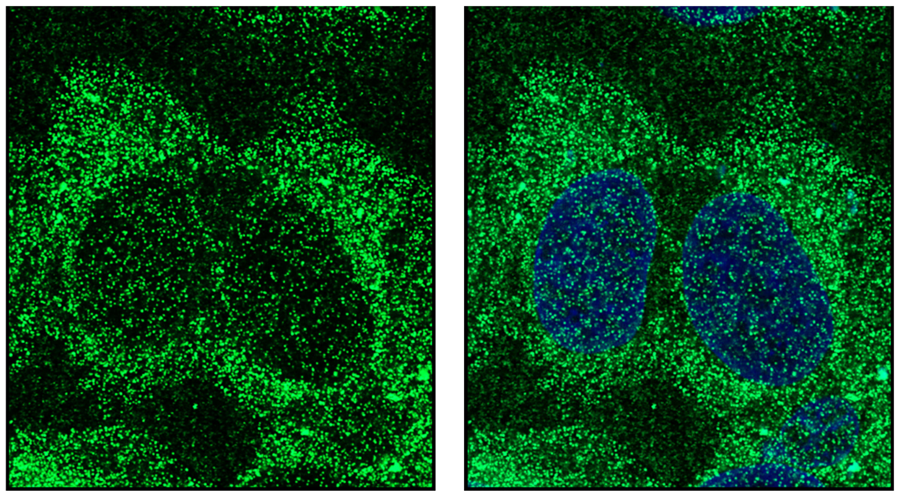
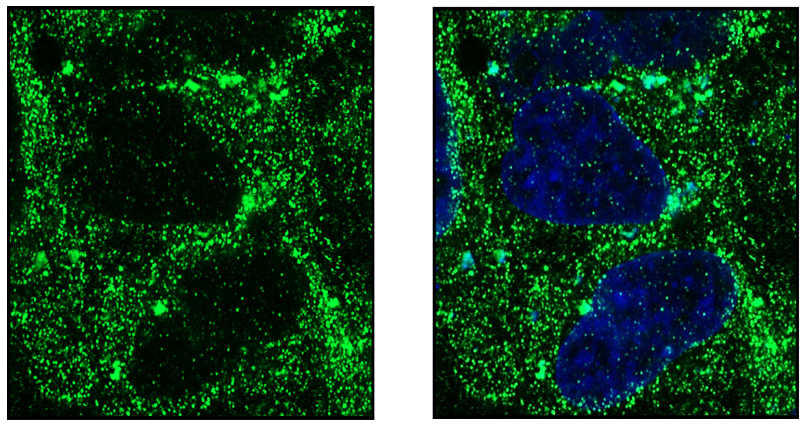
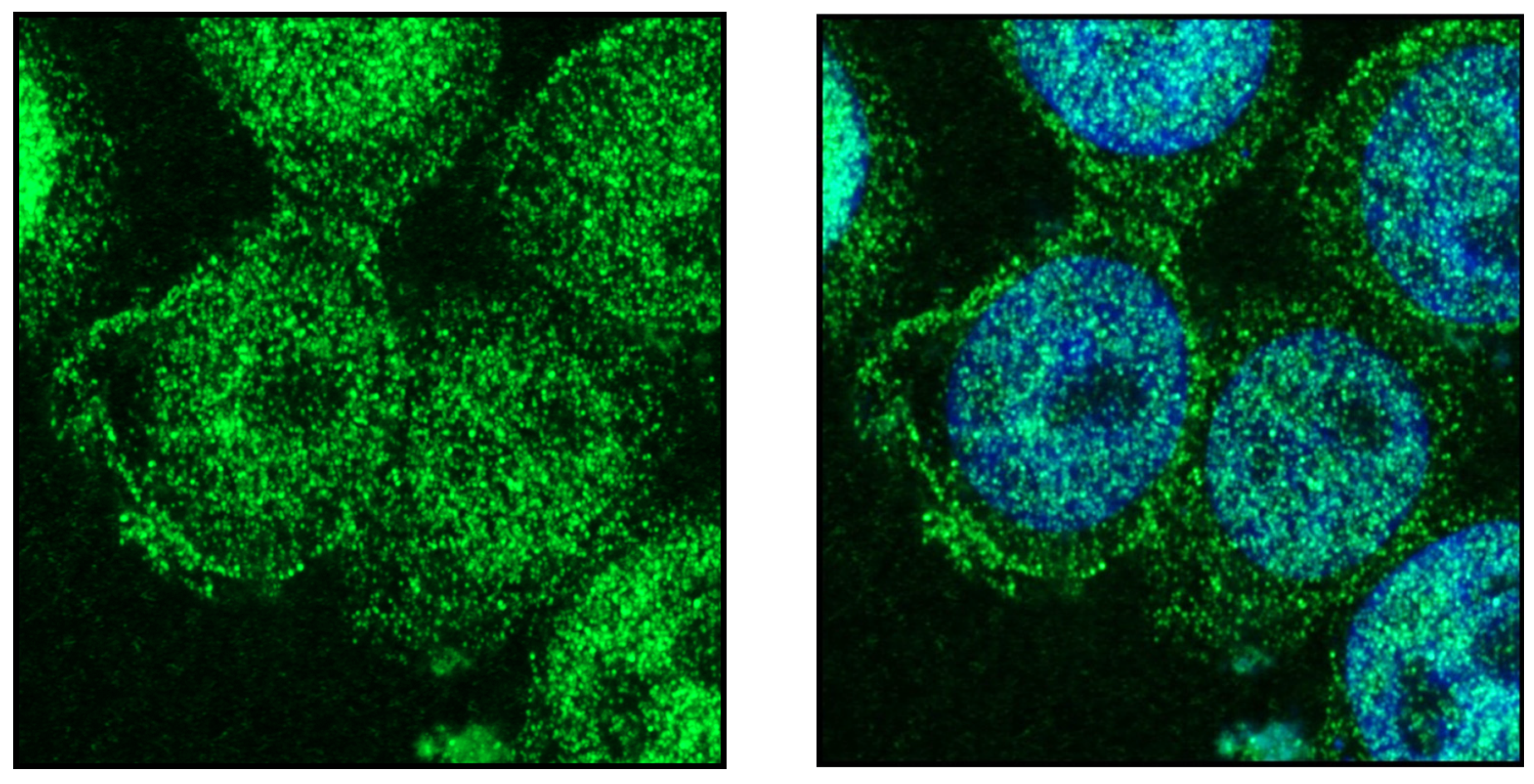
14-3-3s are a family of structurally similar proteins that bind to phosphoserine or phosphothreonine residues, forming the central signaling hub that coordinates or integrates various cellular functions, thereby controlling many pathways important in cancer, cell motility, cell death, cytoskeletal remodeling, neuro-degenerative disorders and many more. Their targets are present in all cellular compartments, and when they bind to proteins they alter their subcellular localization, stability, and molecular interactions with other proteins. Changes in environmental conditions that result in altered homeostasis trigger the interaction between 14-3-3 and other proteins to retrieve or rescue homeostasis. In circumstances where these regulatory proteins are dysregulated, it leads to pathological conditions. Therefore, deeper understanding is needed on how 14-3-3 proteins bind, and how these proteins are regulated or modified. This will help to detect disease in early stages or design inhibitors to block certain pathways. Recently, more research has been devoted to identifying the role of MicroRNAs, and long non-coding RNAs, which play an important role in regulating gene expression. Although there are many reviews on the role of 14-3-3 proteins in cancer, they do not provide a holistic view of the changes in the cell, which is the focus of this review. The unique feature of the review is that it not only focuses on how the 14-3-3 subunits associate and dissociate with their binding and regulatory proteins, but also includes the role of micro-RNAs and long non-coding RNAs and how they regulate 14-3-3 isoforms. The highlight of the review is that it focuses on the role of 14-3-3, actin, actin binding proteins and Rho GTPases in cancer, and how this complex is important for cell migration and invasion. Finally, the reader is provided with super-resolution high-clarity images of each subunit of the 14-3-3 protein family, further depicting their distribution in HeLa cells to illustrate their interactions in a cancer cell.

1. Introduction

Cellular functions, coordinated by multiple proteins such as receptors, channels, and structural proteins, have indispensable roles in maintaining cell integrity. To function in a coordinated fashion, cells should not only be aware of their surroundings, but also be flexible to protein–protein interactions. Credit can be given to the 14-3-3 protein family that is involved in almost every function of the cell, right from cell division to cell death. They are highly conserved family of proteins expressed in eukaryotes, and mammals have 7 isoforms alpha/beta (α/β), eta (η), epsilon (ε), gamma (γ), sigma (σ), theta (θ) and Zeta/delta (ζ/δ). α and δ are the phosphorylated forms of β and ζ. These subunits are encoded by their respective genes YWHAB, YWHAE, YWHAH, YWHAG, SFN, YWHAQ and YWHAZ. 14-3-3 proteins have a molecular mass of 30 kDa, and can form homo or heterodimers [1]. Each monomer contains an amphipathic groove that allows the dimer to bind to two ligands simultaneously. They can bind to same proteins or different proteins by forming a bridge between the two. The ligand-binding sites are conserved in all subunits with exactly the same consensus sequences. Mode I binding sites have the consensus sequence R-S-X-pS/pT-X-P, whereas mode II binding sites have the consensus sequence R-X-F/Y-X-pS/pT-X-P. The third binding site, or mode III, has the consensus sequence R-X-X-pS/pT-X-COOH, which is usually present at the C-terminus of the protein [2]. Crystal structures revealed that each 14-3-3 polypeptide contains nine helices, arranged to form a right-angled corner and two subunits associate with each other to form a saddle-shaped dimer [3]. While the N-terminus forms the floor of the central groove, the C-terminus form the walls. In some isoforms, the distorted C-terminal loop may form a tenth helix that regulates access to the central groove. 14-3-3 dimers form a cup-shaped structure with a negatively charged central passage that has a diameter of 35 Å and a depth of 20 Å [4]. Each monomer contains one amphipathic groove in which the binding sites are formed by the sidechains of Lys49, Arg56, Arg127 and Tyr128. Positioning the substrate within this groove is accomplished by hydrophobic interactions with residues Leu172, Val176, Leu216, Ile217, Leu220, Leu227, and Trp228 belonging to the C-terminal helices of 14-3-3 [5]. Proteins such as FOXO and AANAT have two binding sites for 14-3-3 proteins. The primary site is indispensable for the formation of stable interactions, the second site serves only as an enhancer that has low affinity to the protein. Once the primary site is phosphorylated and bound, the proximity enhances the chance of the secondary binding site [6]. Phosphorylation of proteins trigger transitions in conformational changes which help in protein regulation [7]. Because 14-3-3 proteins do not have intrinsic kinase activity, the vast majority of 14-3-3 partners must be phosphorylated to interact with 14-3-3. Due to their inherent rigid structure, they can stabilize the conformation of client proteins, or physically mask certain residues, or simply facilitate protein interactions by acting as a scaffold [8]. Even though the residues involved in dimer formation and protein binding are highly conserved in all the 14-3-3 subunits, the isotypes specifically target their partners by forming a complex tertiary structure via homo or hetero dimerization. This creates structural differences, position, and specificity of the binding grove [9]. Although the core of 14-3-3 proteins have a rigid structure, flexibility is imparted by the highly unstructured C- terminus. In the unbound state, the C-terminus of 14-3-3 occupies the ligand-binding groove, which is displaced upon ligand binding [10]. Removal of the C-terminus increases the binding affinity of 14-3-3 to various ligands, thereby validating its auto inhibitory function [11] (Figure 1).
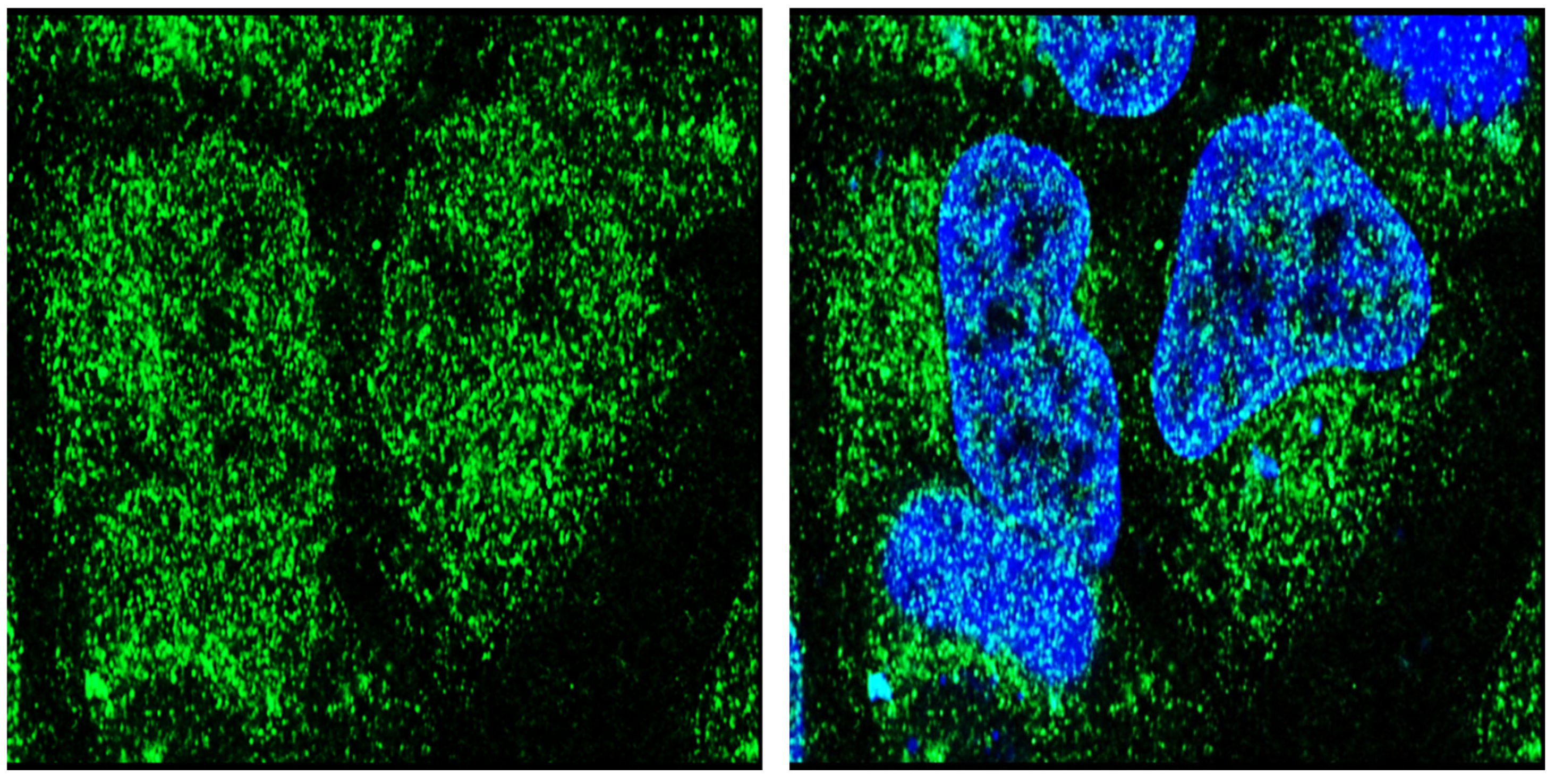
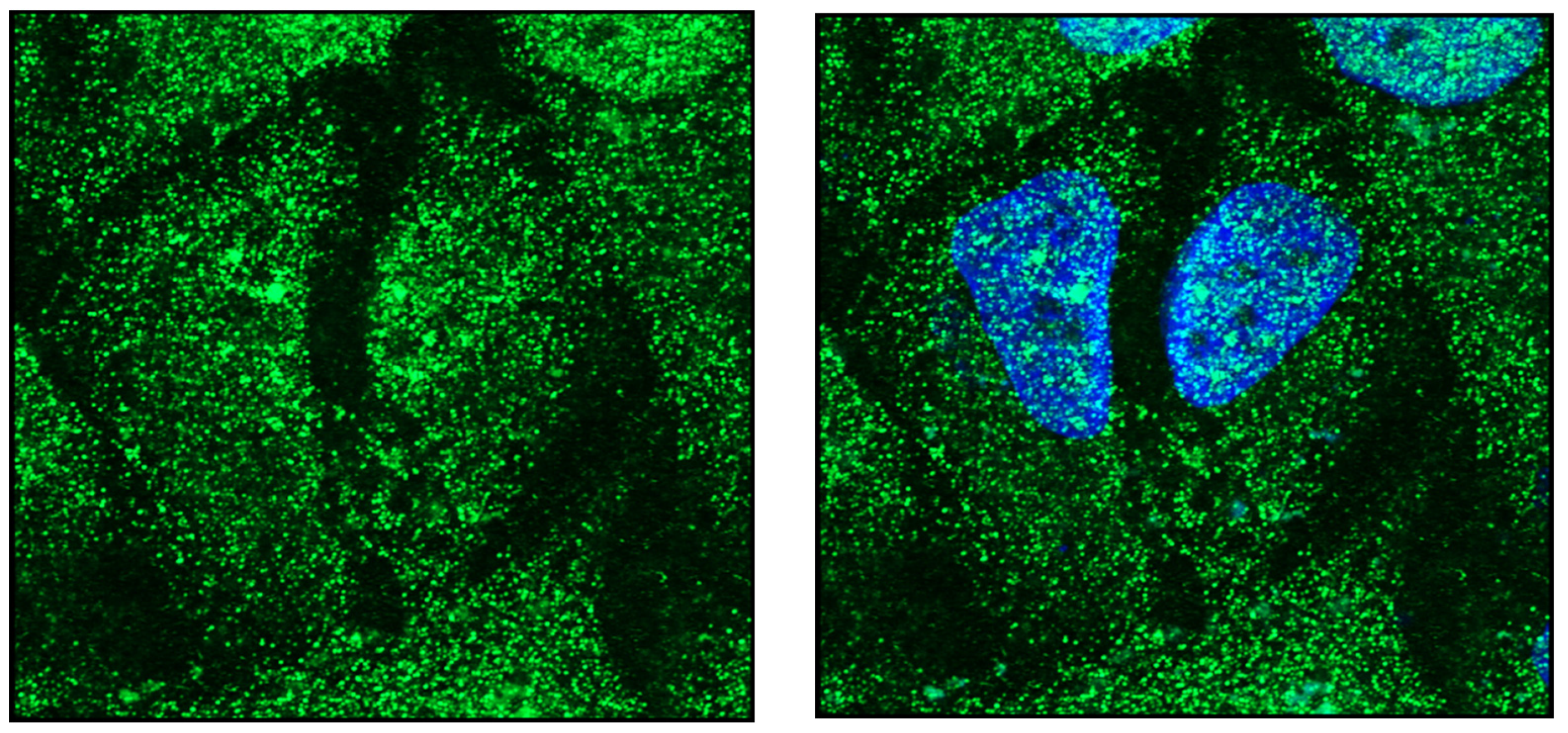
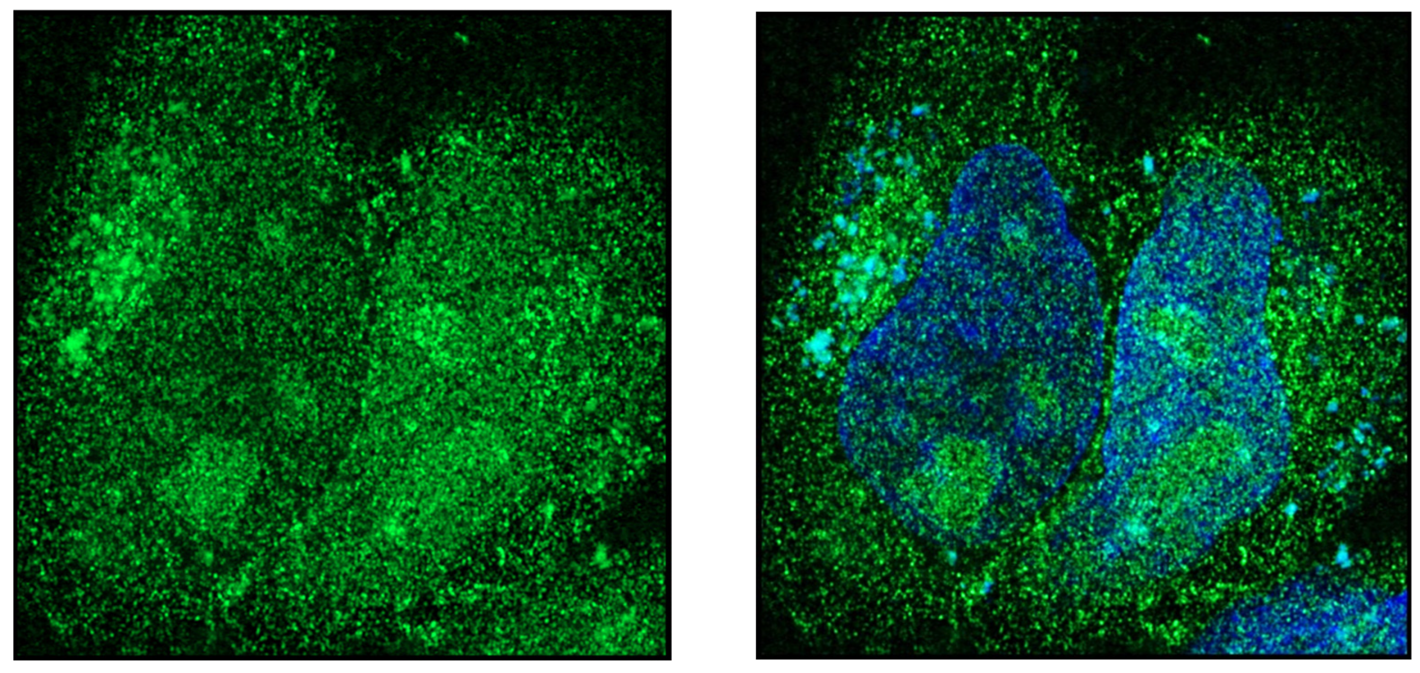
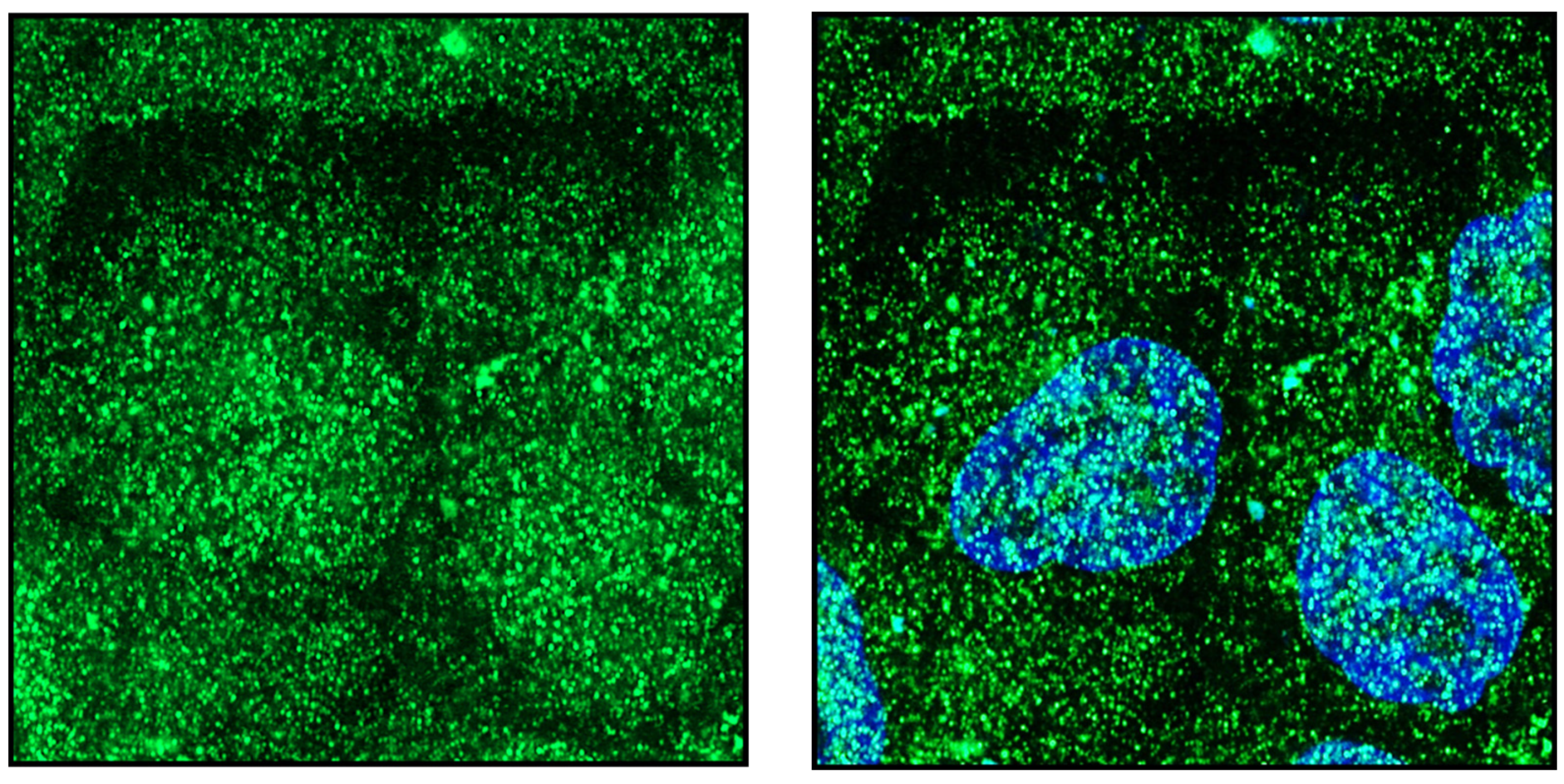
14-3-3 serves as a central hub for various signaling pathways and participate in cell trafficking, regulation of cytoskeletal dynamics, cell plasticity and apoptosis. Dysregulation leads to diseases such as cancer, neurological disorders and metabolic disorders, to name a few. The purpose of this review is to provide a holistic view of the role of 14-3-3 isoforms in cancer, their interactions with binding partners, signaling pathways involved, and how they are regulated. Additionally, this review uniquely discusses the role of MicroRNAs and Long Non-coding RNAs in regulating 14-3-3 subunits. To that end, high-resolution confocal images of each subunit of the 14-3-3 family and its distribution in HeLa cells are presented for better clarity on cellular interactions. For immunofluorescence images, confluent HeLa cells were fixed in 4% paraformaldehyde and stained with the respective 13-3-3 subunit antibodies. 14-3-3α/β (Abcam), 14-3-3η (Novus biologicals), 14-3-3ε (cell signaling technology), 14-3-3γ (Novus biologicals), 14-3-3σ (Sigma Aldrich), 14-3-3θ (abcam), 14-3-3ζ/δ (biolegend). Respective secondary antibodies were Alexa-488-conjugated and standard protocol was followed.
2. 14-3-3s in Cancer
Post-translational modifications (PTM) of proteins are essential for proper regulation, localization, and function. They act as molecular codes to generate functional diver-sity beyond replication and transcription [13]. Kinases play a crucial role in PTMs, as they phosphorylate almost one-third of the proteins, thus playing a crucial role in intracellular signaling. These kinases are regulated by phosphorylated motifs which involve associations with scaffolding proteins such as 14-3-3 [14]. Many researchers have dedicated their study to identifying the role of 14-3-3 in cancer, because they can either be tumor promoters and tumor suppressors, depending on the cell and the associated proteins exclusively. Since the expression of these proteins differ significantly in the normal and disease states, they can act as biomarkers in bodily fluids and in tissues.

3. Oncogenic Role of 14-3-3α/β in Cancer

14-3-3α/β serves as an adapter protein that is implicated in the regulation of signaling pathways. It binds to target proteins by recognizing the phosphoserine or phosphothreonine motif and acts as an oncogene. Immunoflourescence expression in HeLa cells shows the distribution of 14-3-3α/β throughout the cell, highlighting its importance in cell function (Figure 2). Clinical significance of 14-3-3β was reported by Liu et al., in Hepato-Cellular Carcinoma (HCC) patients. Almost 70% of the analyzed tumors had increased expression of 14-3-3β, and 37% of these patients had increased extrahepatic metastasis. The expression in metastatic samples was higher than in primary tumors and led to low survival rates. When expressed in vitro, it increased cell proliferation, anchorage-independent cell growth and migration [15]. Serum levels of 14-3-3β were significantly higher in HCC patients than those with liver cirrhosis and chronic hepatitis, and high levels of 14-3-3β were associated with vascular invasion, TNM stage, BCLC stage, early recurrence, and poor prognosis. Uni and multivariate analysis showed independent association between serum 14-3-3β and HCC which could be used to effectively discriminate between HCC and other liver diseases [16]. Urinary expression 14-3-3α/β in patients with clear cell renal cell carcinoma (ccRCC) was higher than in healthy volunteers even when the tumor size was very small. Higher expression was associated with cancer stage, metastasis, and poor survival. It can be used to distinguish between ccRCC from angiomyolipoma (AML), since AML had lesser expression than ccRCC, which could be used to effectively distinguish between metastatic and benign tumor of kidney [17]. 14-3-3α/β can be used as an effective marker in renal cell carcinoma (RCC), because it is also detected in cyst fluid. Proteomics from cyst fluid identified 14-3-3α/β as one of the proteins that was over-expressed in RCC but not in normal tissues. In addition, the expression was high in urine samples from RCC than from healthy controls [18]. Methylation of YWHAB was higher in HCC than in normal tissues, and a positive relationship was found between increased expression of 14-3-3β and immune filtration. In vitro knockdown by siRNA suppressed proliferation, colony formation and invasion [19]. Expression of antisense 14-3-3β cDNA in nude mice led to decreased tumor size, mitotic rate and angiogenesis. Histological examination showed mitosis decreased by 47%, VEGF by 38%, Raf-1 by 29%, and c-Myc protein by 73%. In this study, the authors also found over-expression of 14-3-3β mRNA in various murine tumor cell lines including hepatomas, mammary tumor, kidney tumor, adrenal pheochromocytoma, peripheral nerve tumors, glioblastoma, gliosarcoma, neuroblastoma cells [20]. NIH 3T3 cells over-expressing the β isoform stimulated cell growth and supported anchorage independent growth through the activation of MAP Kinase pathway by interacting with Raf-1, which was abrogated with overexpression of MEK-1 in these cells [21]. RNA and protein expression of 14-3-3β was found to be increased in human osteosarcoma tissues or in osteosarcoma cell lines. SiRNA knockdown of endogenous protein decreased cell viability, inhibited cell proliferation and invasion by decreasing the S and G2 m phases of the cell cycle. In addition, it also decreased the expression of β-catenin, cyclin D1 and c-myc which may be responsible for decreased cell proliferation [22]. In Lewis lung carcinoma cells, FBI1/Akirin2 was identified as a functional partner of 14-3-3β which acted as a transcriptional suppressor, by binding to the promoter region of MAPK phosphatase1, which dephosphorylates extracellular signal-regulated kinases (ERK) and functions as a negative feedback factor [23]. Proteomic analysis of tissues taken from lung adenocarcinoma patients who had good and poor prognosis revealed 14-3-3α/β and calnexin were involved in proliferation, invasion, and migration of cancer cells. The authors also studied the functions of proteins in vitro using A549 cells and found that targeted depletion of 14-3-3α/β reduced proliferation and invasion as seen in patient tissues [24]. Overexpression of miR-129-5p promotes apoptosis in lung cancer. Target scan identified YWHAB as a candidate, since the 3’UTR of YWHAB mRNA contained a complementary site for miR-129-5p. Luciferase reporter assay confirmed that miR-129-5p could repress the expression of YWHAB at the mRNA and protein level. This shows YWHAB is a downstream target of miR-129-5p and a key mediator in lung cancer cells [25]. In glioma patients, 14-3-3β expression was significantly higher than in normal tissues and these patients had shorter survival times [26]. High expression of 14-3-3β in glioma cells increased cell proliferation and invasion. 14-3-3β knockdown increased the accumulation of cells in the subG1 phase, subsequently decreasing the number of cells in the G2/M phase. It also inhibited the release of Cytochrome C from the mitochondria, induced ER stress, released calcium, and decreased the expression of XBP1S mRNA. Western blot showed increased expression of CHOP and caspase-4 in the siRNA-14-3-3β-transfected cells. Downregulation of 14-3-3β decreased the nuclear localization of β-catenin and inhibited Topflash activity. Knocking down 14-3-3β in human glioma cell lines decreased cell proliferation by inducing cell cycle arrest at G1 and inducing apoptosis. Trans-well assays showed decreased cell migration. When U373-MG cells were injected into nude mice and treated with siRNA, it decreased the tumor size, volume, and inhibited gliomas in mice [27]. Noncoding RNA RP11-732M18.3, which is highly expressed in glioma cells, interacts with 14-3-3α/β to promote tumor growth via cell proliferation through the degradation of p21 mediated by UBE2E1. Pull down assays showed that 14-3-3β/α might interact with the N terminus of lncRNA RP11-732M18.3 and colocalize. Immunofluorescence showed that the overexpression of lncRNA RP11-732M18.3 promoted recruitment of UBE2E1 to 14-3-3α/β [28]. 14-3-3β is specifically expressed in astrocytoma, and their expression frequencies and levels increased with malignancy [29]. Many microRNAs have been found to be involved in microglial activation. miRNA expression profiles showed GCH1, MAP4K5 and YWHAB acted as hub genes, whose expression increased pro inflammatory cytokines during microglial activation [30].
In cervical cancer cell lines, upregulation of 14-3-3β was caused by increased expression of Staphylococcal nuclease domain-containing 1 (SND1). Co-IP showed that both the proteins were associated with each other. Knocking down SND1 downregulated the expression of YWHAB and abrogated its tumorigenic potential. Over expression of YWHAB significantly reversed the effects of SND1 [31]. High cytoplasmic levels of 14-3-3β in vulvar carcinoma patients correlated with cancer aggression and poor disease-free survival [32]. 14-3-3β was one of the differentially regulated proteins in androgen-dependent to androgen-independent stages of prostate cancer, which was also implicated in RNA splicing and TGF-β-beta receptor signaling. Increased expression in patients correlated with shorter survival rates [33]. Multivariate analysis in colorectal cancer (CRC) patients showed high prognostic score of 14-3-3β to be a significant predictor of poor prognosis [34]. The hub genes in AML prognosis from TCGA database showed that the highly expressed gene ACTR2 and YWHAB play an important role in the pathogenesis of AML [35] Triple-negative breast cancer (TNBC) is an aggressive subtype of breast cancer with high rates of metastasis and recurrence. Differentially expressed genes in TNBC and non-TNBC samples revealed 11 genes including YWHAB to be involved in the spliceosome pathway [36]. Molecular Dynamics (MD) simulation used to analyze the residues interacting with YWHAB in melanoma cells revealed YWHAB had more copy number than any other protein and was predicted to be the most important hub with 27 residues as a core factor [37]. Analysis of the upregulated genes in cancer of the liver, lung, stomach, kidney, prostate, and thyroid cancer showed that 14-3-3β was one the most upregulated proteins and differed by a factor of 2.5, when compared with other genes. Gene network revealed it decreased the ubiquitination of β-catenin, increasing its accumulation in the cytosol eventually leading to upregulation of EMT genes [38]. Wee1 plays an important role in cell cycle by arresting the cells in the G2 m phase by inhibiting the activity of Cdc-2 activity. Wee1 truncated at NH2-terminal had a longer half-life than the full-length protein and when bound to 14-3-3β, the enzymatic activity and the half-life of the protein increased, suggesting that 14-3-3β brings about G2 m arrest by binding to Wee1 [39]. It is a well-known fact that 14-3-3β regulates mitogenic signaling as well as development of cancer. Yeast two-hybrid screen identified that 14-3-3β binds to the cytoplasmic domain of integrin β1 in residues 776–790 in a phosphorylation-independent manner. Immunofluorescence showed that the interaction between proteins was spatially and temporally regulated. Wound healing assays showed an increased rate of wound healing upon 14-3-3β overexpression, and was due to an increased cell migration and not by altered cell proliferation. These results suggest a novel cellular function for the 14-3-3 protein family in the regulation of integrin-mediated cell adhesion and signaling events [40].
14-3-3 protein α/β is regulated by miRNA and lncRNAs. Over-expression of miR-152 downregulated the expression of β/α in cancer tissues. This was accompanied by upregulation of Bax, resulting in the pro-apoptotic phenotype. Over-expression of 14-3-3α/β reduced the expression of Bax and increased cell proliferation by increasing the expression of Bcl2 protein and conferred resistance to paclitaxel treatment. Microarray results from clinical samples showed that increased expression resulted in poor prognosis and increased tumorigenicity [41]. The miR-200 family promotes EMT by suppressing the Zeb1/Zeb2 transcriptional repressors. 14-3-3β and 14-3-3γ were two of the 12 targets that were experimentally validated. The Smad family bound to miR-200 formed a complex with Zeb2 and 14-3-3β, whereas 14-3-3γ formed a complex with Snail1. Over-expression of miR-200 induced localization of RNA polymerase II, which reduced the binding of Zeb2 and Snail1 to epithelial gene promoters, eventually reducing EMT. Knockdown of Samd and 14-3-3α/β significantly decreased the cell invasion, suggesting that miR-200 family regulates EMT by interacting with various genes [42]. The relationship between circ_0006282 and GC cell malignant phenotypes was reported by Hua et al. Regulatory network showed that the circ_0006282/miR-144-5p/YWHAB axis is present in GC. Circ_0006282 knockdown decreased cell proliferation and metastasis by targeting miR-144-5p, and upregulation of miR-144-5p decreased the levels of YWHAB by binding to the protein. Circ_0006282 knockdown led to a marked reduction in YWHAB in AGS and HGC-27 cells, whereas miR-144-5p inhibition abolished the impact. Elevation of 14-3-3α/β rescued the downregulation of Cyclin D1 and MMP9 in cells mediated by circ_0006282 silencing. These results indicate that circ_0006282 functions as the sponge for miR-144-5p to positively modulate YWHAB expression in GC cells [43]. All these reports affirm 14-3-3β as a potential biomarker in all types of cancer.

4. Oncogenic Role of 14-3-3 Eta

14-3-3η has an oncogenic function and its expression is found to be increased in almost all cancers. 14-3-3η plays an important role in carcinogenesis by decreasing apoptosis of cancer cells. Immunofluorescence shows that the expression of 14-3-3η in HeLa is distributed throughout the cell, with an increased concentration in the nucleus when compared to cytoplasm (Figure 3). Depletion of 14-3-3η in cells increased cell death by 30%, when compared to the control. The cells were less condensed, had segregation defects, and damaged DNA, increased PARP cleavage, increased H2X and TdT-mediated dUTP nick-end labelling positive cells. Cell death was caused by both caspase-dependent and -independent pathways, and sensitized cells to treatment with microtubule inhibitors [44]. Depletion of 14-3-3η in HCT116 and U87MG cells led to reduction in aneuploidy and enhanced cell death [45]. Proteomic analysis from normal and breast cancer tissues showed differential expression of 14-3-3η in different stages of cancer. The expression increased by 85%, 80% and 100% in stage II, stage III and grade III patients, respectively [46]. Tumor and intra tumoral vessels showed positive expression of 14-3-3η in HCC and vascular endothelial cells. In these tissues, increased phosphorylation of extracellular signal-regulated kinase1/2 stimulated proliferation and angiogenesis. Blocking the expression 14-3-3η or phosphorylation of ERK1/2 inhibited tumor growth [47]. Resistance to the sorafenib, seen in HCC patients, is caused by stabilization of 14-3-3η though its interaction with hypoxia-inducible factor 1α (HIF-1α). miR-16 can reverse this effect by targeting the 3′-UTR of 14-3-3η [48]. In HCC cells, increased activation of NF-κB transcriptionally activated 14-3-3η to promote MDA5-dependent IFNβ induction pathway [49]. In HCC, 14-3-3η is a direct target of miR-660-5p. Upregulation showed correlation with tumor size, tumor number, TNM stage and histological grade. In vitro studies have shown that it significantly enhances proliferation rate, clone formation, migration, invasion, and tumorigenic capacity. The tumorigenic effect could be mitigated by knocking down the expression of 14-3-3η. MiR-660-5p modulates the tumorigenic of function 14-3-3η by binding to it and deactivating the PI3K/AKT pathway [50]. In high-grade serous carcinoma, the expression was correlated with higher levels of CA125 and class III β-tubulin [51]. Tissue microarray of prostate cancer tissues revealed the colocalization of 14-3-3η and androgen receptor in nuclei, which was consistent with the activation of androgen receptor irrespective of the clinical type—castration-recurrent prostate cancer, androgen-stimulated prostate cancer, or benign prostatic hyperplasia [52]. In HNSCC, a microarray of 214 patient samples showed that 14-3-3η increases the expression of inhibitor of growth gene 4 (ING4), causing the protein to translocate from the cytoplasm to nucleus where it increased the expression of genes involved in malignant progression [53]. The cytoplasmic expression of inhibitor of growth gene 3 (ING3) in 173 HNSCC samples positively correlated with the expression of 14-3-3ηs and lymph node metastasis. Decreased expression of 14-3-3η led to the nuclear translocation of ING3, where it increased the transcription of genes involved promoting apoptosis through interactions with p300 and p21 [54]. 14-3-3η enhanced PI3K/Akt signaling, increased the nuclear accumulation of β-catenin, and decreased von Hippel-Lindau protein expression in 786-0 cells. Inhibition of Akt or 14-3-3η decreased cell proliferation by decreasing the nuclear accumulation of β-catenin [55].
A human cervical cancer-related gene, the proliferation-inducing gene 2 (PIG-2)—was found to be associated with YWHAH. Yeast two-hybrid screen and immunoprecipitation studies confirmed the physical association of both these proteins. The binding site in YWHAH was located between residues 61 and 80 which includes α helix C and α helix D linker that may play an important role in this intracellular interaction [56]. The transcription factor Miz1 is required for DNA-damage-induced cell-cycle arrest. 14-3-3η inhibits its function by binding directly to the two consensus sites and partially suppressing Miz1-induced accumulation of cells in G1 phase. 14-3-3η and Miz1 control many genes that are essential for cell proliferation, or genes that are overexpressed in human tumors [57]. Proteomic analysis and RT-PCR showed that 14-3-3η is upregulated in various subtypes of pituitary adenomas including pituitary oncocytomas. Mitochondria-impaired HEK293T showed that 14-3-3η induced mitochondrial biogenesis, inhibited glycolysis, and mitochondrial respiration through the impairment of complex I. 14-3-3η inhibited lactate dehydrogenase A, HIF-1α and increased expression of p-AMPKα and c-Myc. This sheds a novel role for 14-3-3η in mitochondrial biogenesis [58]. Co-IP and nanoLC-MS analysis identified 443 binding partners of 14-3-3η in pituitary oncocytomas. One of its binding partners PRAS40 is upregulated in pituitary oncocytomas by Akt, which reduces its ability to inhibit mTORC1. Uninhibited mTORC1 binds to 14-3-3η to promote tumorigenesis [59]. Proteomic analysis of surgically resected pituitary oncocytomas showed increased expression of 14-3-3η [58]. 14-3-3η restrained apoptosis by increasing the translocation of Bcl-2 to the mitochondrial outer membrane, preventing mitochondrial permeability transition and pore opening, decreasing cytochrome c release, and preventing caspase-3 activation. Downregulating 14-3-3η levels by siRNA14-3-3η transfection attenuated the above-mentioned effects [60].
Immunohistochemistry of CCA tissues revealed that 14-3-3η was highly expressed [61], and in endothelial progenitor cells it promoted proliferation and invasion but suppressed apoptosis by mediating the action of Bcl2. In addition, it also inhibited ROS and attenuated mitochondrial injury [53]. In breast cancer cells, oxidative stress increased the interactions between the p38 kinase and the GSK-3β/β-catenin complex which then binds to 14-3-3η to bring about EMT. Knocking down 14-3-3η inhibited EndMT by decreasing the formation of ROS and p38 signaling [62]. Breast-cancer cells highly express the fructose transporter GLUT5 and metastasize in response to high fructose. To understand the molecular mechanism, proteins binding to Ketohexokinase A (KHK-A) were analyzed in the breast cancer cell line. 14-3-3-η was commonly identified as one of the proteins interacting with KHK-A in a fructose-dependent manner. In vitro binding assay showed KHK-A-induced Ser/Thr-phosphorylation of 14-3-3-η. Injecting 14-3-3-η in overexpressing cell lines increased tumor growth and metastasis to other organs of the body [63]. Bone marrow samples collected from ITP and control subjects showed down regulation of apoptosis-related proteins including 14-3-3-η. The downregulated proteins were related to the PI3K-Akt signaling pathway, which could interact with other pathways to play a role in the pathogenesis of ITP [64].
Overexpression of miR-107 abrogated cell proliferation, induced apoptosis, and inhibited cell invasion of diffuse large B-cell lymphoma and repressed tumor growth in vivo. MiR-107 targeted 14-3-3η to suppress tumorigenesis by binding to the 3′-UTR of 14-3-3η, thereby decreasing the downstream signaling. 14-3-3η was upregulated in lymphoma cell lines, exosomes and tissues of DLBCL patients. The expression decreased when cells were transfected with an agonist of miR-107 but increased when the cells were transfected with an antagonist, suggesting that it is negatively regulated by miR-107 [65]. In thyroid cancer, MAPKAPK5-AS1 negatively regulated miR -519e-5p and one of its targets was YWHAH that was significantly increased in thyroid cancer. Western blot analysis and immunofluorescence showed that ectopic expression of miR-519e-5p decreased expression levels of 14-3-3η. In thyroid cancer cells, MAPKAPK5-AS1 knockdown induced apoptosis which was nullified by miR-519e-5p inhibitor, whereas knockdown of YWHAH antagonized the impact of the miR-519e-5p inhibitor, confirming that miR-519e-5p/YWHAH axis is targeted by MAPKAPK5-AS1 [66]. 14-3-3-η is also regulated by mir-31-5p which acts as a tumor suppressor. Proliferation, invasion, migration and apoptosis in 22RV1 cells is mediated through upregulation of PI3K/AKT/Bcl-2 signaling pathway. This suggests that miRNA could be used as novel prognostic markers and therapeutic targets [67]. The oncogenic role of circ_0000144 in Papillary Thyroid Cancer (PTC) was studied using PTC samples and cell lines. Circular RNA interactome tool predicted that miR-1178-3p was one of the candidates associated with PTC. Knockdown of circ_0000144 elevated miR-1178-3p expression levels, thus confirming the association between the two. Target scan predicted YWHAH as a downstream target for miR-1178-3p. Over-expression of miR-1178-3p significantly downregulated the expression of YWHAH in vitro. YWHAH over-expression overrode the inhibitory effects of miR-1178-3p and restored levels of Cyclin-D1, p21, Bcl-2 and Bax in these cells [68].

5. Opposing Role of 14-3-3ε in Cancer

Depending on the organ and tumor type, 14-3-3ε can function either as tumor suppressor or activator. Immunofluorescence shows the expression of 14-3-3ε in HeLa cells is distributed throughout these cells, with an increased concentration in the cytoplasm when compared to nucleus (Figure 4). More than 60% reduction in 14-3-3ε expression was seen gastric cancer (GC) patients. 14-3-3ε functions as tumor suppressor by inhibiting cell proliferation, invasion, and migration through reduced expression of MYC and CDC25B. In tissues, Myc increased tumor formation by reducing the expression of 14-3-3ε. Diffuse-type GC had reduced expression when compared to intestinal-type GC, and no significant difference was observed in mRNA between tumor and non-tumor samples, indicating post translational regulation. This inverse relationship between 14-3-3ε, Myc and CDC 25B plays an important role in gastric carcinogenesis [69]. Peritoneal metastasis occurs in more than half of the patients with gastric cancer. Jiang et al. found that 14-3-3ε could be used as a marker for peritoneal metastasis. When immunohistochemistry was performed for patients with and without peritoneal metastasis, the differential expression of 14-3-3ε was in accordance with the emergence of peritoneal metastasis. In vitro assays showed 14-3-3ε suppressed cell proliferation [70]. In larynx squamous cell carcinoma, decreased expression of 14-3-3ε contributes to carcinogenesis. Gene and protein expression were significantly lower in tumor tissue than at the surgical margin. Stage III or stage IV tumors had significantly lower levels of expression, when compared to stage I or stage II tumors [71].
In breast cancer, over-expression of 14-3-3ε was associated with tumor size, lymph node metastasis, and poor patient survival. It also increased cell proliferation, migration, and invasion in vitro. Knockdown reduced EMT by decreasing the expression of Snail and Twist and increased resistance to chemotherapeutic agents [29]. Microarray from breast cancer patients, with and without relapse within 72 months of surgery, showed 14-3-3ε was associated with disease free survival [72]. Upregulation of 14-3-3ε in ovarian cancer tissues and cell lines positively correlated with HE4 expression. 14-3-3ε increased proliferation by increasing cells in the G2/M phase and inhibited apoptosis by increasing the expression of Bcl-2 and reducing expression of Bax. Immunohistochemistry of tumor biopsies revealed higher expression of PI3K/AKT/MAPK with concomitant increase in 14-3-3ε. Analysis of the primary tissue samples showed 96% were positive for 14-3-3ε. Malignant samples (stage III–IV) showed higher expression when compared with benign and control samples. Higher expression was associated with poor survival and lymph node metastasis. The same pattern of expression was seen in ovarian cancer cell lines and immunoprecipitation results showed that both the proteins were associated with each other. Knockdown using siRNA reduced the levels of both 14-3-3ε and HE4 [73].
Based on the expression of 14-3-3ε, endometrial stromal neoplasms are classified into different groups, where fusion with other genes is monomorphic. These cells are rounded with eosinophilic cytoplasm, vesicular nuclei, nucleoli, and mitotic figures. Histologically, the tumor was hypercellular with haphazard fascicles, microcysts, and destructive myometrial invasion with increased expression of cyclin-D1 and subset CD10 [74]. Human endometrial tissue sections and mouse tumor samples showed high expression of 14-3-3ε (88%) in cancer tissues when compared to control. Patients had histological stage 2 or 3, deeper myoinvasion and lymph-vascular space invasion [75]. 14-3-3ε and BCOR translocations are common in high-grade endometrial stromal sarcoma. The tumors had uniform round to spindle morphology, with increased expression of cyclin D1 and CD10 and YWHAE translocation [76]. 124 uterine sarcomas harboring YWHAE, BCOR and BCORL1 fusions/ rearrangements showed positive expression for p53 and mutation of this gene could be seen in many samples. Positive sarcoma cells were oval, which may represent a mechanism of progression in these tumors [77]. In high-grade endometrial stromal sarcoma (HG-ESS), YWHAE fuses with NUTM2 to function as a fusion protein. It forms a complex with BRAF/RAF1 and YAP/TAZ to increase the oncogenic effect. Knockdown of 14-3-3ε results in decreased RAF/MEK/MAPK phosphorylation, cyclin D1 expression, and cell proliferation. This shows that the hippo pathway is dysregulated in HG-ESS [78]. The fusion protein YWHAE-NUTM2 resides in the cytoplasm. Increased expression of EGFR, PDGFα, AKT and PI3K were associated with increased migration and recurrent rearrangement [79]. Tumors with YWHAE-FAM22 rearrangements constitute a distinct group of Endometrial Stromal Sarcoma (ESS). The cells had large nuclei, irregular contours, increased mitotic activity and focal necrosis. In primary and secondary metastatic tumors, spindle cells were positive for estrogen, progesterone receptors and CD10, which could be an important marker for prognostic and therapeutic purposes [80]. Fusion between YWHAE and FAM22A/B genes have been identified in patients with endometrial stromal sarcomas that have high grade histology and uniform morphology. They had spindle or round epithelioid cells, low CD10 expression, and high expression of Cyclin D1 and p53 [81].
In HCC, significant correlation was found between FAK and 14-3-3ε in primary and metastatic tumor. Over-expression of 14-3-3ε induced FAK by binding to its promoter, enhanced NFκB activation and increased its nuclear translocation. Targeting FAK alone, or in combination with 14-3-3ε, could be used as a potential therapeutic strategy for preventing HCC tumor progression [82]. Patient survival, metastasis and disease free survival was proportional to the expression of the protein and patients with increased metastasis had a 4.6-fold increase in expression of 14-3-3ε than in healthy controls [83]. 14-3-3ε induces aldo-keto reductase family 1 member B10 (AKR1B10) expression through activation of β-catenin, which induces cell proliferation. Knockdown of AKR1B10 abolished the 14-3-3ε-induced cell proliferation and tumor growth. In patients, loss of AKR1B10 exhibited high invasiveness, which suggests that the combination of 14-3-3ε with AKR1B10 acts in concert in HCC [84]. Increased expression of 14-3-3ε promotes EMT in HCC by inducing the expression of N cadherin, snail and vimentin, concomitantly decreasing the expression of E cadherin. Knocking down 14-3-3ε reduced EMT and cell migration by decreasing the expression of Zeb-1 or Snail. In patients, this is associated with higher metastasis, poor prognosis and overall 5-year survival rates [85]. Higher expression of 14-3-3ε was significantly associated with decreased MT-1 expression in HCC. These patients had higher risk of metastasis than 14-3-3ε-positive HCC patients with unchanged MT-1 expression [22]. Proteomic analysis of HCC cells after BLM treatment showed that 14-3-3ε associates itself with multiple pathway-specific proteins involved in chromosome remodeling, DNA/RNA binding/processing, DNA repair, cell cycle arrest, protein ubiquitination/degradation, signal transduction and apoptosis. Increased interaction between 14-3-3ε and TAK1, inhibits the anti-apoptotic activity of TAK1, leading to increased cell apoptosis [86].
Immunohistochemical analysis of skin cancer showed increased expression of 14-3-3ε was responsible for skin cancer development and progression. In normal skin, the expression is localized to the nuclei, but in cancer tissue it is mislocalized to cytoplasm. Silencing 14-3-3ε increased apoptosis by 65% and inhibited phosphorylation of Bad and AKT. Targeted deletion in mice decreased development of skin tumors by 75% [87]. Quantitative proteomics of renal carcinoma tissues revealed the expression was increased by 1.4-fold in renal cancerous tissues compared to controls. This was further confirmed by RT-PCR and immunohistochemistry. Over-expression of 14-3-3ε can induce abnormal growth of renal tumor cells [88]. Apoptosis in colorectal cancer was suppressed by the NSAID sulindac, which inhibits promoter activity of 14-3-3ε and protein expression PPAR delta, accompanied by reduced expression of Bad in the cytosol [89]. Gene signature for ferroptosis cell death in lung adenocarcinoma found 15 genes including YWHAE, RELA, and ACSL3, etc. These genes had strong prediction not only in the training cohort but also in validation cohorts. Univariant cox regression model highlighted that resting mast cells, resting dendritic cells, and M0 macrophages impacted the prognosis indication [90]. KEGG pathway enrichment identified PI3K/Akt pathway being enriched in cholangiocarcinoma. Knocking down 14-3-3ε with siRNA, attenuated the phosphorylation of PI3K/p85 subunit and Akt, whereas forced expression of 14-3-3ε showed the opposite effects [91]. In HEK 293T cells, TNFα stimulation increased the interaction between 14-3-3ε and the MAPK pathway, NF-kappaB, and Transforming Growth Factor-beta, while activating kinase-1 (TAK1), and protein phosphatase 2C beta (PPM1B). These interactions increased translocation of NF-kB to nucleus to induce transcription of genes involved in cell proliferation and migration [92].
In addition to its role in cancer, 14-3-3ε also plays an important role in apoptosis and cell cycle. Decreased expression of 14-3-3ε was accompanied by a higher apoptosis index in rats. Flow cytometry of 14-3-3ε siRNA-treated GC-1 spg cells showed a higher apoptotic rate, marked by Bax and Bcl-2 expression [93]. A positive correlation was observed between 14-3-3ε expression and proteosome inhibitor response in multiple myeloma cells. 14-3-3ε promoted protein synthesis by inhibiting TSC1/TSC2 complex, as well as directly interacting with and promoting phosphorylation of mTORC1. 14-3-3ε depletion reduced the intracellular abundance of MM light chains [94]. 14-3-3ε is critical for mediating protein−protein interactions in MAPK signal module for modulating NF-κB translocation/activity. This modulation is mediated through its interaction with TAK1 and PPM1B. Zuo et al. showed that 14-3-3ε played an important role in pathway cross talks to modulate its interactions with its coordinating partners [92]. Sai et al., reported the role of 14-3-3ε in autophagy using PC12 cells, wherein knockdown of 14-3-3ε enhanced cell damage by formation of ROS and decreased ATP production. Transfection of 14-3-3ε enhanced the formation of autophagosome and ratio of LC3-II/LC3-I [95]. In 293T cells, HCV core protein induced apoptosis by interacting with 14-3-3ε. Interaction between these two proteins causes Bax to dissociate from the Bax/14-3-3ε complex in cytosol, which then translocates to mitochondria, where it causes alteration in mitochondrial membrane potential and releases Cytochrome c. This in turn signals the activation of caspase-9 and caspase-3, eventually leading to apoptosis. All these effects could be potentially reversed by over-expression of 14-3-3ε [96]. 14-3-3ε plays a role in cell cycle by inhibiting MPF activity which then prevents normal G2/M transition. 14-3-3ε and CDC25B co-localize in the cytoplasm at the G1, S, and early G2 and M phases, and in the late G2 phase CDC25B accumulated in the nucleus to regulate cell cycle. This effect was completely reversed by over-expression of 14-3-3ε, suggesting that 14-3-3ε might directly modulate CDC25B distribution to control cell cycle (Cui et al., 2014). 14-3-3ε regulates actin polymerization and cytoskeletal reorganization by inhibiting by the MAPK-activated protein kinase 5 and its interaction with HSP27 [97].
Interaction between microRNA (miR)-29b-3p and YWHAE play important roles in influencing proliferation and apoptosis of prostate cancer cells. Upregulation of miR-29b-3p inhibited the expression of YWHAE, resulting in decreased p-BAD/BAD and full-length caspase 3/cleaved caspase 3 ratios, and an increased ratio of BAX/BCL-2. Similar results were obtained in vivo, confirming regulation of YWHAE by miR-29b-3p [98]. Bioinformatics analysis suggested that YWHAE lncRNA sponges miR-323a-3p and miR-532-5p which have a binding site for K-Ras 3’UTR sequence. Overexpression of YWHAE lncRNA upregulated K-Ras/Erk1/2 and PI3K/Akt signaling, which was suppressed by over-expression of miR-323a-3p and miR-532-5p. Opposite effects of miR-323a-3p and miR-532-5p and YWHAE lncRNA were seen in cell proliferation, cyclin D1 expression and wound healing in vitro [99]. lncRNA LINC00920 is upregulated in prostate tumors, which impacts cell proliferation, colony formation and migration. Mass spectrometry revealed the interaction between LINC00920 and 14-3-3ε, which enhanced the tumor suppressive function of FOXO1 [100].

6. Oncogenic Role of 14-3-3 Gamma

Over-expression of 14-3-3γ in lung cancer cell line H322 results in abnormal DNA replication and polyploidization. These cells were resistant to microtubule inhibitors and were able to reenter the cell cycle in the absence of mitosis, enabling them to bypass the mitotic checkpoint promoting genomic instability [101]. Immunofluorescence on HeLa showed increased nuclear expression of 14-3-3γ compared to cytoplasm, confirming its nuclear functions (Figure 5). In lung cancer, 14-3-3γ is involved in metastasis by inducing the expression of EMT proteins. Knocking down 14-3-3γ by siRNA decreased the expression of Vimentin by 77%, Snail by 43% and β-catenin by 21%. Matrigel invasion and cell migration assays showed a strong reduction in invasion and migration of cells when compared to control. Gelatin zymography revealed the down regulation of MMP2 and MMP9, confirming the role of 14-3-3γ in inducing EMT [102]. In lung cancer 14-3-3γ expression correlated with p53 overexpression. Ectopic expression of p53 suppressed both endogenous and exogenous 14-3-3γ by binding to the C-terminal domain and inducing its ubiquitination, therefore, suggesting that upregulation of 14-3-3γ in lung cancers is primarily due to the loss of p53 [103]. Tumors, generated by Ba/F3 cells over-expressing 14-3-3γ, had nuclear expression of 14-3-3γ, Myc, and PCNA in more than 50% of the tumor cells. Tumors showed high cellularity with spindle cells and atypical nuclei. When 14-3-3γ was knocked out, it resulted in delayed wound closure. Over-expression of 14-3-3γ increased the rate of colony formation and the number of colonies in cell culture increased 1.8-fold. These cells had increased growth rates, saturation density, and ability to form foci due to upregulation of MAPK signaling. USP37, one of deubiquitinating enzymes, prevents the deubiquitination of 14-3-3γ, thereby preventing its degradation and increasing its stability [104]. Over-expression of 14-3-3γ caused the appearance of polyploid cells and promotes tumor formation. Polyploid cells occur when diploid cells failed to enter mitosis and undergo endoreduplication. Polyploid cells experience prolonged mitosis, since they proceed through mitosis at a delayed rate with increased duration from prophase to metaphase and metaphase to anaphase, leading to error-prone mitosis. This shows that 14-3-3γ promotes tumorigenesis through the production (of a) polyploid intermediate [105].
Gene silencing in glioblastomas inhibited migration and invasion by decreasing the surface expression of Anoctamin-1. Yeast two-hybrid system and pull-down assays showed the two proteins were associated with each other. Immunofluorescence and proximity ligation assay revealed the expression of 14-3-3γ on plasma membrane of cells. shRNA against 14-3-3γ or Anoctamin-1 led to decreased cell proliferation, invasion, and migration [106]. 58% nasopharyngeal carcinoma tumor tissues showed higher expression of 14-3-3γ than non-cancerous tissues and it positively correlated with N classification, distant metastasis, and clinical stage and overall survival [107]. Immunohistochemical analysis of hepatocellular carcinoma tissues showed over-expression of 14-3-3γ in 69% of primary tumors and predicted higher probability of extrahepatic metastasis. 14-3-3γ over-expression was associated with worse 5-year overall survival and worse 5-year progression free survival suggesting that it can be used as a biomarker and a potential target [83]. Immunohistochemical analysis of breast cancer tissues showed higher expression of 14-3-3γ in the cancer samples when compared to the adjacent areas. This increased expression correlated with tumor size and grade and worst overall survival rate, leading to poor prognosis [108]. 14-3-3γ regulates UV induced cell death by binding to Zyxin which promotes cell migration, adhesion, and cell survival. It promotes the nuclear localization of Zyxin, maintains the stability of homeodomain-interacting protein kinase 2 (HIPK2) and promotes UV-induced cell death [109]. Centrosome amplification and clustering is a common feature in malignant tumors which arise from aberrations in the centrosome duplication cycle, which strictly coordinates with the DNA-replication cycle. Mukhopadhyay et al., found that 14-3-3γ plays an important role in this process and 14-3-3γ loss leads to centrosome amplification by phosphorylation of NPM1 at Thr-199. This causes centriole disjunction and centrosome hyper-duplication leading to aneuploidy and increased tumor formation [110]. 14-3-3γ is necessary for normal mitosis in cells. Knocking out 14-3-3γ results in centrosome clustering and pseudo-bipolar mitoses. These cells had compromised desmosome function and decreased keratin levels, ultimately leading to decreased cell stiffness. Restoration of desmosome function increased the formation of multipolar mitoses and knockdown of plakoglobin or keratin 5 led to decreased cell stiffness and increased the formation of pseudo-bipolar mitoses [111]. 14-3-3γ-dependent autophagy was studied in LPS-induced injury in cardiomyocytes. LPS injury decreased cell viability, increase creatine kinase activity and decreased the expression of 14-3-3γ. Cap pretreatment upregulated 14-3-3γ expression and activated AMPK and unc-51-like autophagy-activating kinase 1 and suppressed mTOR. It also decreased the oxidative stress levels, restored the balance between GSH/GSSG, stabilized mitochondrial membrane potential and protected cardiomyocytes [112]. Overexpression of 14-3-3γ also reversed the detrimental effect of LPS through regulating Bcl-2 and Bad. Over-expressed Bax increased the phosphorylation of Bad, which then dissociates from the Bcl2 complex and inhibits apoptosis by preventing of mPTP opening and maintaining ΔΨm [113]. In human uterine leiomyoma cells, 14-3-3γ acts as a tumor suppressor. Over expression of 14-3-3γ inhibited cell proliferation and induced apoptosis. Panther data base and GeneMANIA analysis found that the proteins involved in cytoskeletal remodeling and apoptosis were affected the most. Over-expression of 14-3-3γ reduced the phosphorylation of AKT, pan, ERK1/2, GSK-3 α/β, MEK1/2, FOXO1 and Vimentin, suggesting these pathways can be targeted to reduce uterine leiomyoma [114].
Pseudopodia are actin-rich protrusions that are associated with cell motility and cancer cell invasion. 14-3-3γ was one among the 46 candidate that was specific to pseudopodia. In breast cancer cells, 14-3-3γ was specifically located in the pseudopodia which colocalized with actin. Over-expression of 14-3-3γ significantly increased pseudopodial length, invasion of cells, whereas downregulation decreased both length and invasion. In breast cancer tissues, expression of 14-3-3γ was seen at sites of lymphatic invasion. Migration and wound healing assays showed forced expression and increased the number of underside cells and acellular areas in MDA-MB-23 [115]. Loss of cell adhesion was observed in HCT116 cells when the expression of 14-3-3γ was knocked by siRNA. These cells had decreased cell–cell adhesion, and decreased adhesion to fibronectin and collagen IV. Western blot showed decreased expression of plakoglobin, PKP3, desmoplakin, DSC2 and DSC3, and DSG2 in 14-3-3γ, suggesting that 14-3-3γ is required for the localization of plakoglobin to cell borders and desmosome formation (Sehgal et al., 2014). In xenopus oocytes, inhibition of 14-3-3γ blocked the exchange of keratin subunits into filaments and blocked keratin filament recruitment toward cell–cell contact This shows that 14-3-3γ is essential not only for normal cell division but also for cell integrity and plays a role in cell adhesion [116]. Angiopoietin-like 4 (ANGPTL4) influences the expression of 14-3-3 proteins during wound healing, which is reminiscent of EMT. Decreased expression of ANGPTL4 resulted in a significant decrease in 14-3-3γ expression. ANGPTL4 regulates the expression of 14-3-3γ via PI3K/AKT and MAPK pathways to increase the transcription factors CREB, cFOS and STAT3. The increased energy required for EMT is regulated via ANGPTL4/14-3-3γ signaling axis. ANGPTL4 deficiency reduces cancer metastasis to the lung and liver [117].
14-3-3γ participates in the DNA damage response through the signaling pathway that links Chk1 to Cdc25. Strong BubR1 signals were observed in a majority of mitotic cells, and mitotic index increased upon 14-3-3γ depletion, which was reversed either by silencing BubR1 or Mad2, or both. Phosphorylation of Polo-like kinase 1 (Plk1) at Ser99 creates a docking site for 14-3-3γ, and this interaction stimulates the catalytic activity of Plk1, which controls mitosis. Knockdown of 14-3-3γ, or PI3K or AKT leads to prometaphase/ metaphase-like arrest [118]. The stability of p21 is regulated by various proteins throughout the cell cycle and in response to extracellular signals. One of the proteins is MDMX, which mediates the proteasomal degradation of p21. 14-3-3γ binds to MDMX in a domain adjacent to p21 binding site. Over-expression of 14-3-3γ increased the levels of p21, extending its half-life by preventing the MDMX mediated degradation. Knockdown of 14-3-3γ reversed the effects, confirming that 14-3-3γ is involved in MDMX-mediated p21 turnover [119]. During hypoxia, 14-3-3γ can be induced by activation of p53. Hypoxia induces phosphorylation of MDMX at Ser-367, which enhances its binding to 14-3-3γ, consequently leading to p53 activation. Knockdown of Chk1 under hypoxic condition decreased the phosphorylation of MDMX at Ser-367 and abolished the interaction between MDMX and 14-3-3γ. The same effect was observed when ATR was depleted from the cells, suggesting that the ATR-Chk1-MDMX-14-3-3γ pathway is involved in the hypoxic activation of p53, and 14-3-3γ forms a functional link between ATR-Chk1 kinase and the MDMX-p53 pathway [80]. 14-3-3γ plays an important role in starvation-activated neuronal autophagic influx signaling. 14-3-3γ colocalizes with Beclin-1 and increased the LC3 levels in ischemic brains. Suppressing 14-3-3γ abolished Beclin-1 induction and LC3 activation. Overexpression of 14-3-3γ reactivates β-catenin and suppressed Beclin-1-LC3 signaling [120]. Transfecting NIH3T3 cells with 14-3-3γ resulted in the formation of foci that were similar to c-myc, suggesting that it can function as an oncogene. The number of foci increased when both the proteins were transfected into the same cell. 14-3-3γ transfected cells showed marked anchorage-independent growth. When these cells were subcutaneously implanted in SCID mice, they developed tumors which were recognizable from day 7 and continued to grow until the experiment was terminated. Co-immunoprecipitation reveled that raf-1 was associated with 14-3-3γ, which could phosphorylate MEK. These effects could be decreased when either MAPK or PI3K pathways were inhibited, indicating that it behaves as an oncogene [121]. Cells starved for IL3 and then stimulated with IL-3 exhibited physical characteristic-like tumor cells in culture and had higher expression of 14-3-3γ, COP9 signalosome subunit 4, and Arhgdib. Western blots from starved cells showed increased expression of PI3K, Akt, MEK1, p-ERK, p-p38, p-JNK, and ηPKC. Endogenous 14-3-3γ also bound to c-Raf-1 and p-Raf 259. Since RAF is critical in controlling growth, differentiation, and death, its association with 14-3-3γ could be responsible for modulating these cellular effects [122]. CyclinB1-cdk1 complex is inactivated during interphase by phosphorylation at tyrosine residues which is then dephosphorylated by cdc25C. Since cdc25C is the DNA replication stress-and-damage checkpoint, activation of the stress pathways by Chk1 and Chk2 results in the generation of a mode-1 consensus 14-3-3 binding site on cdc25C. When bound to 14-3-3γ, cdc25C accumulates in the cytoplasm and prevents mitotic progression [123]. 14-3-3γ binds to actin during cell division and apoptotic death, and plays a role in cytoskeletal function [124].
The role of microRNA miR-509-5p in non-small lung cancer (NSCLC) malignancy was studied by Wang et al. who found down regulation of miR-509-5p in NSCLC. Overexpression of miR-509-5p inhibited cell migration, invasion, and phosphorylation of Akt. Western blot and luciferase assay confirmed that miR-509-5p suppressed 14-3-3γ expression by directly binding to the 3′-UTR region. Knocking down 14-3-3γ suppressed cell proliferation, migration and invasion, mimicking the effect as upregulation of miR-509-5p [125]. Several data base prediction results suggest 14-3-3γ as a direct target for microRNA-222 (miR-222). Over-expression of miR-222 increased the cell proliferation and invasion in an osteosarcoma (OS) cell line. The inhibitory effects of miR-222 mimics on cell proliferation were rescued by over-expressing 14-3-3γ, confirming the fact that miR-222 effected OS cell function via regulation of 14-3-3γ [126]. Another miRNA that targets 14-3-3γ is miR-182, which was upregulated in ESCC. Inhibiting miR-182 significantly upregulated the expression of 14-3-3γ, induced apoptosis by increasing the cell proportion at the G0/G1 phase, and decreased cell proportion at the S phase. Dual luciferase assay confirmed its binding to 3′UTR of 14-3-3γ. Silencing 14-3-3γ counteracted the effects of miR-182 by increasing the proliferation and invasive capacities of the cell, proving that miR-182 inhibits YWHAG expression [127]. Centrosome aberrations induce chromosome segregation errors and promote tumor development. Single nucleotide polymorphisms in two loci 15p13.3 and 7q11.23 significantly increased GC risk. Increased promoter and mRNA expression of CEP72 and 14-3-3γ was found in gastric cancer. Knocking down either CEP72 or YWHAG inhibited GC cell proliferation, migration and invasion, and promoted GC cell apoptosis by increasing the Ras signaling pathway, p21 and p27 [128].
LncRNAs play essential regulatory roles in pancreatic cancer (PC) tumorigenesis and progression. LncRNA CERS6-AS1 was highly expressed in PC tissues and cells and was associated with poor prognosis. Downregulation of CERS6-AS1 significantly inhibited PC cell growth by sponging miR-217. MiR-217 directly interacted with 14-3-3γ and increased the phosphorylation of RAF1 leading to translocation of ERK from the cytoplasm to nucleus to increase the expression of genes involved in tumorigenesis [129]. LncRNA-LYPLAL1-2 (lysophospholipase-like 1-2) is expressed more in early stage glioma (I-II) than in advanced stage glioma (III-IV). Tumor size and TNM stage correlated with the expression of lncRNA-LYPLAL1-2. MiR-217, which binds to the 3′UTR of 14-3-3γ, also binds strongly to lncRNA-LYPLAL1-2. LYPLAL1-2 acts as a molecular sponge and inhibits glioma cell migration and invasion by downregulating miR-217 [130]. Phosphorylation of MDM4 by 14-3-3γ increased the rate of degradation of p53 and this could be a potential mechanism by which miR-217 promotes tumor in glioblastoma cells [131]. The biological function and mechanisms of PTPRG-AS1 in HCC was studied by Chen et al. miR-199a-3p, which binds to the 3′UTR of 14-3-3γ, was a direct target of PTPRG-AS1. Down regulation of 14-3-3γ decreased proliferative and invasive capacity of the cells, suggesting that PTPRG-AS1 mediated miR-199a-3p/14-3-3γ axis promote HCC [132]. Another miRNA that induced EMT in breast cancer was miR-181b-3p, which upregulated Snail E-cadherin. Ectopic expression of 14-3-3γ abrogated the effects of miR-181b-3p, confirming the direct association between the two proteins. In situ hybridization and immunohistochemical analysis revealed that both the proteins were inversely associated with each other in breast cancer tissues, suggesting that targeting Snail may reduce the EMT process in breast cancer [132].

7. Opposing Roles of 14-3-3 Sigma in Cancer

14-3-3σ is the most extensively studied of all 14-3-3 subunits due to its opposing role in cancer, depending on the tissue involved. In some it behaves as antiapoptotic and in others it functions as proapoptotic. Expression of 14-3-3σ in oral malignant lesions showed 51% less expression than the adjacent nonmalignant tissue. In cancer tissue sections, the expression was mainly in the cytoplasm, whereas in non-cancerous tissues it was expressed in the granular and spinous layers. Immunofluorescence detects the cytoplasmic expression of 14-3-3σ with minimal expression in the nucleus (Figure 6). Significant correlation was observed between the protein expression and Ki-67 labeling index in oral cancer [133]. The expression gradually decreased, as the lesion progressed from benign to malignant with 8% loss in ductal hyperplasia, 35% in ductal carcinoma in situ, and 77% in invasive ductal carcinoma (IDC) lesions [134]. The asymptomatic nature of early stage esophageal squamous cell carcinoma (ESCC) results in late presentation and consequent dismal prognosis. To identify biomarker for early diagnosis, the expression of 14-3-3σ was analyzed in ESCC patients. IHC and Western blot were carried out in normal esophageal epithelium (NEE), in low-grade intraepithelial neoplasia (LGIN), and high-grade intraepithelial neoplasia (HGIN) patients. In normal tissues, the expression of 14-3-3σ was predominantly in the nuclei of basal cells and in the cytoplasm and membrane of suprabasal cells. As the disease progressed, there was intense staining in the nucleus but to a lesser extent when compared to normal tissues which led to poor prognosis and survival. Western Blot showed the expression of 14-3-3σ decreased as the disease progressed, with the lowest level seen in HGIN. The study demonstrated that the dysregulation of 14-3-3σ starts from the pre-malignant stages which could be used as a prognostic marker in ESCC [135]. In cutaneous melanoma, enhanced CpG methylation of 14-3-3σ gene was present in the lymph node, and metastases was associated with reduced mRNA and protein expression [136]. Upregulation of 14-3-3σ was observed in a p53-dependent manner as early as 3 h after colorectal cancer cells were exposed to ionizing radiation. This was followed by G2/M arrest during which 14-3-3σ was in the cytoplasm, and in some cases perinuclear, accompanied by nuclear enlargement. Over-expression of 14-3-3σ sequesters the phosphorylated Cdc25 protein and prevents entry into mitosis [137]. Over-expression in breast cancer cell lines inhibited cell proliferation and anchorage-dependent growth. It obstructs cell cycle entry by inhibiting the cyclin-CDK activity, in addition to CDC2 sequestration [138]. In HCT116 cells, 14-3-3σ exerted inhibitory effects on p53 activation, thereby preventing p21 gene expression after cisplatin treatment [139]. The average expression of 14-3-3σ in tissues of adenocarcinoma of gallbladder was higher than that of control tissues. This was associated with lower rates of distant metastasis and lower rates of tumor recurrence following resection, which suggests that the expression of 14-3-3σ could be used to monitor the outcome of treatment regimens [140]. 14-3-3σ is a negative regulator of cell-cycle progression. It induces G2 arrest by sequestering the mitotic initiation complex, cdc2-cyclin B1 and binding to CDK2 and CDK4. It is also a p53-inducible gene that is responsive to DNA-damaging agents [138]. 14-3-3σ expression in primary invasive prostate adenocarcinoma decreased by 90%, and in high-grade prostatic intraepithelial neoplasia (PIN) it decreased by 76%, when compared to adjacent normal prostate epithelium. 90% of PIN samples either had no or very little expression, and the total expression progressively decreased as the cell epithelium went from normal to invasive type, concomitant with high grade metastasis [141]. Total protein expression of 14-3-3σ was reduced in cervical squamous cell carcinoma samples. The distribution of the protein was found both in the nucleus and the cytoplasm (50%), or only in the cytoplasm (48%), or only in the nucleus (38%), suggesting that the nuclear translocation of the protein plays a role in carcinogenesis of cervical squamous cell carcinoma [142].
Aberrant expression of 14-3-3σ was evaluated in invasive periampullary adenocarcinomas including pancreaticoduodenal resection, infiltrating pancreatic adenocarcinomas, ampullary adenocarcinomas, and noninvasive intraductal papillary mucinous neoplasms. 82% of primary infiltrating adenocarcinomas of the pancreas had strong expression, whereas 15% had weak expression and 3% were negative for 14-3-3σ. Similar results were obtained for ampullary cancers. The expression of 14-3-3σ was associated with poor survival rate and poor prognosis [143]. Tanaka et al. studies in gastric and 35 colorectal cancers suggests that there may be a p53-independent mechanism for the 14-3-3σ. RT-PCR and Western Blots showed over-expression of 14-3-3σ in gastric and colorectal cancer tissues when compared to normal. Intense cytoplasmic expression correlated with lymph node metastasis in colorectal cancer and Ki-67 labeling in gastric cancer [144]. A functional link between 14-3-3σ and p53 has been proposed by Mhawech in their study of ovary, prostate and endometrial carcinomas, where they found that during DNA damage p53 induced 14-3-3σ expression. 14-3-3σ may directly interact and stabilize p53, suggesting a positive feedback loop [145]. The physiological function and therapeutic significance of 14-3-3σ in cholangiocarcinoma was investigated by Wu et al., by immunohistochemistry. The results revealed that 86% of the samples had intense staining for 14-3-3σ in the cytoplasm, whereas 56% had expression in the adjacent tissues. The expression correlated with tumor size, lymph node metastasis, and tumor stage with a worse 5-year survival rate. Knocking down 14-3-3σ with siRNA in TFK-1 cells decreased the metastatic potential by downregulation of p-AKT and Cyclin D1 and upregulation of p27, p21, Bim and Bax [50]. Increased expression of 14-3-3σ in gastric cancer cells increased the expression of MMP1, phosphorylated ERK and p38, which led to increased cell proliferation, invasion and metastasis in vitro. These effects were abrogated when 14-3-3σ was knocked down using siRNA. Serum of gastric cancer patients showed increased levels of 14-3-3σ, which significantly decreased following gastrectomy in patients with stage II or stage III cancer (Jung et al., 2022). Keratinocyte-releasable form of 14-3-3σ acts as a potent MMP1 stimulatory factor in dermal fibroblasts. It induces the expression of MMP3, MMP8 and MMP24 through the activation of MAPK pathway [146]. In samples collected from patients who underwent resection for gastric cancer, 14-3-3σ was over-expressed in 48% of the samples. This significantly correlated with the pT, pN and UICC stages. Concomitantly, p53 over-expression was also found in these patients, with the likelihood that both the proteins may be linked [147].
In human lung adenocarcinoma, 14-3-3σ enhances RTK stabilization through abnormal USP8 regulation. It colocalizes with USP8 to increase the proliferation and invasion of cells. Increased interaction between SFN and USP8 is trival for USP8 to exert its autodeubiquitination function and avoid dephosphorylation by PP1. The destabilization of RTKs by downregulation of 14-3-3σ or USP8 led to increased lysosomal degradation and decreased proliferation and invasion of cells [148]. Sun et al., reported the expression of 14-3-3σ is associated with the histological grade in lung squamous cell carcinoma. Expression of 14-3-3σ was higher when compared with precancerous and cancerous tissues, which correlated with differentiation grade and poor prognosis [149]. Microarray data from 71 NSCLC tumor specimens showed patients with good prognosis had significantly lower expression of 14-3-3σ than patients who had poor prognosis and high expression was associated with overall decreased survival. Immunohistochemistry studies showed expression of SFN was higher in the primary tumor tissue than the adjacent normal tissues and over-expression conferred cisplatin resistance through the upregulation of PI3K/AKT pathway. It increased the expression of Cyclin D1 and Bcl-2 and inhibited p21, Bax, and cleaved caspase-3 expression [150]. Methylation-specific PCR revealed that 14-3-3σ expression is lost in prostate cancer cell lines. Co-Ip results showed that MBD2 binds to the methylated CpG Island in the 14-3-3σ promoter and not to the unmethylated promoters [151]. Analysis of cytosine methylation in CpG dinucleotides at 5’ promoter regions in urinary bladder carcinoma showed that 57% of high-grade transitional cell carcinomas (TCC) had promoter methylation when compared to normal tissues or low grade TCC. Highly differentiated squamous cell carcinomas had 80% higher promoter methylation [152]. Loss of 14-3-3σ expression, mainly by methylation-mediated silencing, is seen in renal carcinoma. In 87% of cases analyzed (renal carcinoma tissues and para-cancerous tissues), 90% of renal carcinoma tissue had complete hypermethylation and 95% of the para cancerous tissue had complete methylation when compared to controls. Weak mRNA expression was found in OS-RC-2 cells, which gradually increased after treatment with the demethylation agent, 5-aza-2’-deoxycytidine, suggesting methylation frequency and status play an important role in renal carcinogenesis [88]. 14-3-3σ is frequently inactivated by promoter methylation in NPC and this aberrant methylation correlates with lymph node and distant metastasis. Proteomics of tissues from nasopharyngeal carcinoma (NPC) showed expression of sigma was downregulated or lost when compared to non-cancerous nasopharyngeal epithelial tissue (NNET). Promoter methylation was found in (84%) of cancer samples compared to (28%) of adjacent tissue, which correlated with metastasis and survival. Methylation-specific PCR in nasopharyngeal epithelial cell line NP69 showed 100% promoter methylation of the cancer tissues, whereas none was detected in the normal samples. Treatment with methyltransferase inhibitor 5-aza-2’-dC resulted in the demethylation and upregulation of 14-3-3σ [153]. 14-3-3σ promoter methylation with clinicopathological characteristics was studied by Ye et al. in breast cancer patients, where they found higher promotor methylation in cancer tissues when compared with normal tissues. In addition, because the methylation levels were higher in serum of breast cancer patients, this could be used as a biomarker for early diagnosis [154]. Regulation of 14-3-3σ can be mediated by methylation or by ubiquitin-mediated proteolysis by estrogen-responsive ring finger protein, which promotes proliferation of breast cancer cells via accelerated destruction of 14-3-3σ [155]. Analysis of promoter methylation levels of 14-3-3σ in lung adenocarcinoma showed an inverse relationship between SFN transcript and protein. Invasive adenocarcinomas showed lower SFN promoter methylation than adenocarcinoma in situ. Immunohistochemistry of tumor tissues revealed higher methylation in tumor tissue when compared to control, indicating that not only the gene but also the protein level was regulated by methylation of the promoter. As the disease progressed, promoter methylation gradually decreased from stage I to IV, suggesting that a methylation-free promoter status facilitates SFN expression in invasive adenocarcinoma [156]. Correlation between 14-3-3σ methylation levels was assessed in 36 paired tumors of non-small cell lung cancer (NSCLC) and matched serum using methylation-specific polymerase chain. While 14-3-3σ methylation was present in all samples analyzed, the level of methylation was higher in serum (87%) when compared to tissues (46%) [157]. Methylation specific PCR showed a direct relationship between 14-3-3σ CpG island methylation and low protein expression levels of 14-3-3σ [158]. Hyper methylation of 14-3-3σ promoter was observed in human salivary gland adenoid cystic carcinoma (ACC) and mucoepidermoid carcinoma (MEC), with decreased expression of the protein. In cultured ACC cells, hemimethylation of the promoter was responsible for down regulation of the protein, which increased after treatment with the demethylating agent. Irradiation enhanced expression of 14-3-3σ and induced G2/M arrest in normal salivary gland cells. However, ACC cells were resistant to irradiation confirming that downregulation of 14-3-3σ might play critical roles in neoplastic development and radiosensitivity of ACC [159]. Methylation of 14-3-3σ led decreased protein expression in epithelial ovarian cancer. The median value of gene expression was significantly lower with methylation than without. Normal, benign, and borderline tissues were positive for 14-3-3σ but inversely proportional to the methylation status. This was associated with age, grade, high Ki-67 and positive p53 status [160]. Analysis of squamous carcinomas (SCC) of the vulva, vulval pre-malignant lesion and vulval intraepithelial neoplasia (VIN) tissues revealed no mutation in 14-3-3σ gene, but methylation-specific PCR showed around 60% of the cases had methylated CpG islands, accompanied by loss of sigma and p16INK4a, suggesting that epigenetic silencing is an early event in vulval neoplasia [161].
Differential expression of 14-3-3σ in different types of cancer was able to distinguish one from the other. Tissue microarray from 13 different neoplasms was constructed using 350 samples and highest expression was seen in squamous cell carcinoma of the cervix and urothelial bladder carcinoma, followed by prostatic and endometrial adenocarcinoma. The odds ratio for distinguishing between prostate adenocarcinoma from urothelial bladder carcinoma was 0.028, seminoma from embryonal carcinoma of the testis was 0.061, and renal clear cell carcinoma from papillary carcinoma was 0.470, thus making it a valid epithelial marker in cancer [158]. Immunohistochemical expression of 14-3-3σ in various histological subtypes of uterine cervical cancers revealed that 100% of cervical dysplasia and squamous cell carcinomas, 81% of adeno squamous carcinomas, and 87% of adenocarcinomas of the cervix showed sigma expression [162]. In total, 22 differentially expressed proteins were identified by mass spectrophotometry, when primary and metastatic tumor cells in human were analyzed. One of differentially expressed protein—14-3-3σ—was downregulated in lymph node metastasis, when compared to the primary tumor tissue. SiRNA knockdown of 14-3-3σ in HTB-182 and A549 cell lines decreased its invasive ability, whereas overexpression had the opposite effect [163].
Mass spectrophotometry results of an anoikis-resistant Cholangiocarcinoma (CCA) cells revealed that while proteins involved in stress response, cytoskeleton rearrangement, proapoptosis, and cell proliferation were abundant, 14-3-3σ was the most upregulated transcript among them. Silencing 14-3-3σ with siRNA in CCA cells resulted in decreased cell death in detached culture [164]. Expression of 14-3-3σ was evaluated by Okada et al., in intraductal papillary-mucinous tumor (IPMT) and invasive ductal carcinoma (IDC) samples by immunohistochemistry. Increased expression was found in all IDC samples, whereas only 70% of IPMT showed positive expression. Heterogenous immunoreactivity was present in nucleus and cytoplasm or both depending on the area in IPMT [115]. The relationship between 14-3-3σ and AKT was studied in vivo and in vitro by Yang et al. The study showed an inverse relationship between AKT and 14-3-3σ. 14-3-3σ binds to the COOH terminal of AKT to decrease its expression. The physical binding was confirmed by a Co-IP pull-down experiment. Phosphorylation of AKT increased in HCT116 cells which were deficient in 14-3-3σ, thus confirming a direct relationship between the two proteins. In mice, targeted disruption of AKT1 gene led to increased sensitivity to apoptosis-inducing stimuli. When they were injected with Ad-HA-14-3-3σ cells, the tumor volume and phosphor-AKT levels were decreased compared to control, providing evidence that 14-3-3σ is directly involved in inhibiting tumor growth. In addition to AKT, 14-3-3σ mediates cell cycle arrest with upregulation of p27, a cyclin-CDK inhibitor that arrests the cell cycle at G1 phase. It increases the cytoplasmic location of p27 (Kip1), which eventually blocks activation of cyclin E-CDK2 complex, preventing improper entry to cell cycle [165]. The role of 14-3-3σ in EMT came from studies in various stages and grades of bladder transitional cell carcinomas (TCCs). TCCs that were undergoing EMT conversion showed decreased expression of 14-3-3σ. The downregulation in high-grade neoplasms may be due greater plasticity of these cells to differentiation, and its effect of the tumor [166]. These studies suggest that 14-3-3σ might be a context-dependent gene, and that its functions may vary among organs or tissues.

8. Oncogeinc Role of 14-3-3 Theta in Cancer

14-3-3θ or tau plays an important role in neurons and is mainly associated with neurological diseases. It is the least studied 14-3-3 proteins in cancer, so only a few studies have reported its role. Immunofluorescence detects cytoplasmic expression 14-3-3θ, with minimal expression in the nucleus (Figure 7). In Hepatocellular carcinoma (HCC), 70% of the tissues had over-expression of 14-3-3θ, activated by regulatory factor X5 (RFX5). Over-expression of 14-3-3θ recovered decreased colony number in RFsg-treated HepG2 cell line and increased tumor size in mice, proving its oncogenic activity [167]. In cholangiocarcinoma cells, increased expression of 14-3-3θ promotes cell proliferation through NOTCH signaling. SiRNA knockdown increased apoptosis by decreasing the levels of Mcl-1 protein [168]. Analysis of prostate cancer samples by mass spectrophotometry revealed 14-3-3θ was one of 33 differentially expressed genes [169]. Differential expression of many genes has been observed in children with acute lymphoblastic leukemia, which has been linked to various cancer. Systematic analysis of 21 genes involved in apoptosis showed promoter variations in YWHAQ that increased growth and survival of tumor cells [170]. Peripheral blood transcriptome profiling from breast lesion and breast cancer identified 10 biomarkers including 14-3-3θ which are involved in apoptosis, TGF-beta signaling and gene transcription [171]. Autoantibodies to 14-3-3θ were found in human lung adenocarcinoma, which could be used as a prognostic marker [172]. Mass spectrometry of lung adenocarcinoma revealed the high expression of 14-3-3θ and significant autoantibody in sera of patients which can be used in discriminating lung cancer at the preclinical stage from matched controls [173]. During cell apoptosis, 14-3-3θ binds to Bax and releases it through caspase-dependent and independent mechanisms. In isolated mitochondria, 14-3-3θ inhibited the Bax induced cytochrome C release, preventing the cell from apoptosis, thus suggesting that 14-3-3θ negatively regulates Bax activity [174].
14-3-3θ induces G2 arrest and inhibits mitosis by preventing CDC2-cyclin B1 entry into the nucleus. Hypermethylation of CpG islands downregulates its expression in gastric cancer. Cancer Genome Atlas (TCGA) data visualization web tool revealed higher expression in advanced stages of pancreatic cancer than in the early stage, which was confirmed by Western blot in PC-1.0 cell line [113]. Genetic complementation assays identified 14-3-3θ as a component of TLR signaling complex, which negatively regulates TLR2-dependent NF-κB activity [175]. Knockdown of 14-3-3θ resulted in the loss of N-cadherin and β catenin from the cell–cell boundaries. It is a part of a large multiprotein complex that contains dynein-dynactin components that mediate its endoplasmic reticulum export [176]. Yeast two-hybrid screening revealed that 14-3-3θ is associated with uncoupling proteins, and binding occurs at the C-terminal intermembrane space domain of both hUCP2 and hUCP3. When co-expressed in yeast, 14-3-3θ protein could potentiate the inhibitory effect of UCP3 over-expression on cell growth [177]. Proteasome inhibitors are a new class of anticancer drugs that are being studied in various cancers. 14-3-3θ expression is downregulated in glioma cells treated with MG132 in vitro. Western Blot analysis showed the same in primary cells isolated from glioma tissues of human patients. RT-PCR demonstrated down regulation of 14-3-3θ mRNA, confirming that MG132 downregulates the expression of 14-3-3θ at the transcriptional level. Over-expression of 14-3-3θ inhibited the cell death mediated by MG132, by suppressing the activation of ASK1 and p38 and JNK pathways [178]. 14-3-3θ plays a role in tumorigenesis of breast cancer and can be used as a new drug target. Dicaffeoylquinic acids (DCQAs) have antioxidant, antimicrobial, and anti-inflammatory activities. It binds to 14-3-3θ and prevents breast cancer progression. It reduced the activation of Jak/PI3K/Akt and Raf/ERK pathway and induced apoptosis via the Bad/Bax/caspase9 signaling pathway [179]. Allelic differences in soft-tissue sarcomas showed the greatest effects were seen for SNP in YWHAQ. Univariate analysis suggested that cell with different genotypes of the YWHAQ SNP significantly differed in their growth response. Homozygotes for YWHAQ SNP differed 5.4-fold on average in their respective drug sensitivities and were associated with poor overall survival [180].

9. Oncogenic Roles of 14-3-3 Zeta/Delta in Cancer

14-3-3ζ protein has emerged as a critical regulator of diverse cellular pathways in multiple cancers. After sigma, it is the most studied for its role in cancer. 14-3-3ζ was identified as a potential oncogene, owing to its central role in regulating various pathways that contribute to cancer progression, chemotherapy resistance and cancer recurrence. High expression of 14-3-3ζ in glioblastoma (GBM) tissues, especially in stage IV, was related to tumor recurrence and poor survival. Over-expression of 14-3-3ζ in glioma cells induced proliferation, migration, and invasion. Knocking down 14-3-3ζ decreased the above-mentioned effects by decreasing the protein levels of p-PI3K, p-AKT, and Snail, confirming that it acts through the PI3K/AKT signaling pathway [181]. Immunofluorescence in HeLa cells exclusively detects nuclear expression of 14-3-3ζ/δ, with almost no expression in the cytoplasm, confirming that its function is more nuclear than cytoplasmic (Figure 8). Upregulation of 14-3-3ζ in glioblastoma residual cells was confirmed by Western Blot. Knockdown of the protein in residual cells increased the number of mitochondria and its DNA content. Proteins involved in cellular metabolism and oxidative stress were found as interacting partners of GST tagged 14-3-3ζ, which suggests that 14-3-3ζ might have a cellular role as well [182]. The role of 14-3-3ζ in hypoxia and apoptosis was studied in malignant brain tumors, by evaluating the expressions of 14-3-3ζ, HIF-1α, and VEGF in glioma samples. Immunoreactivity scores (IRSs) of 14-3-3ζ increased with tumor grade. Similar results were found for HIF-1α, and VEGF, and a positive correlation was found between all three proteins indicating their coordinated function in GBM [183]. Yeast two-hybrid analysis revealed 14-3-3ζ binds to Raf in the non-catalytic sequences without inhibiting the Ras-Raf association or Raf-catalyzed MEK phosphorylation. Over-expression of 14-3-3ζ enhances expression of recombinant Raf to regulate ERK signaling pathway [184]. Expression of 14-3-3ζ was elevated in CCA samples when compared to benign bile duct tissues. High expression was observed in 61% of tissues. On the contrary, E-cadherin was significantly lower in CCAs than in peritumoral or choledochocyst tissues. 14-3-3ζ was overexpressed in 41% of stage I and II, and 82% in stage III and IV. Silencing 14-3-3ζ expression in CCA cells showed lower invasive abilities. When injected into mice, it significantly inhibited tumor growth, invasion, and metastasis [185]. Autoantibodies against 14-3-3ζ were detected in serum from liver cirrhosis patients 9 months before the diagnosis of HCC, which increased as the disease progressed [35]. Antibody titer was higher in the sera of HCC when compared with liver cirrhosis and normal samples. Immunohistochemical analysis showed that 70% of HCC tissues had a higher level of 14-3-3ζ. Differential expression was found in different cell lines with hepatocellular carcinoma, epitheloid cervix carcinoma, urinary bladder carcinoma. Out of these, small cell lung cancer had the highest expression, whereas epidermoid carcinoma cell lines had the least [186]. In HCC, αB-Crystallin complexes with 14-3-3ζ protein and activates EMT through an ERK1/2/Fra-1/Slug signaling pathway. Patients with increased 14-3-3ζ and αB-Crystallin had poor survival outcome and resistance to sorafenib treatment [187]. 14-3-3ζ was highly expressed in HCC tissues when compared with normal tissues and high expression of mRNA was found by the Oncomine database. Clinicopathological features indicate increased expression was associated with large tumor size, poor differentiation and the terminal TNM stage [188]. Gene Ontology in gastric cancer samples found co-expressed genes, TMX3, HIPK3, and 14-3-3ζ correlated with patient survival. Rate of survival was low in patients expressing high levels of 14-3-3ζ. 14-3-3ζ negatively regulates apoptosis by binding and sequestering Bax into cytoplasm. Phosphorylation of 14-3-3ζ at Ser-184 reduces its affinity for Bax and redistributes it to mitochondria where it exhibits a pro apoptotic effect. 14-3-3ζ upregulation also resulted in chemoresistance [189]. Over-expression of 14-3-3ζ was detected in 85% of cell lines and 72 % of tumor samples which correlated with tumor size, invasion, higher pathologic state, and recurrence and survival rate. Higher expression is associated with low expression of miR-375 in primary GC tissues confirming that miR-375 can lead to downregulation of 14-3-3ζ [190]. When Siewert type II and Siewert type III patient samples with lymph node metastasis of esophagogastric junction (AEG) were analyzed for expression of YWHAZ, varying levels of cytoplasmic expression was found. Higher expression was found in Siewert type III, with larger tumor, invasion, lymph node metastasis, and recurrence, suggesting YWHAZ immunoreactivity can be an independent predictor of overall survival [191]. The prevalence of autoantibodies against 14-3-3ζ was 16.7 % in HCC, which was higher than liver cirrhosis (LC), chronic hepatitis (CH), and normal human sera (NHS) [192]. 14-3-3ζ expression in intrahepatic cholangiocarcinoma (ICC) was very high compared to peritumoral tissues, which positively correlated with metastasis and EMT markers. Inhibition of 14-3-3ζ expression impaired cell proliferation, migration, and invasion, and decreased phosphorylation of ERK in cells. ICC patients with high expression of 14-3-3ζ had poor prognosis, low survival and high recurrence rate of the disease [193]. Quantitative proteomic profiling identified 29 proteins that were differentially expressed between metastatic and primary renal cell carcinoma. One of them was 14-3-3ζ, whose expression increased 2.2-fold in primary ccRCC when compared with the control, and 1.9-fold in metastatic tissues when compared to primary tissue. IHC of 22 cases of primary ccRCC and 26 metastatic RCC tissues revealed that the expression of 14–3-3ζ was increased by 28-36% in primary ccRCC tumor samples and by 67% in metastatic tissues. Highest level of expression was seen in tumors with poor prognosis which resembled metastatic ccRCC. The differential expression between primary and metastatic tumor could be used as a prognostic marker in renal carcinogenesis [194]. Mass spectrophotometry of proteins from paclitaxel-sensitive cell lines, and their paclitaxel-resistant counterparts, revealed that 14–3-3ζ was differentially expressed and had high peptide matching score. Western Blot showed high expression of 14–3-3ζ in mitochondria enriched fraction in paclitaxel-sensitive cell lines, when compared with resistant cell lines. Resistance to paclitaxel increased when 14–3-3ζ was knocked down using siRNA, but no significant difference was observed between chemosensitive and chemoresistant groups. Paclitaxel-resistant patients had low expression of 14-3-3ζ when compared with the paclitaxel-sensitive group [195].
Genomic alteration analysis of bladder cancer tissues revealed YWHAZ gene had a unique amplicon at chromosome 8q22.3 in more advanced stages but was rarely found in the early stages of cancer. This was confirmed by fluorescence in situ hybridization (FISH) in advanced-stage cancer with lymph node invasion and high mitotic activity.
Tissues with high 14–3-3ζ levels showed increased metastasis to the lymphatic and vascular systems, and correlated with low survival rates and metastasis-free survival. STRING network analysis of mutually exclusive genes confirmed downregulation of genes involved in endopeptidase-mediated cell death. Genes involved in apoptosis initiation and activation, such as pro-apoptosis Bcl2 proteins, caspases, and PARP 1, were downregulated. These cells were also chemo resistant, suggesting an oncogenic role of 14–3-3ζ [196]. ChIP and CAGE analysis identified an ARBS in the downstream region of 14-3-3ζ gene and an androgen-regulated CAGE tag cluster around 14-3-3ζ gene promoter, suggesting that the androgen may regulate 14-3-3ζ in prostate cancer. Over-expression of 14-3-3ζ in LN-Cap cells increased cell survival, proliferation, and migration through decreased apoptosis. Luciferase assay showed increased transcription of the androgen receptor and immuno-fluorescence detected colocalization of the androgen receptor with 14-3-3ζ in the nucleus [197]. 14–3-3ζ inhibited ubiquitination and proteasome-mediated degradation of HO-1 facilitating its stabilization, which led to increased cell proliferation through activation of JAK2 and STAT3 signaling pathway. Tumors grown from 14-3-3ζ stable knockdown cells were smaller and had a lower average weight than control group. These data establish the fact that 14–3-3ζ-HO-1-STAT3 axis is an important regulatory mechanism of cancer cell growth [198]. In silico analysis demonstrated 14-3-3ζ expression was higher at tumor site in HNSCC as well as in laryngeal cancer. Silencing expression in vitro reduced cell proliferation and colony formation of Hep-2 cells, by increasing the population of cells in the G2/M phase of cell cycle. Increased SA-β-gal-positive population of cells indicated that cells underwent premature senescence and morphologically these cells were large and flat. They also had increased expression of p27 with no detectable changes in p21 and p16 levels. When 14-3-3ζ-depleted cells were injected in nude mice, tumor mass decreased after 14 days of injection when compared with control mice. These results show that 14-3-3ζ negatively regulates senescence in Hep-2 cells [199]. Tissue microarrays in HNSCC confirmed low YWHAZ copy number gain and protein overexpression. Western Blots showed upregulation of N-cadherin and vimentin and decreased E-cadherin levels confirming EMT leading to metastasis. Silencing protein by siRNA increased cells in G1/G0-phase and decreased number of cells in S-phase, thus confirming an oncogenic role of 14-3-3ζ [200]. Positive immunohistochemistry staining for 14-3-3ζ was found in cytoplasm of non–small cell lung cancers (NLSCC) and over-expression was 77% in adenocarcinoma, 70% in squamous cell carcinoma (SSC) and 100% in large cell carcinoma (LC) tissues. These correlated with the histologic grade and clinical stage of the patients. The overall survival rate was 0.36, compared with patients who had negative 14-3-3ζ expression. Knockdown of 14-3-3ζ enhanced the sensitivity to cisplatin and cells were more sensitive to G2 m arrest and rate of apoptosis increased by 40%. Injecting knockdown cells in mice decreased tumor size by half compared with the control group, consistent with the in vitro data, which suggests that inhibition of14-3-3ζ can sensitize tumor to cisplatin [201]. Upregulation of 14-3-3ζ is seen in aggressive oral carcinomas and NSCLCs. Mass Spectrophotometry of NSCLC patients and cell lines revealed that it forms a complex with Hsp40, Hsp60, Hsp70, and Hsp90. Expression of 14-3-3ζ and Hsp27 was exclusively confined to tumor cells and high expression of 14-3-3ζ was found in 54% of samples. 55% of samples showed positive Hsp27 expression which correlated with tumor size, stage, and lymph node metastasis. Survival rate for patients with low expression of 14-3-3ζ was 52%, when compared to patients with high expression (27%). High expression also correlated with low levels of E-cadherin in tissues, confirming that 14-3-3ζ and HSP27 function coordinately in EMT processes [202]. The role of 14-3-3ζ in tongue squamous cell carcinoma (TSCC) progression and survival was analyzed in 42 TSCC and matched normal para-carcinoma sections. No expression was observed in normal tissues, whereas 85% of carcinoma tissues had expression, which correlated with tumor grade, lymph node metastasis and poor prognosis. SiRNA inhibition in TSCC cells lines inhibited invasion and migration of cells via FOXO3a signaling. Increased apoptosis in siRNA transfection was mediated through increased caspase 3 and 9 [203].
Analysis of 14-3-3ζ in human lung SCC samples showed that 81% of cancer tissues had localized expression in the nucleus and cytoplasm. The expression was more in metastatic samples that had lymph node metastasis. These samples also expressed high levels of TGFβR1 and pSMAD3 (76.5% and 35.8%), which was associated with poor overall survival and worse disease-free survival. Knockdown of the protein in vitro reduced proliferation and colony formation, and decreased EMT, TGFβR1 and pSMAD3 expression [112]. In human glioblastoma, 76% of immunopositive tumor cells showed cytoplasmic labeling for 14-3-3ζ. Knocking down the protein decreased mRNA and protein levels of Cox2, c-Myc, Survivin, β-catenin and E-cadherin. These cells also had a lower rate of proliferation and migration [204]. Tissue microarray of epithelial ovarian cancer tissue showed a 62% higher expression of 14-3-3ζ, when compared to normal tissues. M-RNA and protein expression of many enzymes involved in the glycolytic pathway were reduced, accompanied by decreased lactate and NADH production. Reduced pAKT and pPI3K expression was seen, confirming PI3K/AKT pathway’s involvement in ovarian carcinogenesis. Xenograft models showed smaller tumor size and decreased lung metastasis when 14-3-3ζ silenced cells were injected [205]. Immunohistochemistry and Western Blot on samples from ESCC showed increased expression than control samples. Phosphorylation of NF-κBp65 and sphingosine 1-phosphate (S1P) signaling were also involved. Inverse relationship was found between S1PR2 and 14-3-3ζ. Over-expressing S1PR2 in cells was able to abrogate the carcinogenic activity of 14-3-3ζ [206]. Analysis of tissues from breast cancer patients, who were estrogen receptor (ER)-positive, HER2-positive and triple negative, revealed 53% of tissues stained positive for 14-3-3ζ. Almost all samples were ductal and were high-grade tumors. Bivariant analysis showed 14-3-3ζ positivity correlated with Erα, tumor size, histologic grade, and metastasis, and particularly was a significant factor in recurrence. Overall, 75 % of recurred tumors showed high expression of 14-3-3ζ regardless of the ER status [207]. In breast cancer cell lines and tissues, 14-3-3ζ overexpression led to phosphorylation of AKT, which was associated with decreased survival rates. 14-3-3ζ promotes membrane localization of PI3K, by binding and phosphorylating serine 83 of the p85 subunit. Immunoprecipitation studies confirmed the association of 14-3-3ζ with p85 subunit of PI3K. Mutation of the S83 motif reduced PI3K activation and impaired cellular transformation [208]. ErbB2 over-expression is observed in many breast cancer tissues and cell lines, but by itself it does not have higher oncogenic potential. It partners with 14-3-3ζ to progress into invasive breast cancers. 32% of samples analyzed had higher expression of both ErbB2 and 14-3-3ζ, and they developed metastasis when compared to the ones with lower expression. EMT was increased in these cells due to loss of cell adhesion protein, Src phosphorylation, and repressed transcription of E cadherin. Upregulation of ZFHX1B-activated TGFβ/Smad pathway and decreased proteasomal degradation of TβRI. In addition to ZFHX1B, 14-3-3ζ also increased the activation of TGFβ/Smad pathway [209]. ErbB2-dependent signaling upregulated FoxM1, a member of the Forkhead family of transcription factors, whose targets include MMP-2 which enhances EMT [210].
14-3-3ζ expression was primarily observed in the cytoplasm of ovarian cancer cells, although in few cases the nucleus was also positive. Over-expression was found in 53.4% of ovarian cancers and 65% in serous samples. Significant differences in expression were found between the serous and non-serous histologic subtype, which was associated with stage and disease-free survival and poor clinical outcome, suggesting that 14-3-3ζ might play an important role in ovarian cancer progression [211] (Kim et al., 2018). 14-3-3ζ was recently identified as a potential therapeutic target in castration-resistant prostate cancer (CRPC). The average staining intensity of tumor foci correlated to Gleason score and development of CRPC and PCa-specific death. Kaplan–Meier analysis indicated that patients with higher expression had higher death rate, reduced survival, and increased risk of developing the CRPC subtype than the lower expressing ones [212]. 14-3-3ζ plays a role in prostate cancer cell motility and transendothelial migration through activation of RAC and PAK phosphorylation. RAC1 helped in lamellipodi formation of and enhanced extracellular matrix recognition. Cells expressing the dimer resistant mutant had the opposite effect and inhibited phosphorylation of p21-activated kinase-1 and 2, confirming the involvement of Rac1-Pak1/2 pathway in oncogenic transformation [213]. Exposing keratinocyte cell line to radiation spontaneously induced apoptosis in 14-3-3ζ downregulated cells by activating p38 and JNK pathways, and inducing the translocation of Bax to mitochondria. Downregulation of 14-3-3ζ releases BAD, which translocates to the mitochondria and neutralizes the function of Bcl-XL. Increased cell migration was aided by increased expression of intracellular cell adhesion molecule 1, E-cadherin and decreased expression of γ-catenin [214]. Increased expression of 14-3-3ζ was seen in vincristine-resistant and vincristine sensitive acute myeloid leukemia cells in cytoplasm and nucleus. In addition, mdr-1, Bcl2 and Mcl-1 protein expression were observed. SiRNA knockdown significantly increased the sensitivity to vincristine through the inhibition of Bcl2 and mdr-1, suggesting that it may be a potential target for MDR AML [215]. Silencing 14-3-3ζ in vitro decreased resistance to anoikis, and increased apoptosis by increasing the expression of Bad, by decreasing mcl-1, and by dysregulating Bim [216].

10. Interactions between 14-3-3 Proteins and Actin Cytoskeleton in Cancer

Cell migration requires sustained forward movement at the leading edge of the cell. Many different molecules and signaling pathways coordinate this process in which actin cytoskeleton and its regulators play a very crucial role. They are spatially and temporally regulated, and are fine-tuned according to the needs of the cell. Abnormal binding of proteins or its regulation leads to pathological diseases. Cell movement encompasses a series of steps, such as formation of a leading edge, adhesion to the extracellular matrix, movement of the cell body and retraction of the trailing edge. Lamellipodia and filopodia are two structures that help in cell movement, and are precisely controlled by three Rho GTPases–Cdc42, Rac and Rho [217]. While there are many reports and reviews on the role actin cytoskeleton, there is no review that links 14-3-3 proteins and actin cytoskeleton in cancer.
Cdc42 participates in malignant transformation, tumor progression and metastasis, in addition to its role in regulating dynamics of actin cytoskeleton. During genotoxic stress, Cdc42 forms a ternary complex with PPP2R1A-SOS1-RASGRF1/14-3-3η and enhances genomic instability. Apart from this, 14-3-3η also binds and colocalizes with Cdc42 in cytoplasm and perinuclear region after moderate or high genotoxic stress [218]. Lamellipodia formation is regulated by Rac1, a member of the Rho GTPase family. Inhibition of Rac1 would lead to inhibition of cell migration. One such inhibitor is UTKO1, which inhibited the second EGF-induced wave of Rac1 activation. Immunoprecipitation and Western Blot showed that UTKO1 directly inhibits 14-3-3ζ by binding to C-terminus of the protein and inhibits its binding to Tiam1. SiRNA knockdown of 14-3-3ζ confirmed it could suppress EGF-induced cell migration, lamellipodia formation, and Rac1 activation, indicating that 14-3-3ζ acts upstream of the second EGF-induced wave of Rac1 activation [219]. 14-3-3ζ promoted cell migration and cancer metastasis through actin remodeling. It binds to ezrin, an actin-binding protein involved in cell morphology, adhesion, and motility. Both the proteins colocalize with actin bundle fibers in membrane ruffles, where reorganization of actin takes place [220]. Integrins are heterodimeric proteins that function as transmembrane linkers between extracellular matrix and actin cytoskeleton. Phosphorylation of Thr758 in β2 subunit helps in binding to 14-3-3 proteins in vivo and in vitro. Thr758 phosphorylation also leads to activation of cytoskeleton modulators, Rac1/Cdc42. Mutation of this residue blocks their interaction and leads to abrogation of actin rearrangements, cell spreading and adhesion [221]. Deakin et al. reported a novel interaction between α4 integrin cytoplasmic domain and 14-3-3ζ, which depended on phosphorylation of ser-978 of α4 integrin. This interaction is enhanced by binding paxillin to form a ternary complex that stimulates Cdc42 activity at the lamellipodial leading edge to direct cell movement. Direct association between integrin α4 and 14-3-3ζ was confirmed by FRET, suggesting the distance between both proteins is less than 10 nm [222]. Biophysical techniques were used to characterize interactions between 14-3-3ζ and cytoplasmic tails of α4, β1, β2 and β3 integrins. X-ray crystallography of the 14-3-3-ζ/α4 complex indicates a canonical binding mode for α4 phospho-peptide and, apart from this, residues outside 14-3-3ζ binding sites are essential for efficient interaction. 14-3-3ζ binds preferentially with β1A and β2 integrins that contain two independent, non-overlapping binding sites. Out of the two integrin tails, strongest interaction with 14-3-3ζ was observed for β1A variant [223]. 14-3-3σ, which is generally a tumor suppressor, and whose expression is decreased due to methylation, is increased in breast cancer cell line and patients with malignant progression. It regulates cell migration and invasion independent of cell proliferation. It functions as a cofactor that stabilizes and maintains a complex of actin and keratin K5/K17 filaments, readily available to be assembled into the cytoskeleton when needed for cell migration. The ternary complex between actin/keratin/14-3-3σ is regulated by PKCζ-dependent phosphorylation [224]. 14-3-3 has also been identified as a regulator of IRSp53. It acts as signaling platform to connect Cdc42 and Rac GTPases to actin regulatory proteins. 14-3-3 binding to IRSp53 prevents docking of proteins and terminates IRSp53 signaling [225]. Phosphorylation of IRSp53, directly or indirectly, depends on AMPK. Phosphorylation-dependent inhibition of IRSp53 resulted in downregulation of filopodial dynamics and cell migration. Mutating the phosphorylation site of AMPK reversed the inhibition of filopodia dynamics and cancer cell chemotaxis [226].
Cofilin plays an important role in actin remodeling by acting as a severing protein to generate free barbed ends. Phosphorylation of cofilin on S3 disrupts this function, which is enhanced when it is dephosphorylated by slingshot phosphatases. F-actin binding can activate the function of SSH1L and its binding to F actin is inhibited by 14-3-3 proteins [227]. LIM kinase mediates the phosphorylation of cofilin on Ser3 which is maintained by binding to 14-3-3ζ. The phosphorylation-dependent interaction was confirmed by Co-IP. Expression of 14-3-3ζ increases phosphocofilin levels, and its co-expression with LIMK further elevates phosphocofilin levels and potentiates LIMK-dependent effects on actin cytoskeleton. 14-3-3ζ maintains actin disassembly by maintaining phosphocofilin levels in cell [228]. Yeast two-hybrid assays and glutathione S-transferase pull-down experiments demonstrated that 14-3-3ζ interacts with cofilin and LIMK1. LIMK regulates actin polymerization by phosphorylating cofilin and inhibiting its depolymerizing activities [229]. Filamin A promotes orthogonal branching of actin filaments and links actin filaments to membrane glycoproteins and serves as a scaffold for various cytoplasmic proteins. Interactions between Filamin A and its partners participates in cytoskeletal remodeling, cell motility and invasion. 14-3-3σ binds to Filamin A and colocalizes with it in cytoplasm. Silencing Filamin A increased expression of 14-3-3σ and impaired cell migration [230]. Another actin binding protein is Cortactin which can be found in membrane ruffles, lamellipodia and invadopodia of invasive cells. It interacts with Arp2/3 complex and WAVE2 to enhance actin polymerization. Protein Kinase D-mediated phosphorylation of cortactin on S298 generates a 14-3-3 docking motif and this binding negatively regulates cortactin function [231]. Yeast two-hybrid screening revealed interaction between integrin and 14-3-3β by a non-phosphoserine mechanism. This interaction was confirmed in vitro and in vivo by Co-IP studies. 14-3-3β co-localized with integrin β1 during the early stage of cell spreading, suggesting a role in regulation of cell adhesion [40]. EGF treatment in cells led to colocalization of 14-3-3 proteins in regions close to the cell membrane, possibly lamellipodium. With regard to specific subunits, 14-3-3β and 14-3-3ε had little colocalization with actin but preferred strongly to be associated with tubulin. In contrast, 14-3-3θ preferred to be strongly colocalized with actin rather than tubulin [232]. In vitro and in vivo analysis confirmed inhibition of cell migration by 14-3-3ε induced by MAPK-5. Disorganization of the actin cytoskeleton was observed when cells were transiently or stably transfected after over-expression of MAPK-5. Cell migration is inhibited when 14-3-3ε inhibits phosphorylation of Hsp27, which is a substrate for MAPK-5. Hsp27 phosphorylation regulates actin polymerization, cytoskeletal organization and dynamics of actin filament [97].
Testicular protein kinase 1(TESK1) phosphorylates cofilin/ADF specifically at Ser-3, which leads to rapid depolymerization of actin cytoskeleton, leading to its instability, thus favoring cell migration. Over-expression of TESK1 increases the formation of actin stress fibers and focal adhesions. Yeast two-hybrid system identified binding of 14-3-3β to TESK1, which was later confirmed by Co-IP assays. TESK1 kinase activity is inhibited when bound to 14-3-3β which then leads to decreased phosphorylation of cofilin and stabilizes actin cytoskeleton [233]. In immature astrocytes, endogenous 14-3-3γ colocalized with filamentous actin, and during mitosis it forms a ring-like structure enclosing F actin. In apoptotic astrocytes, the amount of 14-3-3γ in cytoplasm decreased and it aggregated around the nucleus and dissociated from actin, confirming its role in cytoskeletal function [234]. The transcriptional cofactor yes-associated protein (YAP) is involved in many diseases such as cancer, inflammation, and tissue regeneration. ECM stiffness activates YAP, which promotes activation of cancer-associated fibroblasts, leading to ECM remodeling and stiffening, thus providing a positive-feedback loop that favors cancer progression. Interaction between 14-3-3η and YAP prevents its nuclear translocation and retains it in the cytosol, preventing cytoskeletal remodeling [235]. Hippo pathway must be tightly regulated for cell proliferation. It functions as a tumor suppressor by restricting cell proliferation and promoting apoptosis. During canonical Hippo signaling, activation of the kinase cascade phosphorylates transcriptional coactivators YAP and TAZ on sites that 14-3-3 binds. Binding 14-3-3 to these sites inactivates the proliferative function of YAP and TAZ by sequestering them into the cytoplasm and preventing interaction with TEAD transcription factors [236]. During cell proliferation, RAF proteins function as signaling intermediates in receptor tyrosine kinases (RTKs). All RAF proteins contain two 14-3-3 binding domains, one at the N terminus and the other at the C terminus. In the absence of signals, 14-3-3 proteins bind and inactive RAF monomers and retain them in cytosol. During abnormal signaling, RAF is activated and is dimerized as clusters at the membrane leading to abnormal cell proliferation [237]. Another critical effector of RTK/Ras signaling is the PI3K/PDK/AKT cascade in which AKT forms the central role as a kinase that phosphorylates numerous substrates to which 14-3-3 proteins bind. PACS-2 functions both as a sorting and pro-apoptotic protein. Phosphorylation of this protein at S437 by AKT facilitated binding of 14-3-3 proteins and this repressed the pro-apoptotic activity of PACS-2 [238]. Over-expression of Kank gene resulted in the disorganization of actin in G-402 cells. Mass spectrophotometry revealed almost all subunits of 14-3-3 protein interacted with Kank protein. Phosphorylation of Ser167 of Kank by AKT is essential for 14-3-3 binding, suggesting that the interaction of Kank and 14-3-3 may occur downstream of growth factors and can be controlled by PI3K–Akt signaling [239]. RhoA-guanine nucleotide exchange factor, ARHGEF2, plays a role in RAS-mediated transformation. It is kept in an inactive state, by binding to 14-3-3 protein after it is phosphorylated at Ser886 in its C terminus. In the inactive state, it is tightly bound to cytoskeleton, which inhibits its role in RAS-mediated transformation [240]. The small heat-shock protein 20 (Hsp20), when phosphorylated at Ser16, translocates from cytosol to cytoskeleton where it binds to 14-3-3. This dissociates the binding between 14-3-3 and cofilin and enhances actin polymerization. When the inhibitory effect of 14-3-3 on cofilin is lost, it is dephosphorylated by phosphatases, thereby enhancing the depolymerization of actin which eventually helps in cell migration [241].

11. Conclusions

This review provides a succinct yet comprehensive picture of the 14-3-3 protein family, its isoforms and binding partners, and signaling pathways that are dysregulated in cancer. Significant progress has been made in understanding the molecular interactions of the 14-3-3′ protein family, thus making it a valuable diagnostic or prognostic marker. Although its involvement in cancer is currently exploited as an avenue for drug discovery and development, the challenge lies in elucidating specific interactions for each isoform, with the aim of design a drug or molecule having isoform specificity with minimal off-target effects. Although mass spectrophotometry is an invaluable tool in identifying associated proteins, testing the biological functions of the hits is indeed a daunting task. Furthermore, the glaring gap of knowledge in terms of functional redundancy among the 14-3-3 protein family members, or how each versatile subunit compensates the other by way of loss or gain of function, continues to stimulate the minds of researchers. Because research is progressing by leaps and bounds, it is just a matter of time before we completely unravel this enigma of a protein, in the hope of designing more effective inhibitors in cancer therapy.

Funding

This research received no external funding.

Institutional Review Board Statement

Not applicable.

Informed Consent Statement

Not applicable.

Data Availability Statement

Not applicable.

Acknowledgments

I would like to thank John O’Brien, Department of Ophthalmology and Visual Science, University of Texas Health Science Center at Houston for giving me permission to use the HeLa immunofluorescent images. I would also like to thank Portland press for permitting to use 14-3-3 structure.

Conflicts of Interest

The authors declare no conflict of interest.

References

  1. Pennington, K.L.; Chan, T.Y.; Torres, M.P.; Andersen, J.L. The dynamic and stress-adaptive signaling hub of 14-3-3: Emerging mechanisms of regulation and context-dependent protein–protein interactions. Oncogene 2018, 37, 5587–5604. [Google Scholar] [CrossRef] [PubMed] [Green Version]
  2. Smith, A.J.; Daut, J.; Schwappach, B. Membrane Proteins as 14-3-3 Clients in Functional Regulation and Intracellular Transport. Physiology 2011, 26, 181–191. [Google Scholar] [CrossRef] [PubMed] [Green Version]
  3. Chaudhri, M.; Scarabel, M.; Aitken, A. Mammalian and yeast 14-3-3 isoforms form distinct patterns of dimers in vivo. Biochem. Biophys. Res. Commun. 2002, 300, 679–685. [Google Scholar] [CrossRef]
  4. Fan, X.; Cui, L.; Zeng, Y.; Song, W.; Gaur, U.; Yang, M. 14-3-3 Proteins Are on the Crossroads of Cancer, Aging, and Age-Related Neurodegenerative Disease. Int. J. Mol. Sci. 2019, 20, 3518. [Google Scholar] [CrossRef] [Green Version]
  5. Sluchanko, N.; Gusev, N.B. Oligomeric structure of 14-3-3 protein: What do we know about monomers? FEBS Lett. 2012, 586, 4249–4256. [Google Scholar] [CrossRef] [PubMed]
  6. Yaffe, M.B. How do 14-3-3 proteins work?—Gatekeeper phosphorylation and the molecular anvil hypothesis. FEBS Lett. 2001, 513, 53–57. [Google Scholar] [CrossRef]
  7. Johnson, L.N.; Barford, D. The Effects of Phosphorylation on the Structure and Function of Proteins. Annu. Rev. Biophys. Biomol. Struct. 1993, 22, 199–232. [Google Scholar] [CrossRef]
  8. Pair, F.S.; Yacoubian, T.A. 14-3-3 Proteins: Novel Pharmacological Targets in Neurodegenerative Diseases. Trends Pharmacol. Sci. 2021, 42, 226–238. [Google Scholar] [CrossRef]
  9. Cho, E. Emerging roles of 14-3-3γ in the brain disorder. BMB Rep. 2020, 53, 500–511. [Google Scholar] [CrossRef]
  10. Silhan, J.; Obšilová, V.; Vecer, J.; Herman, P.; Sulc, M.; Teisinger, J.; Obsil, T. 14-3-3 Protein C-terminal Stretch Occupies Ligand Binding Groove and Is Displaced by Phosphopeptide Binding. J. Biol. Chem. 2004, 279, 49113–49119. [Google Scholar] [CrossRef]
  11. Obsilova, V.; Herman, P.; Vecer, J.; Sulc, M.; Teisinger, J.; Obsil, T. 14-3-3zeta C-terminal Stretch Changes Its Conformation upon Ligand Binding and Phosphorylation at Thr232. J. Biol. Chem. 2004, 279, 4531–4540. [Google Scholar] [CrossRef] [PubMed] [Green Version]
  12. Mackintosh, C. Dynamic interactions between 14-3-3 proteins and phosphoproteins regulate diverse cellular processes Carol Mackintosh. Biochem. J. 2004, 381, 329–342. [Google Scholar] [CrossRef] [PubMed] [Green Version]
  13. Bagwan, N.; El Ali, H.H.; Lundby, A. Proteome-wide profiling and mapping of post translational modifications in human hearts. Sci. Rep. 2021, 11, 2184. [Google Scholar] [CrossRef]
  14. Obsilova, V.; Obsil, T. The 14-3-3 Proteins as Important Allosteric Regulators of Protein Kinases. Int. J. Mol. Sci. 2020, 21, 8824. [Google Scholar] [CrossRef]
  15. Liu, T.-A.; Jan, Y.-J.; Ko, B.-S.; Chen, S.-C.; Liang, S.-M.; Hung, Y.-L.; Hsu, C.; Shen, T.-L.; Lee, Y.-M.; Chen, P.-F.; et al. Increased Expression of 14-3-3β Promotes Tumor Progression and Predicts Extrahepatic Metastasis and Worse Survival in Hepatocellular Carcinoma. Am. J. Pathol. 2011, 179, 2698–2708. [Google Scholar] [CrossRef]
  16. Lin, H.; Jiao, X.; Yu, B.; Du, J.; Xu, H.; Dong, A.; Wan, C. Clinical significance of serum 14-3-3 beta in patients with hepatocellular carcinoma. Cancer Biomark. 2017, 20, 143–150. [Google Scholar] [CrossRef] [PubMed]
  17. Kaneko, S.; Matsumoto, K.; Minamida, S.; Hirayama, T.; Fujita, T.; Kodera, Y.; Iwamura, M. Incremental Expression of 14-3-3 Protein Beta/Alpha in Urine Correlates with Advanced Stage and Poor Survival in Patients with Clear Cell Renal Cell Carcinoma. Asian Pac. J. Cancer Prev. 2016, 17, 1399–1404. [Google Scholar] [CrossRef] [Green Version]
  18. Minamida, S.; Iwamura, M.; Kodera, Y.; Kawashima, Y.; Tabata, K.; Matsumoto, K.; Fujita, T.; Satoh, T.; Maeda, T.; Baba, S. 14-3-3 Protein beta/alpha as a urinary biomarker for renal cell carcinoma: Proteomic analysis of cyst fluid. Anal. Bioanal. Chem. 2011, 401, 245–252. [Google Scholar] [CrossRef]
  19. Hu, X.; Bao, M.; Huang, J.; Zhou, L.; Zheng, S. Identification and Validation of Novel Biomarkers for Diagnosis and Prognosis of Hepatocellular Carcinoma. Front. Oncol. 2020, 10, 541479. [Google Scholar] [CrossRef]
  20. Sugiyama, A.; Miyagi, Y.; Komiya, Y.; Kurabe, N.; Kitanaka, C.; Kato, N.; Nagashima, Y.; Kuchino, Y.; Tashiro, F. Forced expression of antisense 14-3-3 RNA suppresses tumor cell growth in vitro and in vivo. Carcinogenesis 2003, 24, 1549–1559. [Google Scholar] [CrossRef]
  21. Takihara, Y.; Matsuda, Y.; Hara, J. Role of the beta isoform of 14-3-3 proteins in cellular proliferation and oncogenic transformation. Carcinogenesis 2000, 21, 2073–2077. [Google Scholar] [CrossRef] [PubMed] [Green Version]
  22. Wu, C.-Y.; Jan, Y.-J.; Ko, B.-S.; Wu, Y.-J.; Wu, Y.-J.; Liou, J.-Y. Prognostic Significance of 14-3-3ε, Aldo-keto Reductase Family 1 B10 and Metallothionein-1 in Hepatocellular Carcinoma. Anticancer Res. 2018, 38, 6855–6863. [Google Scholar] [CrossRef] [PubMed]
  23. Komiya, Y.; Akiyama, H.; Sakumoto, R.; Tashiro, F. FBI1/Akirin2 promotes tumorigenicity and metastasis of Lewis lung carcinoma cells. Biochem. Biophys. Res. Commun. 2014, 444, 382–386. [Google Scholar] [CrossRef] [PubMed]
  24. Okayama, A.; Miyagi, Y.; Oshita, F.; Nishi, M.; Nakamura, Y.; Nagashima, Y.; Akimoto, K.; Ryo, A.; Hirano, H. Proteomic Analysis of Proteins Related to Prognosis of Lung Adenocarcinoma. J. Proteome Res. 2014, 13, 4686–4694. [Google Scholar] [CrossRef]
  25. Xu, C.; Du, Z.; Ren, S.; Liang, X.; Li, H. MiR-129-5p sensitization of lung cancer cells to etoposide-induced apoptosis by reducing YWHAB. J. Cancer 2020, 11, 858–866. [Google Scholar] [CrossRef] [Green Version]
  26. Liu, L.; Liu, Z.; Wang, H.; Chen, L.; Ruan, F.; Zhang, J.; Hu, Y.; Luo, H.; Wen, S. 14-3-3β exerts glioma-promoting effects and is associated with malignant progression and poor prognosis in patients with glioma. Exp. Ther. Med. 2017, 15, 2381–2387. [Google Scholar] [CrossRef]
  27. Cao, L.; Lei, H.; Chang, M.-Z.; Liu, Z.-Q.; Bie, X.-H. Down-regulation of 14-3-3β exerts anti-cancer effects through inducing ER stress in human glioma U87 cells: Involvement of CHOP–Wnt pathway. Biochem. Biophys. Res. Commun. 2015, 462, 389–395. [Google Scholar] [CrossRef]
  28. Kang, C.-M.; Bai, H.-L.; Li, X.-H.; Huang, R.-Y.; Zhao, J.-J.; Dai, X.-Y.; Zheng, L.; Qiu, Y.-R.; Hu, Y.-W.; Wang, Q. The binding of lncRNA RP11-732M18.3 with 14-3-3 β/α accelerates p21 degradation and promotes glioma growth. eBioMedicine 2019, 45, 58–69. [Google Scholar] [CrossRef] [Green Version]
  29. Yang, Y.-F.; Lee, Y.; Wang, Y.; Wang, C.; Hou, M.; Yuan, S.F. YWHAE promotes proliferation, metastasis, and chemoresistance in breast cancer cells. Kaohsiung J. Med. Sci. 2019, 35, 408–416. [Google Scholar] [CrossRef]
  30. Jia, S.; Chen, G.; Liang, Y.; Liang, X.; Meng, C.Y. GCH1-regulated miRNAs are potential targets for microglial activation in neuropathic pain. Biosci. Rep. 2021, 41, BSR20210051. [Google Scholar] [CrossRef]
  31. Zhang, X.; Zhang, Q.; Zhang, K.; Wang, F.; Qiao, X.; Cui, J. Circ SMARCA5 Inhibited Tumor Metastasis by Interacting with SND1 and Downregulating the YWHAB Gene in Cervical Cancer. Cell Transplant. 2021, 30, 963689720983786. [Google Scholar] [CrossRef]
  32. Wang, Z.; Nesland, J.M.; Suo, Z.; Trope, C.G.; Holm, R. The Prognostic Value of 14-3-3 Isoforms in Vulvar Squamous Cell Carcinoma Cases: 14-3-3β and ε Are Independent Prognostic Factors for These Tumors. PLoS ONE 2011, 6, e24843. [Google Scholar] [CrossRef] [PubMed]
  33. Singh, A.N.; Sharma, N. Quantitative SWATH-Based Proteomic Profiling for Identification of Mechanism-Driven Diagnostic Biomarkers Conferring in the Progression of Metastatic Prostate Cancer. Front. Oncol. 2020, 10, 00493. [Google Scholar] [CrossRef] [PubMed]
  34. Ahluwalia, P.; Mondal, A.K.; Bloomer, C.; Fulzele, S.; Jones, K.; Ananth, S.; Gahlay, G.K.; Heneidi, S.; Rojiani, A.M.; Kota, V.; et al. Identification and Clinical Validation of a Novel 4 Gene-Signature with Prognostic Utility in Colorectal Cancer. Int. J. Mol. Sci. 2019, 20, 3818. [Google Scholar] [CrossRef] [Green Version]
  35. Wang, T.; Huang, X.-Y.; Zheng, S.-J.; Liu, Y.-Y.; Chen, S.-S.; Ren, F.; Lu, J.; Duan, Z.-P.; Liu, M. Serum Anti-14-3-3 Zeta Autoantibody as a Biomarker for Predicting Hepatocarcinogenesis. Front. Oncol. 2021, 11, 733680. [Google Scholar] [CrossRef] [PubMed]
  36. Cao, W.; Jiang, Y.; Ji, X.; Guan, X.; Lin, Q.; Ma, L. Identification of novel prognostic genes of triple-negative breast cancer using meta-analysis and weighted gene co-expressed network analysis. Ann. Transl. Med. 2021, 9, 205. [Google Scholar] [CrossRef] [PubMed]
  37. Yin, S.-J.; Qian, G.-Y.; Yang, J.-M.; Lee, J.; Park, Y.-D. Detection of Melanogenesis and Anti-Apoptosis-Associated Melanoma Factors: Array CGH and PPI Mapping Integrating Study. Protein Pept. Lett. 2021, 28, 1408–1424. [Google Scholar] [CrossRef]
  38. Barbosa-Silva, A.; Magalhães, M.; Da Silva, G.F.; Da Silva, F.A.B.; Carneiro, F.R.G.; Carels, N. A Data Science Approach for the Identification of Molecular Signatures of Aggressive Cancers. Cancers 2022, 14, 2325. [Google Scholar] [CrossRef]
  39. Wang, Y.; Jacobs, C.; E Hook, K.; Duan, H.; Booher, R.N.; Sun, Y. Binding of 14-3-3beta to the carboxyl terminus of Wee1 increases Wee1 stability, kinase activity, and G2-M cell population. Cell Growth Differ. 2000, 11, 211–219. [Google Scholar]
  40. Han, D.C.; Rodriguez, L.G.; Guan, J.-L. Identification of a novel interaction between integrin β1 and 14-3-3β. Oncogene 2001, 20, 346–357. [Google Scholar] [CrossRef] [Green Version]
  41. Jasinski-Bergner, S.; Stehle, F.; Gonschorek, E.; Kalich, J.; Schulz, K.; Huettelmaier, S.; Braun, J.; Seliger, B. Identification of 14-3-3β Gene as a Novel miR-152 Target Using a Proteome-based Approach. J. Biol. Chem. 2014, 289, 31121–31135. [Google Scholar] [CrossRef] [PubMed]
  42. Perdigão-Henriques, R.; Petrocca, F.; Altschuler, G.; Thomas, M.P.; LE, T.N.M.; Tan, S.M.; Hide, W.; A Lieberman, J. miR-200 promotes the mesenchymal to epithelial transition by suppressing multiple members of the Zeb2 and Snail1 transcriptional repressor complexes. Oncogene 2015, 35, 158–172. [Google Scholar] [CrossRef] [PubMed]
  43. Hua, Y.; Wang, H.; Wang, H.; Wu, X.; Yang, L.; Wang, C.; Li, X.; Jin, Y.; Li, M.; Wang, L.; et al. Circular RNA Circ_0006282 Promotes Cell Proliferation and Metastasis in Gastric Cancer by Regulating MicroRNA-144-5p/Tyrosine 3-Monooxygenase/Tryptophan 5-Monooxygenase Activation Protein β Axis. Cancer Manag. Res. 2021, 13, 815–827. [Google Scholar] [CrossRef] [PubMed]
  44. Park, G.-Y.; Han, J.Y.; Han, Y.K.; Kim, S.D.; Kim, J.S.; Jo, W.S.; Chun, S.H.; Jeong, D.H.; Lee, C.-W.; Yang, K.; et al. 14-3-3 eta depletion sensitizes glioblastoma cells to irradiation due to enhanced mitotic cell death. Cancer Gene Ther. 2014, 21, 158–163. [Google Scholar] [CrossRef]
  45. Lee, C.G.; Park, G.-Y.; Han, Y.K.; Lee, J.H.; Chun, S.H.; Park, H.-Y.; Lim, K.-H.; Kim, E.-G.; Choi, Y.-J.; Yang, K. Roles of 14-3-3η in mitotic progression and its potential use as a therapeutic target for cancers. Oncogene 2012, 32, 1560–1569. [Google Scholar] [CrossRef] [Green Version]
  46. Liang, S.; Singh, M.; Gam, L.-H. The Differential Expression of Aqueous Soluble Proteins in Breast Normal and Cancerous Tissues in Relation to Stage and Grade of Patients. J. Biomed. Biotechnol. 2010, 2010, 516469. [Google Scholar] [CrossRef] [Green Version]
  47. Shen, J.; Jiang, F.; Yang, Y.; Huang, G.; Pu, F.; Liu, Q.; Chen, L.; Ju, L.; Lu, M.; Zhou, F.; et al. 14-3-3η is a novel growth-promoting and angiogenic factor in hepatocellular carcinoma. J. Hepatol. 2016, 65, 953–962. [Google Scholar] [CrossRef]
  48. Qiu, Y.; Shan, W.; Yang, Y.; Jin, M.; Dai, Y.; Yang, H.; Jiao, R.; Xia, Y.; Liu, Q.; Ju, L.; et al. Reversal of sorafenib resistance in hepatocellular carcinoma: Epigenetically regulated disruption of 14-3-3η/hypoxia-inducible factor-1α. Cell Death Discov. 2019, 5, 120. [Google Scholar] [CrossRef] [Green Version]
  49. Lin, J.-P.; Fan, Y.-K.; Liu, H.M. The 14-3-3η chaperone protein promotes antiviral innate immunity via facilitating MDA5 oligomerization and intracellular redistribution. PLoS Pathog. 2019, 15, e1007582. [Google Scholar] [CrossRef] [Green Version]
  50. Wu, Q.; Fan, H.; Lang, R.; Li, X.; Zhang, X.; Lv, S.; He, Q. Overexpression of 14-3-3σ Modulates Cholangiocarcinoma Cell Survival by PI3K/Akt Signaling. BioMed Res. Int. 2020, 2020, 3740418. [Google Scholar] [CrossRef]
  51. Davidson, B.; Holth, A.; Wang, Z.; Hellsylt, E.; Tropé, C.G.; Falkenthal, T.E.H.; Holm, R. Expression of 14-3-3 sigma and eta proteins is unrelated to survival in metastatic high-grade serous carcinoma. APMIS 2018, 126, 309–313. [Google Scholar] [CrossRef] [PubMed]
  52. Titus, M.A.; Tan, J.-A.; Gregory, C.W.; Ford, O.H.; Subramanian, R.R.; Fu, H.; Wilson, E.M.; Mohler, J.L.; French, F.S. 14-3-3η Amplifies Androgen Receptor Actions in Prostate Cancer. Clin. Cancer Res. 2009, 15, 7571–7581. [Google Scholar] [CrossRef] [PubMed] [Green Version]
  53. Li, X.-H.; Kikuchi, K.; Zheng, Y.; Noguchi, A.; Takahashi, H.; Nishida, T.; Masuda, S.; Yang, X.-H.; Takano, Y. Downregulation and translocation of nuclear ING4 is correlated with tumorigenesis and progression of head and neck squamous cell carcinoma. Oral Oncol. 2011, 47, 217–223. [Google Scholar] [CrossRef] [PubMed]
  54. Li, X.; Zhang, Q.; Zhang, M.; Luo, Y.; Fu, Y. Downregulation of nuclear ING3 expression and translocalization to cytoplasm promotes tumorigenesis and progression in head and neck squamous cell carcinoma (HNSCC). Histol. Histopathol. 2019, 35, 681–690. [Google Scholar] [CrossRef]
  55. Castañeda, A.; Serrano, C.; Hernández-Trejo, J.A.; Gutiérrez-Martínez, I.Z.; Montejo-López, W.; Gómez-Suárez, M.; Hernández-Ruiz, M.; Betanzos, A.; Candelario-Martínez, A.; Romo-Parra, H.; et al. pVHL suppresses Akt/β-catenin-mediated cell proliferation by inhibiting 14-3-3ζ expression. Biochem. J. 2017, 474, 2679–2689. [Google Scholar] [CrossRef] [PubMed]
  56. Namkoong, H.; Shin, S.M.; Kim, H.K.; Ha, S.-A.; Cho, G.W.; Hur, S.Y.; Kim, T.E.; Kim, J.W. The bone morphogenetic protein antagonist gremlin 1 is overexpressed in human cancers and interacts with YWHAH protein. BMC Cancer 2006, 6, 74. [Google Scholar] [CrossRef] [Green Version]
  57. Wanzel, M.; Kleine-Kohlbrecher, D.; Herold, S.; Hock, A.; Berns, K.; Park, J.; Hemmings, B.; Eilers, M. Akt and 14-3-3η regulate Miz1 to control cell-cycle arrest after DNA damage. Nat. Cell Biol. 2004, 7, 30–41. [Google Scholar] [CrossRef]
  58. Feng, J.; Zhang, Q.; Li, C.; Zhou, Y.; Zhao, S.; Hong, L.; Song, Q.; Yu, S.; Hu, C.; Wang, H.; et al. Enhancement of mitochondrial biogenesis and paradoxical inhibition of lactate dehydrogenase mediated by 14-3-3η in oncocytomas. J. Pathol. 2018, 245, 361–372. [Google Scholar] [CrossRef] [Green Version]
  59. Zhao, S.; Li, B.; Li, C.; Gao, H.; Miao, Y.; He, Y.; Wang, H.; Gong, L.; Li, D.; Zhang, Y.; et al. The Apoptosis Regulator 14-3-3η and Its Potential as a Therapeutic Target in Pituitary Oncocytoma. Front. Endocrinol. 2019, 10, 797. [Google Scholar] [CrossRef] [Green Version]
  60. Zhang, Z.; He, H.; Qiao, Y.; Huang, J.; Wu, Z.; Xu, P.; Yin, D.; He, M. Tanshinone IIA Pretreatment Protects H9c2 Cells against Anoxia/Reoxygenation Injury: Involvement of the Translocation of Bcl-2 to Mitochondria Mediated by 14-3-3η. Oxid. Med. Cell. Longev. 2018, 2018, 3583921. [Google Scholar] [CrossRef] [Green Version]
  61. Haonon, O.; Rucksaken, R.; Pinlaor, P.; Pairojkul, C.; Chamgramol, Y.; Intuyod, K.; Onsurathum, S.; Khuntikeo, N.; Pinlaor, S. Upregulation of 14-3-3 eta in chronic liver fluke infection is a potential diagnostic marker of cholangiocarcinoma. Proteom.-Clin. Appl. 2015, 10, 248–256. [Google Scholar] [CrossRef] [PubMed]
  62. Zhen, J.; Jiao, K.; Yang, K.; Wu, M.; Zhou, Q.; Yang, B.; Xiao, W.; Hu, C.; Zhou, M.; Li, Z. The 14-3-3η/GSK-3β/β-catenin complex regulates EndMT induced by 27-hydroxycholesterol in HUVECs and promotes the migration of breast cancer cells. Cell Biol. Toxicol. 2020, 37, 515–529. [Google Scholar] [CrossRef] [PubMed]
  63. Kim, J.; Kang, J.; Kang, Y.-L.; Woo, J.; Kim, Y.; Huh, J.; Park, J.-W. Ketohexokinase-A acts as a nuclear protein kinase that mediates fructose-induced metastasis in breast cancer. Nat. Commun. 2020, 11, 5436. [Google Scholar] [CrossRef]
  64. Liu, S.-Y.; Yuan, D.; Sun, R.-J.; Zhu, J.-J.; Shan, N.-N. Significant reductions in apoptosis-related proteins (HSPA6, HSPA8, ITGB3, YWHAH, and PRDX6) are involved in immune thrombocytopenia. J. Thromb. Thrombolysis 2020, 51, 905–914. [Google Scholar] [CrossRef] [PubMed]
  65. Liu, J.; Han, Y.; Hu, S.; Cai, Y.; Yang, J.; Ren, S.; Zhao, Y.; Lu, T.; Zhou, X.; Wang, X. Circulating Exosomal MiR-107 Restrains Tumorigenesis in Diffuse Large B-Cell Lymphoma by Targeting 14-3-3η. Front. Cell Dev. Biol. 2021, 9, 667800. [Google Scholar] [CrossRef]
  66. Zhou, Y.; Liu, S.; Luo, Y.; Zhang, M.; Jiang, X.; Xiong, Y. IncRNA MAPKAPK5-AS1 promotes proliferation and migration of thyroid cancer cell lines by targeting miR-519e-5p/YWHAH. Eur. J. Histochem. 2020, 64, 3177. [Google Scholar] [CrossRef]
  67. Zhao, J.; Xu, H.; Duan, Z.; Chen, X.; Ao, Z.; Chen, Y.; Ruan, Y.; Ni, M. miR-31-5p Regulates 14-3-3 ɛ to Inhibit Prostate Cancer 22RV1 Cell Survival and Proliferation via PI3K/AKT/Bcl-2 Signaling Pathway. Cancer Manag. Res. 2020, 12, 6679–6694. [Google Scholar] [CrossRef]
  68. Ma, Y.; Yang, D.; Guo, P. Circ_0000144 acts as a miR-1178-3p decoy to promote cell malignancy and angiogenesis by increasing YWHAH expression in papillary thyroid cancer. J. Otolaryngol.-Head Neck Surg. 2022, 51, 28. [Google Scholar] [CrossRef]
  69. Leal, M.F.; Ribeiro, H.F.; Rey, J.A.; Pinto, G.R.; Smith, M.C.; Moreira-Nunes, C.A.; Assumpção, P.P.; Lamarão, L.M.; Calcagno, D.Q.; Montenegro, R.C.; et al. YWHAE silencing induces cell proliferation, invasion and migration through the up-regulation of CDC25B and MYC in gastric cancer cells: New insights about YWHAE role in the tumor development and metastasis process. Oncotarget 2016, 7, 85393–85410. [Google Scholar] [CrossRef] [Green Version]
  70. Jiang, Y.; Chen, F.; Ren, X.; Yang, Y.; Luo, J.; Yuan, J.; Yuan, J.; Tong, Q. RNA-Binding Protein COL14A1, TNS1, NUSAP1 and YWHAE Are Valid Biomarkers to Predict Peritoneal Metastasis in Gastric Cancer. Front. Oncol. 2022, 12, 830688. [Google Scholar] [CrossRef]
  71. Che, X.-H.; Chen, H.; Xu, Z.-M.; Shang, C.; Sun, K.-L.; Fu, W.-N. 14-3-3epsiloncontributes to tumour suppression in laryngeal carcinoma by affecting apoptosis and invasion. BMC Cancer 2010, 10, 306. [Google Scholar] [CrossRef] [PubMed]
  72. Cimino, D.; Fuso, L.; Sfiligoi, C.; Biglia, N.; Ponzone, R.; Maggiorotto, F.; Russo, G.; Cicatiello, L.; Weisz, A.; Taverna, D.; et al. Identification of new genes associated with breast cancer progression by gene expression analysis of predefined sets of neoplastic tissues. Int. J. Cancer 2008, 123, 1327–1338. [Google Scholar] [CrossRef] [PubMed]
  73. Li, X.; Wang, C.; Wang, S.; Hu, Y.; Jin, S.; Liu, O.; Gou, R.; Nie, X.; Liu, J.; Lin, B. YWHAE as an HE4 interacting protein can influence the malignant behaviour of ovarian cancer by regulating the PI3K/AKT and MAPK pathways. Cancer Cell Int. 2021, 21, 302. [Google Scholar] [CrossRef] [PubMed]
  74. Ullah, A.; Arbindi, H.; Zola, F.; Mattox, S.N.; Velasquez Zarate, L.; Munroe, J.; Heneidi, S.; Sharma, S.; Ghamande, S.; Kleven, D.T. Pleomorphic high grade endometrial stromal sarcoma with YWHAE gene amplification may be a novel variant with poor prognosis. Int. J. Clin. Exp. Pathol. 2022, 15, 72–78. [Google Scholar]
  75. Yang, Y.; Sang, Z.-Y.; Ma, J.; Zhu, Y.-P.; Wu, S.-F. KRAS, YWHAE, SP1 and MSRA as biomarkers in endometrial cancer. Transl. Cancer Res. 2021, 10, 1295–1312. [Google Scholar] [CrossRef]
  76. Sun, L.; Zhao, W.; Zhao, Z.; Zhu, Y. JAZF1, YWHAE and BCOR gene translocation in primary extrauterine low-grade and high-grade endometrial stromal sarcomas. Histopathology 2021, 80, 809–819. [Google Scholar] [CrossRef]
  77. Mohammad, N.; Stewart, C.J.; Chiang, S.; Turashvili, G.; Dickson, B.C.; Ng, T.L.; Köbel, M.; McCluggage, W.G.; Croce, S.; Lee, C. p53 immunohistochemical analysis of fusion-positive uterine sarcomas. Histopathology 2020, 78, 805–813. [Google Scholar] [CrossRef]
  78. Ou, W.-B.; Lundberg, M.Z.; Zhu, S.; Bahri, N.; Kyriazoglou, A.; Xu, L.; Chen, T.; Mariño-Enriquez, A.; Fletcher, J.A. YWHAE-NUTM2 oncoprotein regulates proliferation and cyclin D1 via RAF/MAPK and Hippo pathways. Oncogenesis 2021, 10, 37. [Google Scholar] [CrossRef]
  79. Li, C.; Wang, C. LG-ESSs and HG-ESSs: Underlying molecular alterations and potential therapeutic strategies. J. Zhejiang Univ. Sci. B 2021, 22, 633–646. [Google Scholar] [CrossRef]
  80. Lee, C.-H.; Mariño-Enriquez, A.; Ou, W.; Zhu, M.; Ali, R.H.; Chiang, S.; Amant, F.; Gilks, C.B.; van de Rijn, M.; Oliva, E.; et al. The Clinicopathologic Features of YWHAE-FAM22 Endometrial Stromal Sarcomas: A histologically high-grade and clinically aggressive tumor. Am. J. Surg. Pathol. 2012, 36, 641–653. [Google Scholar] [CrossRef] [Green Version]
  81. Croce, S.; Hostein, I.; Ribeiro, A.; Garbay, D.; Velasco, V.; Stoeckle, E.; Guyon, F.; Floquet, A.; Neuville, A.; Coindre, J.-M.; et al. YWHAE rearrangement identified by FISH and RT-PCR in endometrial stromal sarcomas: Genetic and pathological correlations. Mod. Pathol. 2013, 26, 1390–1400. [Google Scholar] [CrossRef] [PubMed] [Green Version]
  82. Ko, B.-S.; Jan, Y.-J.; Chang, T.-C.; Liang, S.-M.; Chen, S.-C.; Liu, T.-A.; Wu, Y.-M.; Wang, J.; Liou, J.-Y. Upregulation of Focal Adhesion Kinase by 14-3-3ε via NFκB Activation in Hepatocellular Carcinoma. Anti-Cancer Agents Med. Chem. 2013, 13, 555–562. [Google Scholar] [CrossRef] [PubMed]
  83. Ko, B.-S.; Lai, I.-R.; Chang, T.-C.; Liu, T.-A.; Chen, S.-C.; Wang, J.; Jan, Y.-J.; Liou, J.-Y. Involvement of 14-3-3γ overexpression in extrahepatic metastasis of hepatocellular carcinoma. Hum. Pathol. 2011, 42, 129–135. [Google Scholar] [CrossRef] [PubMed]
  84. Liu, T.-A.; Jan, Y.-J.; Ko, B.-S.; Wu, Y.-J.; Lu, Y.-J.; Liang, S.-M.; Liu, C.-C.; Chen, S.-C.; Wang, J.; Shyue, S.-K.; et al. Regulation of Aldo-keto-reductase family 1 B10 by 14-3-3ε and their prognostic impact of hepatocellular carcinoma. Oncotarget 2015, 6, 38967–38982. [Google Scholar] [CrossRef] [Green Version]
  85. Liu, T.-A.; Jan, Y.-J.; Ko, B.-S.; Liang, S.-M.; Chen, S.-C.; Wang, J.; Hsu, C.; Wu, Y.-M.; Liou, J.-Y. 14-3-3ε Overexpression Contributes to Epithelial-Mesenchymal Transition of Hepatocellular Carcinoma. PLoS ONE 2013, 8, e57968. [Google Scholar] [CrossRef] [Green Version]
  86. Tang, S.; Bao, H.; Zhang, Y.; Yao, J.; Yang, P.; Chen, X. 14-3-3ε Mediates the Cell Fate Decision-Making Pathways in Response of Hepatocellular Carcinoma to Bleomycin-Induced DNA Damage. PLoS ONE 2013, 8, e55268. [Google Scholar] [CrossRef] [Green Version]
  87. Holmes, T.R.; Al Matouq, J.; Holmes, M.; Sioda, N.; Rudd, J.C.; Bloom, C.; Nicola, L.; Palermo, N.Y.; Madson, J.G.; Lovas, S.; et al. Targeting 14-3-3ε activates apoptotic signaling to prevent cutaneous squamous cell carcinoma. Carcinogenesis 2020, 42, 232–242. [Google Scholar] [CrossRef]
  88. Liang, S.; Xu, Y.; Shen, G.; Liu, Q.; Zhao, X.; Xu, Z.; Xie, X.; Gong, F.; Li, R.; Wei, Y. Quantitative protein expression profiling of 14-3-3 isoforms in human renal carcinoma shows 14-3-3 epsilon is involved in limitedly increasing renal cell proliferation. Electrophoresis 2009, 30, 4152–4162. [Google Scholar] [CrossRef]
  89. Liou, J.-Y.; Ghelani, D.; Yeh, S.; Wu, K.K. Nonsteroidal Anti-inflammatory Drugs Induce Colorectal Cancer Cell Apoptosis by Suppressing 14-3-3epsilon. Cancer Res. 2007, 67, 3185–3191. [Google Scholar] [CrossRef] [Green Version]
  90. Zhang, A.; Yang, J.; Ma, C.; Li, F.; Luo, H. Development and Validation of a Robust Ferroptosis-Related Prognostic Signature in Lung Adenocarcinoma. Front. Cell Dev. Biol. 2021, 9, 616271. [Google Scholar] [CrossRef]
  91. Jin, M.; Wu, L.; Chen, S.; Cai, R.; Dai, Y.; Yang, H.; Tang, L.; Li, Y. Arsenic trioxide enhances the chemotherapeutic efficiency of cisplatin in cholangiocarcinoma cells via inhibiting the 14-3-3ε-mediated survival mechanism. Cell Death Discov. 2020, 6, 92. [Google Scholar] [CrossRef] [PubMed]
  92. Zuo, S.; Xue, Y.; Tang, S.; Yao, J.; Du, R.; Yang, P.; Chen, X. 14-3-3 Epsilon Dynamically Interacts with Key Components of Mitogen-Activated Protein Kinase Signal Module for Selective Modulation of the TNF-α-Induced Time Course-Dependent NF-κB Activity. J. Proteome Res. 2010, 9, 3465–3478. [Google Scholar] [CrossRef] [PubMed]
  93. He, Q.Q.; Wu, X.; Liu, X.P.; Yang, X.J.; Yuan, Z.M.; Zhang, Y. 14-3-3 epsilon plays an important role in testicular germ cell apoptosis: A functional proteomic study of experimental varicocele. Andrologia 2019, 51, e13275. [Google Scholar] [CrossRef] [PubMed]
  94. Xu, Y.; Fulciniti, M.; Samur, M.K.; Ho, M.; Deng, S.; Liu, L.; Wen, K.; Yu, T.; Chyra, Z.; Dereibal, S.; et al. YWHAE/14-3-3ε expression impacts the protein load, contributing to proteasome inhibitor sensitivity in multiple myeloma. Blood 2020, 136, 468–479. [Google Scholar] [CrossRef] [PubMed]
  95. Sai, Y.; Peng, K.; Ye, F.; Zhao, X.; Zhao, Y.; Zou, Z.; Cao, J.; Dong, Z. 14-3-3 Proteins in the Regulation of Rotenone-Induced Neurotoxicity Might be via Its Isoform 14-3-3Epsilon’s Involvement in Autophagy. Cell. Mol. Neurobiol. 2013, 33, 1109–1121. [Google Scholar] [CrossRef]
  96. Lee, S.K.; Park, S.O.; Joe, C.O.; Kim, Y.S. Interaction of HCV core protein with 14-3-3ε protein releases Bax to activate apoptosis. Biochem. Biophys. Res. Commun. 2007, 352, 756–762. [Google Scholar] [CrossRef]
  97. Tak, H.; Jang, E.; Kim, S.B.; Park, J.; Suk, J.; Yoon, Y.S.; Ahn, J.K.; Lee, J.-H.; Joe, C.O. 14-3-3epsilon inhibits MK5-mediated cell migration by disrupting F-actin polymerization. Cell. Signal. 2007, 19, 2379–2387. [Google Scholar] [CrossRef]
  98. Zhao, J.; Ma, X.; Xu, H. miR-29b-3p inhibits 22Rv1 prostate cancer cell proliferation through the YWHAE/BCL-2 regulatory axis. Oncol. Lett. 2022, 24, 289. [Google Scholar] [CrossRef]
  99. Bjeije, H.; Soltani, B.M.; Behmanesh, M.; Zali, M.R. YWHAE long non-coding RNA competes with miR-323a-3p and miR-532-5p through activating K-Ras/Erk1/2 and PI3K/Akt signaling pathways in HCT116 cells. Hum. Mol. Genet. 2019, 28, 3219–3231. [Google Scholar] [CrossRef]
  100. Angeles, A.K.; Heckmann, D.; Flosdorf, N.; Duensing, S.; Sültmann, H. The ERG-Regulated LINC00920 Promotes Prostate Cancer Cell Survival via the 14-3-3ϵ–FOXO Pathway. Mol. Cancer Res. 2020, 18, 1545–1559. [Google Scholar] [CrossRef]
  101. Qi, W.; Liu, X.; Chen, W.; Li, Q.; Martinez, J.D. Overexpression of 14-3-3γ causes polyploidization in H322 lung cancer cells. Mol. Carcinog. 2007, 46, 847–856. [Google Scholar] [CrossRef] [PubMed]
  102. Raungrut, P.; Wongkotsila, A.; Champoochana, N.; Lirdprapamongkol, K.; Svasti, J.; Thongsuksai, P. Knockdown of 14-3-3γ Suppresses Epithelial–Mesenchymal Transition and Reduces Metastatic Potential of Human Non-small Cell Lung Cancer Cells. Anticancer Res. 2018, 38, 3507–3514. [Google Scholar] [CrossRef] [PubMed]
  103. Chen, D.-Y.; Dai, D.-F.; Hua, Y.; Qi, W.-Q. p53 suppresses 14-3-3γ by stimulating proteasome-mediated 14-3-3γ protein degradation. Int. J. Oncol. 2014, 46, 818–824. [Google Scholar] [CrossRef] [PubMed] [Green Version]
  104. Kim, J.-O.; Kim, S.-R.; Lim, K.-H.; Kim, J.-H.; Ajjappala, B.; Lee, H.-J.; Choi, J.-I.; Baek, K.-H. Deubiquitinating enzyme USP37 regulating oncogenic function of 14-3-3γ. Oncotarget 2015, 6, 36551–36576. [Google Scholar] [CrossRef] [Green Version]
  105. Gomes, C.J.; Centuori, S.M.; Harman, M.W.; Putnam, C.W.; Wolgemuth, C.W.; Martinez, J.D. The induction of endoreduplication and polyploidy by elevated expression of 14-3-3γ. Genes Cancer 2017, 8, 771–783. [Google Scholar] [CrossRef] [Green Version]
  106. Lee, Y.-S.; Lee, J.K.; Bae, Y.; Lee, B.-S.; Kim, E.; Cho, C.-H.; Ryoo, K.; Yoo, J.; Kim, C.-H.; Yi, G.-S.; et al. Suppression of 14-3-3γ-mediated surface expression of ANO1 inhibits cancer progression of glioblastoma cells. Sci. Rep. 2016, 6, 26413. [Google Scholar] [CrossRef] [Green Version]
  107. Wu, Z.; Weng, D.; Li, G. Quantitative proteome analysis of overexpressed Cripto-1 tumor cell reveals 14-3-3γ as a novel biomarker in nasopharyngeal carcinoma. J. Proteom. 2013, 83, 26–36. [Google Scholar] [CrossRef]
  108. Song, Y.; Yang, Z.; Ke, Z.; Yao, Y.; Hu, X.; Sun, Y.; Li, H.; Yin, J.; Zeng, C. Expression of 14-3-3γ in patients with breast cancer: Correlation with clinicopathological features and prognosis. Cancer Epidemiol. 2012, 36, 533–536. [Google Scholar] [CrossRef]
  109. Zhao, Y.; Yue, S.; Zhou, X.; Guo, J.; Ma, S.; Chen, Q. O-GlcNAc transferase promotes the nuclear localization of the focal adhesion–associated protein Zyxin to regulate UV-induced cell death. J. Biol. Chem. 2022, 298, 101776. [Google Scholar] [CrossRef]
  110. Mukhopadhyay, A.; Sehgal, L.; Bose, A.; Gulvady, A.; Senapati, P.; Thorat, R.; Basu, S.; Bhatt, K.; Hosing, A.S.; Balyan, R.; et al. 14-3-3γ Prevents Centrosome Amplification and Neoplastic Progression. Sci. Rep. 2016, 6, 26580. [Google Scholar] [CrossRef] [Green Version]
  111. Tilwani, S.; Gandhi, K.; Narayan, S.; Ainavarapu, S.R.K.; Dalal, S.N. Disruption of desmosome function leads to increased centrosome clustering in 14-3-3γ-knockout cells with supernumerary centrosomes. FEBS Lett. 2021, 595, 2675–2690. [Google Scholar] [CrossRef] [PubMed]
  112. Zhao, Y.; Qiao, W.; Wang, X.; Yin, H.; Cui, J.; Cui, Y.; Chen, X.; Hu, J.; Lu, H.; Meng, Q.; et al. 14-3-3ζ/TGFβR1 promotes tumor metastasis in lung squamous cell carcinoma. Oncotarget 2016, 7, 82972–82984. [Google Scholar] [CrossRef] [PubMed]
  113. Liu, P.; Kong, L.; Liang, K.; Wu, Y.; Jin, H.; Song, B.; Tan, X. Identification of dissociation factors in pancreatic Cancer using a mass spectrometry-based proteomic approach. BMC Cancer 2020, 20, 45. [Google Scholar] [CrossRef] [PubMed] [Green Version]
  114. Shen, Q.; Hu, X.; Zhou, L.; Zou, S.; Sun, L.-Z.; Zhu, X. Overexpression of the 14-3-3γ protein in uterine leiomyoma cells results in growth retardation and increased apoptosis. Cell. Signal. 2018, 45, 43–53. [Google Scholar] [CrossRef] [PubMed]
  115. Hiraoka, E.; Mimae, T.; Ito, M.; Kadoya, T.; Miyata, Y.; Ito, A.; Okada, M. Breast cancer cell motility is promoted by 14-3-3γ. Breast Cancer 2019, 26, 581–593. [Google Scholar] [CrossRef] [Green Version]
  116. Mariani, R.A.; Paranjpe, S.; Dobrowolski, R.; Weber, G.F. 14-3-3 targets keratin intermediate filaments to mechanically sensitive cell–cell contacts. Mol. Biol. Cell 2020, 31, 930–943. [Google Scholar] [CrossRef]
  117. Teo, Z.; Sng, M.K.; Chan, J.S.K.; Lim, M.M.K.; Li, Y.; Li, L.; Phua, T.; Lee, J.Y.H.; Tan, Z.W.; Zhu, P.; et al. Elevation of adenylate energy charge by angiopoietin-like 4 enhances epithelial–mesenchymal transition by inducing 14-3-3γ expression. Oncogene 2017, 36, 6408–6419. [Google Scholar] [CrossRef] [Green Version]
  118. Kasahara, K.; Goto, H.; Izawa, I.; Kiyono, T.; Watanabe, N.; Elowe, S.; A Nigg, E.; Inagaki, M. PI 3-kinase-dependent phosphorylation of Plk1–Ser99 promotes association with 14-3-3γ and is required for metaphase–anaphase transition. Nat. Commun. 2013, 4, 1882. [Google Scholar] [CrossRef] [Green Version]
  119. Lee, J.-H.; Lu, H. 14-3-3γ Inhibition of MDMX-mediated p21 Turnover Independent of p53. J. Biol. Chem. 2011, 286, 5136–5142. [Google Scholar] [CrossRef] [Green Version]
  120. Xiong, X.X.; Hu, D.X.; Xu, L.; Lin, H.; Zhang, Y.; Li, C.Y.; Chen, X.Q. Selective 14-3-3γ Upregulation Promotes Beclin-1-LC3-Autophagic Influx via β-Catenin Interaction in Starved Neurons In Vitro and In Vivo. Neurochem. Res. 2019, 44, 849–858. [Google Scholar] [CrossRef]
  121. Radhakrishnan, V.M.; Martinez, J.D. 14-3-3γ Induces Oncogenic Transformation by Stimulating MAP Kinase and PI3K Signaling. PLoS ONE 2010, 5, e11433. [Google Scholar] [CrossRef] [PubMed] [Green Version]
  122. Ajjappala, B.S.; Kim, Y.-S.; Kim, M.-S.; Lee, M.-Y.; Lee, K.-Y.; Ki, H.-Y.; Cha, D.H.; Baek, K.-H. 14-3-3γ Is Stimulated by IL-3 and Promotes Cell Proliferation. J. Immunol. 2009, 182, 1050–1060. [Google Scholar] [CrossRef] [PubMed]
  123. Hosing, A.S.; Kundu, S.; Dalal, S.N. 14-3-3 gamma is required to enforce both the incomplete S phase and G2 DNA damage checkpoints. Cell Cycle 2008, 7, 3171–3179. [Google Scholar] [CrossRef] [PubMed] [Green Version]
  124. Chen, X.Q.; Chen, J.G.; Zhang, Y.; Hsiao, W.W.L.; Yu, A.C.H. 14-3-3γ Is upregulated by in vitro ischemia and binds to protein kinase Raf in primary cultures of astrocytes. Glia 2003, 42, 315–324. [Google Scholar] [CrossRef]
  125. Wang, P.; Deng, Y.; Fu, X. MiR-509-5p suppresses the proliferation, migration, and invasion of non-small cell lung cancer by targeting YWHAG. Biochem. Biophys. Res. Commun. 2017, 482, 935–941. [Google Scholar] [CrossRef]
  126. Chu, Y.-W.; Wang, C.-R.; Weng, F.-B.; Yan, Z.-J.; Wang, C. MicroRNA-222 contributed to cell proliferation, invasion and migration via regulating YWHAG in osteosarcoma. Eur. Rev. Med. Pharmacol. Sci. 2018, 22, 2588–2597. [Google Scholar]
  127. Bai, Q.; Yu, J.; Li, Y.; Ma, J.; Gou, Y. MicroRNA-182 promoted esophageal squamous cell carcinoma cell growth and metastasis via targeting YWHAG. J. BUON 2018, 23, 1439–1447. [Google Scholar]
  128. Ni, J.; Wang, J.; Fu, Y.; Yan, C.; Zhu, M.; Jiang, Y.; Chen, J.; Ding, Y.; Fan, X.; Li, G.; et al. Functional genetic variants in centrosome-related genes CEP72 and YWHAG confer susceptibility to gastric cancer. Arch. Toxicol. 2020, 94, 2861–2872. [Google Scholar] [CrossRef]
  129. Xu, J.; Wang, J.; He, Z.; Chen, P.; Jiang, X.; Chen, Y.; Liu, X.; Jiang, J. LncRNA CERS6-AS1 promotes proliferation and metastasis through the upregulation of YWHAG and activation of ERK signaling in pancreatic cancer. Cell Death Dis. 2021, 12, 648. [Google Scholar] [CrossRef]
  130. Zheng, Y.; Miao, Y.; Xie, J.; Lin, Y.; Yao, Q.; Cai, J.; Yang, X. Long noncoding RNA lysophospholipase-like 1-2 as ceRNA modulates glioma metastasis by regulating miR-217/YWHAG. Am. J. Transl. Res. 2020, 12, 4204–4215. [Google Scholar]
  131. Wang, H.; Zhi, H.; Ma, D.; Li, T. MiR-217 promoted the proliferation and invasion of glioblastoma by repressing YWHAG. Cytokine 2017, 92, 93–102. [Google Scholar] [CrossRef]
  132. Chen, G.; Zhang, Z.; Li, Y.; Wang, L.; Liu, Y. LncRNA PTPRG-AS1 Promotes the Metastasis of Hepatocellular Carcinoma by Enhancing YWHAG. J. Oncol. 2021, 2021, 3624306. [Google Scholar] [CrossRef] [PubMed]
  133. Bhawal, U.K.; Sugiyama, M.; Nomura, Y.; Kuniyasu, H.; Tsukinoki, K. Loss of 14-3-3 Sigma Protein Expression and Presence of Human Papillomavirus Type 16 E6 in Oral Squamous Cell Carcinoma. Arch. Otolaryngol.—Head Neck Surg. 2008, 134, 1055–1059. [Google Scholar] [CrossRef] [PubMed] [Green Version]
  134. Simooka, H.; Oyama, T.; Sano, T.; Horiguchi, J.; Nakajima, T. Immunohistochemical analysis of 14-3-3 sigma and related proteins in hyperplastic and neoplastic breast lesions, with particular reference to early carcinogenesis. Pathol. Int. 2004, 54, 595–602. [Google Scholar] [CrossRef]
  135. Qi, Y.-J.; Wang, M.; Liu, R.-M.; Wei, H.; Chao, W.-X.; Zhang, T.; Lou, Q.; Li, X.-M.; Ma, J.; Zhu, H.; et al. Downregulation of 14-3-3σ Correlates with Multistage Carcinogenesis and Poor Prognosis of Esophageal Squamous Cell Carcinoma. PLoS ONE 2014, 9, e95386. [Google Scholar] [CrossRef]
  136. Schultz, J.; Ibrahim, S.M.; Vera, J.; Kunz, M. 14-3-3σ gene silencing during melanoma progression and its role in cell cycle control and cellular senescence. Mol. Cancer 2009, 8, 53. [Google Scholar] [CrossRef] [PubMed] [Green Version]
  137. Hermeking, H.; Lengauer, C.; Polyak, K.; He, T.-C.; Zhang, L.; Thiagalingam, S.; Kinzler, K.W.; Vogelstein, B. 14-3-3σIs a p53-Regulated Inhibitor of G2/M Progression. Mol. Cell 1997, 1, 3–11. [Google Scholar] [CrossRef]
  138. Laronga, C.; Yang, H.-Y.; Neal, C.; Lee, M.-H. Association of the Cyclin-dependent Kinases and 14-3-3 Sigma Negatively Regulates Cell Cycle Progression. J. Biol. Chem. 2000, 275, 23106–23112. [Google Scholar] [CrossRef] [Green Version]
  139. Han, Z.; Dimas, K.; Tian, X.; Wang, Y.; Hemmi, H.; Yamada, K.; Kato, N.; Pantazis, P.; Ramanujam, R.J.; Anant, S.; et al. 14-3-3sigma-dependent resistance to cisplatin. Anticancer Res. 2009, 29, 2009–2014. [Google Scholar]
  140. Sirivatanauksorn, V.; Dumronggittigule, W.; Dulnee, B.; Srisawat, C.; Sirivatanauksorn, Y.; Pongpaibul, A.; Masaratana, P.; Somboonyosdech, C.; Sripinitchai, S.; Kositamongkol, P.; et al. Role of stratifin (14-3-3 sigma) in adenocarcinoma of gallbladder: A novel prognostic biomarker. Surg. Oncol. 2019, 32, 57–62. [Google Scholar] [CrossRef]
  141. Cheng, L.; Pan, C.-X.; Zhang, J.-T.; Zhang, S.; Kinch, M.S.; Li, L.; Baldridge, L.A.; Wade, C.; Hu, Z.; Koch, M.O.; et al. Loss of 14-3-3σ in Prostate Cancer and Its Precursors. Clin. Cancer Res. 2004, 10, 3064–3068. [Google Scholar] [CrossRef] [PubMed] [Green Version]
  142. Holm, R.; Ali, T.; Svendsrud, D.H.; Nesland, J.M.; Kristensen, G.B.; Lyng, H. Expression of 14-3-3σ in cervical squamous cell carcinomas: Relationship with clinical outcome. Oncol. Rep. 2009, 22, 11–15. [Google Scholar] [CrossRef] [PubMed] [Green Version]
  143. Hustinx, S.R.; Fukushima, N.; Zahurak, M.L.; Riall, T.S.; Maitra, A.; Brosens, L.; Cameron, J.L.; Yeo, C.J.; Offerhaus, G.J.A.; Hruban, R.H.; et al. Expression and prognostic significance of 14-3-3 sigma and ERM family protein expression in periampullary neoplasms. Cancer Biol. Ther. 2005, 4, 596–601. [Google Scholar] [CrossRef] [PubMed] [Green Version]
  144. Tanaka, K.; Hatada, T.; Kobayashi, M.; Mohri, Y.; Tonouchi, H.; Miki, C.; Nobori, T.; Kusunoki, M. The clinical implication of 14-3-3 sigma expression in primary gastrointestinal malignancy. Int. J. Oncol. 2004, 25, 1591–1597. [Google Scholar] [CrossRef] [PubMed]
  145. Mhawech, P.; Greloz, V.; Assaly, M.; Herrmann, F. Immunohistochemical expression of 14-3-3 sigma protein in human urological and gynecological tumors using a multi-tumor microarray analysis. Pathol. Int. 2005, 55, 77–82. [Google Scholar] [CrossRef]
  146. Ghaffari, A.; Li, Y.; Kilani, R.T.; Ghahary, A. 14-3-3σ associates with cell surface aminopeptidase N in the regulation of matrix metalloproteinase-1. J. Cell Sci. 2010, 123, 2996–3005. [Google Scholar] [CrossRef]
  147. Mühlmann, G.; Öfner, D.; Zitt, M.; Müller, H.M.; Maier, H.; Moser, P.; Schmid, K.W.; Zitt, M.; Amberger, A. 14-3-3 sigma and p53 expression in gastric cancer and its clinical applications. Dis. Markers 2010, 29, 21–29. [Google Scholar] [CrossRef]
  148. Kim, Y.; Shiba-Ishii, A.; Nakagawa, T.; Iemura, S.-I.; Natsume, T.; Nakano, N.; Matsuoka, R.; Sakashita, S.; Lee, S.; Kawaguchi, A.; et al. Stratifin regulates stabilization of receptor tyrosine kinases via interaction with ubiquitin-specific protease 8 in lung adenocarcinoma. Oncogene 2018, 37, 5387–5402. [Google Scholar] [CrossRef]
  149. Sun, N.; Wu, Y.; Huang, B.; Liu, Q.; Dong, Y.; Ding, J.; Liu, Y. Decreased expression of 14-3-3 σ, an early event of malignant transformation of respiratory epithelium, also facilitates progression of squamous cell lung cancer. Thorac. Cancer 2015, 6, 715–721. [Google Scholar] [CrossRef]
  150. Ma, Y.-S.; Hou, L.-K.; Yao, S.-H.; Liu, J.-B.; Yu, X.-C.; Shi, Y.; Yang, X.-L.; Wu, W.; Wu, C.-Y.; Jiang, G.-X.; et al. Elevated Stratifin promotes cisplatin-based chemotherapy failure and poor prognosis in non-small cell lung cancer. Mol. Ther.-Oncolytics 2021, 22, 326–335. [Google Scholar] [CrossRef]
  151. Pulukuri, S.M.; Rao, J.S. CpG island promoter methylation and silencing of 14-3-3σ gene expression in LNCaP and Tramp-C1 prostate cancer cell lines is associated with methyl-CpG-binding protein MBD2. Oncogene 2006, 25, 4559–4572. [Google Scholar] [CrossRef] [PubMed] [Green Version]
  152. Kunze, E.; Wendt, M.; Schlott, T. Promoter hypermethylation of the 14-3-3 σ, SYK and CAGE-1 genes is related to the various phenotypes of urinary bladder carcinomas and associated with progression of transitional cell carcinomas. Int. J. Mol. Med. 2006, 18, 547–557. [Google Scholar] [CrossRef] [PubMed]
  153. Yi, B.; Tan, S.-X.; Tang, C.-E.; Huang, W.-G.; Cheng, A.-L.; Li, C.; Zhang, P.-F.; Li, M.-Y.; Li, J.-L.; Yi, H.; et al. Inactivation of 14-3-3 σ by promoter methylation correlates with metastasis in nasopharyngeal carcinoma. J. Cell. Biochem. 2009, 106, 858–866. [Google Scholar] [CrossRef]
  154. Ye, M.; Huang, T.; Ying, Y.; Li, J.; Yang, P.; Ni, C.; Zhou, C.; Chen, S. Detection of 14-3-3 sigma (σ) promoter methylation as a noninvasive biomarker using blood samples for breast cancer diagnosis. Oncotarget 2016, 8, 9230–9242. [Google Scholar] [CrossRef] [PubMed] [Green Version]
  155. Ko, S.; Kim, J.Y.; Jeong, J.; Lee, J.E.; Yang, W.I.; Jung, W.H. The Role and Regulatory Mechanism of 14-3-3 Sigma in Human Breast Cancer. J. Breast Cancer 2014, 17, 207–218. [Google Scholar] [CrossRef] [PubMed] [Green Version]
  156. Shiba-Ishii, A. Significance of stratifin in early progression of lung adenocarcinoma and its potential therapeutic relevance. Pathol. Int. 2021, 71, 655–665. [Google Scholar] [CrossRef]
  157. Raungrut, P.; Petjaroen, P.; Geater, S.L.; Keeratichananont, W.; Phukaoloun, M.; Suwiwat, S.; Thongsuksai, P. Methylation of 14-3-3σ gene and prognostic significance of 14-3-3σ expression in non-small cell lung cancer. Oncol. Lett. 2017, 14, 5257–5264. [Google Scholar] [CrossRef] [Green Version]
  158. Mhawech, P.; Benz, A.; Cerato, C.; Greloz, V.; Assaly, M.; Desmond, J.C.; Koeffler, H.P.; Lodygin, D.; Hermeking, H.; Herrmann, F.; et al. Downregulation of 14-3-3σ in ovary, prostate and endometrial carcinomas is associated with CpG island methylation. Mod. Pathol. 2004, 18, 340–348. [Google Scholar] [CrossRef] [Green Version]
  159. Uchida, D.; Begum, N.-M.; Almofti, A.; Kawamata, H.; Yoshida, H.; Sato, M. Frequent downregulation of 14-3-3 σ protein and hypermethylation of 14-3-3 σ gene in salivary gland adenoid cystic carcinoma. Br. J. Cancer 2004, 91, 1131–1138. [Google Scholar] [CrossRef] [Green Version]
  160. Akahira, J.-I.; Sugihashi, Y.; Suzuki, T.; Ito, K.; Niikura, H.; Moriya, T.; Nitta, M.; Okamura, H.; Inoue, S.; Sasano, H.; et al. Decreased Expression of 14-3-3σ Is Associated with Advanced Disease in Human Epithelial Ovarian Cancer: Its correlation with aberrant DNA methylation. Clin. Cancer Res. 2004, 10, 2687–2693. [Google Scholar] [CrossRef] [Green Version]
  161. Gasco, M.; Bell, A.K.; Heath, V.; Sullivan, A.; Smith, P.; Hiller, L.; Yulug, I.; Numico, G.; Merlano, M.; Farrell, P.J.; et al. Epigenetic inactivation of 14-3-3 sigma in oral carcinoma: Association with p16(INK4a) silencing and human papillomavirus negativity. Cancer Res. 2002, 62, 2072–2076. [Google Scholar] [PubMed]
  162. Sano, T.; Shimooka, H.; Weixa, P.; Segawa, A.; Jian, Z.; Motegi, A.; Nakayama, H.; Oyama, T.; Nakajima, T. Immunohistochemical expression of 14-3-3 sigma protein in various histological subtypes of uterine cervical cancers. Pathol. Int. 2004, 54, 743–750. [Google Scholar] [CrossRef] [PubMed]
  163. Li, D.-J.; Deng, G.; Xiao, Z.-Q.; Yao, H.-X.; Li, C.; Peng, F.; Li, M.-Y.; Zhang, P.-F.; Chen, Y.-H.; Chen, Z.-C. Identificating 14-3-3 sigma as a lymph node metastasis-related protein in human lung squamous carcinoma. Cancer Lett. 2009, 279, 65–73. [Google Scholar] [CrossRef]
  164. Khongmanee, A.; Lirdprapamongkol, K.; Tit-Oon, P.; Chokchaichamnankit, D.; Svasti, J.; Srisomsap, C. Proteomic analysis reveals important role of 14-3-3σ in anoikis resistance of cholangiocarcinoma cells. Proteomics 2013, 13, 3157–3166. [Google Scholar] [CrossRef] [PubMed]
  165. Yang, H.; Wen, Y.-Y.; Zhao, R.; Lin, Y.-L.; Fournier, K.; Yang, H.-Y.; Qiu, Y.; Diaz, J.; Laronga, C.; Lee, M.-H. DNA Damage–Induced Protein 14-3-3 σ Inhibits Protein Kinase B/Akt Activation and Suppresses Akt-Activated Cancer. Cancer Res. 2006, 66, 3096–3105. [Google Scholar] [CrossRef] [PubMed] [Green Version]
  166. Moreira, J.M.A.; Gromov, P.; Celis, J.E. Expression of the Tumor Suppressor Protein 14-3-3σ Is Down-regulated in Invasive Transitional Cell Carcinomas of the Urinary Bladder Undergoing Epithelial-to-Mesenchymal Transition. Mol. Cell. Proteom. 2004, 3, 410–419. [Google Scholar] [CrossRef] [PubMed]
  167. Chen, D.-B.; Zhao, Y.-J.; Wang, X.-Y.; Liao, W.-J.; Chen, P.; Deng, K.-J.; Cong, X.; Fei, R.; Wu, X.; Shao, Q.-X.; et al. Regulatory factor X5 promotes hepatocellular carcinoma progression by transactivating tyrosine 3-monooxygenase/tryptophan 5-monooxygenase activation protein theta and suppressing apoptosis. Chin. Med. J. 2019, 132, 1572–1581. [Google Scholar] [CrossRef]
  168. Singrang, N.; Kittisenachai, S.; Roytrakul, S.; Svasti, J.; Kangsamaksin, T. NOTCH1 regulates the viability of cholangiocarcinoma cells via 14-3-3 theta. J. Cell Commun. Signal. 2018, 13, 245–254. [Google Scholar] [CrossRef]
  169. Zhang, Y.; Huang, Z.; Zhu, Z.; Liu, J.; Zheng, X.; Zhang, Y. Network analysis of ChIP-Seq data reveals key genes in prostate cancer. Eur. J. Med. Res. 2014, 19, 47. [Google Scholar] [CrossRef] [Green Version]
  170. Sanchez, R.; St-Cyr, J.; LaLonde, M.-E.; Healy, J.; Richer, C.; Gagné, V.; Laverdière, C.; Silverman, L.B.; Sallan, S.E.; Neuberg, D.; et al. Impact of promoter polymorphisms in key regulators of the intrinsic apoptosis pathway on the outcome of childhood acute lymphoblastic leukemia. Haematologica 2013, 99, 314–321. [Google Scholar] [CrossRef] [Green Version]
  171. Hou, H.; Lyu, Y.; Jiang, J.; Wang, M.; Zhang, R.; Liew, C.-C.; Wang, B.; Cheng, C. Peripheral blood transcriptome identifies high-risk benign and malignant breast lesions. PLoS ONE 2020, 15, e0233713. [Google Scholar] [CrossRef]
  172. Qiu, J.; Choi, G.; Li, L.; Wang, H.; Pitteri, S.J.; Pereira-Faca, S.R.; Krasnoselsky, A.L.; Randolph, T.W.; Omenn, G.S.; Edelstein, C.; et al. Occurrence of Autoantibodies to Annexin I, 14-3-3 Theta and LAMR1 in Prediagnostic Lung Cancer Sera. J. Clin. Oncol. 2008, 26, 5060–5066. [Google Scholar] [CrossRef] [Green Version]
  173. Pereira-Faca, S.R.; Kuick, R.; Puravs, E.; Zhang, Q.; Krasnoselsky, A.L.; Phanstiel, D.; Qiu, J.; Misek, D.E.; Hinderer, R.; Tammemagi, M.; et al. Identification of 14-3-3θ as an Antigen that Induces a Humoral Response in Lung Cancer. Cancer Res. 2007, 67, 12000–12006. [Google Scholar] [CrossRef] [Green Version]
  174. Nomura, M.; Shimizu, S.; Sugiyama, T.; Narita, M.; Ito, T.; Matsuda, H.; Tsujimoto, Y. 14-3-3 Interacts Directly with and Negatively Regulates Pro-apoptotic Bax. J. Biol. Chem. 2003, 278, 2058–2065. [Google Scholar] [CrossRef] [Green Version]
  175. Schuster, T.B.; Costina, V.; Findeisen, P.; Neumaier, M.; Ahmad-Nejad, P. Identification and Functional Characterization of 14-3-3 in TLR2 Signaling. J. Proteome Res. 2011, 10, 4661–4670. [Google Scholar] [CrossRef]
  176. Nakamura, T.; Hayashi, T.; Mimori-Kiyosue, Y.; Sakaue, F.; Matsuura, K.; Iemura, S.-I.; Natsume, T.; Akiyama, T. The PX-RICS-14-3-3ζ/θ Complex Couples N-cadherin-β-Catenin with Dynein-Dynactin to Mediate Its Export from the Endoplasmic Reticulum. J. Biol. Chem. 2010, 285, 16145–16154. [Google Scholar] [CrossRef]
  177. Pierrat, B.; Ito, M.; Hinz, W.; Simonen, M.; Erdmann, D.; Chiesi, M.; Heim, J. Uncoupling proteins 2 and 3 interact with members of the 14.3.3 family. Eur. J. Biochem. 2000, 267, 2680–2687. [Google Scholar] [CrossRef]
  178. Yan, Y.; Xu, Y.; Gao, Y.-Y.; Zong, Z.-H.; Zhang, Q.; Li, C.; Wang, H.-Q. Implication of 14-3-3ε and 14-3-3θ/τ in proteasome inhibition-induced apoptosis of glioma cells. Cancer Sci. 2012, 104, 55–61. [Google Scholar] [CrossRef]
  179. Zhou, Y.; Fu, X.; Guan, Y.; Gong, M.; He, K.; Huang, B. 1,3-Dicaffeoylquinic acid targeting 14-3-3 tau suppresses human breast cancer cell proliferation and metastasis through IL6/JAK2/PI3K pathway. Biochem. Pharmacol. 2020, 172, 113752. [Google Scholar] [CrossRef]
  180. Vazquez, A.; Grochola, L.F.; Bond, E.E.; Levine, A.J.; Taubert, H.; Müller, T.H.; Würl, P.; Bond, G.L. Chemosensitivity Profiles Identify Polymorphisms in the p53 Network Genes 14-3-3τ and CD44 That Affect Sarcoma Incidence and Survival. Cancer Res. 2010, 70, 172–180. [Google Scholar] [CrossRef] [Green Version]
  181. Li, J.; Xu, H.; Wang, Q.; Wang, S.; Xiong, N. 14-3-3ζ promotes gliomas cells invasion by regulating Snail through the PI3K/AKT signaling. Cancer Med. 2019, 8, 783–794. [Google Scholar] [CrossRef] [PubMed]
  182. Rajendra, J.; Ghorai, A.; Dutt, S. 14-3-3ζ negatively regulates mitochondrial biogenesis in GBM residual cells. Heliyon 2021, 7, e08371. [Google Scholar] [CrossRef]
  183. Cao, W.-D.; Kawai, N.; Miyake, K.; Zhang, X.; Fei, Z.; Tamiya, T. Relationship of 14-3-3zeta (ζ), HIF-1α, and VEGF expression in human brain gliomas. Brain Tumor Pathol. 2013, 31, 1–10. [Google Scholar] [CrossRef]
  184. Luo, Z.-J.; Zhang, X.-F.; Rapp, U.; Avruch, J. Identification of the 14.3.3 ζ Domains Important for Self-association and Raf Binding. J. Biol. Chem. 1995, 270, 23681–23687. [Google Scholar] [CrossRef] [PubMed] [Green Version]
  185. Yang, Y.; Liu, Y.; He, J.-C.; Wang, J.-M.; Schemmer, P.; Ma, C.-Q.; Qian, Y.-W.; Yao, W.; Zhang, J.; Qi, W.-P.; et al. 14-3-3ζ and aPKC-ι synergistically facilitate epithelial-mesenchymal transition of cholangiocarcinoma via GSK-3β/snail signaling pathway. Oncotarget 2016, 7, 55191–55210. [Google Scholar] [CrossRef] [PubMed] [Green Version]
  186. Liu, D.; Yi, B.; Liao, Z.; Tang, L.; Yin, D.; Zeng, S.; Yao, J.; He, M. 14-3-3γ protein attenuates lipopolysaccharide-induced cardiomyocytes injury through the Bcl-2 family/mitochondria pathway. Int. Immunopharmacol. 2014, 21, 509–515. [Google Scholar] [CrossRef]
  187. Huang, X.-Y.; Ke, A.-W.; Shi, G.-M.; Zhang, X.; Zhang, C.; Shi, Y.-H.; Wang, X.-Y.; Ding, Z.-B.; Xiao, Y.-S.; Yan, J.; et al. αB-crystallin complexes with 14-3-3ζ to induce epithelial-mesenchymal transition and resistance to sorafenib in hepatocellular carcinoma. Hepatology 2013, 57, 2235–2247. [Google Scholar] [CrossRef]
  188. Wang, X.; Shen, H.; Zhangyuan, G.; Huang, R.; Zhang, W.; He, Q.; Jin, K.; Zhuo, H.; Zhang, Z.; Wang, J.; et al. 14-3-3ζ delivered by hepatocellular carcinoma-derived exosomes impaired anti-tumor function of tumor-infiltrating T lymphocytes. Cell Death Dis. 2018, 9, 159. [Google Scholar] [CrossRef] [Green Version]
  189. Zhao, J.; Fu, X.; Chen, H.; Min, L.; Sun, J.; Yin, J.; Guo, J.; Li, H.; Tang, Z.; Ruan, Y.; et al. G3BP1 interacts with YWHAZ to regulate chemoresistance and predict adjuvant chemotherapy benefit in gastric cancer. Br. J. Cancer 2020, 124, 425–436. [Google Scholar] [CrossRef]
  190. Nishimura, Y.; Komatsu, S.; Ichikawa, D.; Nagata, H.; Hirajima, S.; Takeshita, H.; Kawaguchi, T.; Arita, T.; Konishi, H.; Kashimoto, K.; et al. Overexpression of YWHAZ relates to tumor cell proliferation and malignant outcome of gastric carcinoma. Br. J. Cancer 2013, 108, 1324–1331. [Google Scholar] [CrossRef] [Green Version]
  191. Watanabe, N.; Komatsu, S.; Ichikawa, D.; Miyamae, M.; Ohashi, T.; Okajima, W.; Kosuga, T.; Konishi, H.; Shiozaki, A.; Fujiwara, H.; et al. Overexpression of YWHAZ as an independent prognostic factor in adenocarcinoma of the esophago-gastric junction. Am. J. Cancer Res. 2016, 6, 2729–2736. [Google Scholar]
  192. Liu, M.; Liu, X.; Ren, P.; Li, J.; Chai, Y.; Zheng, S.-J.; Chen, Y.; Duan, Z.-P.; Li, N.; Zhang, J.-Y. A cancer-related protein 14-3-3ζ is a potential tumor-associated antigen in immunodiagnosis of hepatocellular carcinoma. Tumor Biol. 2014, 35, 4247–4256. [Google Scholar] [CrossRef] [Green Version]
  193. Zhang, C.; Liu, L.-X.; Dong, Z.-R.; Shi, G.-M.; Cai, J.-B.; Zhang, P.-F.; Ke, A.-W.; Yu, J.-X.; Zhou, J.; Fan, J. Up-regulation of 14-3-3ζ expression in intrahepatic cholangiocarcinoma and its clinical implications. Tumor Biol. 2014, 36, 1781–1789. [Google Scholar] [CrossRef]
  194. Masui, O.; White, N.M.A.; DeSouza, L.V.; Krakovska, O.; Matta, A.; Metias, S.; Khalil, B.; Romaschin, A.D.; Honey, R.J.; Stewart, R.; et al. Quantitative Proteomic Analysis in Metastatic Renal Cell Carcinoma Reveals a Unique Set of Proteins with Potential Prognostic Significance. Mol. Cell. Proteom. 2013, 12, 132–144. [Google Scholar] [CrossRef] [Green Version]
  195. Chen, M.; Huang, H.; He, H.; Ying, W.; Liu, X.; Dai, Z.; Yin, J.; Mao, N.; Qian, X.; Pan, L. Quantitative proteomic analysis of mitochondria from human ovarian cancer cells and their paclitaxel-resistant sublines. Cancer Sci. 2015, 106, 1075–1083. [Google Scholar] [CrossRef] [Green Version]
  196. Yu, C.; Li, C.; Chen, I.; Lai, M.; Lin, Z.; Korla, P.K.; Chai, C.; Ko, G.; Chen, C.; Hwang, T.; et al. YWHAZ amplification/overexpression defines aggressive bladder cancer and contributes to chemo-/radio-resistance by suppressing caspase-mediated apoptosis. J. Pathol. 2019, 248, 476–487. [Google Scholar] [CrossRef] [PubMed] [Green Version]
  197. Murata, T.; Takayama, K.-I.; Urano, T.; Fujimura, T.; Ashikari, D.; Obinata, D.; Horie-Inoue, K.; Takahashi, S.; Ouchi, Y.; Homma, Y.; et al. 14-3-3ζ, a Novel Androgen-Responsive Gene, Is Upregulated in Prostate Cancer and Promotes Prostate Cancer Cell Proliferation and Survival. Clin. Cancer Res. 2012, 18, 5617–5627. [Google Scholar] [CrossRef] [PubMed]
  198. Song, J.; Zhang, X.; Liao, Z.; Liang, H.; Chu, L.; Dong, W.; Zhang, X.; Ge, Q.; Liu, Q.; Fan, P.; et al. 14–3-3ζ inhibits heme oxygenase-1 (HO-1) degradation and promotes hepatocellular carcinoma proliferation: Involvement of STAT3 signaling. J. Exp. Clin. Cancer Res. 2019, 38, 3. [Google Scholar] [CrossRef] [Green Version]
  199. Bin Seo, S.; Baek, J.-Y.; Lim, J.-H.; Jin, X.; Lee, M.-Y.; Lee, J.-H. 14-3-3ζ targeting induced senescence in Hep-2 laryngeal cancer cell through deneddylation of Cullin1 in the Skp1-Cullin-F-box protein complex. Cell Prolif. 2019, 52, e12654. [Google Scholar] [CrossRef]
  200. Lin, M.; Morrison, C.D.; Jones, S.; Mohamed, N.; Bacher, J.; Plass, C. Copy number gain and oncogenic activity of YWHAZ/14-3-3ζ in head and neck squamous cell carcinoma. Int. J. Cancer 2009, 125, 603–611. [Google Scholar] [CrossRef]
  201. Fan, T.; Li, R.; Todd, N.W.; Qiu, Q.; Fang, H.-B.; Wang, H.; Shen, J.; Zhao, R.Y.; Caraway, N.P.; Katz, R.L.; et al. Up-regulation of 14-3-3ζ in Lung Cancer and Its Implication as Prognostic and Therapeutic Target. Cancer Res. 2007, 67, 7901–7906. [Google Scholar] [CrossRef] [PubMed] [Green Version]
  202. Zhao, G.-Y.; Ding, J.-Y.; Gu, J.; Lu, C.-L.; Lin, Z.-W.; Guo, J.; Ge, D. The overexpression of 14-3-3ζ and Hsp27 promotes non-small cell lung cancer progression. Cancer 2013, 120, 652–663. [Google Scholar] [CrossRef] [Green Version]
  203. Jin, L.M.; Han, X.H.; Jie, Y.Q.; Meng, S.S. 14-3-3ζ silencing retards tongue squamous cell carcinoma progression by inhibiting cell survival and migration. Cancer Gene Ther. 2016, 23, 206–213. [Google Scholar] [CrossRef]
  204. Yang, X.; Cao, W.; Wang, X.; Zhang, X.; Zhang, W.; Li, Z.; Fu, H. Down-regulation of 14-3-3zeta reduces proliferation and increases apoptosis in human glioblastoma. Cancer Gene Ther. 2019, 27, 399–411. [Google Scholar] [CrossRef]
  205. Shi, J.; Ye, J.; Fei, H.; Jiang, S.; Wu, Z.; Chen, Y.; Zhang, L.; Yang, X. YWHAZ promotes ovarian cancer metastasis by modulating glycolysis. Oncol. Rep. 2018, 41, 1101–1112. [Google Scholar] [CrossRef] [Green Version]
  206. Tong, S.; Chen, S.-C.; Xu, K.-Y.; Fang, B.; Wang, S.-H.; Wang, J.-J. 14-3-3ζ promotes esophageal squamous cell carcinoma invasion by repressing S1PR2 protein expression through NF-κB signaling. Arch. Biochem. Biophys. 2018, 643, 7–13. [Google Scholar] [CrossRef]
  207. Bergamaschi, A.; Frasor, J.; Borgen, K.; Stanculescu, A.; Johnson, P.; Rowland, K.; Wiley, E.L.; Katzenellenbogen, B.S. 14-3-3ζ as a predictor of early time to recurrence and distant metastasis in hormone receptor-positive and -negative breast cancers. Breast Cancer Res. Treat. 2012, 137, 689–696. [Google Scholar] [CrossRef]
  208. Neal, C.L.; Xu, J.; Li, P.; Mori, S.; Yang, J.; Neal, N.N.; Zhou, X.; Wyszomierski, S.L.; Yu, D. Overexpression of 14-3-3ζ in cancer cells activates PI3K via binding the p85 regulatory subunit. Oncogene 2011, 31, 897–906. [Google Scholar] [CrossRef] [Green Version]
  209. Lu, J.; Guo, H.; Treekitkarnmongkol, W.; Li, P.; Zhang, J.; Shi, B.; Ling, C.; Zhou, X.; Chen, T.; Chiao, P.J.; et al. 14-3-3ζ Cooperates with ErbB2 to Promote Ductal Carcinoma In Situ Progression to Invasive Breast Cancer by Inducing Epithelial-Mesenchymal Transition. Cancer Cell 2009, 16, 195–207. [Google Scholar] [CrossRef] [Green Version]
  210. Kambach, D.M.; Sodi, V.L.; I Lelkes, P.; Azizkhan-Clifford, J.; Reginato, M.J. ErbB2, FoxM1 and 14-3-3ζ prime breast cancer cells for invasion in response to ionizing radiation. Oncogene 2013, 33, 589–598. [Google Scholar] [CrossRef] [Green Version]
  211. Kim, H.-J.; Sung, S.H.; Kim, C.Y.; Bae, M.K.; Cho, M.S.; Kim, Y.H.; Kim, S.C.; Ju, W. 14-3-3ζ Overexpression is Associated with Poor Prognosis in Ovarian Cancer. Yonsei Med. J. 2018, 59, 51–56. [Google Scholar] [CrossRef]
  212. Rüenauver, K.; Menon, R.; A Svensson, M.; Carlsson, J.; Vogel, W.; Andrén, O.; Nowak, M.; Perner, S. Prognostic significance of YWHAZ expression in localized prostate cancer. Prostate Cancer Prostatic Dis. 2014, 17, 310–314. [Google Scholar] [CrossRef] [PubMed]
  213. Goc, A.; Abdalla, M.; Al-Azayzih, A.; Somanath, P.R. Rac1 Activation Driven by 14-3-3ζ Dimerization Promotes Prostate Cancer Cell-Matrix Interactions, Motility and Transendothelial Migration. PLoS ONE 2012, 7, e40594. [Google Scholar] [CrossRef] [PubMed] [Green Version]
  214. Niemantsverdriet, M.; Wagner, K.; Visser, M.; Backendorf, C. Cellular functions of 14-3-3ζ in apoptosis and cell adhesion emphasize its oncogenic character. Oncogene 2007, 27, 1315–1319. [Google Scholar] [CrossRef] [Green Version]
  215. Liang, R.; Chen, X.-Q.; Bai, Q.-X.; Wang, Z.; Zhang, T.; Yang, L.; Dong, B.-X.; Gao, G.-X.; Gu, H.-T.; Zhu, H.-F. Increased 14-3-3ζ Expression in the Multidrug-Resistant Leukemia Cell Line HL-60/VCR as Compared to the Parental Line Mediates Cell Growth and Apoptosis in Part through Modification of Gene Expression. Acta Haematol. 2014, 132, 177–186. [Google Scholar] [CrossRef] [PubMed]
  216. Li, Z.; Zhao, J.; Du, Y.; Park, H.R.; Sun, S.-Y.; Bernal-Mizrachi, L.; Aitken, A.; Khuri, F.R.; Fu, H. Down-regulation of 14-3-3ζ suppresses anchorage-independent growth of lung cancer cells through anoikis activation. Proc. Natl. Acad. Sci. USA 2008, 105, 162–167. [Google Scholar] [CrossRef] [Green Version]
  217. Aseervatham, J. Cytoskeletal Remodeling in Cancer. Biology 2020, 9, 385. [Google Scholar] [CrossRef]
  218. Silva, L.E.; Souza, R.C.; Kitano, E.S.; Monteiro, L.F.; Iwai, L.K.; Forti, F.L. Proteomic and Interactome Approaches Reveal PAK4, PHB-2, and 14-3-3η as Targets of Overactivated Cdc42 in Cellular Responses to Genomic Instability. J. Proteome Res. 2019, 18, 3597–3614. [Google Scholar] [CrossRef]
  219. Kobayashi, H.; Ogura, Y.; Sawada, M.; Nakayama, R.; Takano, K.; Minato, Y.; Takemoto, Y.; Tashiro, E.; Watanabe, H.; Imoto, M. Involvement of 14-3-3 Proteins in the Second Epidermal Growth Factor-induced Wave of Rac1 Activation in the Process of Cell Migration. J. Biol. Chem. 2011, 286, 39259–39268. [Google Scholar] [CrossRef] [Green Version]
  220. Chen, M.; Liu, T.; Xu, L.; Gao, X.; Liu, X.; Wang, C.; He, Q.; Zhang, G.; Liu, L. Direct Interaction of 14-3-3ζ with Ezrin Promotes Cell Migration by Regulating the Formation of Membrane Ruffle. J. Mol. Biol. 2014, 426, 3118–3133. [Google Scholar] [CrossRef]
  221. Takala, H.; Nurminen, E.; Nurmi, S.M.; Aatonen, M.; Strandin, T.; Takatalo, M.; Kiema, T.; Gahmberg, C.G.; Ylänne, J.; Fagerholm, S.C.; et al. β2 integrin phosphorylation on Thr758 acts as a molecular switch to regulate 14-3-3 and filamin binding. Blood 2008, 112, 1853–1862. [Google Scholar] [CrossRef] [PubMed] [Green Version]
  222. Deakin, N.O.; Bass, M.D.; Warwood, S.; Schoelermann, J.; Mostafavi-Pour, Z.; Knight, D.; Ballestrem, C.; Humphries, M.J. An integrin-α4–14-3-3ζ–paxillin ternary complex mediates localised Cdc42 activity and accelerates cell migration. J. Cell Sci. 2009, 122, 1654–1664. [Google Scholar] [CrossRef] [PubMed] [Green Version]
  223. Bonet, R.; Vakonakis, I.; Campbell, I.D. Characterization of 14-3-3-ζ Interactions with Integrin Tails. J. Mol. Biol. 2013, 425, 3060–3072. [Google Scholar] [CrossRef] [PubMed] [Green Version]
  224. Boudreau, A.; Tanner, K.; Wang, D.; Geyer, F.C.; Reis-Filho, J.S.; Bissell, M.J. 14-3-3σ stabilizes a complex of soluble actin and intermediate filament to enable breast tumor invasion. Proc. Natl. Acad. Sci. USA 2013, 110, E3937–E3944. [Google Scholar] [CrossRef] [Green Version]
  225. Robens, J.M.; Yeow-Fong, L.; Ng, E.; Hall, C.; Manser, E. Regulation of IRSp53-Dependent Filopodial Dynamics by Antagonism between 14-3-3 Binding and SH3-Mediated Localization. Mol. Cell. Biol. 2010, 30, 829–844. [Google Scholar] [CrossRef] [Green Version]
  226. Kast, D.J.; Dominguez, R. IRSp53 coordinates AMPK and 14-3-3 signaling to regulate filopodia dynamics and directed cell migration. Mol. Biol. Cell 2019, 30, 1285–1297. [Google Scholar] [CrossRef]
  227. Nagata-Ohashi, K.; Ohta, Y.; Goto, K.; Chiba, S.; Mori, R.; Nishita, M.; Ohashi, K.; Kousaka, K.; Iwamatsu, A.; Niwa, R.; et al. A pathway of neuregulin-induced activation of cofilin-phosphatase Slingshot and cofilin in lamellipodia. J. Cell Biol. 2004, 165, 465–471. [Google Scholar] [CrossRef]
  228. Gohla, A.; Bokoch, G.M. 14-3-3 Regulates Actin Dynamics by Stabilizing Phosphorylated Cofilin. Curr. Biol. 2002, 12, 1704–1710. [Google Scholar] [CrossRef] [Green Version]
  229. Birkenfeld, J.; Betz, H.; Roth, D. Identification of cofilin and LIM-domain-containing protein kinase 1 as novel interaction partners of 14-3-3zeta. Biochem. J. 2003, 369, 45–54. [Google Scholar] [CrossRef] [Green Version]
  230. Ji, Z.-M.; Yang, L.-L.; Ni, J.; Xu, S.-P.; Yang, C.; Duan, P.; Lou, L.-P.; Ruan, Q.-R. Silencing Filamin A Inhibits the Invasion and Migration of Breast Cancer Cells by Up-regulating 14-3-3σ. Curr. Med. Sci. 2018, 38, 461–466. [Google Scholar] [CrossRef]
  231. Eiseler, T.; Hausser, A.; De Kimpe, L.; Van Lint, J.; Pfizenmaier, K. Protein Kinase D Controls Actin Polymerization and Cell Motility through Phosphorylation of Cortactin. J. Biol. Chem. 2010, 285, 18672–18683. [Google Scholar] [CrossRef] [PubMed] [Green Version]
  232. Abdrabou, A.; Brandwein, D.; Wang, Z. Differential Subcellular Distribution and Translocation of Seven 14-3-3 Isoforms in Response to EGF and During the Cell Cycle. Int. J. Mol. Sci. 2020, 21, 318. [Google Scholar] [CrossRef] [Green Version]
  233. Toshima, J.Y.; Toshima, J.; Watanabe, T.; Mizuno, K. Binding of 14-3-3β Regulates the Kinase Activity and Subcellular Localization of Testicular Protein Kinase 1. J. Biol. Chem. 2001, 276, 43471–43481. [Google Scholar] [CrossRef] [PubMed] [Green Version]
  234. Chen, X.Q.; Yu, A.C.H. The association of 14-3-3γ and actin plays a role in cell division and apoptosis in astrocytes. Biochem. Biophys. Res. Commun. 2002, 296, 657–663. [Google Scholar] [CrossRef]
  235. Moreno-Vicente, R.; Pavón, D.M.; Martín-Padura, I.; Català-Montoro, M.; Díez-Sánchez, A.; Quílez-Álvarez, A.; Lopez, J.A.; Sanchez-Alvarez, M.; Vázquez, J.; Strippoli, R.; et al. Caveolin-1 Modulates Mechanotransduction Responses to Substrate Stiffness through Actin-Dependent Control of YAP. Cell Rep. 2018, 25, 1622–1635.e6. [Google Scholar] [CrossRef] [Green Version]
  236. Freeman, A.K.; Morrison, D.K. 14-3-3 Proteins: Diverse functions in cell proliferation and cancer progression. Semin. Cell Dev. Biol. 2011, 22, 681–687. [Google Scholar] [CrossRef] [Green Version]
  237. Rushworth, L.K.; Hindley, A.D.; O’Neill, E.; Kolch, W. Regulation and Role of Raf-1/B-Raf Heterodimerization. Mol. Cell. Biol. 2006, 26, 2262–2272. [Google Scholar] [CrossRef]
  238. Aslan, J.E.; You, H.; Williamson, D.M.; Endig, J.; Youker, R.T.; Thomas, L.; Shu, H.; Du, Y.; Milewski, R.L.; Brush, M.H.; et al. Akt and 14-3-3 Control a PACS-2 Homeostatic Switch that Integrates Membrane Traffic with TRAIL-Induced Apoptosis. Mol. Cell 2009, 34, 497–509. [Google Scholar] [CrossRef] [Green Version]
  239. Kakinuma, N.; Roy, B.C.; Zhu, Y.; Wang, Y.; Kiyama, R. Kank regulates RhoA-dependent formation of actin stress fibers and cell migration via 14-3-3 in PI3K–Akt signaling. J. Cell Biol. 2008, 181, 537–549. [Google Scholar] [CrossRef]
  240. Sandí, M.-J.; Marshall, C.B.; Balan, M.; Coyaud, É.; Zhou, M.; Monson, D.M.; Ishiyama, N.; Chandrakumar, A.A.; La Rose, J.; Couzens, A.L.; et al. MARK3-mediated phosphorylation of ARHGEF2 couples microtubules to the actin cytoskeleton to establish cell polarity. Sci. Signal. 2017, 10, aan3286. [Google Scholar] [CrossRef] [Green Version]
  241. Vafiadaki, E.; Arvanitis, D.A.; Eliopoulos, A.G.; Kranias, E.G.; Sanoudou, D. The Cardioprotective PKA-Mediated Hsp20 Phosphorylation Modulates Protein Associations Regulating Cytoskeletal Dynamics. Int. J. Mol. Sci. 2020, 21, 9572. [Google Scholar] [CrossRef] [PubMed]
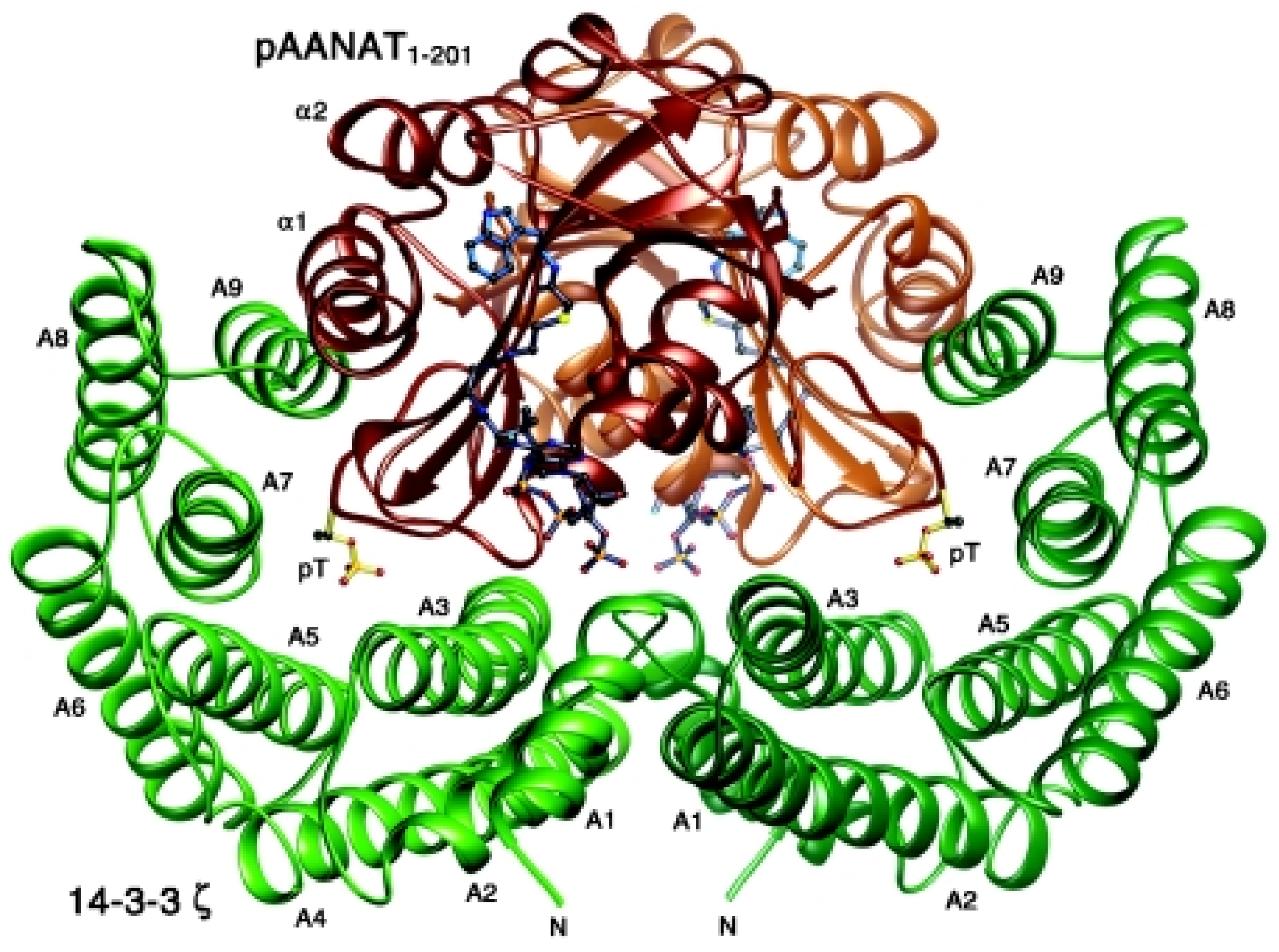
Figure 1. Crystal structure of a 14-3-3 dimer bound to phosphorylated AANAT. Structure of the 14-3-3–pAANAT complex, indicating the pThr31 of AANAT (brown/yellow), the proline twist C-terminal to pThr31, and the loop in AANAT (α1/α2 in brown) whose movement is restricted by the 14-3-3 dimer (green). Reproduced with permission from Portland press from the manuscript, Dynamic interactions between 14-3-3 proteins and phosphoproteins regulate diverse cellular processes Carol Mackintosh, Biochem. J. 2004 Jul 15; 381: 329–342 [12].
Endocrines 03 00057 g001
Figure 2. Immunofluorescence of HeLa cells showing the distribution of 14-3-3α/β both in nucleus and cytoplasm. PFA fixed HeLa cells were stained 14-3-3α/β with Alexa 488 conjugated secondary antibody. Images were taken using airy scan mode in Zeiss 800 confocal microscope using 63× objective. Nucleus is shown in blue (blue). Scale bar = 5 µm.
Endocrines 03 00057 g002
Figure 3. Immunofluorescence of HeLa cells showing the distribution of 14-3-η in the nucleus and cytoplasm. PFA fixed HeLa cells were stained 14-3-3-3η with Alexa 488 conjugated secondary antibody. Images were taken using airy scan mode in Zeiss 800 confocal microscope using 63× objective. Nucleus is shown in blue (blue). Scale bar = 5 µm.
Endocrines 03 00057 g003
Figure 4. Immunofluorescence of HeLa cells showing the distribution of 14-3-3ε both in the cytoplasm and nucleus. PFA fixed HeLa cells were stained 14-3-3-3ε with Alexa 488 conjugated secondary antibody. Images were taken using airy scan mode in Zeiss 800 confocal microscope using 63× objective. Nucleus is shown in blue (blue). Scale bar = 5 µm.
Endocrines 03 00057 g004
Figure 5. Immunofluorescence of HeLa cells showing the distribution of 14-3-3γ mainly in the nucleus with moderate levels in the cytoplasm. PFA fixed HeLa cells were stained 14-3-3-3γ with Alexa 488 conjugated secondary antibody. Images were taken using airy scan mode in Zeiss 800 confocal microscope using 63× objective. Nucleus is shown in blue (blue). Scale bar = 5 µm.
Endocrines 03 00057 g005
Figure 6. Immunofluorescence of HeLa cells showing the cytoplasmic distribution of 14-3-3σ with minimal expression in the nucleus. PFA fixed HeLa cells were stained 14-3-3-3σ with Alexa 488 conjugated secondary antibody. Images were taken using airy scan mode in Zeiss 800 confocal microscope using 63× objective. Nucleus is shown in blue (blue). Scale bar = 5 µm.
Endocrines 03 00057 g006
Figure 7. Immunofluorescence of HeLa cells showing the distribution of 14-3-3θ in the cytoplasm with little expression in the nucleus. PFA fixed HeLa cells were stained 14-3-3-3θ with Alexa 488 conjugated secondary antibody. Images were taken using airy scan mode in Zeiss 800 confocal microscope using 63× objective. Nucleus is shown in blue (blue). Scale bar = 5 µm.
Endocrines 03 00057 g007
Figure 8. Immunofluorescence of HeLa cells showing the distribution of 14-3-3θ mainly in the nucleus. PFA fixed HeLa cells were stained 14-3-3-3θ with Alexa 488 conjugated secondary antibody. Images were taken using airy scan mode in Zeiss 800 confocal microscope using 63× objective. Nucleus is shown in blue (blue). Scale bar = 5 µm.
Endocrines 03 00057 g008
Publisher’s Note: MDPI stays neutral with regard to jurisdictional claims in published maps and institutional affiliations.