
Actinomyces in Pregnancy: A Rare and Silent Cause of Preterm Delivery—Case Report

 ,
, 
Abstract
1. Introduction
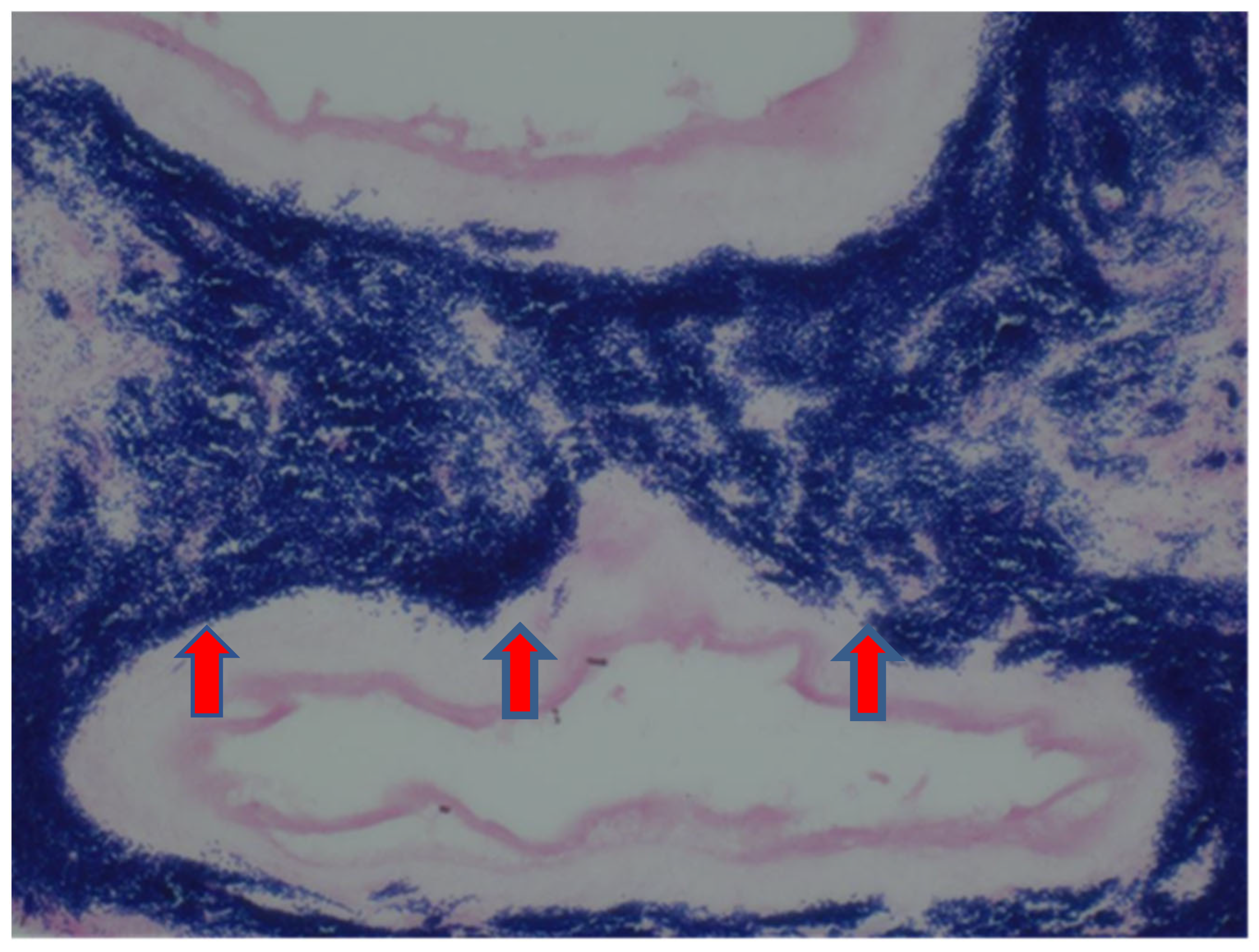
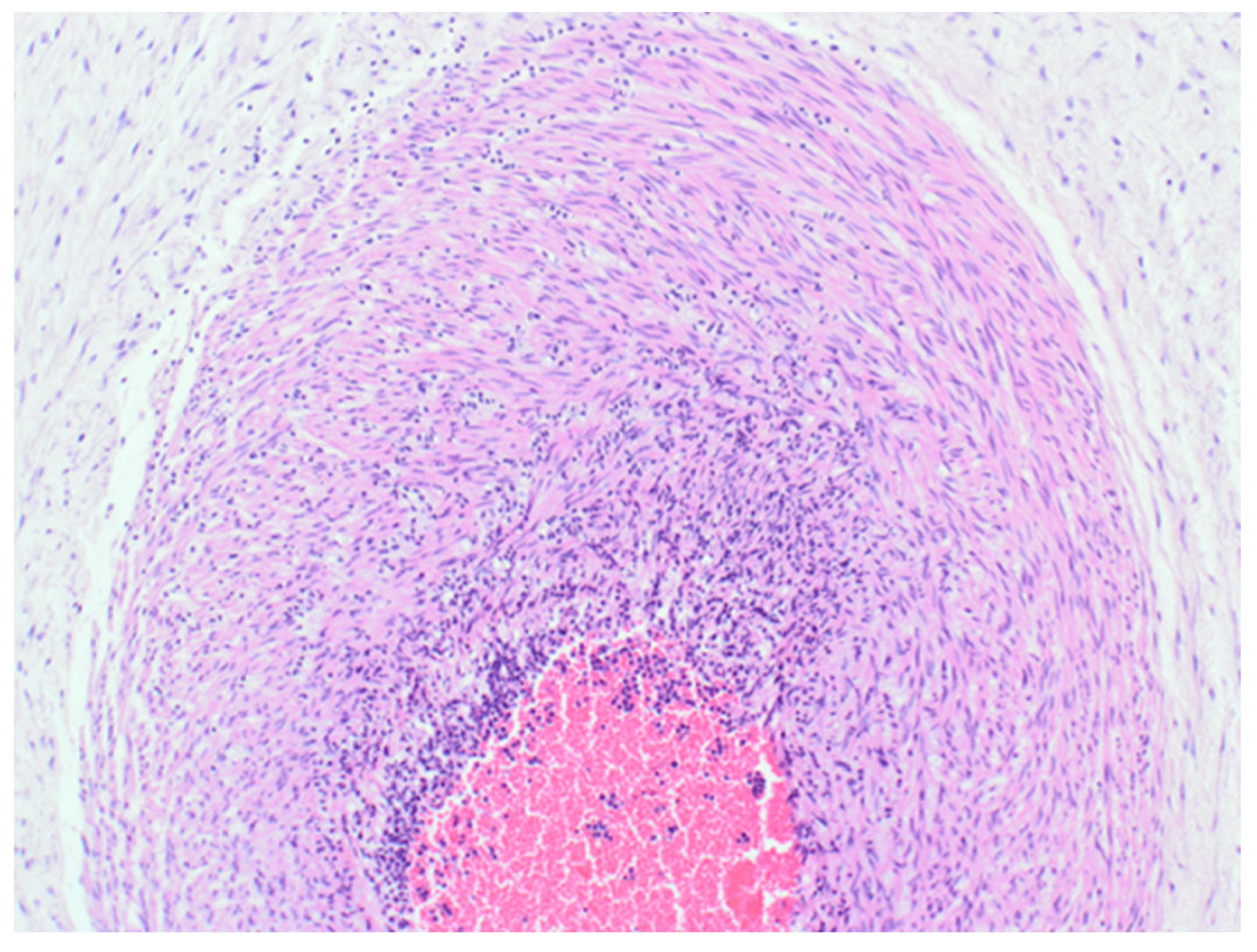
2. Case Report
3. Discussion
4. Conclusions
Author Contributions
Funding
Institutional Review Board Statement
Informed Consent Statement
Data Availability Statement
Conflicts of Interest
References
- Rueda, M.S.; Hefter, Y.; Stone, B.; Hahn, A.; Jantausch, B. A Premature Infant With Neonatal Actinomyces odontolyticus Sepsis. J. Pediatr. Infect. Dis. Soc. 2021, 10, 533–535. [Google Scholar] [CrossRef] [PubMed]
- Ong, H.-C.; Ling, A.C.-K.; Ng, D.S.-W.; Ng, R.-X.; Wong, P.-L.; Omar, S.F.S. Case report: Actinomyces naeslundii complicating preterm labour in a trisomy-21 pregnancy. ID Cases 2021, 23, e01051. [Google Scholar] [CrossRef] [PubMed]
- Villar, J.; Cavoretto, P.I.; Barros, F.C.; Romero, R.; Papageorghiou, A.T.; Kennedy, S.H. Etiologically Based Functional Taxonomy of the Preterm Birth Syndrome. Clin. Perinatol. 2024, 51, 475–495. [Google Scholar] [CrossRef] [PubMed]
- Zhang, Y.; Ye, Z.; Miao, Q.; Xu, H.; Pang, W. Actinomyces meyeri-induced brain abscess in pregnancy: A case report. BMC Neurol. 2023, 23, 401. [Google Scholar] [CrossRef] [PubMed]
- Gajdács, M.; Urbán, E. The Pathogenic Role of Actinomyces spp. and Related Organisms in Genitourinary Infections: Discoveries in the New, Modern Diagnostic Era. Antibiotics 2020, 9, 524. [Google Scholar] [CrossRef] [PubMed]
- Alsohime, F.; Assiri, R.A.; Al-Shahrani, F.; Bakeet, H.; Elhazmi, M.; Somily, A.M. Premature labor and neonatal sepsis caused by Actinomyces neuii. J. Infect. Public Health 2019, 12, 282–284. [Google Scholar] [CrossRef] [PubMed]
- Alghamdi, A.; Tabb, D.; Hagan, L. Preterm Labor Caused by Hemolysis, Elevated Liver Enzymes, Low Platelet Count (HELLP) Syndrome and Postpartum Infection Complicated with Actinomyces Species: A Case Report. Am. J. Case Rep. 2018, 19, 1350–1353. [Google Scholar] [CrossRef] [PubMed]
- Neuz-Zaragoza, W.; Sanfeliu, I. Acute necritizing chorioaminionitis caused by Actinomyces Neuii. Enfermedades Infecc. Y Microbiol. Clínica 2022, 40, 455–464. [Google Scholar]
- Wong, V.K.; Turmezei, T.D.; Weston, V.C. Actinomycosis. BMJ 2011, 343, d6099. [Google Scholar] [CrossRef] [PubMed]
- Gajdács, M.; Urbán, E.; Terhes, G. Microbiological and Clinical Aspects of Cervicofacial Actinomyces Infections: An Overview. Dent. J. 2019, 7, 85. [Google Scholar] [CrossRef] [PubMed]
- Kim, Y.J.; Youm, J.; Kim, J.H.; Jee, B.C. Actinomyces-like organisms in cervical smears: The association with intrauterine device and pelvic inflammatory diseases. Obstet. Gynecol. Sci. 2014, 57, 393–396. [Google Scholar] [CrossRef] [PubMed]
- Ohuma, E.O.; Moller, A.-B.; Bradley, E.; Chakwera, S.; Hussain-Alkhateeb, L.; Lewin, A.; Okwaraji, Y.B.; Mahanani, W.R.; Johansson, E.W.; Lavin, T.; et al. National, regional, and global estimates of preterm birth in 2020, with trends from 2010: A systematic analysis. Lancet 2023, 402, 1261–1271, Erratum in Lancet 2024, 403, 618. [Google Scholar] [CrossRef] [PubMed]
- Yu, H.-R.; Tsai, C.-C.; Chan, J.Y.H.; Lee, W.-C.; Wu, K.L.H.; Tain, Y.-L.; Hsu, T.-Y.; Cheng, H.-H.; Huang, H.-C.; Huang, C.-H.; et al. A Higher Abundance of Actinomyces spp. in the Gut Is Associated with Spontaneous Preterm Birth. Microorganisms 2023, 11, 1171. [Google Scholar] [CrossRef] [PubMed]
- Kahle, P.J.; Mellinger, G.T. Renal actinomycosis in pregnancy; report of a case. Urol. Cutan. Rev. 1949, 53, 720–723. [Google Scholar] [PubMed]


Disclaimer/Publisher’s Note: The statements, opinions and data contained in all publications are solely those of the individual author(s) and contributor(s) and not of MDPI and/or the editor(s). MDPI and/or the editor(s) disclaim responsibility for any injury to people or property resulting from any ideas, methods, instructions or products referred to in the content. |
© 2025 by the authors. Licensee MDPI, Basel, Switzerland. This article is an open access article distributed under the terms and conditions of the Creative Commons Attribution (CC BY) license (https://creativecommons.org/licenses/by/4.0/).
Share and Cite
Idaewor, P.E.; Ozua, P.; Jaiyesimi, R.A.K.; Al-Zawi, A.S.A. Actinomyces in Pregnancy: A Rare and Silent Cause of Preterm Delivery—Case Report. Reprod. Med. 2025, 6, 7. https://doi.org/10.3390/reprodmed6010007
Idaewor PE, Ozua P, Jaiyesimi RAK, Al-Zawi ASA. Actinomyces in Pregnancy: A Rare and Silent Cause of Preterm Delivery—Case Report. Reproductive Medicine. 2025; 6(1):7. https://doi.org/10.3390/reprodmed6010007
Chicago/Turabian StyleIdaewor, Philip E., Peter Ozua, Rotimi A. K. Jaiyesimi, and Abdalla SAAD Abdalla Al-Zawi. 2025. "Actinomyces in Pregnancy: A Rare and Silent Cause of Preterm Delivery—Case Report" Reproductive Medicine 6, no. 1: 7. https://doi.org/10.3390/reprodmed6010007
APA StyleIdaewor, P. E., Ozua, P., Jaiyesimi, R. A. K., & Al-Zawi, A. S. A. (2025). Actinomyces in Pregnancy: A Rare and Silent Cause of Preterm Delivery—Case Report. Reproductive Medicine, 6(1), 7. https://doi.org/10.3390/reprodmed6010007