Review

Ovarian Cancer: Treatment and Resistance to Pharmacotherapy

1
Department of Gynecology, Division of Gynecology and Obstetrics, Ljubljana University Medical Center, Zaloška 2, 1000 Ljubljana, Slovenia
2
Department of Gynecology and Obstetrics, Faculty of Medicine, University of Ljubljana, Šlajmerjeva 3, 1000 Ljubljana, Slovenia
3
Institute of Pharmacology and Experimental Toxicology, Faculty of Medicine, University of Ljubljana, Korytkova 2, 1000 Ljubljana, Slovenia
*
Author to whom correspondence should be addressed.
Reprod. Med. 2022, 3(2), 127-140; https://doi.org/10.3390/reprodmed3020011
Submission received: 24 February 2022 / Revised: 28 April 2022 / Accepted: 5 May 2022 / Published: 9 May 2022
(This article belongs to the Special Issue Recent Advances in Gynecologic Oncology)

Abstract

:
Despite advances in surgical techniques and chemotherapy, ovarian cancer is still a leading cause of death among gynecological cancers. In addition to the late detection of the disease, the main reason for poor prognosis is resistance to pharmacotherapy, mostly platinum compounds. About a third of patients do not respond to primary platinum-based chemotherapy treatment, and over time, eventually, 80% of other patients develop chemoresistance, which makes the recurrence of disease incurable. In this review, we describe a difficult clinical hurdle faced in ovarian cancer therapy as a result of platinum resistance, as well as resistance to newer targeted therapy with PARP inhibitors and bevacizumab. We, furthermore, give attention also to the role of the tumor microenvironment as it is less well understood than the tumor cell-intrinsic mechanism. Because a central goal in ovarian cancer research is the development of novel strategies to overcome chemoresistance, treatment for cancer is moving toward personalized therapy.

1. Introduction

Ovarian cancer (OC) is a malignant neoplasm in one or both ovaries. It is one of the most common gynecological malignancies and has the highest mortality rate. In 2018, there were 295,414 cases of OC newly diagnosed across the globe and 184,799 OC patients died [1,2,3]. Based on information from the Slovenian Cancer Registry, OC ranks eighth among common types of cancer in women in Slovenia. On average, 166 women (15.9 per 100,000) developed OC and 141 (13.6 per 100,000) died from it per year between 2010 and 2014, which ranks Slovenia at the EU average [4,5,6]. This unfavorable prognosis is the result of asymptomaticity or late occurrence of symptoms and the lack of an effective screening method. Consequently, a diagnosis is made at an advanced stage, when treatment is less effective (stages III and IV per the International Federation of Gynecology and Obstetrics [FIGO]). The 5-year survival rate for women that are diagnosed with OC in the early stage is, therefore, 93%, and the rate for those that are diagnosed with OC in the advanced stage is 29% [6,7,8].
A mix of both genetic and epigenetic changes, plus increasing genetic diversity found in tumor cells as cancer progresses, are the major factors hindering a cure [6,9]. Platinum-based chemotherapy agents (platins) have been deemed fundamental chemotherapy treatment for four decades now. Moreover, despite the development of novel targeted drugs, carboplatin with additional paclitaxel is still the standard first-line treatment used for OC in the advanced stage [6,10]. Nonetheless, from 20% to 40% of patients show no response to this primary therapy. Moreover, 80% of patients that exhibit a positive initial response, especially those with the subtype classified as high-grade serous ovarian cancer (HGSOC), develop a platinum-resistant recurrence over time [11]. Based on the most recent consensus conference manuscript on ovarian cancer from the European Society for Gynecological Oncology (ESGO) and European Society for Medical Oncology (ESMO), which was published in May 2019, at present, there are no validated molecular predictive biomarkers for platinum resistance [10].
This review sums up current information about OC and treatment protocols for it. Moreover, it addresses the resistance mechanism to the most established and what are still the most effective categories of chemotherapeutics for treating OC—platinum agents and taxanes—as well as resistance to newer targeted therapy with poly (ADP-ribose) polymerase (PARP) inhibitors and bevacizumab, a vascular endothelial growth factor (VEGF) inhibitor. Attention is also paid to the role of the tumor microenvironment because it is less well understood than the tumor cell-intrinsic mechanism. This is also evidenced by the much-lesser-known resistance to bevacizumab, a drug that affects the tumor microenvironment.

2. Histological Subtypes of OC

Ovarian cancer can form from any type of cells that make up the ovary. A distinction is made between epithelial, germ cell, stromal, and other types (mesothelial–mesenchymal, mixed cell, and secondary tumors). Epithelial ovarian cancer is the most frequent type, and it accounts for 90% of ovarian malignancies. It is further divided into two groups of histopathological subtypes. The first group comprises endometrioid, clear cell, mucinous, and low-grade serous carcinoma (LGSOC), and the second group includes high-grade serous carcinoma (HGSOC) as the most common subtype (75%) of epithelial OC [12,13]. It is important to determine the subtype because there are differences between them, in terms of origin, risk factors, patterns of spread, treatment, and responses to treatment [14].
Group one subtypes usually have an indolent clinical course and are genetically more stable. They are often characterized by mutations in regulators of the mitogen-activated protein kinase (MAPK) pathway, such as BRAF (v-Raf murine sarcoma viral oncogene homolog B) and KRAS (Kirsten rat sarcoma), p-catenin genes, CDKN2A (cyclin-dependent kinase inhibitor 2A), and PIK3CA (phosphatidylinositol-4,5-bisphosphate 3-kinase, catalytic alpha). Mutations in TP53 (tumor protein P53) are rare, with the exception of the mucinous subtype. Clear cell carcinomas are characterized by a high frequency of inactivating mutations in the tumor suppressor gene ARID1A (AT-rich interaction domain 1A). In addition, amplification of the ERBB2 gene that encodes for HER2 (human epidermal growth factor receptor 2) is more frequent in mucinous and clear cell carcinomas [15,16].
HGSOC in group two has a more aggressive clinical course and is genetically less stable. It is generally only diagnosed once it has reached an advanced stage, and it has a poor prognosis. TP53 mutations are typical (present in 96% of tumors) and defective homologous recombination DNA repair is also common (50%) [17]. Recurrent mutations in other genes are uncommon, with the exception of breast cancer genes BRCA1 and BRCA2, which primarily manifest as HGSOC (13). Amplification of CCNE1 (cyclin-E12), MYC (myelocytomatosis oncogene), and TPX2 (targeting protein X2) is also more frequent [15].
OC differs from the majority of other cancer types in terms of how it spreads because it rarely metastasizes hematogenously. It basically metastasizes through a passive mechanism, whereby ovarian cancer cells that are shed from the surface of the primary tumor are carried via the physiological movement of the peritoneal fluid and transported to the peritoneum and omentum (i.e., transcoelomic metastasis) [18]. In this, cancer cells transform from an epithelial to a mesenchymal form (known as epithelial to mesenchymal transition, or EMT), which allows them to detach from their sister cells and attach to and penetrate the basement membrane [19]. Pathologic accumulation of peritoneal fluid or the formation of ascites may facilitate the spread of cancer cells. Ascites forms due to an imbalance in the formation and reabsorption of peritoneal fluid. Cancer cells are believed to prevent its reabsorption via the subperitoneal lymphatic ducts. In the early stages, this involves nonobstructive mechanisms, including contraction of lymph vessels induced by the secretion of tumor cell products, and an obstruction of lymphatic ducts at later periods [12]. In addition, cancer cells secrete numerous factors that can increase the formation of peritoneal fluid. The most researched factor in this regard is the effect of vascular endothelial growth factor (VEGF), which increases the permeability of vessels and, thus, the accumulation of ascites [18,20].

Tumor Heterogeneity

OC is a clinically diverse and histologically and molecularly highly heterogeneous disease. Increasingly, more studies are showing differences both between and within individual ovarian cancer types and subtypes. In addition, they demonstrate the existence of tumor heterogeneity at the level of an individual patient, which can be either spatial or temporal. Spatial heterogeneity can be intertumoral due to differences between primary and metastatic lesions, or intratumoral, which is not unexpected due to subclonal tumor evolution. The term temporal heterogeneity refers to changes that may occur over time as the tumor progresses (e.g., at diagnosis vs. relapse) [15,16]. Studies using next-generation sequencing have determined that intratumoral differences are already present before treatment and are not limited exclusively to high-grade tumors. Most relapse characteristics or metastases are already present as subclonal populations within the primary tumor [21].

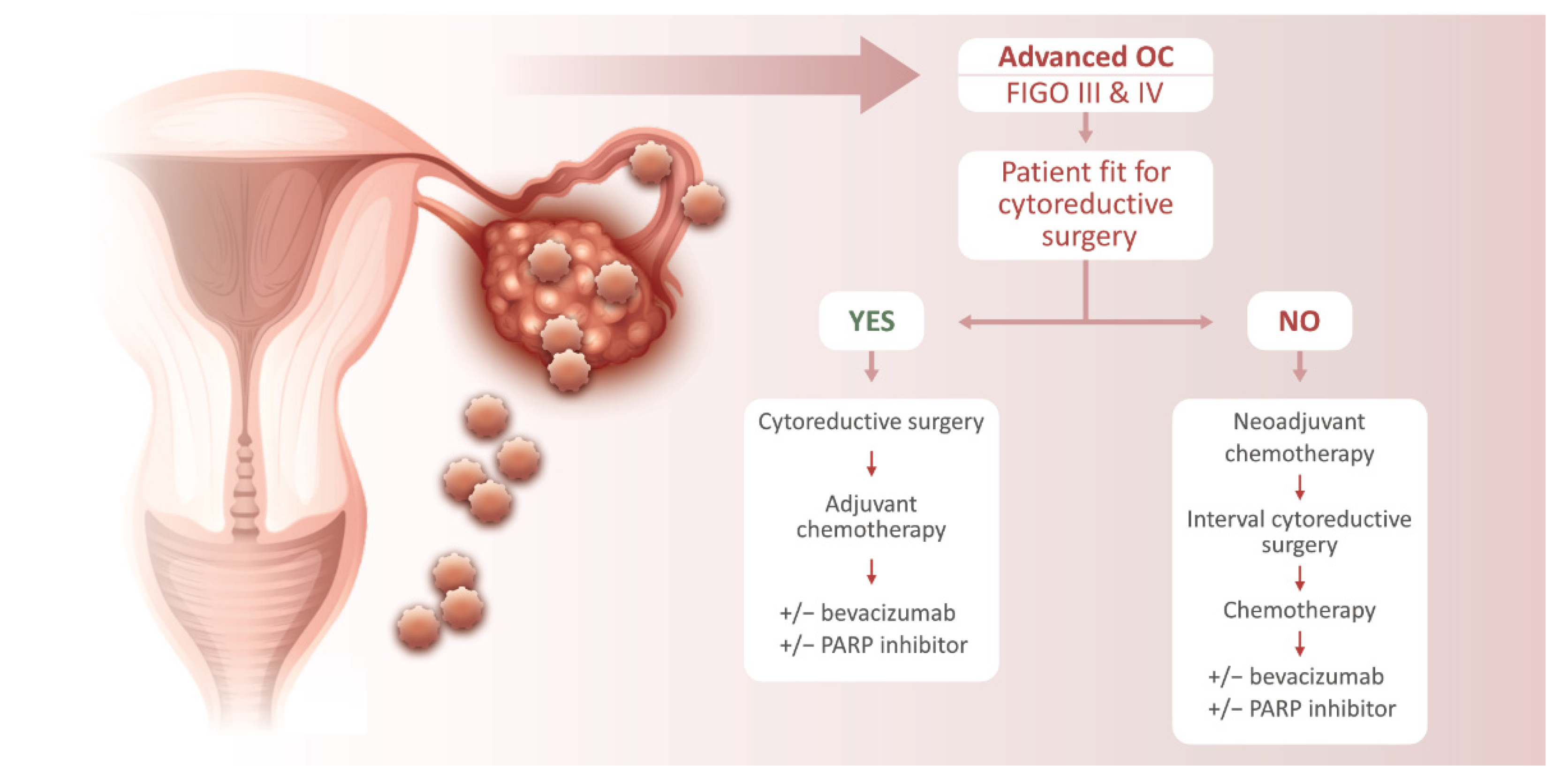
3. Ovarian Cancer Treatment

Treatment for OC includes surgical removal of the tumor (cytoreductive surgery) and systemic chemotherapy. Since the mid-1970s, platinum compounds have formed the basis for chemotherapy. Initially, this was cisplatin, which, however, was associated with a range of adverse effects. Therefore, second-generation platinum compounds soon began to be developed, resulting in the 1989 introduction of carboplatin, which is just as effective as cisplatin but has fewer serious adverse effects, especially primarily in terms of nephrotoxicity. The addition of targeted therapies in the 2010s brought the possibility of a better safety profile, but even this therapy is not without serious adverse effects. PARP inhibitors, which have generally been found to be safe and well tolerated, are associated with a risk of serious hematological toxicities. Bevacizumab, another targeted drug, increases the risk of even fatal gastrointestinal perforation, and so patients that have a history of treatment for inflammatory bowel disease, or bowel resection, should be excluded from such therapy. In addition, hypertensive patients should be closely monitored [22].
According to established guidelines, cytoreductive surgery is followed by postoperative (adjuvant) chemotherapy with paclitaxel and carboplatin. Patients are usually administered six to eight cycles every 21 days [4]. Contraindications for a combination of paclixatel and carboplatin are poor overall performance status (PS ≥ 3 according to WHO), significant comorbidities (heart failure, ischemic heart disease, neuropathy, etc.), uncontrollable hypersensitivity to the medication, and high biological age. Pegylated liposomal doxorubicin (PLD) can be used as an alternative to paclitaxel, but it has demonstrated a higher incidence of hematological and dermatological toxicity and stomatitis. However, it causes less neurotoxicity and alopecia [23]. In patients with poor performance status, comorbidities, and high age, it is to be expected that they will have difficulty enduring combined systemic treatment in full three-week doses and it is less likely that they will complete the course of therapy. In this case, a weekly form of combined therapy can be administered alternating paclitaxel and carboplatin, or monotherapy with carboplatin [23,24,25]. In some cases, treatment begins with preoperative (neoadjuvant) chemotherapy followed by surgery. This method may be considered for patients with advanced ovarian cancer (stages III–IV), for whom the surgeon decides that radical removal is not possible, or if a patient is unable to undergo surgical treatment due to comorbidities [26]. The purpose of preoperative therapy is to improve the patient’s status or clinical response that would increase the likelihood of radical surgery. The same combination of drugs is applied in three to six cycles, after which interval surgery follows with maximum cytoreduction and three to four cycles of postoperative systemic therapy. A schematic diagram showing a treatment algorithm for advanced OC is shown in Figure 1.
Bevacizumab can be added to standard chemotherapy (from the second round on) [4]. In patients with advanced disease (stages IIIB, IIIC, and IV), there are two types of target agents available for maintenance therapy: bevacizumab and PARP inhibitor [4]—Table 1. PARP inhibitor maintenance therapy has resulted in the improvement of progression-free survival, particularly in patients with homologous recombinant deficiency (HRD)-positive status, defined by either BRCA1/2 mutation and/or genomic instability. However, prerequisites for using PARP inhibitors are the following: (a) complete or partial response to the first platin therapy in patients that have primary OC, or (b) complete or partial response to the most recently applied regimen in patients that have platinum-sensitive recurrent OC [6,27]. Patients in whom there is a relapse of OC should be evaluated as candidates for further platinum therapy if it is not contraindicated or if they do not exhibit definite platinum resistance [28]. Because of the basic role of platins in treating OC, platinum sensitivity remains a major determinant for prognosis. Nonetheless, predicting true platinum chemoresistance is not currently possible, chiefly because the exact molecular mechanisms of platinum resistance remain unclear. An additional reason connected with enigmatic mechanisms is the lack of validated molecular predictive biomarkers for platinum resistance [6,10,28]. As a result, significant effort is currently being invested to overcome this clinical obstacle.
Although targeted therapy has brought advances in treatment, it still has some limitations and challenges. Despite considerable advances in prolonging the time to first progression, overall survival of EOC patients remains unsatisfactory. Resistance may also occur with targeted therapy. There are no validated predictive molecular biomarkers of bevacizumab benefit. Angiogenic markers, such as CD31 expression, microvessel density, and tumor VEGF-A levels, may provide prognostic information. However, these findings need to be further validated for routine clinical practice. Attempts to identify robust predictive biomarkers of response to PARP inhibitors in HGSC beyond key HG gene mutation have proven difficult (e.g., testing on RAD51C/D, BRIP1, and PALB2 should be considered). Moreover, a major limitation of current HR assays is that they are largely insensitive to reversion of HRD, which may occur upon development of resistance to Pt-based medicines and PARP inhibitors [10].

4. Resistance to Treatment

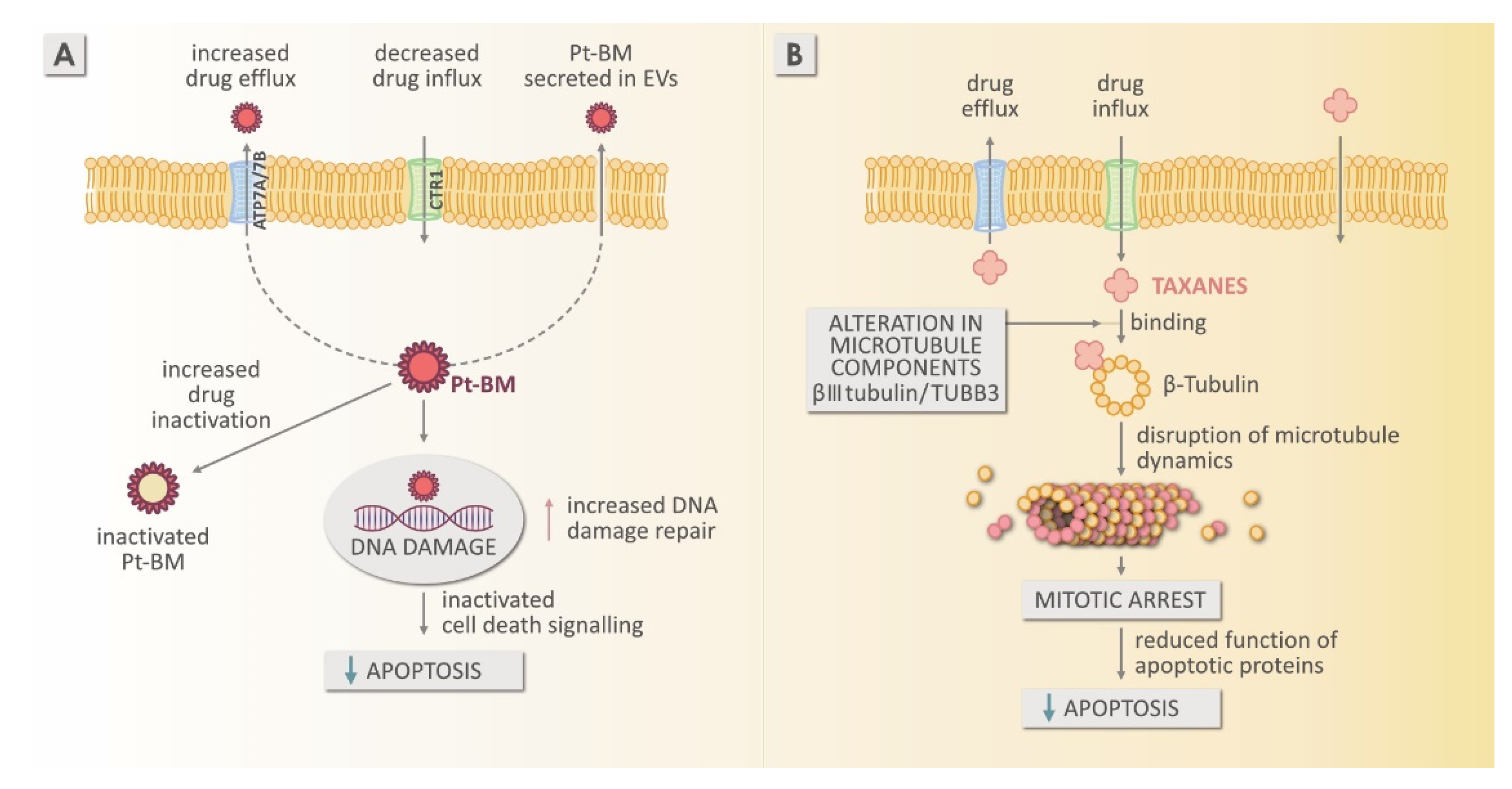
4.1. Resistance to Platinum-Based Chemotherapy

The first-choice platin (Pt) used at present for clinically managing OC is carboplatin. It replaced cisplatin as a result of its superior safety profile. There are a greater number of studies on cisplatin due to its status as an older medicine, but cisplatin and carboplatin are considered to operate in a similar manner to create an antineoplastic effect [6,29]. Consequently, the chemoresistance mechanisms may also be similar. Nonetheless, caution is advised because they have different chemical structures; this aspect is reflected in their differing pharmacokinetic parameters [6,30].
Chemoresistance occurs in 20% to 30% of patients during primary treatment [31]. The remainder of patients respond well to treatment, but in 80%, the median progression-free survival is only 18 months. When retreating patients that have relapsed after more than 12 months, only 50% respond to treatment. The percentage is even lower (10–20%) in patients that relapsed less than 6 months after initial treatment. With each new recurrence, the interval to the next recurrence tends to be shorter, and the likelihood of resistant disease tends to increase [32].
The problem of Pt resistance typically occurs in patients that receive neoadjuvant therapy with carboplatin before surgery. In their in vitro study, Matsuo et al. established a greater resistance to carboplatin in patients that received neoadjuvant therapy (33.3%) compared to patients that underwent primary cytoreductive surgery (9.2%) [33]. Similarly, Rauh-Hain et al. determined more frequent resistance to carboplatin in patients that received neoadjuvant therapy (88.8%) compared to patients that underwent primary cytoreductive surgery (55.3%) [34]. However, there is no consensus regarding a more suitable primary treatment. Some studies contest the superiority of primary cytoreductive surgery over neoadjuvant chemotherapy. The CHORUS randomized trial [35], which included 550 patients, compared the results of both treatment methods. Among the patients, 276 underwent standard primary cytoreductive surgery, followed by adjuvant Pt-based chemotherapy, and 274 first received Pt-based neoadjuvant therapy, followed by interval cytoreduction. The second method has been connected with increased optimal debulking, reduced early mortality, and survival similar to that in standard treatment [35].
The treatment type is based on the amount of time that has elapsed since the end of the primary treatment (the platinum-free interval, PFI), and it is split into four categories [11]. The PFI is the primary indicator for tumor classification (Pt-sensitive or Pt-resistant), on the basis of a 6-month cutoff from the most recent Pt treatment. This definition developed when there were limited options beyond Pt re-challenge for recurrent disease treatment. It has a number of shortcomings and it was, therefore, abandoned during the Gynecologic Cancer Intergroup’s (GCIG) Fifth Ovarian Cancer Consensus Conference (OCCC) [6,10]. Recently the concept has shifted to whether “platinum is an option” or “platinum is not an option”.
The literature most often describes two types of resistance with different development mechanisms, although precisely distinguishing between the two can be difficult. These are two forms, intrinsic resistance and acquired (extrinsic) resistance [36]. Intrinsic resistance refers to the inherent ability of cancer cells to remain resistant due to their characteristics that already existed before their first exposure to treatment. A subtype of tumor cells with such resistant characteristics is referred to as cancer stem cells (CSC), which are capable of self-renewal, persistent sustenance of tumor growth, and long-term dormancy. The characteristics of cells that demonstrate intrinsic chemoresistance include the ability to reduce drug uptake, enhance drug efflux, and increase the activity of detoxification enzymes, such as cytochrome P450 or glutathione transferases [32]. Almost one out of three HGSOC patients do not respond to initial treatment and, therefore, the disease progresses during or immediately after first-line therapy, so that patients die within 1 year of being diagnosed [6,37]. The second resistance type is acquired resistance, in which cancer cells gradually acquire resistance over the course of treatment, and genetic changes and epigenetic alterations to key genes develop that allow cancer cells to adapt to the effects of chemotherapeutics. This can be thought of as microevolution, in which, through the acquired alteration, a cell gains an advantage over others, survives, and is, therefore, clonally selected to proliferate [16]. Among the molecular mechanisms involved in intrinsic or acquired chemoresistance are extracellular matrix proteins, oncogene and tumor suppressor gene mutations (TP53 and PAR-4), mitochondrial alteration, insensitivity to and repair of DNA damage, autophagy, cancer stemness, transporter proteins, epithelial–mesenchymal transition (EMT), and alterations in glutathione reductase expression/activity [6].

Mechanism of Resistance to Platinum-Based Medicines

It is known that reduced in vitro intracellular Pt accumulation is one of the most typical features of cell lines that are cisplatin resistant [38]. The finding that cultured cancer cells that are resistant to cisplatin are also cross-resistant to Cu and vice versa implies that Pt-based medicines (Pt-BMs) may be a substrate for Cu transporters [6]. It has been demonstrated that Pt chemotherapeutics can cross cell membranes, either through passive diffusion or through the assistance of transporters, among which are well-characterized copper transporters and other mechanisms yet to be defined [38]. Several mechanisms are involved in Pt chemoresistance; these include lower drug accumulation in the intracellular space and/or an increase in drug efflux, drug inactivation through augmented levels of cellular thiols, changes in drug targets, processing damage induced by drugs through greater nucleotide excision-repair activity, and lower mismatch-repair activity and apoptosis evasion—Figure 2. Moreover, changed gene expression, alterations in DNA copy number, and greater genomic instability may be a contributing factor to Pt chemoresistance [6,394041424344].
Although copper is an essential cofactor for many enzymes, like Pt, it can have high toxicity and, therefore, a complex protein network has evolved in order to regulate copper transport to and from cells and also for chaperoning it inside cells. Among these proteins are copper transport receptors CTR1 and CTR2 and copper-transporting P-type ATPases ATP7A and ATP7B. These assist in maintaining copper homeostasis and they have been connected with Pt efflux [38]. In cells that are resistant, membrane transporter changes can impact the accumulation of Pt drugs by causing increased drug efflux or decreased drug uptake [6]. Indeed, several studies of cisplatin’s cellular pharmacology suggest that specialized membrane-bound proteins are involved in mediating drug uptake and efflux, and that nearly all cell lines that have been selected for cisplatin resistance show alterations in accumulation of drugs [45]. In general, the copper efflux transporters ATP7A and ATP7B and the copper importer CTR1 seem to play a significant role in cisplatin’s pharmacokinetics and cytotoxicity, together with the copper chaperone ATOX1, which helps mediate copper transfer from CTR1 to ATP7A and ATP7B [41,45,46,47,48,49,50,51] The history showed that expression of both transporters, ATP7A and ATP7B, changes during tumorigenesis of OC. Samimi et al. (2003) concluded that it is, nonetheless, possible that increased expression of ATP7A may offer some significant in vivo growth advantages, and that in some tumors, the quantity of ATP7A-expressing cells increases regardless of any effect of the therapy [48]. The literature [6] indicates that there is a correlation between increased expression of the Cu efflux transporters ATP7A and ATP7B in tumor tissue and a poor response to Pt-BMs and low survival in OC patients. This indicates that these transporters are some of the important targets in what is otherwise multifactorial resistance to Pt. The association between an inadequate response to Pt-BMs and greater expression of ATP7B at initial diagnosis or with ATP7A during treatment points to the potential of both Cu-ATPases for deciding upon the best treatment for an individual patient [6].

4.2. Mechanism of Resistance to Taxanes

Taxanes, including paclitaxel, target not only microtubules but also self-assembled alpha- and beta-tubulin heterodimers. These are dynamic components of the cytoskeleton, and during cell division, they play essential roles in intracellular transport and the mitotic spindle. Aggregates of dysfunctional microtubules disrupt normal processes of cellular transport and also interfere in mitosis, later leading to mitotic arrest—Figure 2. In the cell culture, taxanes cause both mitotic arrest and apoptosis. In OC, the emergence of cisplatin/carboplatin plus paclitaxel probably shows the impact of microtubule disruption drugs on the trafficking of DNA repair proteins to the nucleus. Disruptions of translocation to the nucleus, critical for repairing the DNA damage that is caused by the Pt agent, is probably the basis for the significant activity of the combination of the Pt compound with paclitaxel in OC [37]. The multidrug-resistant (MDR) phenotype is characterized by a decreased concentration intracellular drug, an increase in the expression of drug-metabolizing enzymes, changed progression of cell cycle checkpoints, altered pathways for apoptosis or survival, and signal transduction pathway deregulation. As a specific feature of taxane-treated tumors, unusual expression of microtubule subunits and proteins associated with them would come into direct conflict with the taxane mechanism of action and result in reduced bioactivity of the drug [52]. Taxanes are good substrates for MDR1 efflux pump; however, the extent of the role that efflux plays in clinical drug resistance remains unclear. It was discovered that high MDR1 expression is linked to a poor prognosis in multiple series [38]. Therefore, making use of MDR1 and other ABC transporter proteins as biomarkers for taxane response could have greater utility than directly targeting the proteins [52]. Taxane resistance is not completely understood; however, the following mechanisms are seen as major factors and are even often observed in different types of cancer. The reduced effectiveness of taxanes’ method of action culminates in a lower concentration of intracellular drug (as a result of increased drug export), greater metabolism of taxanes (due to upregulating CYP enzymes), and a changed composition of tubulin subunits (in particular, III-tubulin upregulation). If chemotherapy is applied at low levels or is not able to properly bind to the target, it may not be possible for cells to properly arrest, and no apoptosis is induced. Moreover, it is possible that hypoxic conditions found in solid tumors play a greater role in activating these pathways to resistance [38,52].

4.3. Mechanism of Resistance to PARP Inhibitors

The clinical use of PARP inhibitors in the treatment of epithelial OC has increased dramatically. As mentioned in Table 1, PARP inhibitors are now EMA approved across all lines of treatment of epithelial OC, and they are used as maintenance therapy after neoadjuvant chemotherapy in patients with OC [4,53,54,55]. Despite their use in the clinic, PARP inhibitor resistance is common and develops through multiple mechanisms. Cells with BRCA1/2 deficiency were found to be particularly sensitive to PARP inhibitors. Importantly, BRCA1/2-deficient tumor cells can become resistant to PARP inhibitors by restoring homologous recombination (HR) repair and/or by stabilizing their replication forks [56]. Aside from the restoration of HR, other mechanisms have been identified in vitro but have not yet been confirmed in large clinical studies (Shieldin complex, which blocks DNA repair by the NHEJ pathway and inactivation, confers PARP inhibitor resistance, and loss of proteins PTIP and EZH2) [40,54,55]. As concluded by Lee and Matulonis [55], many mechanisms, already described, require clinical validation. Moreover, the multiple resistance mechanisms described can also arise within one individual, which makes a simple conclusion very difficult and, therefore, it is important that clinicians attempt to determine which patients are most likely to benefit from PARP inhibitors. There are different strategies (combination with chemotherapies, targeting acquired vulnerabilities associated with resistance to PARP inhibitors, or suppressing genomic instability) that might overcome these mechanisms in an attempt to identify the optimal treatment regimens [57].

4.4. Mechanism of Resistance to Bevacizumab

By establishing a new vascular network, angiogenesis is an important contributor to tumor development and metastatic dissemination. Targeting VEGF offers an advance in the therapeutic management of cancer patients. However, complete responses are rare, and tumors counteract this inhibition through various processes [58]. Bevacizumab is a monoclonal humanized antibody against VEGF that is the most potent proangiogenic factor for mediating multiple steps of tumor angiogenesis. Because it targets the process that is essential for the growth of a solid tumor, it is no surprise that bevacizumab has a special place in therapy; it has been implemented in front-line treatment with Pt but also in Pt-sensitive and -resistant recurrent OC (Table 1). However, resistance to bevacizumab is inevitable because angiogenesis is a multifactor process, but molecular mechanisms of resistance are, nonetheless, not fully understood [58,59]. Several mechanisms exist and involve a wide range of processes, which are categorized from earliest to latest. The earliest address the upregulation of genes involved in angiogenic redundancy, epithelial–mesenchymal transition, or the lysosomal sequestration of drugs. On the other hand, the latest contain an adaptation of the tumor microenvironment, reflected by the recruitment of progenitor cells, lymphangiogenesis, and adapted neovascularization modalities [58]. Wieser and Marth [59] and Yang et al. [60] described, in detail, that mechanisms of resistance to anti-VEGF therapy might be mediated by tumor cells and by elements of the microenvironment. VEGF pathway inhibition can cause hypoxia, which is a major molecular controller of the angiogenic switch. Hypoxia inducible factor 1-alpha (HIF-1α) and interleukin 8 (IL-8) have been shown to support angiogenesis and resistance to apoptosis. Another consequence of VEGF inhibition is the promotion and recruitment of vascular progenitors (e.g., endothelial and pericyte progenitors) and vascular modulators, such as tumor-associated macrophages (TAM), immature monocytes, VEGFR-1, CD11b-myeloid cells, and hemangiocytes. Growing evidence indicates that inflammation controls angiogenesis because TAMs have been linked to the escape from anti-angiogenic therapy. TAMs of the M2 phenotype promote tumor transforming growth factor (TGF-β) and VEGF, and they attract leukocytes to further enhance angiogenesis [59,60]. All these mechanisms allow for tumor metastasis and serve as limitations to anti-angiogenic drug efficacy, highlighting that targeting only one mechanism involved in cancer development is insufficient [58,61].

5. The Role of the Tumor Microenvironment

A tumor is not merely a clonal expansion of mutant cells. It should also be viewed as an organ-like structure that communicates with the environment it lives in [16]. The role that the tumor microenvironment plays in mediating resistance to chemotherapy effects is less well understood than the tumor cell-intrinsic mechanism discussed above [38]. Tumor heterogeneity, disease progression, and development of therapeutic resistance are likely not only the consequence of the (intra) cellular features of the tumor, but also its extracellular environment or microenvironment [62]. This is composed of leukocytes, endothelial cells, and stromal myofibroblasts. Stromal myofibroblasts produce proteases, angiogenic factors, growth factors, signaling molecules, immunomodulatory proteins, antiapoptotic proteins, and extracellular matrix. They are also referred to as cancer-associated fibroblasts (CAF) because they respond to the stimuli of cancer cells and facilitate their invasion through bidirectional communication [16]. In experimental models, CAF can increase Pt resistance through the release of glutathione and cysteine into the microenvironment [38]. Tumor progression is associated with a disruption in tissue architecture and organization; the tumor overrides the homeostatic processes and adapts the environment to its own needs, ensuring its survival [63].
One way in which normal and cancer cells communicate, both between themselves and the environment, is through extracellular vesicles (EVs). These are a heterogeneous group of structures that are released from the cells and enclosed in membranes, and they can transport proteins, lipids, and nucleic acids (DNA, mRNA, and miRNA) [16]. They have been isolated from a variety of bodily fluids, including plasma, ascites, cerebrospinal fluid, urine, and amniotic fluid. EVs include exosomes 30 to 100 nm in size and microvesicles 100 to 1000 μm in size. Cancer cells also secrete oncosomes, which are 1 to 10 μm in diameter [64]. Microvesicles can form and be released through budding from the plasma membrane of cells into the environment. Exosomes are initially multivesicular bodies or endosomes (intracellular vesicles) that fuse with the membrane and are released as exosomes. EVs are formed and released under the influence of many endogenous and exogenous factors that may change the number, type, and content of the vesicle. All EVs have surface molecules that make it possible for them to target recipient cells. EVs can then cause signaling through receptor–ligand interaction or they can be internalized via endocytosis and/or phagocytosis. It is even possible for them to fuse with the membrane of the target cell to release their material into the cell [65].
In cancer cells, the quantity of EVs released changes, as does their size and even the type of molecules they carry. By carrying bioactive substances, they influence the tumor’s microenvironment and contribute to its spread and the development of chemoresistance. Three chemoresistance mechanisms are frequently described [66]. One of them is increased drug export from cells via EVs, as shown by Safaei et al. They compared the concentration of Pt in EVs released from Pt-resistant cell lines to that in EVs released from Pt-sensitive cell lines, and they established that the EVs released from the former contained 2.6-times more Pt. In addition, EVs from Pt-resistant cells also contained more Pt transporters [67]. EV-mediated transfer of miRNA is also mentioned as a drug resistance mechanism. It is presumed that EVs function as a genetic exchange vector in the tumor microenvironment. miRNA is a small non-coding RNA molecule that binds to mRNA and performs posttranscriptional gene modification. Pink et al. compared the expression of a variety of miRNAs and identified miR-21-3p as a possible cause of resistance. They also showed that resistance can be transferred from resistant to sensitive cell lines via the EVs of the former [68]. Another interesting drug-resistance mechanism is the neutralization of antibody-based (immunotherapy) drugs. The tumor also releases drug-targeted proteins onto EVs. These proteins bind with the drug before it manages to bind to the cancer cells and, thus, neutralize it. The latest conclusion by Heredia-Soto et al. [69] is that it is now well established that the micro-environment’s influence on tumor development is crucial for understanding disease evolution. The exhaustive characterization of the specific role of each cellular component and its interactions (the connection between tumor cells and stromal components, such as endothelial cells, fibroblasts, and immune cells, which has an influence on tumor development) has become a priority challenge, with the focus on designing effective combination therapies in OC [69].

6. Resistance Study Models

Ovarian cancer is a heterogeneous disease of a largely epithelial origin that has five subtypes. These display key differences, meaning these subtypes should be treated as independent diseases. Knowing their characteristics and identifying possible markers might contribute to the development of new target drugs. Preclinical studies, thus, require well-defined cell-line models that match the known clinical phenotypes. Since 1990, around 100 articles a year have been published that rely on commercially obtained cell lines. The problem is that their origin and histology are largely poorly defined, which makes it difficult to categorize the study results under a specific histological and molecular subtype that would be truly representative. Analyses of the most commonly used cell lines for HGSOC studies to date have shown that these cell lines in fact only poorly reflect the genetic characteristics of HGSOC [6,16,70]. On the other hand, clinically, epithelial tumors account for 75% of all tumors and as much as 90% to 95% of malignant ovarian tumors. Consequently, most guidelines for histopathological treatment involving ovarian tumors primarily address epithelial tumors. In addition, this applies to primary peritoneal carcinoma and to epithelial tumors of the fallopian tube [4].
Beauford et al. [71]. conducted a detailed analysis of 39 ovarian cancer cell lines to elucidate their background. This way, they formed the basis for selecting the models of various histological and molecular ovarian cancer types [71]. There is an additional shortage of cell lines that could serve as a good model for studying chemoresistance. Only a few of them are known to originate from patients with a clinical resistance to carboplatin. Most in vitro studies are performed on cell lines whose resistance was established through long-term exposure to Pt, but their similarity to the actual clinical picture is questionable. Good models for studying chemoresistance would be multiple cell lines harvested from the same patient during her disease until the onset of chemoresistance, so that the genetic and molecular changes accompanying resistance onset can be identified [72].

7. Conclusions

Ovarian cancer is the most frequent gynecological malignancy that has a fatal outcome. Most studies focus on the predominant type, HGSOC, but unfortunately, the most common cell lines used to study it to date have proved insufficiently representative. Due to the great heterogeneity of the disease, in vitro studies require well-defined models to make the findings comparable and transferable to the clinical environment. The main reason for the poor prognosis of ovarian cancer and its unsuccessful treatment is primarily the emergence of chemoresistance to carboplatin. Although there is a good response to primary treatment, the disease recurs in 80%, at which point, it is largely resistant to carboplatin. Pt-resistant ovarian cancer is still an aggressive disease that has very limited options for effective treatment. Research is shedding light on the various intracellular mechanisms that, in combination with an immunosuppressive tumor microenvironment, changed angiogenesis, and stroma, lie behind chemoresistance. These mechanisms are multifactorial and they evolve over time, which makes detection in the clinic difficult. Developing new strategies to overcome chemoresistance is a key goal of research on OC because it is already evident that targeting only one mechanism involved in cancer development is insufficient. Increasing the availability of novel (mechanistically distinct) treatment approaches in OC and the selection of patients that benefit from particular treatment modalities may improve OC survival, and cancer treatment is, therefore, moving toward personalized therapy. The proper choice of a treatment strategy is essential for improved survival of advanced OC patients; however, the criteria for selection are not completely clear. Addressing all these issues will require further clinical investigations and identification of biomarkers, which could be successfully validated for routine clinical practice to predict the risk of individual patients developing resistance to Pt-BMs.

Funding

Ljubljana University Medical Center tertiary project with grant number 20210037 and Slovenian Research Agency (research core founding Pharmacology and Pharmacogenomics, no. P3-067).

Institutional Review Board Statement

Not applicable.

Informed Consent Statement

Not applicable.

Acknowledgments

Thanks to Miha Brvar for graphic design and Dawn Reindl for assistance with copyediting this manuscript.

Conflicts of Interest

The authors declare no conflict of interest.

References

  1. Bray, F.; Ferlay, J.; Soerjomataram, I.; Siegel, R.L.; Torre, L.A.; Jemal, A. Global cancer statistics 2018: GLOBOCAN estimates of incidents and mortality worldwide for 36 cancers in 185 countries. CA Cancer J. Clin. 2018, 68, 394–424. [Google Scholar] [CrossRef] [PubMed] [Green Version]
  2. Sant, M.; Chirlaque Lopez, R.; Agresti, R.; Sanchez Perez, M.J.; Holleczek, B.; Bielska-Lasota, M.; Dimitrova, N.; Innos, K.; Katalinic, A.; Langseth, H.; et al. EUROCARE-5 Working Group, Survival of women with cancers of breast and genital organs in Europe 1999–2007: Results of the EUROCARE-5 study. Eur. J. Cancer 2015, 51, 2191–2205. [Google Scholar] [CrossRef]
  3. Kobal, B. Operativno zdravljenje v ginekološki onkologiji. In Ginekologija in Perinatologija, 1st ed.; Takac, I., Gersak, K., Borko, E., Rakar, S., Sabec, N., Valh Lopert, A., Kobal, G., Rems, V., Eds.; Medicinska Fakulteta: Maribor, Slovenia, 2016; pp. 320–331. [Google Scholar]
  4. Zobec Logar, H.B.; Smrkolj, Š.; Merlo, S.; Bebar, S.; Kobal, B.; Škof, E.; Cerar, O.; Cvjetićanin, B.; Vakselj, A.; Šešek, M.; et al. Smernice za Obravnavo Bolnic z Rakom Jajčnikov, Jajcevodov in s Primarnim Peritonealnim Seroznim Rakom, 2nd ed.; Združenje za Radioterapijo in Onkologijo SZD, Združenje za Ginekološko Onkologijo, Kolposkopijo in Cervikalno Patologijo SZD, Onkološki Inštitut: Ljubljana, Slovenia, 2016. [Google Scholar]
  5. Zadnik, V.; Primic Zakelj, M.; Lokar, K.; Jarm, K.; Ivanus, U.; Zagar, T. Cancer burden in Slovenia with the time trends analysis. Radiol. Oncol. 2017, 51, 47–55. [Google Scholar] [CrossRef] [PubMed] [Green Version]
  6. Lukanović, D.; Herzog, M.; Kobal, B.; Černe, K. The contribution of copper efflux transporters ATP7A and ATP7B to chemoresistance and personalized medicine in ovarian cancer. Biomed. Pharm. 2020, 129, 110401. [Google Scholar] [CrossRef] [PubMed]
  7. Reid, B.M.; Permuth, J.B.; Sellers, T.A. Epidemiology of ovarian cancer: A review. Cancer Biol. Med. 2017, 14, 9–32. [Google Scholar]
  8. Momenimovahed, Z.; Tiznobaik, A.; Taheri, S.; Salehiniya, H. Ovarian cancer in the world: Epidemiology and risk factors. Int. J. Womens Health 2019, 11, 287–299. [Google Scholar] [CrossRef] [PubMed] [Green Version]
  9. Alberts, A.; Johnson, J.; Lewis, M.; Raff, K.; Roberts, P.W. Molecular Biology of the Cell; Garland Science: New York, NY, USA, 2002; pp. 1091–1144. [Google Scholar]
  10. Colombo, N.; Sessa, C.; du Bois, A.; Ladermann, J.; McCluggage, W.G.; McNeish, I.; Morice, P.; Pignata, S.; Ray-Coquard, I.; Vergote, I.; et al. ESMO–ESGO Ovarian Cancer Consensus Conference Working Group, ESMO–ESGO consensus conference recommendations on ovarian cancer: Pathology and molecular biology, early and advanced stages, borderline tumours and recurrent disease. Ann. Oncol. 2019, 30, 672–705. [Google Scholar] [CrossRef] [Green Version]
  11. Cornelison, R.; Llaneza, D.C.; Landen, C.N. Emerging therapeutics to overcome chemoresistance in epithelial ovarian cancer: A mini-review. Int. J. Mol. Sci. 2017, 18, 2171. [Google Scholar] [CrossRef] [Green Version]
  12. Berek, J.S.; Kehoe, S.T.; Kumar, L.; Friedlander, M. Cancer of the ovary, fallopian tube, and peritoneum. Int. J. Gynaecol. Obs. 2018, 143 (Suppl. S2), 59–78. [Google Scholar] [CrossRef]
  13. Kroeger, P.T., Jr.; Drapkin, R. Pathogenesis and heterogeneity of ovarian cancer. Curr. Opin. Obs. Gynecol. 2017, 29, 26–34. [Google Scholar] [CrossRef]
  14. Prat, J. Pathology of cancers of the female genital tract. Int. J. Gynaecol. Obs. 2015, 131 (Suppl. S2), S132–S145. [Google Scholar] [CrossRef] [PubMed] [Green Version]
  15. Rojas, V.; Hirshfield, K.M.; Ganesan, S.; Rodriguez-Rodriguez, L. Molecular characterization of epithelial ovarian cancer: Implications for diagnosis and treatment. Int. J. Mol. Sci. 2016, 17, 2113. [Google Scholar] [CrossRef] [PubMed] [Green Version]
  16. Vodnik, L. Ovarian cancer: Resistance to treatment. Med. Razgl. 2021, 60, 55–64. [Google Scholar]
  17. Cancer Genome Atlas Research Network. Integrated genomic analyses of ovarian carcinoma. Nature 2011, 474, 609–615. [Google Scholar] [CrossRef]
  18. Yeung, T.L.; Leung, C.S.; Yip, K.P.; Yueng, C.L.A.; Wong, S.T.C.; Mok, S.C. Cellular and molecular processes in ovarian cancer metastasis. A review in the theme: Cell and molecular processes in cancer metastasis. Am. J. Physiol. Cell Physiol. 2015, 309, C444–C456. [Google Scholar] [CrossRef] [Green Version]
  19. Moffitt, L.; Karimnia, N.; Stephens, A.; Bilandzic, M. Therapeutic targeting of collective invasion in ovarian cancer. Int. J. Mol. Sci. 2019, 20, 1466. [Google Scholar] [CrossRef] [Green Version]
  20. Černe, K.; Kobal, B. Ascites in advanced ovarian cancer. In Ovarian Cancer; Devaja, O., Papadopoulos, A.J., Eds.; IntechOpen: London, UK, 2017; pp. 1–20. [Google Scholar]
  21. Paracchini, L.; Mannarino, L.; Craparotta, I.; Romualdi, C.; Fruscio, R.; Grassi, T.; Fotia, V.; Caratti, G.; Perego, P.; Calura, E.; et al. Regional and temporal heterogeneity of epithelial ovarian cancer tumor biopsies: Implications for therapeutic strategies. Oncotarget 2016, 12, 2404–2417. [Google Scholar] [CrossRef] [Green Version]
  22. McGuire, W.P., 3rd; Markman, M. Primary ovarian cancer chemotherapy: Current standards of care. Br. J. Cancer 2003, 89 (Suppl. S3), S3–S8. [Google Scholar] [CrossRef] [Green Version]
  23. Lawrie, T.A.; Rabbie, R.; Thoma, C.; Morrison, J.; Cochrane Gynaecological, Neuro-oncology and Orphan Cancer Group. Pegylated liposomal doxorubicin for first-line treatment of epithelial ovarian cancer. Cochrane Database Syst. Rev. 2013, 10, CD010482. [Google Scholar]
  24. Pignata, S.; Scambia, G.; Katsaros, D.; Gallo, C.; Pujade-Lauraine, E.; De Placido, S.; Bologna, A.; Weber, B.; Raspagliesi, F.; Panici, P.B.; et al. Carboplatin plus paclitaxel once a week versus every 3 weeks in patients with advanced ovarian cancer (MITO-7): A randomised, multicentre, open-label, phase 3 trial. Lancet Oncol. 2014, 15, 396–405. [Google Scholar] [CrossRef]
  25. Falandry, C.; Rousseau, F.; Mouret-Reynier, M.A.; Tinquaut, F.; Lorusso, D.; Herrstedt, J.; Savoye, A.M.; Stefani, L.; Bourbouloux, E.; Sverdlin, R.; et al. Efficacy and safety of first-line single-agent carboplatin vs carboplatin plus paclitaxel for vulnerable older adult women with ovarian cancer: A GINECO/GCIG randomized clinical trial. JAMA Oncol. 2021, 7, 853–861. [Google Scholar] [CrossRef] [PubMed]
  26. Kobal, B.; Cvjetičanin, B. Kirurško zdravljenje epitelnega raka jajčnikov (ERJ), ocena resektabilnosti. In Rak Jajčnikov: Zbornik Predavanj; Sola o Ginekološkem Raku: Ljubljana, Slovenia, 2015; pp. 47–53. [Google Scholar]
  27. Jiang, X.; Li, W.; Li, X.; Bai, H.; Zhang, Z. Current status and future prospects of PARP inhibitor clinical trials in ovarian cancer. Cancer Manag. Res. 2019, 11, 4371–4390. [Google Scholar] [CrossRef] [PubMed] [Green Version]
  28. European Medicines Agency. Summary of Product Characteristics. Available online: https://www.ema.europa.eu/en/glossary/summary-product-characteristics (accessed on 18 February 2022).
  29. Harrap, K.R. Preclinical studies identifying carboplatin as a viable cisplatin alternative. Cancer Treat. Rev. 1985, 12, 21–33. [Google Scholar] [CrossRef]
  30. Chatelut, E. Pharmacology of platinum compounds: Differences between the three molecules and factors of interpatient variability. Bull. Cancer 2011, 98, 1253–1261. [Google Scholar] [CrossRef] [PubMed]
  31. Zhang, X.Y.; Zhang, P.Y. Recent perspectives of epithelial ovarian carcinoma. Oncol. Lett. 2016, 12, 3055–3058. [Google Scholar] [CrossRef] [Green Version]
  32. Liu, M.X.; Chan, D.W.; Ngan, H.Y.S. Mechanisms of chemoresistance in human ovarian cancer at a glance. Gynecol. Obs. 2012, 2, e104. [Google Scholar] [CrossRef] [Green Version]
  33. Matsuo, K.; Eno, M.L.; Im, D.D.; Rossenshein, N.B. Chemotherapy time interval and development of platinum and taxane resistance in ovarian, fallopian, and peritoneal carcinomas. Arch. Gynecol. Obs. 2010, 281, 325–328. [Google Scholar] [CrossRef]
  34. Rauh-Hain, J.A.; Nitschmann, C.C.; Worley, M.J., Jr.; Bradford, L.S.; Berkowitz, R.S.; Schorge, J.O.; Campos, S.M.; del Carmen, M.G.; Horowitz, N.S. Platinum resistance after neoadjuvant chemotherapy compared to primary surgery in patients with advanced epithelial ovarian carcinoma. Gynecol. Oncol. 2013, 129, 63–68. [Google Scholar] [CrossRef]
  35. Kehoe, S.; Hook, J.; Nankivell, M.; Jayson, G.C.; Kitchener, H.; Lopes, T.; Luesley, D.; Perren, T.; Bannoo, S.; Mascarenhas, M.; et al. Primary chemotherapy versus primary surgery for newly diagnosed advanced ovarian cancer (CHORUS): An open-label, randomised, controlled, non-inferiority trial. Lancet 2015, 386, 249–257. [Google Scholar] [CrossRef]
  36. Zahreddine, H.; Borden, K.L.B. Mechanisms and insights into drug resistance in cancer. Front. Pharmacol. 2013, 4, 28. [Google Scholar] [CrossRef] [Green Version]
  37. Škof, E.; Cerar, O. Epitelijski rak jajčnikov, rak jajcevodov, primarni peritonealni serozni karcinom (PPSC)—Priporočila za sistemsko zdravljenje. Onkologija 2015, 19, 17–21. [Google Scholar]
  38. Cole, C.B.; Bates, S.; Fojo, A. Overview of mechanisms of chemoresistance in ovarian cancer. In Overcoming Ovarian Cancer Chemoresistance; Samimi, G., Annunziate, C., Eds.; Academic Press: London, UK, 2020; pp. 26–38. [Google Scholar]
  39. Kardos, J.; Héja, L.; Simon, A.; Jablonkai, I.; Kovacs, R.; Jemnitz, K. Copper signalling: Causes and consequences. Cell Commun. Signal. 2018, 16, 71. [Google Scholar] [CrossRef] [PubMed] [Green Version]
  40. Makovec, T. Cisplatin and beyond: Molecular mechanisms of action and drug resistance development in cancer chemotherapy. Radiol. Oncol. 2019, 53, 148–158. [Google Scholar] [CrossRef] [Green Version]
  41. Palm, M.E.; Weise, C.F.; Lundin, C.; Wingsle, G.; Nygren, Y.; Björn, E.; Naredi, P.; Wolf-Watz, M.; Wittung-Stafshede, P. Cisplatin binds human copper chaperone Atox1 and promotes unfolding in vitro. Proc. Natl. Acad. Sci. USA 2011, 108, 6951–6956. [Google Scholar] [CrossRef] [PubMed] [Green Version]
  42. Tadini-Buoninsegni, A.F.; Bartolommei, G.; Moncelli, M.R.; Inesi, G.; Galliani, A.; Sinisi, M.; Losacco, M.; Natile, G.; Arnesano, F. Translocation of platinum anticancer drugs by human copper ATPases ATP7A and ATP7B. Angew. Chem. 2014, 53, 1297–1301. [Google Scholar] [CrossRef] [Green Version]
  43. Safaei, R.; Maktabi, M.H.; Blair, B.G.; Larson, C.A.; Howel, S.B. Effects of the loss of Atox1 on the cellular pharmacology of cisplatin. J. Inorg. Biochem. 2009, 103, 333–341. [Google Scholar] [CrossRef] [Green Version]
  44. Katano, K.; Safaei, R.; Samimi, G.; Holzer, A.; Tomioka, M.; Goodman, M.; Howell, S.B. Confocal microscopic analysis of the interaction between cisplatin and the copper Transporter ATP7B in human ovarian carcinoma cells. Clin. Cancer Res. 2004, 10, 4578–4588. [Google Scholar] [CrossRef] [Green Version]
  45. Arnesano, F.; Natile, G. Interference between copper transport systems and platinum drugs. Semin. Cancer Biol. 2021, 76, 173–188. [Google Scholar] [CrossRef]
  46. Ishida, S.; Lee, J.; Thiele, D.J.; Herskowitz, I. Uptake of the anticancer drug cisplatin mediated by the copper transporter Ctr1 in yeast and mammals. Proc. Natl. Acad. Sci. USA 2002, 99, 14298–14302. [Google Scholar] [CrossRef] [Green Version]
  47. Lin, X.; Okuda, T.; Holzer, A.; Howell, S.B. The copper transporter CTR1 regulates cisplatin uptake in Saccharomyces cerevisiae. Mol. Pharmacol. 2002, 62, 1154–1159. [Google Scholar] [CrossRef] [Green Version]
  48. eIncrease in expression of the copper transporter ATP7A during platinum drug-based treatment is associated with poor survival in ovarian cancer patients. Clin. Cancer Res. 2003, 9, 5853–5859.
  49. Katano, K.; Safaei, R.; Samimi, G.; Holzer, A.; Rochdi, M.; Howell, S.B. The copper export pump ATP7B modulates the cellular pharmacology of carboplatin in ovarian carcinoma cells. Mol. Pharmacol. 2003, 64, 466–473. [Google Scholar] [CrossRef] [PubMed] [Green Version]
  50. Komatsu, M.; Sumizawa, T.; Mutoh, M.; Chen, Z.S.; Terada, K.; Furukawa, T.; Yang, X.L.; Gao, H.; Miura, N.; Sugiyama, T. Copper-transporting P-type adenosine triphosphatase (ATP7B) is associated with cisplatin resistance. Cancer Res. 2000, 60, 1312–1316. [Google Scholar] [PubMed]
  51. Hua, H.; Günther, V.; Georgiev, O.; Schaffner, W. Distorted copper homeostasis with decreased sensitivity to cisplatin upon chaperone Atox1 deletion in Drosophila. BioMetals 2011, 24, 445–453. [Google Scholar] [CrossRef] [PubMed]
  52. Maloney, S.M.; Hoover, C.A.; Morejon-Lasso, L.V.; Prosperi, J.R. Mechanisms of taxane resistance. Cancers 2020, 12, 3323. [Google Scholar] [CrossRef]
  53. Škof, E. Experience with olaparib in the treatment of recurrent ovarian epithelial cancer with mutations in the BRCA 1 and BRCA 2 genes. Oncology 2021, 25, 12–16. [Google Scholar]
  54. D’Andrea, A.D. Mechanisms of PARP inhibitor sensitivity and resistance. DNA Repair 2018, 71, 172–176. [Google Scholar] [CrossRef]
  55. Lee, E.K.; Matulonis, U.A. PARP Inhibitor resistance mechanisms and implications for post-progression combination therapies. Cancers 2020, 12, 2054. [Google Scholar] [CrossRef]
  56. Li, H.; Liu, Z.Y.; Wu, N.; Chen, Y.C.; Cheng, Q.; Wang, J. PARP inhibitor resistance: The underlying mechanisms and clinical implications. Mol. Cancer 2020, 19, 107. [Google Scholar] [CrossRef]
  57. Dias, M.P.; Moser, S.C.; Ganesan, S.; Jonkers, J. Understanding and overcoming resistance to PARP inhibitors in cancer therapy. Nat. Rev. Clin. Oncol 2021, 18, 773–791. [Google Scholar] [CrossRef]
  58. Montemagno, C.; Pagès, G. Resistance to anti-angiogenic therapies: A mechanism depending on the time of exposure to the drugs. Front. Cell Dev. Biol. 2020, 8, 584. [Google Scholar] [CrossRef] [PubMed]
  59. Wieser, V.; Marth, C. Resistance to chemotherapy and anti-angiogenic therapy in ovarian cancer. Memo 2019, 12, 144–148. [Google Scholar] [CrossRef] [Green Version]
  60. Yang, X.; Shen, F.; Hu, W.; Coleman, R.L.; Sood, A.K. New ways to successfully target tumor vasculature in ovarian cancer. Curr. Opin. Obs. Gynecol. 2015, 27, 5865. [Google Scholar] [CrossRef] [PubMed] [Green Version]
  61. Haibe, Y.; Kreidieh, M.; El Hajj, H.; Khalifeh, I.; Mukherji, D.; Temraz, S.; Shamseddine, A. Resistance mechanisms to anti-angiogenic therapies in cancer. Front. Oncol. 2020, 10, 221. [Google Scholar] [CrossRef] [PubMed] [Green Version]
  62. Correia, A.L.; Bissell, M.J. The tumor microenvironment is a dominant force in multidrug resistance. Drug Resist. Updat. 2012, 15, 39. [Google Scholar] [CrossRef] [PubMed] [Green Version]
  63. Senthebane, D.A.; Rovve, A.; Thomford, N.E.; Shipanga, H.; Munro, D.; Mazeedi, M.A.M.A.; Almazyadi, H.A.M.; Kallmeyer, K.; Dandara, C.; Pepper, M.S.; et al. The role of tumor microenvironment in chemoresistance: To survive, keep your enemies closer. Int. J. Mol. Sci. 2017, 18, 1586. [Google Scholar] [CrossRef] [PubMed]
  64. Cesi, G.; Walbrecq, G.; Margue, C.; Kreis, S. Transferring intercellular signals and traits between cancer cells: Extracellular vesicles as “homing pigeons”. Celli Commun. Signal. 2016, 14, 13. [Google Scholar] [CrossRef] [Green Version]
  65. Tkach, M.; Théery, C. Communication by extracellular vesicles: Where we are and where we need to go. Cell 2016, 164, 1226–1232. [Google Scholar] [CrossRef] [Green Version]
  66. Giallombardo, M.; Taverna, S.; Alessandro, R.; Hong, D.; Rolfo, C. Exosome-mediated drug resistance in cancer: The near future is here. Ther. Adv. Med. Oncol. 2016, 8, 320–322. [Google Scholar] [CrossRef] [Green Version]
  67. Trzpis, M.; McLaughlin, P.M.; de Leij, L.M.F.H.; Harmsen, M.C. Epithelial cell adhesion molecule: More than a carcinoma marker and adhesion molecule. Am. J. Pathol. 2007, 171, 386–395. [Google Scholar] [CrossRef] [Green Version]
  68. Schmelzer, E.; Reid, L.M. EpCAM expression in normal, non-pathological tissues. Front. Biosci. 2008, 13, 3096–3100. [Google Scholar] [CrossRef] [PubMed] [Green Version]
  69. Heredia-Soto, V.; López-Guerrero, J.A.; Redondo, A.; Mendiola, M. The hallmarks of ovarian cancer: Focus on angiogenesis and micro-environment and new models for their characterisation. EJC Suppl. 2020, 15, 49–55. [Google Scholar] [CrossRef] [PubMed]
  70. Domcke, S.; Sinha, R.; Levine, D.A.; Sander, C.; Schultz, N. Evaluating cell lines as tumour models by comparison of genomic profiles. Nat. Commun. 2013, 4, 2126. [Google Scholar] [CrossRef]
  71. Beaufort, C.M.; Helmijr, J.C.; Piskorz, A.M.; Hoogstraat, M.; Ruigrok-Ritstier, K.; Besselink, N.; Murtaza, M.; van IJcken, W.F.; Heine, A.A.; Smid, M.; et al. Ovarian cancer cell line panel (OCCP): Clinical importance of in vitro morphological subtypes. PLoS ONE 2015, 10, e0122284. [Google Scholar] [CrossRef]
  72. Lisio, M.A.; Fu, L.; Goyeneche, A.; Gao, Z.H.; Telleria, C. High grade serous ovarian cancer: Basic sciences, clinical and therapeutic standpoints. Int. J. Mol. Sci. 2019, 20, 952. [Google Scholar] [CrossRef] [PubMed] [Green Version]
Figure 1. Treatment algorithm for advanced OC.
Reprodmed 03 00011 g001
Figure 2. Major mechanism of resistance to Pt-BMs is shown on Section (A) and to taxanes on Section (B). Pt-BM = platinum-based medicine; EVs = extracellular vesicles.
Reprodmed 03 00011 g002
Table 1. Therapeutic indications of targeted therapy in advanced epithelial OC approved by European Medicines Agency (EMA) [28].
MedicinesMonotherapyCombinationPrior Response to Special Consideration
Pt-Based Therapy
(Complete or Partial)
Primary/Front-Line Therapy
bevacizumab1. carboplatin/paclitaxel1. FIGO stages IIIB, IIIC, IV.
Maintenance Therapy Following Completion of First-Line Pt-Based CT
olaparibyesyes1. BRCA1/2-mutation.
2. High-grade EOC, FIGO stages III, IV.
olaparib1. bevacizumabyes1. HRD positive status.
2. High-grade EOC, FIGO stages III, IV.
3. Following first-line Pt-based CT in combination with bevacizumab.
niraparibyesyes1. High-grade EOC, FIGO stages III, IV.
Therapy of Relapsed Cancer
bevacizumab1. carboplatin/gemcitabinyes1. First recurrence.
2. carboplatin/Paclitaxel 2. No prior anti-VEGF therapy.
bevacizumab1. paclitaxelno1. No more than 2 prior CT regimens.
2. topotecan 2. No prior anti-VEGF therapy.
3. doxorubicin PL
rucaparibyes1. BRCA1/2-mutation.
2. High-grade EOC.
3. Progression after two or more prior Pt-based CT.
3. Un-tolerant to further Pt-based CT.
Maintenance Treatment of Relapsed Cancer
olaparibyesyes1. BRCA1/2-mutation.
2. High-grade EOC.
niraparibyesyes1. High-grade serous EOC.
rucaparibyesyes1. High-grade EOC.
EOC = epithelial ovarian cancer; PT = platinum; CT = chemotherapy; PL = pegylated liposomal; HRD = homologous recombinant deficiency (defined by either BRCA1/2 mutation and/or genomic instability).
Publisher’s Note: MDPI stays neutral with regard to jurisdictional claims in published maps and institutional affiliations.

