Article

Efficacy of Low Doses of Acetylsalicylic Acid in the Prevention of Preeclampsia in Women with Type 1 and 2 Diabetes Mellitus

by
Roman V. Kapustin
1,2,*,
Elizaveta M. Tcybuk
2,
Andrew V. Korenevsky
3,
Ekaterina V. Kopteeva
1,
Elena N. Alekseenkova
1,
Alena V. Tiselko
1 and
Olga N. Arzhanova
1,2
1
Department of Obstetrics, Division of Maternal-Fetal Medicine, D.O. Ott Research Institute of Obstetrics, Gynecology and Reproductive Medicine, 199034 St. Petersburg, Russia
2
Department of Obstetrics, Gynecology and Reproduction, Faculty of Medicine, St. Petersburg State University, 199034 St. Petersburg, Russia
3
Department of Immunology and Cells Interaction, D.O. Ott Research Institute of Obstetrics, Gynecology and Reproductive Medicine, 199034 St. Petersburg, Russia
*
Author to whom correspondence should be addressed.
Reprod. Med. 2021, 2(4), 144-154; https://doi.org/10.3390/reprodmed2040015
Submission received: 18 September 2021 / Revised: 17 October 2021 / Accepted: 18 October 2021 / Published: 20 October 2021
(This article belongs to the Collection Recent Advances in Preeclampsia)

Abstract

:
Background: The effective approach to preventing preeclampsia (PE) is administering acetylsalicylic acid (ASA) to high-risk patients. However, there are not enough data analyzing the effectiveness of ASA intake by pregnant women with diabetes mellitus (DM). This study aims to evaluate the effect of ASA on perinatal outcomes in pregnant women with different types of pregestational DM. Methods: This retrospective study included 735 pregnant women with DM (types 1 and 2). At 12–14 weeks of gestation, some patients were prescribed daily ASA at a 100–150 mg dose continuously for up to 36 weeks. The effect of ASA on the development of PE and other outcomes of pregnancy was assessed. The times of delivery and the onset of PE were evaluated as well. Results: When taking ASA, PE developed significantly less frequently in pregnant women with DM. This was significantly more evident in patients with type 2 DM (OR 0.65; 95% CI: 0.52–0.79). In patients with type 1 DM, the mean period of development of PE was 1.5 weeks later relative to those pregnant women who did not take the drug and was 35.5 weeks of gestation. The OR for the development of preterm birth was reduced by 3 times (OR 0.33; 95% CI: 0.15–0.62). In women with DM who took ASA during pregnancy, babies were born with greater body weight, and the frequency of small for gestational age births decreased. Conclusions: ASA administration is associated with a reduction of the incidence of PE, a delay in its manifestations, and a mitigating the risk of other adverse perinatal outcomes typical for pregnant women with DM.

1. Introduction

Preeclampsia (PE) is currently a leading cause of maternal and perinatal morbidity and mortality worldwide. This condition complicates 2–8% of pregnancies [1]. As there are no effective treatment strategies for the disease [2], early prediction and prevention of PE are needed [3]. According to the leading international recommendations, women with diabetes mellitus (DM) are at high risk of developing PE [1,4,5].
The prevalence of DM among pregnant women is growing every year. In 2019, one in six pregnancies was accompanied by hyperglycemia [6]. Different types of pregestational DM negatively affect placental development, interfering with normal angiogenesis and maturation of chorionic villi. This is facilitated by chronic subclinical inflammation and systemic vascular lesions in women with DM [7,8]. PE was diagnosed in 15–20% of women with type 1 DM and in 10–14% with type 2 DM [2]. The significant risk factors for PE in pregnant women with pregestational DM types are the duration of DM, chronic arterial hypertension, and microvascular complications [2].
To date, the only effective approach to preventing PE in high-risk women is the administration of low doses of acetylsalicylic acid (ASA) [3]. In 1985, one of the first randomized trials was published that demonstrated the beneficial effects of daily intake of 150 mg of ASA and 300 mg of dipyridamole in pregnant women at high risk of PE and fetal growth restriction (FGR) from 12 weeks gestation to delivery. There were no cases of PE in the antiplatelet group [9]. This effect of ASA was accounted for by its ability to inhibit cyclooxygenase (COX), thus decreasing the synthesis of thromboxane and in turn leading to the prevailing effects of prostacyclin, specifically vasodilation and decreased platelet aggregation [9,10]. A violation of the ratio between these two factors towards the predominance of thromboxane in patients with PE is involved in the development of the disease. Therefore, prescribing ASA may correct this imbalance [10]. It is also known that low doses of ASA are anti-inflammatory. This drug acts primarily through the inhibition of prostaglandin synthesis while not having a teratogenic effect on the fetus [11].
Leading national societies of obstetricians and gynecologists recommend that pregnant women at high risk of PE start taking ASA early in pregnancy. Still, the optimal dose and initiation remain unclear and vary greatly between guidelines [1,4,5]. The International Federation of Gynecology and Obstetrics (FIGO) recommends prescribing ASA in a daily dose of 150 mg in the evening with the start of therapy before 14+6 weeks of gestation [5]. The ASPRE (ASpirin for evidence-based PREeclampsia prevention) trial is a large-scale multicenter study on the use of ASA for the prevention of PE. Pregnant women at high risk of developing PE, were prescribed the drug in a daily dose of 150 mg at gestational ages from 11–14 to 36 weeks. As a result, the incidence of early PE was 62% lower than in the control group (OR 0.38; 95% CI: 0.20–0.74; p = 0.004) [12]. Some studies showed the effectiveness of using ASA to prevent PE, regardless of the dose and initiation [13,14]. However, when studying the response of platelet aggregation to the daily intake of 81 mg of ASA, the expected changes were not found in every third patient, which was easily overcome by increasing the dosage of the drug [15]. According to the results of a large meta-analysis conducted by Roberge S. et al. (2017), it was concluded that it is rational to start taking ASA before 16 weeks of gestation at a dose of at least 100 mg, which significantly reduced the incidence of adverse outcomes: PE (OR 0.57), FGR (OR 0.56). At the same time, a dose-dependent effect of the drug was observed: the relative risk of developing perinatal complications decreased with increasing the dose of ASA. A prophylactic administration of ASA influences one of the key stages of placentation, the impairment of which underlies the pathogenesis of such pregnancy complications as PE and FGR [16].
Many studies demonstrate the benefits of prophylactic ASA in pregnant women at high risk of developing PE [9,11,12,16]. Women with pregestational DM belong to this group and are usually included in such studies. However, there are not enough data analyzing the effectiveness of ASA intake by pregnant women with DM, and the available results are controversial. This study aims to assess the impact of ASA on perinatal outcomes in pregnant women with different types of pregestational DM.

2. Materials and Methods

2.1. Study Design

This single-center retrospective study was performed at D.O. Ott Institute of Obstetrics, Gynecology, and Reproductive Medicine (St. Petersburg, Russia) to evaluate the effect of daily intake of ASA at a dose of 100–150 mg, prescribed before 14 weeks of gestation, on the outcomes of pregnancy and childbirth in patients with pregestational DM. The analysis of the clinical data of pregnant women who were registered at dispensary and inpatient treatment in the 2013 to 2019 period was performed.
The criteria for inclusion in the study were singleton pregnancy, DM (types 1 and 2), informed consent to participate in this research program. The exclusion criteria were multiple pregnancy, cancer, severe somatic pathology, gestational DM, diabetes insipidus, and refusal to participate in the research program. According to FIGO recommendations, some patients at 12–14 weeks of gestation were prescribed daily ASA at a 100–150 mg dose continuously in the evening for up to 36 weeks [5]. This category of patients was compared to pregnant women who did not receive ASA. Differentiation was carried out depending on the type of DM. In total, 735 women were included in the study, making up the following groups:
  • type 1 DM (n = 506)
    taking ASA (n = 100),
    not taking ASA (n = 406).
  • type 2 DM (n = 229)
    taking ASA (n = 96),
    not taking ASA (n = 133).
The patients under study were initially assessed for age, body mass index (BMI) before pregnancy, parity, cigarette smoking, and the presence of chronic arterial hypertension and vascular complications of DM. Delivery terms, newborns’ weight, and the frequency of delivery by caesarean section were compared as pregnancy outcomes.
Initially, a complication of gestation such as PE was assessed, considering the severity and timing of its initial manifestations. The diagnosis of PE was defined according to the definition given by the International Society for the Study of Hypertension in Pregnancy [17]. Additionally, other conditions were evaluated, indicating the pathological functioning of the placenta, namely small for gestational age and premature birth. The frequencies of the birth of a large fetus and postpartum hemorrhage were also determined.

2.2. Statistical Analysis

Statistical data processing was performed using IBM SPSS Statistics Version 23.0. Armonk, NY: IBM Corp. (USA) and GraphPad Prism version 8.0.0, GraphPad Software, San Diego, California (USA) software packages. The sample distribution parameters were estimated using the Kolmogorov-Smirnov test. To determine the statistical significance of the differences between the quantitative parameters of the normally distributed data, the paired Student’s t-test was used, with a 95% confidence interval (CI) calculated. The Mann–Whitney U-test was used for paired comparisons of the study groups, with the interquartile range (IQR) calculated for the nonparametric distribution of data. Statistical processing of qualitative features was carried out using the χ2 criterion. The effect of ASA on the development of PE and other outcomes of pregnancy and childbirth is presented quantitatively as odds ratios with the 95% CI. The times of delivery and the onset of the initial manifestations of PE were assessed using the Kaplan–Meier method.

3. Results

The clinical characteristics of the study groups are presented in Table 1. We found no essential differences between patients who received a daily prophylactic dose of ASA and those who did not take the drug among women with type 1 DM and type 2 DM. However, there were fewer overweight individuals among pregnant women with type 1 DM receiving ASA relative to those not prescribed the drug. In patients with type 2 DM taking ASA, the pregestational BMI values were higher than in pregnant women from the group not receiving the drug (p > 0.05). Chronic arterial hypertension was less widespread among patients with type 1 DM than those with type 2 DM. Among women of all groups, vascular complications of DM (nephropathy, retinopathy) were quite often diagnosed, but they were most typical for patients with type 2 DM (Table 1).
The comparison of the outcomes of diabetic pregnancy is presented in Table 2. When taking ASA, PE developed significantly less frequently in pregnant women with DM. None of the women with type 1 DM who received ASA had a pregnancy complicated by severe PE. However, this trend was not observed in pregnant women with type 2 DM. In women of this group who took ASA, the overall incidence of PE was half as frequent. At the same time, moderate PE cases were registered six times less often. In patients with type 1 DM receiving ASA, the mean period of development of PE was 1.5 weeks later than in pregnant women who did not take the drug and was 35.5 weeks of gestation.
In patients who received ASA during pregnancy, the newborns had a greater body weight and, less often, were small by gestational age. Besides, in women with type 1 DM who received antiplatelet therapy, labor was more often on time than in patients of the control group and occurred later, on average, by 1 1/7 weeks. Nevertheless, every third pregnancy in women of this group was complicated by the development of fetal macrosomia (Table 2).
When assessing the effect of ASA intake on pregnancy outcomes (Table 3), we have found that administration of low-dose ASA decreased the risk of severe PE by 8% (OR 0.92; 95% CI: 0.87–0.99; p = 0.05) in women with type 1 DM after adjusting for differences in excess body weight and chronic arterial hypertension. In patients with type 2 DM after adjusting for differences in BMI and chronic arterial hypertension, ASA intake also resulted in reduction in the incidence of PE (OR 0.65; 95% CI: 0.52–0.79; p = 0.02). Meanwhile, moderate PE was significantly less likely to complicate pregnancy, and the effect of ASA on the incidence of severe PE in this category of patients was not observed.
In patients with type 1 DM who received antiplatelet therapy during pregnancy, the adjusted odds ratio for the development of preterm birth was reduced by 3 times (OR 0.33; 95% CI: 0.15–0.62; p = 0.03). The greatest decrease in the number of preterm births was found at 36+0 – 36+6 weeks of gestation (OR 0.51; 95% CI: 0.39–0.84; p = 0.05). In addition, we found a correlation between the intake of ASA and the weight of the newborn. In patients with type 1 DM receiving ASA, the risk of having a large fetus doubled (OR 1.82; 95% CI: 1.13–2.86; p = 0.009). Conversely, the chance of having a small for gestational age newborn infant was reduced by three times in pregnant women with any of DM types who received ASA (Table 3).
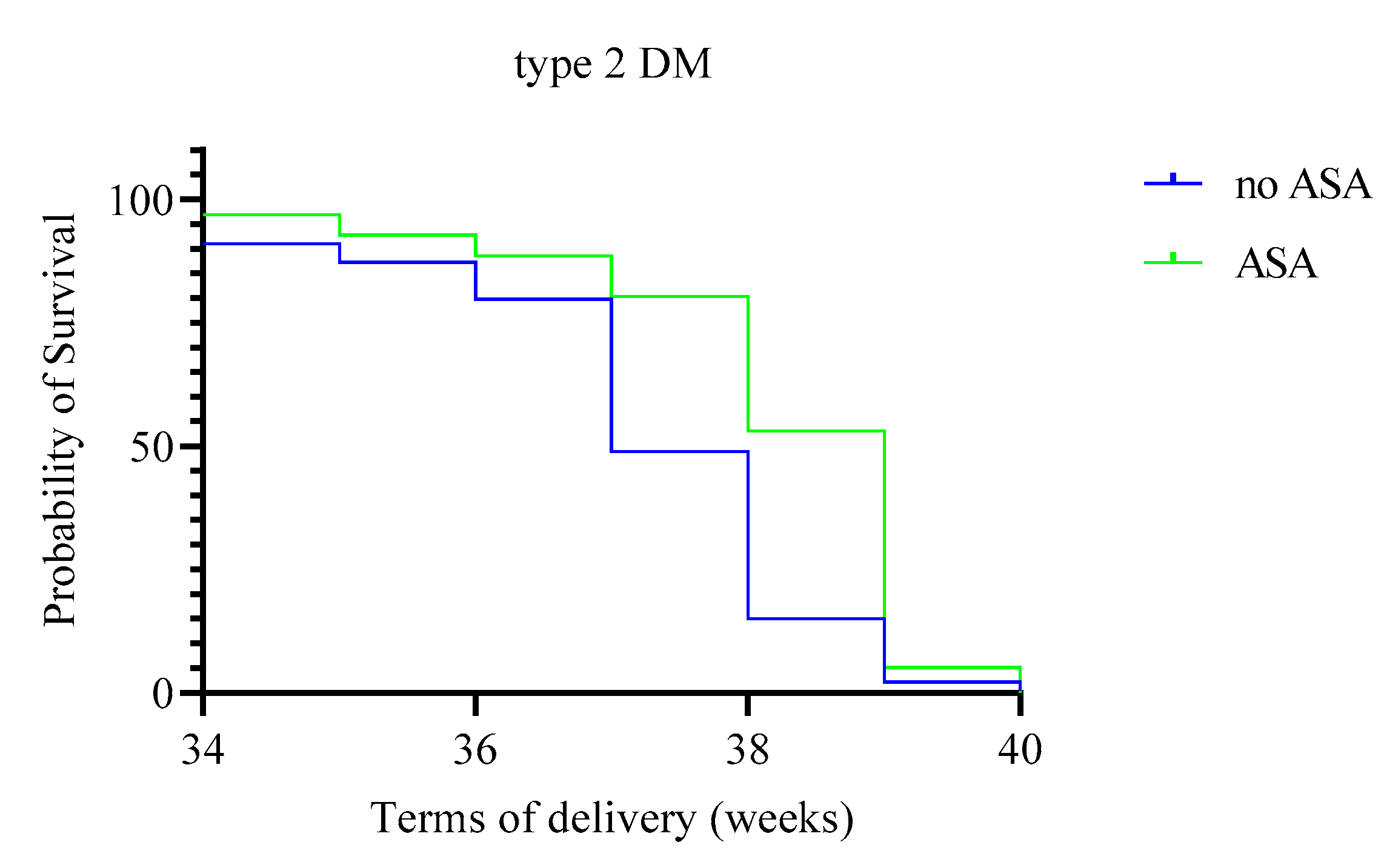
We found that the use of ASA preparations after 12 and up to 36 weeks of pregnancy in patients with pregestational DM types increases the delivery time by an average of 7–12 days. This pattern was determined especially up to 36 weeks of pregnancy, as seen from the Kaplan–Meier plots (Figure 1 and Figure 2).
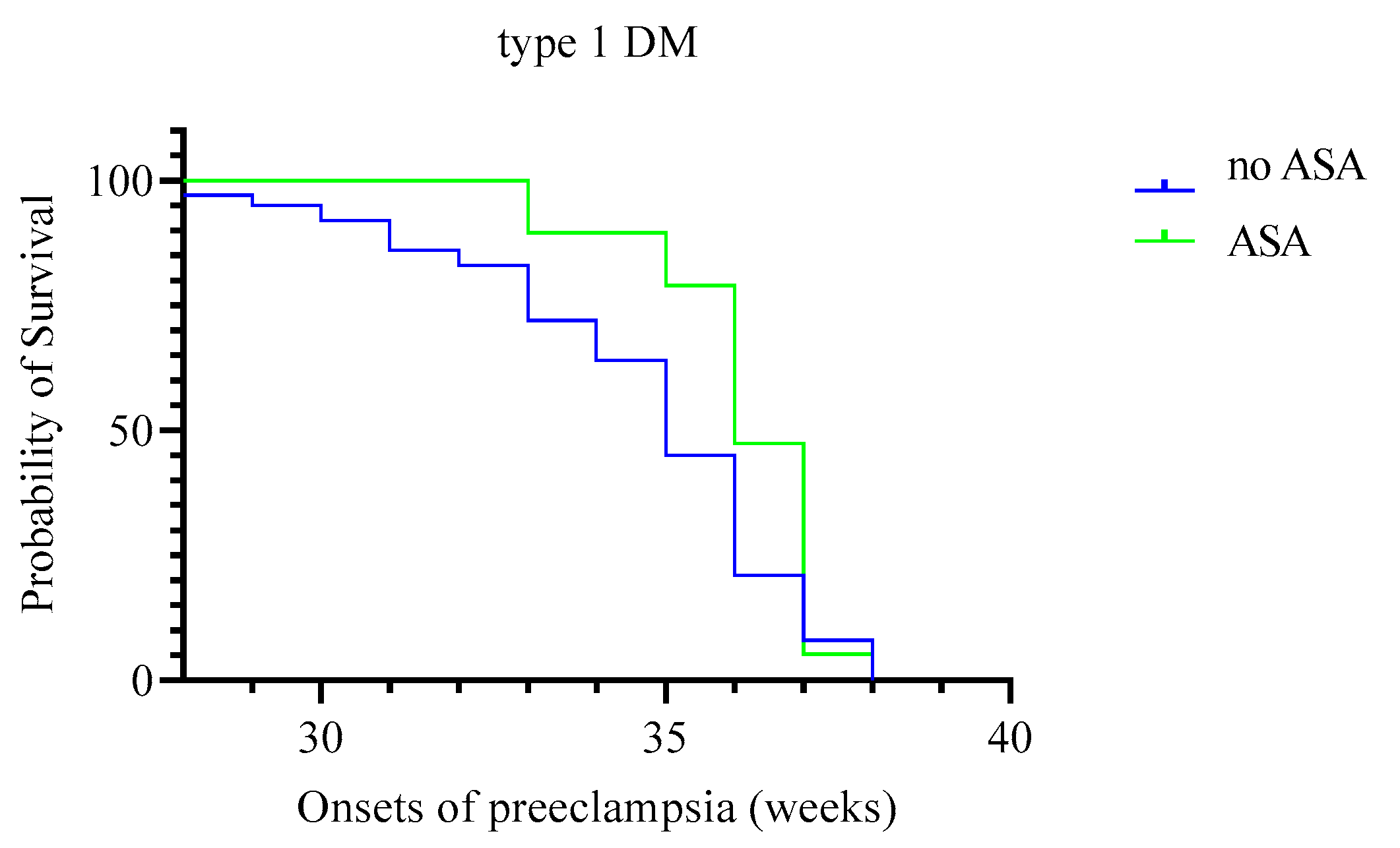
Other important features of assessing the effectiveness of using ASA are the absence of cases of early PE and the later onset of the disease in women with different DM types according to a retrospective analysis of the study (Figure 3 and Figure 4).

4. Discussion

We have shown in this study that pregnancy in women with pregestational DM types is associated with a high incidence of perinatal complications, such as preeclampsia, large fetus, and premature birth.
Our study showed the efficiency of prescribing low doses of ASA from early pregnancy to women with different DM types. This simple preventive measure helps to reduce the risk of several adverse pregnancy outcomes, which are especially common among these patients. Prescribing ASA significantly reduced the likelihood of developing PE in women with carbohydrate metabolism disorders. This effect was more pronounced in pregnant women with type 2 DM, in whom the chance of this complication decreased by 35%. Taking ASA in patients with type 1 DM is associated with the absence of severe forms of PE. In addition, we revealed differences in the timing of the initial manifestations of PE. In women with pregestational DM types who took ASA from 12 to 36 weeks of gestation, there were no cases of early PE, and, in general, those patients were characterized by a later onset of the disease. In patients with type 1 DM who received ASA, the average duration of PE (35.5 weeks of pregnancy) was 1.5 weeks later than in women who did not take the drug.
A potential limitation of the study is that it is not randomized. Prescribing aspirin was offered to all women with DM, but some of them refused to take this drug. Our decision to take the aspirin was based on only the presence of DM [1,4]. However, prescribing ASA would be more justified if we used Bayes’ theorem to combine maternal characteristics with biophysical and biochemical markers [18]. Such screening of high-risk groups was not planned in our study.
Similar data were obtained in a meta-analysis conducted by Bartsch E. et al. (2016) among 23 million pregnant women. The authors reported the relationship between clinical risk factors, which can be determined before 16 weeks of gestation, and the development of PE. The presence of DM increased the likelihood of developing PE by almost four times (OR 3.7; 95% CI: 3.1–4.3). It has also been shown that the use of low doses of ASA for the prevention of PE is justified in women with pregestational DM [19]. A study conducting a secondary analysis of the ASPRE trial [12] examined the differences in the effectiveness of ASA in preventing early PE among a variety of high-risk groups. The study showed that the use of the drug had no positive effect on pregnant women with chronic arterial hypertension. This analysis was not possible for pregnant women with DM due to the small sample and rare cases of PE in the study group [20].
Some studies showed that low-dose ASA does not reduce the incidence of PE in women with DM [21,22]. In the study conducted by Caritis S. et al. (1998), 462 women with DM received daily ASA at a dose of 60 mg on insulin therapy. However, this preventive measure did not decrease the incidence of PE and did not affect other adverse perinatal outcomes (stillbirth, premature birth, small for gestational age) [21]. The disadvantages of this research were the use of too low a dose of ASA and the duration of the drug administration: women were involved in the study at 13–26 weeks of gestation. Large meta-analyses of these days show that ASA can only be effective when taken daily in a dose of at least 100 mg, and prophylaxis is started before the 16th week of pregnancy [16,23]. Subsequently, Moore G. et al. (2016) conducted a secondary analysis of the above study. The authors selected only those pregnant women who received ASA before 17 weeks of gestation. Nevertheless, even with this modification, the positive effects of ASA on the development of obstetric complications in women with DM were not found [24]. In our work, ASA has proved to be an effective means of preventing premature birth. Antiplatelet therapy contributed to an increase in the delivery time, on average, by 7–12 days compared to patients who did not take ASA. This was especially noted with a gestation period of up to 36 weeks. In pregnant women with type 1 DM receiving ASA, the likelihood of preterm birth was reduced by more than three times compared to those patients to whom ASA was not prescribed. Preterm birth was, however, seen primarily as a consequence of preeclampsia.
Later secondary analysis of the Caritis S. study was carried out by Adkins K. et al. (2017) [25]. The authors studied the effect of taking ASA on the weight of the fetus in pregnant women with DM, both in the group of patients as a whole and depending on the presence or absence of vascular complications (according to the White classification) [26]. In the analysis, the involved individuals were not differentiated in terms of ASA prescription, with most pregnant women enrolled in the study after 17 weeks of gestation. Nevertheless, potentially significant results were obtained. In pregnant women without vascular complications, the intake of ASA was associated with higher weights of newborns, large for gestational age fetuses being significantly more often born. At the same time, the incidence of small for gestational age newborns in mothers who took ASA did not differ from the control group, in both women without vascular complications and patients having them [25]. It is known that for women with DM without vasculopathy, one of the most frequent complications of pregnancy is fetal macrosomia, while in diabetic vascular lesions, the frequency of small for gestational age newborns increases [26]. Therefore, the authors are wary of taking ASA by pregnant women with DM, arguing that the drug can significantly increase the transplacental transport of nutrients and accelerate fetal growth [25]. The data obtained by us do not contradict this observation. We found that babies were born with greater body weight in women with DM who took ASA during pregnancy, and the frequency of small for gestational age births decreased. In every third patient with type 1 DM, the gestation was complicated with fetal macrosomia, which often became the cause of unfavorable obstetric and perinatal outcomes during childbirth. The use of ASA in this category of patients increased the risk of developing fetal macrosomia by almost twice. However, the probability of small gestational age births in women with different DM types decreased by more than three times. The patients included in our study constituted a heterogeneous population. They differed in type DM, duration of disease, and presence of vascular complications. Therefore, prescribing aspirin may have advantages among all pregnant with pregestational types of DM, but especially among women with “Class D diabetes” (The White Classification) [27].
In a recent retrospective study, Lah S. et al. (2021) evaluated the effectiveness of ASA for the prevention of PE among 164 pregnant women with type 1 DM and type 2 DM [22]. The initiation of daily intake of ASA at a dose of 100–150 mg was carried out until the 16th week of pregnancy, and the drug intake continued until the 36th week of gestation. When assessing the outcomes, the authors did not find differences in the development of PE between pregnant women who received ASA and patients in the control group (OR 1.7; 95% CI: 0.7–4.3; p = 0.243). These results differ from our data. We have demonstrated in a large homogeneous sample of patients with DM that ASA can reduce the incidence of PE and decrease the incidence of its severe forms. When taking ASA, this hypertensive complication begins later, and early forms of PE are excluded.
According to the same study, among patients taking ASA, the incidence of other gestational complications associated with placental insufficiency (small for gestational age, premature birth) did not decrease. Still, this preventive measure was associated with a threefold increased risk of postpartum hemorrhage [22]. This is inconsistent with our data that have demonstrated the effectiveness of ASA in reducing the risk of developing both preterm and small for gestational age births. At the same time, the incidence of postpartum hemorrhage was minimal in both patients taking ASA and those not. The difference in the data obtained could be because, in the study by Lah S. et al., patients in the ASA group had a higher baseline risk of PE and other manifestations of abnormal placentation. This was because those women were more often diagnosed with diabetic nephropathy, with elevated HbA1c levels from the early stages of pregnancy and higher BMI values. Some of those patients needed anticoagulant therapy during pregnancy [22]. We found no other studies on this topic.
ASA action is not limited to inhibiting COX. Recently, researchers have been drawn to the effect of ASA on the secretion of vasotropic factors, the impaired synthesis of which plays a key role in the development of PE [28]. In a cytotrophoblast cell culture, it was demonstrated that ASA, by inactivating COX-1, inhibits the production of soluble fms-like tyrosine kinase-1 (sFlt-1) induced by hypoxia [29]. Other works report the improved trophoblast integration into endothelial cell monolayers in vitro due to the inhibitory effect of ASA on the expression of the transcription factor AP-1, which also reduces sFlt-1 synthesis, or TNF-α [28,30]. In one of the latest works on this topic, it is reported that taking ASA daily at a dose of 100 mg from early gestation in women at high risk of developing PE increased the serum level of placental growth factor, which is the major angiogenic factor necessary for the establishment of uteroplacental circulation [31]. However, to date, the precise mechanisms of ASA action on placentation are still unidentified.
Thus, our study shows that low-dose ASA administration to women with DM from early pregnancy is warranted. This is associated with a reduction of the incidence of PE, a delay in its manifestations, and mitigating the risk of other adverse perinatal outcomes that are typical for this category of patients. However, there should be vigilance about developing a key complication of diabetic pregnancy, which is fetal macrosomia. Due to the lack of studies devoted to this issue and the differing results obtained by the date, further detailed research of the effect of ASA on the course of pregnancy in women with pregestational DM types is needed.

Author Contributions

R.V.K. conceived of the presented idea, performed the analytic calculations and the numerical simulations, writing the manuscript, supervised the project. E.M.T. performed the analytic calculations and the numerical simulations, writing the manuscript. A.V.K. writing the manuscript. E.V.K., E.N.A., and A.V.T. contributed to the final version of the manuscript. O.N.A. supervised the project. All authors have read and agreed to the published version of the manuscript.

Funding

The study was supported by the grant [AAAA-A20-120041390024-2] from the Ministry of Science and Higher Education, Russian Federation.

Institutional Review Board Statement

The study was conducted according to the guidelines of the Declaration of Helsinki, and approved by the Ethics Committee of D.O. Ott Institute of Obstetrics, Gynecology, and Reproductive Medicine (St. Petersburg, Russia) (protocol code 83 and date of approval: 21 April 2017).

Informed Consent Statement

Informed consent was obtained from all subjects involved in the study.

Conflicts of Interest

The authors declare no conflict of interest. The funders had no role in the design of the study; in the collection, analyses, or interpretation of data; in the writing of the manuscript, or in the decision to publish the results.

References

  1. Gestational Hypertension and Preeclampsia: ACOG Practice Bulletin, Number 222. Obstet. Gynecol. 2020, 135, e237–e260. [CrossRef] [PubMed]
  2. Weissgerber, T.L.; Mudd, L.M. Preeclampsia and Diabetes. Curr. Diabetes Rep. 2015, 15, 1–10. [Google Scholar] [CrossRef] [PubMed] [Green Version]
  3. Poon, L.C.; Sahota, D. Screening and Prevention of Preeclampsia. Matern. Med. 2019, 1, 25–30. [Google Scholar] [CrossRef]
  4. Hypertension in Pregnancy: Diagnosis and Management. NICE Guideline. 2019. Available online: www.nice.org.uk/guidance/ng133 (accessed on 30 March 2021).
  5. Poon, L.C.; Shennan, A.; Hyett, J.A.; Kapur, A.; Hadar, E.; Divakar, H.; McAuliffe, F.; da Silva Costa, F.; von Dadelszen, P.; McIntyre, H.D.; et al. The International Federation of Gynecology and Obstetrics (FIGO) initiative on pre-eclampsia: A pragmatic guide for first-trimester screening and prevention. Int. J. Gynecol. Obstet. 2019, 145 (Suppl. 1), 1–33. [Google Scholar] [CrossRef] [PubMed] [Green Version]
  6. International Diabetes Federation. IDF Diabetes Atlas, 9th ed.; International Diabetes Federation: Brussels, Belgium, 2019; Available online: http://www.diabetesatlas.org/ (accessed on 30 March 2021).
  7. Huynh, J.; Dawson, D.; Roberts, D.; Bentley-Lewis, R. A systematic review of placental pathology in maternal diabetes mellitus. Placenta 2015, 36, 101–114. [Google Scholar] [CrossRef] [Green Version]
  8. Li, X.; Milosavljevic, A.; Elsea, S.H.; Wang, C.C.; Scaglia, F.; Syngelaki, A.; Nicolaides, K.H.; Poon, L.C. Effective Aspirin Treatment of Women at Risk for Preeclampsia Delays the Metabolic Clock of Gestation. Hypertension 2021, 78, 1398–1410. [Google Scholar] [CrossRef]
  9. Beaufils, M.; Donsimoni, R.; Uzan, S.; Colau, J. Prevention of pre-eclampsia by early antiplatelet therapy. Lancet 1985, 1, 840–842. [Google Scholar] [CrossRef]
  10. ACOG Committee Opinion No. 743: Low-Dose Aspirin Use During Pregnancy. Obstet. Gynecol. 2018, 132, e44–e52. [CrossRef]
  11. Loussert, L.; Vidal, F.; Parant, O.; Hamdi, S.M.; Vayssiere, C.; Guerby, P. Aspirin for prevention of preeclampsia and fetal growth restriction. Prenat. Diagn. 2020, 40, 519–527. [Google Scholar] [CrossRef]
  12. Rolnik, D.L.; Wright, D.; Poon, L.C.; O’Gorman, N.N.; Syngelaki, A.A.; Matallana, C.C.D.P.; Akolekar, R.R.; Cicero, S.S.; Janga, D.D.; Singh, M.M.; et al. Aspirin versus Placebo in Pregnancies at High Risk for Preterm Preeclampsia. N. Engl. J. Med. 2017, 377, 613–622. [Google Scholar] [CrossRef]
  13. Meher, S.; Duley, L.; Hunter, K.; Askie, L. Antiplatelet therapy before or after 16 weeks’ gestation for preventing preeclampsia: An individual participant data meta-analysis. Am. J. Obstet. Gynecol. 2017, 216, 121–128.e2. [Google Scholar] [CrossRef] [PubMed]
  14. Roberge, S.; Nicolaides, K.; Demers, S.; Villa, P.; Bujold, E. Prevention of perinatal death and adverse perinatal outcome using low-dose aspirin: A meta-analysis. Ultrasound Obstet. Gynecol. 2013, 41, 491–499. [Google Scholar] [CrossRef] [PubMed]
  15. Caron, N.; Rivard, G.-É.; Michon, N.; Morin, F.; Pilon, D.; Moutquin, J.-M.; Rey, É. Low-dose ASA response using the PFA-100 in women with high-risk pregnancy. J. Obstet. Gynaecol. Can. 2009, 31, 1022–1027. [Google Scholar] [CrossRef]
  16. Roberge, S.; Nicolaides, K.; Demers, S.; Hyett, J.; Chaillet, N.; Bujold, E. The role of aspirin dose on the prevention of preeclampsia and fetal growth restriction: Systematic review and meta-analysis. Am. J. Obstet. Gynecol. 2017, 216, 110–120.e6. [Google Scholar] [CrossRef] [PubMed]
  17. Brown, M.A.; Magee, L.A.; Kenny, L.C.; Karumanchi, S.A.; McCarthy, F.; Saito, S.; Hall, D.R.; Warren, C.E.; Adoyi, G.; Ishaku, S. Hypertensive Disorders of Pregnancy: ISSHP Classification, Diagnosis, and Management Recommendations for International Practice. Hypertension 2018, 72, 24–43. [Google Scholar] [CrossRef] [Green Version]
  18. Tan, M.Y.; Wright, D.; Syngelaki, A.; Akolekar, R.; Cicero, S.; Janga, D.; Singh, M.; Greco, E.; Wright, A.; Maclagan, K.; et al. Comparison of diagnostic accuracy of early screening for pre-eclampsia by NICE guidelines and a method combining maternal factors and biomarkers: Results of SPREE. Ultrasound Obstet. Gynecol. 2018, 51, 743–750. [Google Scholar] [CrossRef] [Green Version]
  19. Bartsch, E.; Medcalf, K.E.; Park, A.L.; Ray, J.G.; High Risk of Pre-Eclampsia Identification Group. Clinical risk factors for pre-eclampsia determined in early pregnancy: Systematic review and meta-analysis of large cohort studies. BMJ 2016, 353, i1753. [Google Scholar] [CrossRef] [Green Version]
  20. Poon, L.C.; Wright, D.; Rolnik, D.L.; Syngelaki, A.; Delgado, J.L.; Tsokaki, T.; Leipold, G.; Akolekar, R.; Shearing, S.; De Stefani, L.; et al. Aspirin for Evidence-Based Preeclampsia Prevention trial: Effect of aspirin in prevention of preterm preeclampsia in subgroups of women according to their characteristics and medical and obstetrical history. Am. J. Obstet. Gynecol. 2017, 217, 585.e1–585.e5. [Google Scholar] [CrossRef] [Green Version]
  21. Caritis, S.; Sibai, B.; Hauth, J.; Lindheimer, M.D.; Klebanoff, M.; Thom, E.; VanDorsten, P.; Landon, M.; Paul, R.; Miodovnik, M.; et al. Low-dose aspirin to prevent preeclampsia in women at high risk. National Institute of Child Health and Human Development Network of Maternal-Fetal Medicine Units. N. Engl. J. Med. 1998, 338, 701–705. [Google Scholar] [CrossRef]
  22. Lah, S.; Cheung, N.W.; Lee, V.; Athayde, N.; Inglis, E.; Padmanabhan, S. Aspirin and preeclampsia prevention in women with pre-existing diabetes: A retrospective study. Intern. Med. J. 2021. [Google Scholar] [CrossRef]
  23. Roberge, S.; Bujold, E.; Nicolaides, K. Aspirin for the prevention of preterm and term preeclampsia: Systematic review and metaanalysis. Am. J. Obstet. Gynecol. 2018, 218, 287–293.e1. [Google Scholar] [CrossRef] [PubMed] [Green Version]
  24. Moore, G.; Allshouse, A.A.; Post, A.; Galan, H.L.; Heyborne, K. Early initiation of low-dose aspirin for reduction in preeclampsia risk in high-risk women: A secondary analysis of the MFMU High-Risk Aspirin Study. J. Perinatol. 2015, 35, 328–331. [Google Scholar] [CrossRef] [PubMed] [Green Version]
  25. Adkins, K.; Allshouse, A.A.; Metz, T.D.; Heyborne, K.D. Impact of aspirin on fetal growth in diabetic pregnancies according to White classification. Am. J. Obstet. Gynecol. 2017, 217, 465.e1–465.e5. [Google Scholar] [CrossRef] [Green Version]
  26. Klemetti, M.M.; Laivuori, H.; Tikkanen, M.; Nuutila, M.; Hiilesmaa, V.; Teramo, K. White’s classification and pregnancy outcome in women with type 1 diabetes: A population-based cohort study. Diabetologia 2015, 59, 92–100. [Google Scholar] [CrossRef] [Green Version]
  27. Sacks, D.A.; Metzger, B.E. Classification of Diabetes in Pregnancy: Time to reassess the alphabet. Obstet. Gynecol. 2013, 121, 345–348. [Google Scholar] [CrossRef] [Green Version]
  28. Lin, L.; Li, G.; Zhang, W.; Wang, Y.; Yang, H. Low-dose aspirin reduces hypoxia-induced sFlt1 release via the JNK/AP-1 pathway in human trophoblast and endothelial cells. J. Cell. Physiol. 2019, 234, 18928–18941. [Google Scholar] [CrossRef]
  29. Li, C.; Raikwar, N.; Santillan, M.; Santillan, D.; Thomas, C. Aspirin inhibits expression of sFLT1 from human cytotrophoblasts induced by hypoxia, via cyclo-oxygenase 1. Placenta 2015, 36, 446–453. [Google Scholar] [CrossRef] [Green Version]
  30. Xu, B.; Shanmugalingam, R.; Chau, K.; Pears, S.; Hennessy, A.; Makris, A. The effect of acetyl salicylic acid (Aspirin) on trophoblast-endothelial interaction in vitro. J. Reprod. Immunol. 2017, 124, 54–61. [Google Scholar] [CrossRef]
  31. Murtoniemi, K.; Vahlberg, T.; Hämäläinen, E.; Kajantie, E.; Pesonen, A.; Räikkönen, K.; Taipale, P.; Villa, P.; Laivuori, H. The effect of low-dose aspirin on serum placental growth factor levels in a high-risk PREDO cohort. Pregnancy Hypertens. 2018, 13, 51–57. [Google Scholar] [CrossRef] [Green Version]
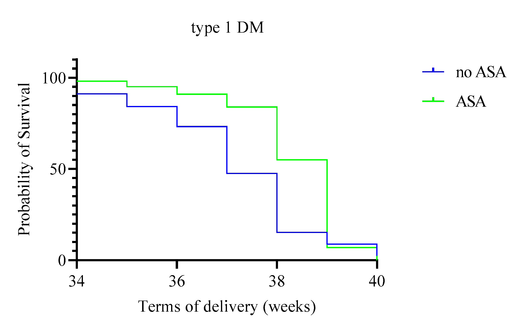
Figure 1. Terms of delivery in patients with type 1 DM.
Reprodmed 02 00015 g001
Figure 2. Terms of delivery in patients with type 2 DM.
Reprodmed 02 00015 g002
Figure 3. Onsets of preeclampsia in patients with type 1 DM.
Reprodmed 02 00015 g003
Figure 4. Onsets of preeclampsia in patients with type 2 DM.
Reprodmed 02 00015 g004
Table 1. Clinical characteristics of the study groups.
Type 1 DM (n = 506)Type 2 DM (n = 229)
GroupASA
(n = 100)
No ASA
(n = 406)
F/U/χ2p-ValueASA
(n = 96)
No ASA
(n = 133)
F/U/χ2p-Value
Age
(years, IQR)
28.1
(27.2; 29.0)
28.5
(27.9; 28.9)
196690.6333.0
(30.0; 38.0)
35.0
(31.5; 38.0)
58620.29
Body-mass index (kg/m2, IQR)27.4
(26.6–28.1) *
27.1
(26.7–27.4) *
2.300.1335.1
(30.2; 38.6)
32.4
(27.9; 36.8)
48070.007
Cigarette smoking
(n, %)
00 5 (5.2)4 (3)0.420.51
Primiparous
(n, %)
29 (29.0)110 (31.8)1.580.8215 (15.6)36 (27.5)1.400.22
Chronic arterial hypertension
(n, %)
5 (5.0)28 (6.9)0.470.4925 (26.0)29 (21.8)0.560.46
Vascular complications
(n, %)
26 (26.0)79 (19.5)1.600.2354 (56.3)83 (62.4)0.880.35
Excess body weight
(n, %) §
39 (39.0)219 (53.9)7.200.00717 (17.7)34 (26.8)2.540.11
Obesity
(n, %)
27 (27.0)76 (18.7)1.750.1975 (78.1)82 (61.7)2.300.1
* Data are presented as 95% CI. § Excess body weight defined as a BMI ≥ 25 kg/m2, but < 30 kg/m2. Obesity defined as a BMI ≥ 30 kg/m2.
Table 2. Comparison of pregnancy outcomes in patients with diabetes mellitus who received or did not receive antiplatelet therapy.
Type 1 DM (n = 506)Type 2 DM (n = 229)
ASA
(n = 100)
No ASA
(n = 406)
F/U/χ2p-ValueASA
(n = 96)
No ASA
(n = 133)
F/U/χ2p-Value
Due date
(weeks, 95% CI)
38.3
(38.1–38.5)
37.1
(36.8–37.3)
9.500.00238.1
(37.7–38.4)
37.1
(36.8–37.4)
0.560.46
Weight of newborns
(g, 95% CI)
3825
(3408–4200)
3550
(3045–3955)
14820<0.00013640
(3160–3840)
3200
(2578–3785)
4545<0.001
Onset of preeclampsia
(weeks)
35.5 (35; 36)34 (33.5; 34)10.00436 (34.5; 36)35 (34; 35)9.50.08
Preeclampsia
(n, %)
29 (29)159 (39.2)3.550.03719 (19.8)72 (54.1)27.46<0.0001
Moderate
(n, %)
29 (29)106 (26.1)0.340.326 (6.3)53 (39.8)32.9<0.0001
Severe
(n, %)
053 (13)14.58<0.000113 (13.5)19 (14.3)0.030.52
Premature birth
(n, %)
9 (9.0)110 (27.1)14.6<0.000111 (11.5)27 (20.3)3.150.07
<30 weeks
(n, %)
06 (1.5)1.500.222 (2.1)2 (1.5)0.110.74
30–34 weeks
(n, %)
015 (3.7)3.800.0506 (4.5)4.450.035
34–36 weeks
(n, %)
5 (5.0)44 (10.8)3.100.085 (5.2)9 (6.8)0.2463
36–37 weeks
(n, %)
4 (4.0)45 (11.1)4.600.0364 (4.2)10 (7.5)1.010.3
Macrosomia
(n, %)
36 (36.0)91 (22.4)7.900.00516 (16.7)22 (16.5)0.0010.98
Caesarean section
(n, %)
51 (51.0)174 (50.1)0.020.8847 (49.0)59 (44.4)0.470,49
Planned
(n, %)
18 (18.0)63 (18.1)0.0010.9821 (21.9)21 (15.8)0.380.24
Emergency
(n, %)
23 (23.0)100 (28.7)1.280.2615 (15.6)18 (13.5)0.200.66
Small for gestational age
(n, %) *
2 (2.0)39 (9.6)6.230.0136 (6.3)36 (27.1)16.1<0.0001
* The condition small for gestational age is defined as a weight below the 10th and above 3rd percentile for the gestational age.
Table 3. Effect of acetylsalicylic acid intake on pregnancy outcomes.
Type 1 DM (n = 506)Type 2 DM (n = 229)
ComplicationsOR
(95% CI)
p-ValueAdjusted OR
(95% CI) *
p-ValueOR
(95% CI)
p-ValueAdjusted OR
(95% CI) **
p-Value
Preeclampsia0.64
(0.39–1.02)
0.060.92
(0.47–1.24)
0.080.57
(0.46–0.71)
<0.010.65
(0.52–0.79)
0.02
Moderate0.90
(0.64–1.28)
0.560.96
(0.72–1.38)
0.720.64
(0.55–0.74)
<0.010.78
(0.57–0.91)
0.04
Severe0.87
(0.84–0.9)
<0.010.92
(0.87–0.99)
0.050.99
(0.89–1.1)
0.871.06
(0.86–1.14)
0.92
Premature birth0.27
(0.13–0.55)
<0.010.33
(0.15–0.62)
0.030.51
(0.24–1.08)
0.070.69
(0.2–1.16)
0.16
<30 weeks0.99
(0.97–1.00)
0.221.21
(0.97–1.45)
0.31.39
(0.19–1.77)
0.741.53
(0.48–2.05)
0.91
30–34 weeks0.96
(0.95–0.98)
0.051.05
(0.97–1.12)
0.090.96
(0.92–0.99)
0.040.98
(0.95–1.13)
0.06
34–36 weeks0.43
(0.17–1.12)
0.070.57
(0.32–1.28)
0.130.76
(0.25–2.34)
0.630.84
(0.54–1.87)
0.71
36–37 weeks0.33
(0.12–0.95)
0.030.51
(0.39–0.84)
0.050.54
(0.16–1.76)
0.300.66
(0.43–1.52)
0.36
Macrosomia1.95
(1.22–3.12)
0.0051.82
(1.13–2.86)
0.0091.00
(0.50–2.00)
0.981.15
(0.75–1.64)
0.85
Postpartum
hemorrhage
Caesarean section1.04
(0.66–1.62)
0.881.28
(0.87–1.75)
1.121.20
(0.70–2.04)
0.491.29
(0.86–1.72)
0.53
Planned0.99
(0.56–1.77)
0.981.16
(0.95–1.44)
0.991.49
(0.76–2.90)
0.241.47
(0.81–2.70)
0.47
Emergency0.74
(0.44–1.25)
0.260.93
(0.54–1.38)
0.671.18
(0.56–2.48)
0.661.31
(0.74–2.29)
0.7
Small for
gestational age
0.19
(0.05–0.81)
0.0130.35
(0.17–0.62)
0.0250.18
(0.07–0.45)
<0.0010.31
(0.19–0.44)
0.003
* Adjusted for differences in excess body weight and chronic arterial hypertension. ** Adjusted for differences in body mass index and chronic arterial hypertension.
Publisher’s Note: MDPI stays neutral with regard to jurisdictional claims in published maps and institutional affiliations.