Review

The History of the Discovery of Ectopic Epithelial Cells in Lower Peritoneal Organs: The So-Called Mucosal Invasion

1
Department of Health Sciences, University Hospitals of Leicester, University of Leicester, Leicester Royal Infirmary, Infirmary Square, Leicester LE1 5WW, UK
2
Department of Experimental and Clinical Medicine, School of Sciences of Human Health, University of Florence, 50134 Florence, Italy
3
Department of Maternal and Child Health, Gynaecology and Urology, Sapienza University of Rome, Piazzale Aldo Moro 5, 00100 Rome, Italy
*
Author to whom correspondence should be addressed.
Reprod. Med. 2021, 2(2), 68-84; https://doi.org/10.3390/reprodmed2020008
Submission received: 8 March 2021 / Revised: 24 March 2021 / Accepted: 4 April 2021 / Published: 7 April 2021

Abstract

:
Through microscopy, early researchers identified the epithelium on the inner surfaces of the uterus, cervix and Fallopian tubes. The identification of ectopic epithelium was gradual, starting from the gross pathology study of unusual cystic lesions. Towards the end of the nineteenth century, attention focused on the epithelium as a critical component. The term ‘adenomyoma’ was coined around eighteen eighty to designate the majority of mucosa-containing lesions. Several theories were advanced to explain its aetiology. In the main, lesions were considered to arise from invasion from uterine epithelium; implantation of endometrium through retrograde menstruation; hematogenous or lymphatic spread; or from embryonic remnants. Although initially widely rejected, around 1920, an almost unanimous consensus formed on the endometrial nature of epithelial invasions. During the following years, adenomyosis and endometriosis came to be used to distinguished lesions within or outside the uterus. Adenomyosis was attributed to direct infiltration of uterine mucosa into the myometrium, and endometriosis to the implantation of endometrial cells and stroma into the peritoneal cavity through retrograde menstruation. Around the same time, ovarian lesions, initially described as ovarian hematomas or chocolate cysts, were regarded as a form of endometriosis. Three variants of endometriosis were thus described: superficial peritoneal, deep nodular and ovarian endometriomas. Ectopic epithelium has long been recognised as having similarities to tubal, or cervical epithelium. Lesions containing mixed epithelium are often termed Müllerianosis. This article demonstrates the stepwise evolution of knowledge, the role of the pioneers and the difficulties that needed to be overcome. It also demonstrates the value of collaboration and the inter-connected nature of the scientific endeavour.

1. Introduction

The introduction of the microscope by Marcello Malpighi, in the 17th century, constituted the most important revolution in the history of biology [1]. It allowed the subsequent discovery by Robert Hooke that organisms are made of ‘cells’ [2] and later of the existence of a great variety of cells in animals and plants [3,4].
The formation of organs from the folding, fusion and growth of the primordial germ layers was detailed by Caspar Friedrich Wolff [5] in 1774 in his thesis “Theoria Generationis”. For this, he is considered the father of embryology. Heinz Pander is credited with the description, in 1817, of the three germ layers (ectoderm, endoderm and mesoderm) in the developing chick embryo [6]. Subsequently, von Baer expanded this work and suggested that the three layers are common to all vertebrates [7].
During embryonic development, the ‘ectoderm’ gives rise to the skin and the nervous system; the ‘endoderm’ to the digestive tract and associated glands and the ‘mesoderm’, which develops between the endoderm and the ectoderm, gives rise to the coelom. The ‘intermediate mesoderm’, which is located between the paraxial mesoderm and the lateral plate mesoderm, gives rise to the urogenital structures (kidneys, gonads and genital tract).
In his pioneering work, Wolff [5] identified an embryonic structure developing from the coelomic epithelium as part of the ‘urogenital ridge’ and intermediate mesoderm, which he referred to as the ‘mesonephros’ (later came to be known as the Wolffian body) with its duct. In 1830, Johannes Peter Müller [8] described the early stages of the formation of female internal genital organs, which originate from two ‘paramesonephric ducts’ (Müllerian ducts). They run caudally lateral to the mesonephric duct to the urogenital ridge and terminate at a small tubercle in the primitive urogenital sinus. The Wolffian ducts develop from the intermediate mesoderm and the Müllerian duct epithelium develops from the rostral mesonephric epithelium, in the form of antero-lateral invaginations of the coelomic epithelium [8,9,10]. There is a close relationship between the developing Müllerian and Wolffian ducts [10,11]. The uterine epithelium, surrounded by its stroma, is formed by fusion of the Müllerian ducts, whereas the myometrium is derived from the mesenchyme. The first detailed description of “The mucous membrane of the womb in its development up to the time of puberty” was published by Engelmann almost 150 years ago [12].
During the first part of the 19th century, researchers identified the presence of an epithelium only on the inner surfaces of the uterus, cervix and Fallopian tubes. Identifying the presence of epithelium at ectopic sites was a gradual discovery that started with the study of unusual cystic lesions. The early lesions attracted attention because of their size and/or unusual macroscopic features. In most early accounts of these large uterine or pelvic cavitated lesions, a distinction was not made on whether they had a mucosal lining and reports focused on gross pathology. In many cases, specimens were not even subjected to histological examination. The aim of this article is to ascertain the progress of knowledge in this field and how adenomyosis and endometriosis were linked and uncoupled through this course. The main landmark contributions are summarised in Table 1.

2. Early Descriptions

Mostly, early reports dealt with descriptions of lesions of large or unusual macroscopic appearance, and not much attention was paid to symptoms. Cullen provided an initial account of his experience in 1919 [13] and a more detailed account in 1920 [14]. Cullen stated that adenomyomas can readily be diagnosed based on clinical grounds. However, in common with other gynecological affections, mucosal invasions (MI) may be asymptomatic, or associated with cyclical pelvic pain and infertility and, despite modern imaging techniques, in a majority of cases, it is not possible to arrive at a definitive diagnosis of MI without histological examination. Thus, a history of the discovery of MI is necessarily distinct from a narrative of how medicine or society addressed affections, or manifestations of diseases of women and should be linked to the use of histological proof of the presence of ectopic epithelial cells.
Our approach to the history of progress in understanding MI contrasts with that taken by Nezhat et al. [15] who attempted to explore the social history of a variety of illnesses affecting women. Their approach creates difficulties, because of the non-specific symptoms linked to MI and because it ascribes to ancient physicians knowledge that they simply could not have had. Therefore, whether diseases caused by MI existed in the early days cannot be confirmed through identifying reports suggestive of women suffering pelvic pain. This means that, on the one hand, there is no reason to believe that diseases caused by MI did not affect women even millennia ago, on the other, it remains speculative whether modern lifestyles and other influences affected the incidence of MI. It is for this reason that endometriosis, which is one of the main MI, has been referred to as a ‘modern disease’ [16]. MI have only been brought to medical attention through advances in diagnostics during the last part of the XIX and during the XX century.
Knapp [17] reported that a number of dissertations dating back to the 17th and 18th centuries mentioned the morphological features of endometriosis, but these theses describe ulcers and inflammations of the uterus. Knapp believed that Daniel Schrön [18] described endometriosis in 1690 when he reported ulcers on the peritoneum, bladder and elsewhere in the pelvis. It is conceivable that some of the lesions observed by Schrön were instances of MI, but he did not recognise them to be due to the presence of ectopic epithelial cells. In addition, he goes on to describe the lesions as pus-filled or forming abscesses rather than having the macroscopic appearance of a mucosal invasion. Similarly, Knapp stated that Crellius [19] mentioned an ovarian endometriotic cyst in 1739 when he described a tumour adhering to the fundus of the uterus (tumorem fundo uteri externe adherentem describit). These words do not entail a recognition of lesions featuring MI.
The wider use of microscopy enabled the recognition of mucosa-lined cysts and of microscopic gland-like structures in the lower peritoneal organs. The first such descriptions were made by Carl Rokitansky, who together with Rudolph Virchow are widely credited as the founders of pathological anatomy. Rokitansky performed microscopical examination of tissues, but this was not routine, or widely practiced at the time. In his manual, first published in 1849, Rokitansky [20] included a reference to ‘chocolate containing cysts of the ovary’, but only as instances of simple cysts, which he attributed to the growth of Graafian follicles. He believed that the trigger for the growth of these cysts was a response to either an inflammatory process or de novo growth. He also provided an account of the existence of uterine hypertrophy, but not of mucosal growths within the myometrium. In 1860, Rokitansky [21] described the new formation of mucosal tissue in the uterus and ovaries and described tumours, or polyps, containing glands that were similar to uterine glands. He used the term ‘sarcoma adenoids uterinum’ to refer to these new growths and the term ‘cystosarcoma adenoids uterinum’ for lesions that contained cysts. An early description of cysts containing chocolate-like material was also provided by Spencer Wells [22]. Twenty-five years after Rokitansky’s description, Gusserow [23] may have been the first to refer to his work when he stated, in discussing fibroid polyps, that lesions containing glandular structures are better viewed as lesions of the mucosa.

3. The Term Adenomyoma

During the second half of the 19th century, the term ‘adenomyoma’ was coined to designate the majority of mucosa-containing lesions. According to Lockyer [24], the first detailed description of an adenomyoma was made by Babeș (Victor Babesiu) [25] who, in 1882, published a case of an intramural myoma containing cysts lined with ‘low cubical epithelium derived from embryonic germs’ and by Diesterweg [26] who, in 1883, described ‘two polypi of the posterior uterine wall containing cysts lined with ciliated epithelium and filled with blood’. Both von Recklinghausen [27,28] and Cullen [29] utilised the term ‘adenomyoma’ and they were followed by Pick [30], Rolly [31] and Iwanoff [32].
Cullen [33] and Lockyer [24] quoted Breus [34] as stating that, for their dissertations, Schröder, [35], Heer [36] and Grosskopf [37] had collected more than 100 cases of adenomyomas from existing medical literature. However, these seemingly uncritical quotations overlooked the fundamental distinction that Breus made, based on whether cystic spaces observed within myomas had an epithelial lining. Breus emphasised that a mucosal lining was demonstrated in only four (possibly five) of all published cases, as the vast majority had not been subjected to microscopic examination or had no mucosal lining when examined. This was in agreement with Fritsch [38] who argued that the presence of epithelial lining in these cystic spaces is very rare. To Breus, the cases described by Babeș [25], Diesterweg [26], Schröder and examined by Ruge [39] were the only instances that had histologically proven mucosa-lined-cysts. Breus added two cases made available to him by Kundrat (Rokitansky’s successor) in Vienna.
Up to Breus’s report, published cases mostly dealt with gross lesions. For instance, one of the cases he described contained 7 liters of fluid, and the size of another was ‘that of a child’s head’. In the case described by Babeș [25], there were two hazelnut-sized cysts in the uterus of a 91-year-old woman. Diesterweg [26] described a tumour the size of a man’s fist containing cysts lined by ciliated epithelium. Thus, none of these reports fits with our current appreciation of uterine adenomyosis or endometriosis.
Real progress in recognising adenomyomas was made when Friedrich von Recklinghausen published two reports in 1893 and 1896 [27,28], describing 34 cases and including extensive histological examination (Figure 1). He acknowledged the work of Rokitansky [21], Kolb [40], Röhrig [41] and Babeș [25] and concluded that MI should be divided based on whether the lesion was found at the periphery or centrally in the uterus and tubes. He described four varieties:
  • Hard: predominately made of muscle tissue.
  • Cystic: containing visible cystic spaces and equal glandular and muscle tissue.
  • Soft: predominately made of glandular tissue.
  • Telangiectatic: soft, very vascular growths that are almost devoid of cysts.
Von Recklinghausen classified glandular structures based on their architecture and epithelial type into straight, tortuous and round-ended. He viewed these as analogous to secreting tubules, ampullae and end-bulbs of Wolffian ducts. Fusion of the tubules formed canals, which were either discrete or aggregated (Figure 2). Structural features and the observation that there were no glands in the fallopian tubes convinced him that adenomyoma originated from Wolffian duct structures.
Another major contributor to the recognition of adenomyoma of the uterus is Thomas Cullen, who presented his first case to the John Hopkins Hospital Medical Society in 1895 [13]. This was shortly after he had completed his training in Germany and had taken up the position of head of gynecological pathology. Cullen added an important contribution to the field, published in German as a tribute to Johannes Orth of Göttingen [42]. In this, he acknowledged the support of von Recklinghausen and described a number of known cases, including those by Paul Locksteadt who was able, through the introduction of Prussian Blue dye, to establish continuity between the glands in the myometrium and the uterine mucosa [43].
The first case of an adenomyoma involving the round ligament was published by Cullen in 1896 [29]. He refered to the histological features that include striations with scattered chocolate-coloured areas varying from 1 to 5 mm in diameter, as the characteristic features of adenomyoma. He observed that scattered endometrial glands were accompanied by a stroma.
In the book, Adenomyoma of the Uterus, published in 1908, Cullen [33] referred to the first case he encountered during his clinical practice in 1894. The description is that of a uterus about four times the natural size with uniform, diffuse thickening of its anterior wall. He viewed it as most unusual and warranting histological examination. That revealed a diffuse myomatous tumor with the uterine mucosa flowing into it at many points. In their book, Kelly and Cullen [44] describe cases of adenomyoma as “diffuse gland containing myomatous thickening” either confined to the anterior, posterior, or lateral uterine muscle wall or totally encircling the uterus. Normal uterine mucosa was observed to flow within the myomatous tissue, either as isolated glands or as large masses of mucosa. The mucosa lining the uterine cavity was normal. These descriptions are more aligned with our current understanding of uterine adenomyosis, with less emphasis on large tumour masses.
In contrast to the infrequent cases reported in the 19th century, Cullen [14] wrote that he was “amazed at the widespread distribution of these tumors”. He classified these lesions based on their site: the body of the uterus, the rectovaginal septum, the uterine horn or the Fallopian tube, the round ligament, the uterosacral ligament, the sigmoid flexure, the rectus muscle and the umbilicus. There is a similarity here with the classification provided in Lockyer’s text, which Cullen had received. Cullen’s description of the lesions is also aligned with our current understanding and includes striations, with scattered chocolate-coloured areas, varying in diameter from 1–5 mm and histologically scattered endometrial glands accompanied by stroma.

4. The Search for the Origin of Mucosal Invasions

Several theories were advances about the aetiology of these lesions. In the main, the mucosa was considered to arise from ‘invasion’ from uterine mucosa, ‘implantation’ of uterine epithelium from retrograde menstruation or following ‘hematogenous’ or ‘lymphatic’ spread, or to originate from ‘embryonic remnants’ (either duplicate, rudimentary or cellular structures). Theories were advanced based on plausibility or histological continuity, or lesion morphology. Many authors were willing to entertain the possibility that diseases at various sites have different aetiologies; others sought a unified theory. Only occasionally did these early researchers undertake a diligent search for tissue continuity, or for experimental studies. Early literature reflects curiosity about the origin of the observed cystic structures and, when recognised, of the mucosal component.
As well as stating their preferred theory, many writers included a critique of alternatives. Von Recklinghausen [28] included histological sections depicting mucosal invasion forming adenomyomas which he classified based on their location at the periphery of the uterus and in the tubes, or centrally within the uterus. He stated that he was unable to find convincing evidence of dissimilarities from the uterine epithelium. Nevertheless, he concluded that whilst it is possible to envisage that lesions close to the uterine cavity originate from endometrial glands, he remained unconvinced that the same origin could apply to lesions closer to the peritoneum. His studies of the shape and branching of the glands led him to the hypothesis that peripheral lesions are derived from Wolffian remnants and that the surrounding stroma is derived from differentiation of local connective tissue. There were many opponents to the Wolffian theory, including Kossmann [45] who argued that aberrant glands originate from accessory Müllerian, rather than Wolffian ducts. Ivanoff [32] proposed that lesions in the peritoneum that do not have a connection to the uterine mucosa arise through metaplasia. This view was supported by Meyer [46] and by more recent authors as the origin of endometriosis [47]. Meyer [46] believed that aberrant glands develop in response to a sequence of inflammation, induration and epithelial hypertrophy. According to this theory, the source of the epithelium is the overlying peritoneum and the surrounding mantle originates from original connective tissue exhibiting response to inflammation.
Cullen [14] took the position that, for the majority of cases, the glands were derived from the uterine mucosa. Von Franqué [48] believed that epithelial growths found in a number of abdominal organs were derived from the ‘mature mucous membrane’ of the uterus that had acquired the ability to infiltrate other organs as a consequence of a process of inflammation. Other early supporters of Cullen’s theory were Baldy and Longcope [49], who refused both von Recklinghausen’s [28] Wolffian hypothesis and Kossmann’s theory [45] of an origin from accessory Müllerian ducts. Schickele [50] was amongst the last to argue in favour of a mesonephric origin of mucosal growth. He wrote, “when I try to take an impartial view of published cases, I am compelled to state that the mucosal theory is not proved”.
In connection with lesions of the rectogenital space, Lockyer [24] described Cullen as being dogmatic in stating that “glands in these growths undoubtedly arise from the uterine mucosa, or from remnants of Muller’s duct”. To Cullen, the presence of a connection between glands within the muscle and the mucosa was sufficient for him to assert that glandular elements of diffuse adenomyoma have undoubtedly arisen from uterine glands [33]. Lockyer drew a distinction, not addressed by Cullen in his assertions, between an origin from ‘dystopic’ (congenital) or ‘orthotopic’ (mature) mucosa and pointed out that it had not been proven that mature uterine mucosa provided any gland-tissue to any etrauterine growth. There are also references stating that the glands derive from the Gartner duct (‘ductus longitudinalis epoophori’), discovered and described in 1822 by Hermann Treschow Gartner as part of the Wolffian duct [51].
Many theories were proposed by the end of the XIX century to explain the origin of ovarian lesions, including a derivation from the germinal epithelium as supported by Waldeyer [52], Marchand [53] and Williams [54] or from the Graafian follicle as supported by Frommel [55] and Williams [54].
In addition to deliberations about pathogenesis, early literature debated nomenclature. Many researchers were not satisfied with the various proposed terms, including those that they themselves had initially supported. A critical factor was the desire to introduce nomenclature that did not imply mechanism or causation. For example, the term Müllerianosis as advocated by Bailey [56] was described by Sampson [57] as inclusive and correct; however, he preferred the term endometriosis to avoid a suggestion of an embryonic origin. It is notable that Sampson also rejected the terms endometrioma or endometriomyoma proposed by Blair Bell [58].

5. Introducing the Term Adenomyosis

As detailed in Section 3, for almost fifty years, lesions featuring MI were described under a unified nomenclature. Then, in 1925, Oskar Frankl [59] distinguished anatomical features of the intrauterine variety of mucosal invasion as a separate nosological entity and he named it ‘adenomyosis uteri’. He reserved the term adenomyoma to localised disease and rejected the terms adenometritis, adenomyositis or adenomyometritis which were in use at the time because they suggest a link to inflammation which he was not able to identify. Frankl stated that adenomyoma may originate independently within the myoma, but that adenomyosis is directly connected to the eutopic endometrium and pointed to similarity with the “menstruating uterine mucosa on the surface of the ovary, first described by Sampson”. He concluded that having studied Sampson’s original slides, he became convinced that in his and in Sampson’s case, “misplaced uterine glands were seen filled with blood, undoubtedly menstrual blood”. Emge [60] reviewed the topic in 1962 and noted that only little advance had been made in understanding disease causation and clinical recognition. Thus, Emge referred to adenomyosis as an elusive disease and pointed out that the majority of students of the subject view invasion of the myometrium by basal endometrial elements as the common mechanism and that a less common mechanism, championed by Sampson [61], Halban [62] and Taussig [63], is metastatic lymphatics and vascular invasion. Emge [60] emphasised the difficulty in diagnosis and felt that at his time there was a risk of overdiagnosis, given the lack of concordance between clinical and histological diagnosis.
Bird [64] provided the currently used definition of adenomyosis: the benign invasion of the endometrium into the myometrium, producing a diffusely enlarged uterus which microscopically exhibits ectopic non-neoplastic, endometrial glands and stroma surrounded by the hypertrophic and hyperplastic myometrium. The exact definition and derivation of the myometrial hyperplasia and hypertrophy referred to by Bird are unclear. Importantly also, he divided adenomyosis into grades based on the depth of gland invasion within the myometrium. The debate that followed focused on the cut-off point for histological diagnosis of adenomyosis, a parameter that remains a source of considerable controversy.
Improved non-invasive diagnostic modalities added an important dimension and options for treatment: Hricak et al. [65] described the junctional zone (JZ) myometrium and Tamai et al. [66] documented the importance of its thickening for non-invasive diagnosis of adenomyosis.
Classification of adenomyosis continues to be debated, but disagreement remains on significant aspects such as the role of imaging and how best to include the range of disease phenotypes. In a recent review, we detailed old and more recent attempts at producing a taxonomy [67]. There remains considerable uncertainty about how to reconcile opposing views about pathogenesis particularly of lesions found in the uterovesical space, the pouch of Douglas and in the outer myometrium. One theory holds that disease found in these areas originates from invasion by uterine adenomyosis; the other holds that lesions present in the outer myometrium originate from peritoneal endometriosis. Much of the debate is based on fragmentary evidence and extrapolations. Because of these limitations and until research can resolve the questions raised, broad agreement on a hypothesis to underpin any proposed classification is unlikely.

6. Identification of Endometriosis

Sampson described the similarities between endometriosis lesions and the uterine mucosa and is widely credited for bringing attention to endometriosis and for popularising the theory of retrograde menstruation for disease in the pelvis [68]. Sampson introduced the term ‘endometriosis’, but he also utilised the term ‘implantation adenomyoma’. Sampson was able to provide detailed illustrations of lesions [57].
It is to be noted that Sampson [68] applied the term ‘endometriosis’ to all variants; thus, he described four routes for endometriosis: (1) direct or primary endometriosis (also described as Müllerianosis) affecting the uterine wall causing adenomyoma, or a similar lesion in the tube; (2) peritoneal implantation endometriosis, which included implantation-like deposits of endometrial or Müllerian tissue; (3) transplantation endometriosis at the site of scars; (4) metastatic endometriosis to distant sites, which he likened to cancer metastasis. Sampson stated that although a possibility, he has not been able to appreciate cases of developmentally misplaced endometrial tissue. In 1925, Sampson [57] wrote that misplaced ‘endometrium-like’ tissue presents a varied structure and that “its origin is not always the same. We believe that it arises from both the uterine and the tubal mucosa and possibly, when situated between the layers of the broad ligament, it may sometimes arise from remnants of the Wolffian body. We also know that gland-like inclusions of the peritoneal mesothelium and of the surface epithelium of the ovary arise from peritoneal irritation and that some of these lesions may simulate atypical endometrial tissue”. In this article, he drew parallels between endometrial tissue spread and the spread of cancer, including the invasion and subsequent spread into the vascular and lymphatic systems and dissemination into the peritoneal cavity. In his 1927 article [68], Sampson enunciated the theory, still considered the most likely, of “peritoneal endometriosis due to the menstrual dissemination of endometrial tissue into the peritoneal cavity”.

6.1. The Ovarian Endometrioma

It is probable that the first variant of what we today call endometriosis to be described was ovarian. With few exceptions [26], this phenotype was not described under the name adenomyoma and, from its early description, it was considered a separate entity. What is not clear is whether its endometrial nature and related implications were recognised in early reports. In terms of priority, Roland Batt [69,70] and Emge [60] stated that Carl Rokitansky [21] was the first to describe an ovarian endometrioma. However, Rokitansky’s account, apart from the unusual name he selected (‘cystosarcoma adenoids uterinum’), is not that of an ovarian endometrioma lined with endometrial mucosa.
Endometriomas of the ovary are not rare and have characteristic macroscopic features. It is the case that the endometrial type epithelium of their lining went unrecognised for many decades. For this reason, it is highly likely that early descriptions of ‘blood-filled’ or ‘tarry’ cysts concerned cases of endometriomas. Perhaps the first clear identification of the presence of endometrial tissue within an ovary is that provided by Russel [71], who described a case which, under the microscope, exhibited a number of “areas, which were an exact prototype of the uterine glands and interglandular connective tissue”. These glands “were arranged as in normal uterine mucous membrane and opened into spaces, their epithelium being continuous with its lining membrane”.
A few additional cases were published soon afterward. Semmelink and De Joselin de Jong [72] reported an ovarian cyst the structure of which was similar to an endometrial stroma. They believed this to be of Wolffian origin. The same year, Pick published four cases [30] and in 1909, Sitzenfrei [73] described two cases associated with recto-uterine adenomyoma. In the first case, he found the presence of “zwei walnuss große cystische mit geronnenen Bluter füllte Hohlräume” (two cystic cavities as big as a walnut full of blood), but he did not describe the nature of the lining. In the second case he described “drüsiger Bildungen and durchbluteten zytogenn Gewebes” (glandular formations and blood-containing cytogenic tissues) in the adherent part of the ovary.
Several descriptions of ‘hematoma of the ovary’ were also published. Savage [74] presented seven cases but did not identify the lining of the cysts as endometrial tissue. Casler [75] reported an unusual presentation of a woman who experienced menstruation following a hysterectomy which was attributed to bleeding from a cyst wall. Smith [76] described several cases of ovarian hematomas and described leakage of chocolate coloured fluid from the soft, fixed mass in the left cul-de-sac and from small ovarian cysts. Cullen [14] published three cases and described a “brownish membrane lining of cylindrical epithelium” and a case associated with “widespread adenomyoma of the recto-vaginal septum”, which he interpreted as an indication that “the uterine mucosa on the surface of the ovary was due to an overflow of the adenomyoma of the recto-vaginal septum”.
Around 1920, there was increased recognition of the similarities between the cyst lining and the endometrium. Norris [77] reported a small ovarian cyst containing free blood and noted congruity in the cycle phase between the endometrium and the mucosal lining of the cyst. Donald [78] published a clear description of ‘ovarian adenomyomas’ and observed that they contain endometrial stroma and smooth muscle and that the lining can exhibit changes similar to those of the endometrium, including a decidual like reaction in pregnancy. He noted that there was a frequent bilateral association with lesions in the rectovaginal space and that these lesions are not malignant. Of interest is his comment that this type of cyst was long known to all gynaecologists but that its true nature had only recently been discovered. Donald reported sixteen cases that he encountered in only one year, which emphasised that these lesions were not rare. In the discussion of the article, Herbert Spencer [78] attributed the identification of stroma in the cyst wall to Pick [79], who referred to this lesion as ‘adenoma endometrioides’. In response to the debate about nomenclature that was ongoing at the time, both Donald and Spencer rejected the use of the term ‘endometrioma’, because they believed it implied acceptance of a theory about the origin of such lesions. They were also keen to emphasise that lesions occur in the utero-rectal space, not in the rectovaginal septum, which they regarded as an erroneous designation. Donald stated that upon reviewing his operative work over many years, he became convinced that many cases he previously assumed to be cases of pelvic inflammation or infected ovarian cysts were instances of ovarian adenomyomas [78].
In 1921, Sampson [80] described 23 cases of “perforating hemorrhagic (chocolate) cysts of the ovary”, which varied from 1-9 cm in diameter. He referred to them as perforating, as he believed that a perforation on the lateral or on the free surface of the ovary was responsible for the leakage of content, for the observed adhesions and for coexisting lesions in the pouch of Douglas. Subsequently, Sampson [81] described additional cases of superficial and deep chocolate cysts. He considered that lesions outside the ovary could occur as a consequence of leaking irritants from the ovarian cysts resulting in metaplasia of the peritoneal epithelium, or the activation of dormant endometrial epithelium. However, he favoured the view that lesions result from implantation analogous to the spread of malignancy from a ruptured cyst. Another view advanced by Sampson [81] is that of the ovary as an incubator or intermediary host that imparts greater virulence to the epithelium enabling pelvic implantation.
Bailey [56], on the one hand, acknowledged Sampson’s contribution to understanding pelvic adenomyomatous growths, linking them to chocolate cysts of the ovary; on the other hand, he disagreed with him on two counts. First, he challenged the notion of a perforating cyst. Through extensive histological examination, Bailey documented the continuity between the epithelium within the cyst and that of the ovarian cortex. He argued that ovarian lesions develop on the outer surface of the ovary forming a cavity, not a cyst. As the invading endometrium erodes the ovary, the mouth of the cavity becomes obstructed by adhesion and by the build-up of material. Second, he challenged the notion of the ovary being an intermediate host for endometriosis as he did not find viable cells capable of implantation within the cyst fluid.

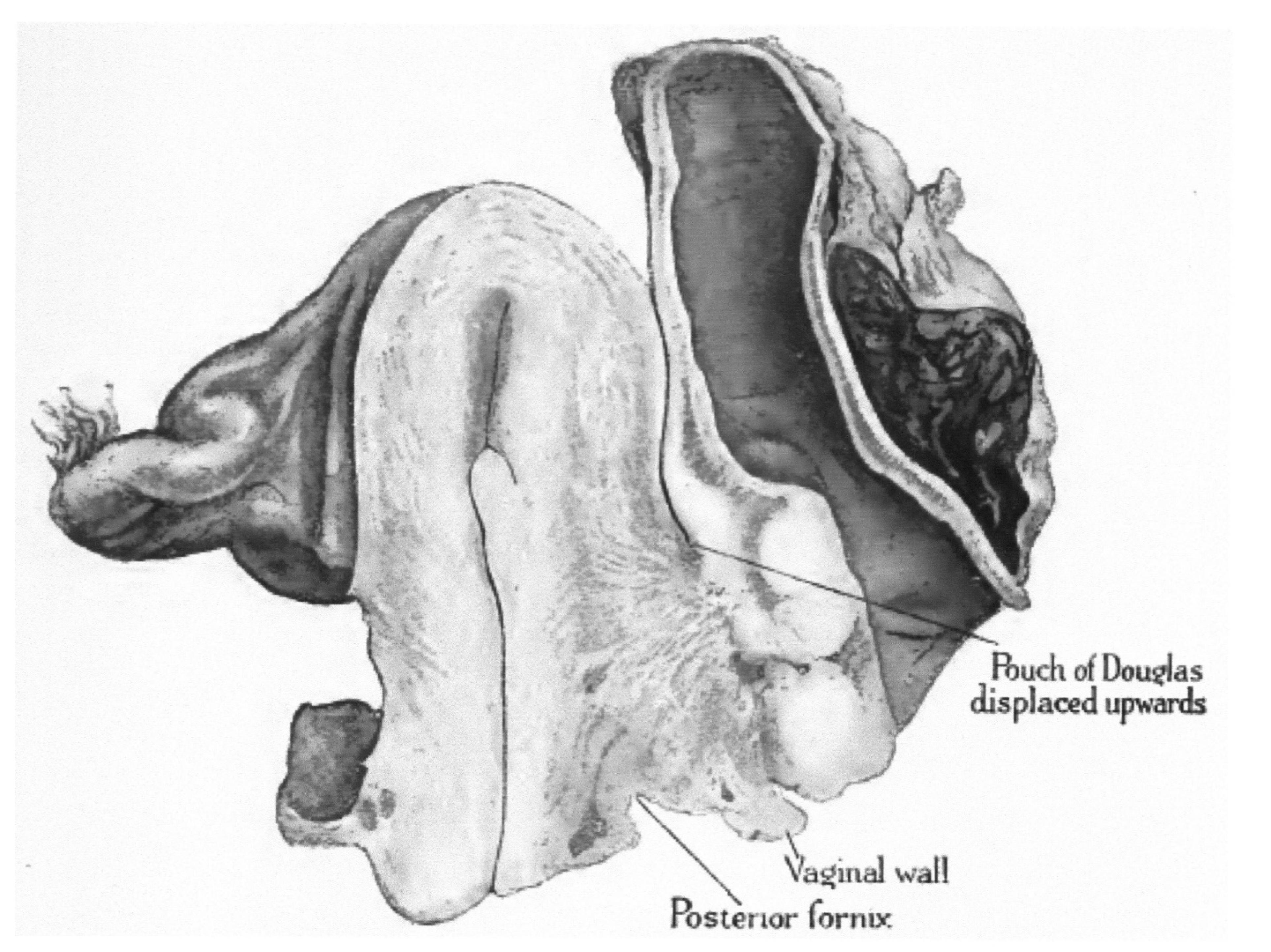
6.2. Deep, Infiltrating Endometriotic Nodules

Initially, these lesions may have been referred to as ‘posterior parametritis’, which is referenced in literature from the late 19th century [82,83] and perhaps even earlier. But these early reports did not associate the condition with the presence of endometrial epithelium. The link between these nodules and ‘parametritis nodosa posterior’ seems to have been first recognised by Meyer [84], who included an early description of epithelial inclusion. Eden and Lockyer [85] referred to these lesions as adenomyomas (Figure 3) and included the reproduction of the image published by Kleinhaus [86] of an adenomyoma affecting the rectogenital septum. They also included a description of the lesion, which was referred to as ‘parametritis chronica atrophicans’ (another name for ‘parametritis nodosa posterior’). They draw a distinction between this type of lesion and other causes of pelvic cellulitis, which can lead to suppuration.
It is interesting that in the description of his first case, which was operated upon in 1910, Sampson [80] identified ovarian ‘chocolate’ cysts and dense adhesions resulting in induration in the rectal wall. He initially attributed this induration to syphilis and only recognised that the lesion is consistent with an adenomyoma a few years later. In line with other observers, Sampson believed that he had overlooked several cases between 1912 and 1918.
In his 1903 report, Cullen [42] did not include a description of the disease affecting the pouch of Douglas, but this entity was a major focus of his article published in 1919–1920, where he wrote “many of you have undoubtedly seen but may not have recognised them”. Cullen credited Lockyer’s article published in 1913 [87] for enabling him to recognise the nature of his first cases. It is notable that Lockyer, assuming the nodules were malignant, advocated hysterectomy and colectomy. In retrospect, he contemplated whether the lesion resulted from Wolffian duct remnants and therefore whether extensive surgery was justified. The discussion that accompanied his publication included descriptions by other contributors of similar cases from their own practice. In his book, Lockyer [24] referred to the earliest described cases as the two published by Pfannenstiel [88] and the one by von Herff [89]. The glandular epithelium was identified in these cases but was considered to be of Wolffian origin. This was followed by a case by Pick [90]. By 1918, Lockyer [24] commented that the literature on the subject was already quite extensive and that the error of treating it as malignant should be avoided, because the condition was, by then, well recognised.

6.3. Superficial Peritoneal Lesions

The identification of peritoneal lesions seems to have occurred around the same time as the other variants and was initially reported as lesions of the organs covered by the peritoneum. Both Lockyer [24] and Cullen [14] include a description of lesions affecting extrauterine locations (e.g. the fallopian tubes, ovaries, round ligament). As mentioned above, Sampson deserves recognition for establishing the link between these lesions and ovarian chocolate cysts.
In 1927, Sampson [68], having studied additional cases of superficial lesions not associated with ovarian lesions, advanced the theory that they may originate from implantation of endometrial or endosalpingeal tissue following retrograde menstruation and coined the expression ‘peritoneal endometriosis’. At the same time, he argued that these were not the primary lesions, since in his view endometrial tissue would first reach the ovary creating an endometrial cyst and, from the perforation of such a cyst, secondarily reach the peritoneum. Alternatively, he believed that ectopic cells and stroma may reach the pelvic peritoneum from a menstrual reaction of endometrial tissue growing on the peritoneal surface of the ovary or other pelvic structures. Sampson documented the plausibility of the various stages of the process of implantation of endometrial mucosa in the pelvis through retrograde menstruation, concluding that peritoneal endometriosis sometimes arises from that route. He did not abandon other possible sources of endometrial tissue.

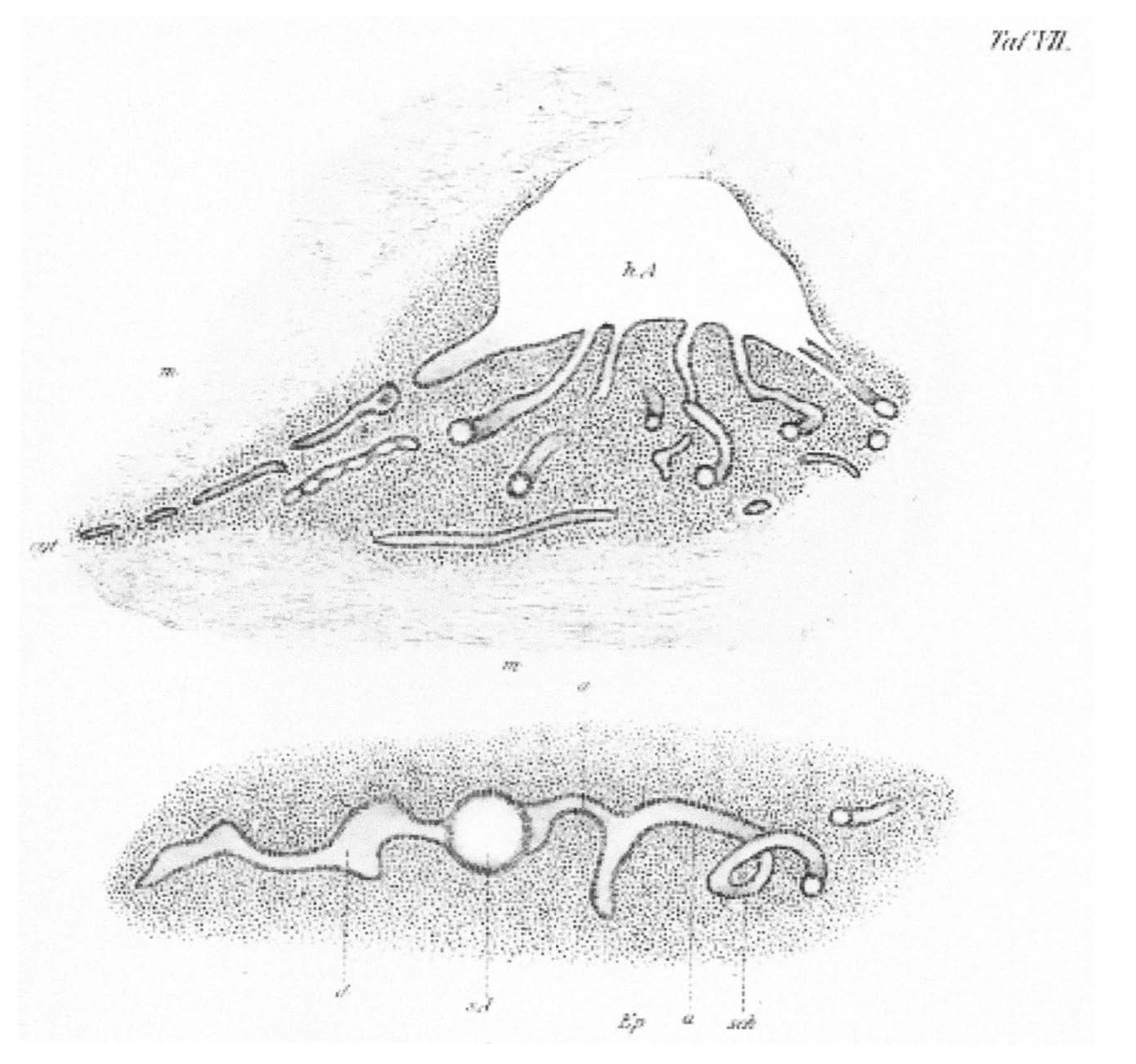
7. Tubal Adenomyosis

This entity, which was also referred to as salpingitis nodosa, adenomyosalpigitis (of Rabinovitz), cornual adenomyomata, salpingitis isthmica nodosa (of Chiari), or adenomyositis tubae, represents the tubal counterpart of a uterine adenomyoma.
A description of this variant was made as early as 1818 by Meckel [91], who identified the presence of nodularity in the fallopian tube. It was also mentioned by Rokitansky [92], Förster [93] and Kelbs [94]. Mostly, these were considered as myomas. Chiari [95] identified tubal epithelium within the uterine musculature and proposed that these lesions originate from chronic inflammation and that the growth of the epithelium induces hyperplasia in the surrounding muscle. Von Recklinghausen [28] proposed that these lesions originate from congenital Wolffian duct remnants, a theory disputed by Meyer [96] and subsequently by Cullen [33]. The controversy continued and in 1917, Rabinovitz [97] went on to argue that the condition is the end result of a chronic inflammatory process due to gonorrhoea or tuberculosis.

8. Other Types of Epithelia Present in Lower Peritoneal Organs

Microscopy enabled the recognition that some ectopic epithelia found in lower peritoneal organs are similar to the tubal, or the endocervical, rather than the uterine lining. These lesions are rare, and most reports date from the end of the 20th century.

8.1. Müllerianosis and Mixed Endometrial-Myometrial Lesions

Habiba et al. [98] collated cases of those rare lesions characterised by the presence of one or more of the Müllerian type epithelia and myometrium outside the normal location. These lesions have been described under the nomenclature uterus-like mass (U-LM), endomyometriosis, adenomyomatous polyp and adenomyomatous polyp. The earliest example of a U-LM may be that described by Oliver in 1912 [99], followed by the case of Cranstoun of 1922 [100]. Similar lesions were described in the small intestine [101] and the nervous system [102]. Extrauterine adenomyomas may arise from the ovary [103], the ovarian ligament [104], the broad ligament [105] or the round ligament [106] and are not connected to the uterus. Rokitansky’s early description may be an instance of an adenomyomatous polyp [21].
As discussed above, the use of the term Müllerianosis was debated in earlier literature but was not favoured as it may imply a developmental origin [56,57]. Mostly, the term is used to refer to lesions that contain more than one type of epithelium [98,107]. Batt and Yeh [108] put forward a theory of Müllerianosis claiming that distinct from the more common acquired type, some MI lesions develop from epithelial rests. According to this hypothesis, these lesions may contain endometrial, endosalpingeal and endocervical tissue singly or in combination as an organoid structure (termed ‘choristoma’) of embryonic origin. Instances include lesions found outside the pelvis and genital tract, or in peritoneal ‘defects’ or pockets. The embryonic origin theories project the root cause to events early in fetal life but do not explain the aberration. These theories have been considered in older literature, but supportive evidence remains fragmentary [109].

8.2. Endosalpingiosis

This variant refers to the presence of ectopic tubal epithelium. Perhaps the earliest account of this type of lesions is that provided by Chiari’s essay on salpingitis isthmica nodosa [95]. In the early days, the presence of ectopic tubal-type epithelium was recognised by Bailey [56] and by Sampson [57] but, as already mentioned, there was reluctance to use the term Müllerianosis when referring to these lesions. It seems that Sampson coined the term endosalpingiosis in 1930 [110]. Endosalpingiosis can occur in various organs and cases of benign lesions identical to endosalpingiosis in women have been described in the lymph nodes of male patients with prostatic adenocarcinoma or urothelial carcinoma, not treated with exogenous hormones [111]. This phenomenon is probably the result of Müllerian metaplasia. The uterine variant is rare and a recent review of the literature identified 18 cases only. Macroscopically, it often appears as multiple cysts of different sizes [112].

8.3. Endocervicosis

The second variant, coined endocervicosis, refers to the presence of ectopic cervical epithelium, mostly in the urinary bladder. It was first described by Clement and Young [107], who stated that it can mimic an adenocarcinoma. A review of the literature on the subject [113] concluded that the lesion is characterised by the presence of mucinous endocervical epithelium within the detrusor muscle of the bladder [114]. In an unusual case [115], surface mucinous epithelium was associated with the endocervicosis glands, prompting immunohistochemical profiling of the case. This showed slightly differing immunohistochemical phenotypes of the surface mucinous and morphologically similar endocervicosis glandular epithelium.

9. Conclusions

Following the initial concept that lower peritoneal organs were of mesenchymal origin and that only their lining was epithelial in nature, further studies detected the existence of more complex lesions. It took some 40 years for the concept to be accepted that most lesions observed were due to an ‘invasion’ by the uterine lining and did not originate from Wolffian or Müllerian remnants.
Everything coalesced during the late nineteen twenties, when complete identification of the variants we know today was made, enabling clinicians to deal with each type separately and to devise relevant diagnostics and therapeutics.
Proof of the existence of ectopic cervical or tubal epithelium represents a more recent achievement, probably because of their relative rarity. Indeed, it is now accepted that the majority of mucosal invasions predominantly contain one type of epithelium (endometrium, endosalpinx or endocervix). Alternatively, they may contain a mixture of epithelial types.
This article demonstrates how knowledge evolved with the accumulation of information in small steps and over time. We highlighted the important role of the pioneers who developed a special interest in the field and how ideas were debated and criticised before being finally accepted. It is notable how research benefitted from the exchange of ideas amongst investigators working across boundaries. This is all the more remarkable considering the technology available to them at the time.

Author Contributions

M.H.: led the literature review and writing up and contributed to developing the idea. G.B.: conceived the idea reviewed and revised reiterations of the manuscript. D.L.: developed the introduction, reviewed and accepted the final version. All authors have read and agreed to the published version of the manuscript.

Funding

No funding was received in connection with this manuscript.

Conflicts of Interest

The authors confirm that they have no direct or indirect conflict of interest in relation to this manuscript.

References

  1. Reverón, R.R. Marcello Malpighi (1628–1694), Founder of Microanatomy. Int. J. Morphol. 2011, 29, 399–402. [Google Scholar] [CrossRef] [Green Version]
  2. Hooke, R. Micrographia; The Royal Society: London, UK, 1665. [Google Scholar]
  3. Schleiden, M.J. Beiträge zur Phytogenesis. In Archiv Anatomie, Physiologie und Wissenschaftliche Medicin; Müller, J., Ed.; Viet: Berlin, Germany, 1838; pp. 137–176. [Google Scholar]
  4. Schwann, T. Mikroskopische Untersuchungen über die Übereinstimmung in der Struktur und dem Wachsthum der Thiere und Pflanzen; Verlag der Sander’schen Buchhandlung: Berlin, Germany, 1839. [Google Scholar]
  5. Wolff, C.F. Theoria Generationis; Halae ad Salam, Litteris Hendelianis: Halle (Saale), Germany, 1759. [Google Scholar]
  6. Pander, H.C. Beiträge zur Entwickelungsgeschichte des Hühnchens im Eye; Bronner: Vista, CA, USA, 1817. [Google Scholar]
  7. von Baer, K.E.; O’Malley, C.D. On the Genesis of the Ovum of Mammals and of Man; Isis: Oxford, UK, 1956; Volume 47, pp. 117–153. [Google Scholar]
  8. Müller, J. Bildungsgeschichte der Genitalien aus Anatomischen Untersuchungen an Embryonen des Menschen und der Thiere; Arnz: Düsseldorf, Germany, 1830. [Google Scholar]
  9. Grünwald, P. Zur entwicklungsmechanik des urogenitalsystems beim huhn. Wilhelm Roux Arch. Entwickl. Mech. Org. 1937, 136, 786–813. [Google Scholar] [CrossRef]
  10. Grünwald, P. The relation of the growing Müllerian duct to the Wolffian duct and its importance for the genesis of malformations. Anat. Rec. 1941, 81, 1–19. [Google Scholar] [CrossRef]
  11. Guioli, S.; Sekido, R.; Lovell-Badge, R. The origin of the Müllerian duct in chick and mouse. Dev. Biol. 2007, 302, 389–398. [Google Scholar] [CrossRef] [Green Version]
  12. Engelmann, G.J. The Mucous Membrane of the Uterus, with Special Reference to the Development and Structure of the Deciduae; William Wood and Co.: New York, NY, USA, 1875. [Google Scholar]
  13. Cullen, T.S. The distribution of adenomyomata containing uterine mucosa. Am. J. Obstet. Dis. Women Child. 1919, 80, 130–138. [Google Scholar]
  14. Cullen, T.S. The distribution of adenomyomas containing uterine mucosa. Arch. Surg. 1920, 1, 215–283. [Google Scholar] [CrossRef] [Green Version]
  15. Nezhat, C.; Nezhat, F.; Nezhat, C. Endometriosis: Ancient disease, ancient treatments. Fertil. Steril. 2012, 98, S1–S62. [Google Scholar] [CrossRef] [PubMed]
  16. Benagiano, G.; Lippi, D.; Brosens, I. Endometriosis: Ancient or Modern Disease? Indian J. Med. Res. 2015, 141, 69–71. [Google Scholar] [CrossRef] [Green Version]
  17. Knapp, V.J. How old is endometriosis? Late 17th- and 18th-century European descriptions of the disease. Fertil. Steril. 1999, 72, 10–14. [Google Scholar] [PubMed]
  18. Schrön, D.C. Disputatio Inauguralis Medica de Ulceribus Uteri; [Inaugural Medical Thesis on Ulcers of the Uterus]; Crause, R.W., Ed.; Literis Krebsianis: Jena, Germany, 1960; pp. 6–17. [Google Scholar]
  19. Crellius, J.F. Tumorem Fundo Uteri Externe Adhaerentem Describit; [Tumor Adhering to the Uterus Fundus is Described]; Ordinis Medici in Academia Vitembergensis: Würtemberg, Germany, 1739. [Google Scholar]
  20. Rokitansky, C.A. Manual of Pathological Anatomy (1849); Adlar: London, UK, 1854. [Google Scholar]
  21. Rokitansky, C. Über Uterusdrüsen-Neubildung in Uterus- und Ovarial-Sarcomen; [On the neoplasm of uterus glands on uterine and ovarian sarcomas]; Zeitschr Gesellschaft Aerzte: Wien, Austria, 1860; Volume 16, pp. 577–581. [Google Scholar]
  22. Wells, T. Diseases of the Ovaries. Their Diagnosis and Treatment; J & A Churchill: London, UK, 1872. [Google Scholar]
  23. Gusserow, A. Die Neubildungen des Uterus; [The Neoplasms of the Uterus]; Verlag von Ferdinand Enke: Stuttgart, Germany, 1886. [Google Scholar]
  24. Lockyer, C. Fibroids and Allied Tumours (Myoma and Adenomyoma); MacMillan: London, UK, 1918. [Google Scholar]
  25. Babeș, V. Über epitheliale Geschwulste in Uterusmyomem. [About epithelial tumors in uterine fibroids]. Allgem. Wiener. Med Ztschr. 1882, 27, 36–48. [Google Scholar]
  26. Diesterweg, B. Ein Fall von Cystofibrom uteri verum; [A case of cystofibroma uteri verum]. Zeitschr. Geburtshilfe. 1883, 9, 191–195. [Google Scholar]
  27. von Recklinghausen, F. Über Adenomyome des Uterus und der Tuba. [About the adenomyomas of the uterus and tube]. Wiener Klin. Wochenschr. 1895, 29, 530. [Google Scholar]
  28. von Recklinghausen, F. Die Adenomyomata und Cystadenomata der Uterus und Tubenwandung: Ihre Abkunft von Resten des Wolffischen Körpers; [The adenomyomas and cystadenomas of the uterus and tube wall: Their origin from remnants of the Wolffian body]; August Hirschwald Verlag: Berlin, Germany, 1896. [Google Scholar]
  29. Cullen, T. Adenomyoma of the round ligament. John’s Hopkins Hosp. Bull. 1896, 7, 112–117. [Google Scholar]
  30. Pick, L. Ein neuer Typus des voluminösen paroophoralen Adenomyoms—Zugleich über eine bisher nicht bekannte Geschwulst Form der Gebärmutter (Adenomyoma psammopapillare) und über totale Verdoppelung des Eileiters [A new type of voluminous paroophoral adenomyoma—At the same time about a previously unknown tumor form of the uterus (adenomyoma psammopapillare) and about total doubling of the fallopian tubes]. Archiv Gynäkol. 1897, 54, 117–206. [Google Scholar]
  31. Rolly, F. Über einen fall von adenomyoma uteri mit übergang in karcinom und metastasenbildung. [About a case of uterine adenomyoma with transition to carcinoma and metastasis]. Archiv Pathol. Anat. Physiol. Klin. Med. 1897, 150, 555–582. [Google Scholar] [CrossRef]
  32. Ivanoff, N.S. Drüsiges cystenhaltiges Uterusfibromyom compliciert durch Sarcom und Carcinom (Adenofibromyoma cysticum sarcomatodes carcinomatosum) [Uterine fibromyoma containing glandular cysts (Adenofibromyoma cysticum sarcomatodes carcinomatosum)]. Monatssch. Gebursthilfe Gynäkol. 1898, 7, 295–300. [Google Scholar]
  33. Cullen, T.S. Adenomyoma of the Uterus; Saunders: London, UK, 1908. [Google Scholar]
  34. Breus, C. Über Wahre epitel Fürende Cysten Bildung in Uterus Myomen; [Over the formation of true epithelial cysts in uterine myomas]; Franz Deuticke: Vienna, Austria, 1894. [Google Scholar]
  35. Schröder, O. Über Cystofibroide des Uterus Speciell Über Einen Fall von Intra-Uterinen Cystofibroid; [On the cystofibroids of the uterus]; Universitäts-Buchdrucker: Strasburg, France, 1873. [Google Scholar]
  36. Heer, O. Über Fibrocysten des Uterus; [Over the Cystic Fibroids of the uterus]; Druck von Zürcher und Furrer: Zürich, Switzerland, 1874. [Google Scholar]
  37. Grosskopf, C. Zur Kenntniss der Cystomyome des Uterus; [On knowledge of the cystomyomas of the uterus]; Druckerei der" Bayerischen Landeszeitung: Munich, Germany, 1884. [Google Scholar]
  38. Fritsch, H. Die Krankheiten der Frauen, fur Ärzte und Studirende; [The Diseases of Women, for Doctors and Students]; Verlag von Friedrich Werden: Berlin, Germany, 1892. [Google Scholar]
  39. Schröder, C. Das adenom des uterus [The adenoma of the uterus]. Ztschr. Geburtshilfe Gynäkol. 1877, 1, 189–219. [Google Scholar]
  40. Kolb, J.M. Pathologische Anatomie der weiblichen Sexualorgane; [Pathological Anatomy of the Female Sexual Organs]; Wilhelm Braumüller: Vienna, Austria, 1864. [Google Scholar]
  41. Röhrig, A. Erfahrungen über verlauf und prognose der uterusfibromyome [Experience with the course and prognosis of uterine fibromyomas]. Zeitschr. Geburtshilfe Gynäkol. 1880, 5, 265–316. [Google Scholar]
  42. Cullen, T.S. Adeno-Myome des Uterus [Uterine Adenomyomas]; Verlag von August Hirschwald: Berlin, Germany, 1903. [Google Scholar]
  43. von Lockstaedt, P. Über Vorkommen und Bedeutung von Drüsenschläuchen in den Myomen des Uterus [About the occurrence and importance of glandular tubes in the fibroids of the uterus]. Monatssch. Geburtshilfe Gynäkol. 1898, 7, 188–232. [Google Scholar]
  44. Kelly, H.A.; Cullen, T.S. Myomata of the Uterus; Saunders: London, UK, 1909. [Google Scholar]
  45. Kossman, R. Die Abstammung der Drüseneinschlüsse in der Uterus und der Tuben [The lineage of the glandular inclusions in the uterus and tubes]. Archiv Gynäkol. 1897, 54, 359–381. [Google Scholar]
  46. Meyer, R. Über eine adenomatose Wucherung der Serosa in einer Bauchnarde [On an adenomatous growth of the serosa around silk ligatures]. Zeitschr Gebursthilfe Gynäkol. 1903, 49, 32–41. [Google Scholar]
  47. Novak, E. Pelvic endometriosis. Spontaneous rupture of endometrial cysts, with a report of three cases. Am. J. Obstet. Gynecol. 1931, 22, 826–837. [Google Scholar] [CrossRef]
  48. von Franqué, O. Salpingitis nodosa isthmica und Adenomyoma tubae. Zeitschr Gebursthilfe Gynäkol. 1900, 42, 41–54. [Google Scholar]
  49. Baldy, J.M.; Longcope, W.T. Adenomyomata of the Uterus. Am. J. Obstet. Dis. Women Child. 1903, 45, 78–92. [Google Scholar]
  50. Schickele, G. Die lehre von den Mesonephrischen Geschülsten [The teaching of the Mesonephric Schools]. Zentralbl. Allg. Pathol. Anat. 1904, 15, 261–302. [Google Scholar]
  51. Meyer, R. Anatomie und histigenese der myome und fibrome [Anatomy and histogenesis of myoma and fibroma]. In Handbuch der Gynäkologie; von Verlag, J.F., Ed.; Bergmann: Weisbaden, Germany, 1907; pp. 413–486. [Google Scholar]
  52. Waldeyer, W. Eierstock und Ei. Ein Beitrag zur Anatomie und Entwicklungeschichte der Sexualorgane [Ovary and Egg. A Contribution to the Anatomy and Developmental History of the Sexual Organs]; Verlag von Wilhelm Engelmann: Leipzig, Germany, 1870. [Google Scholar]
  53. Marchand, J. Beiträge zur Kenntnis der Ovarialtumoren [Contributions to the Knowledge of Ovarian Tumors]. Habilitation Thesis, Habilitationsschrift, Halle, Germany, 1879. [Google Scholar]
  54. Williams, J.W. Papillomatous tumours of the ovary. Johns Hopkins Hosp. Rep. 1894, 3, 1–84. [Google Scholar]
  55. Frommel, R. Das Oberflächenpapillom des Eierstocks, seine Histogenese und, seine Stellung zum papillären Flimmerepithelcystom [The surface papilloma of the ovary, its histogenesis and its position in relation to the papillary ciliated epithelial cystoma]. Zeitschr Geburtsh Gynäkol. 1890, 19, 44–72. [Google Scholar]
  56. Bailey, K.V. The etiology, classification and life history of tumours of the ovary and other female pelvic organs containing aberrant Müllerian elements, with suggested nomenclature. J. Obstet. Gynaecol. Br. Emp. 1924, 31, 539–579. [Google Scholar] [CrossRef]
  57. Sampson, J.A. Inguinal endometriosis (often reported as endometrial tissue in the groin, adenomyoma in the groin, and adenomyoma of the round ligament). Am. J. Obstet. Gynecol. 1925, 10, 462–503. [Google Scholar] [CrossRef]
  58. Blair Bell, W. Endometrioma and Endometriomyoma of the Ovary. J. Obstet. Gynaecol. 1922, 29, 443–446. [Google Scholar] [CrossRef] [Green Version]
  59. Frankl, O. Adenomyosis uteri. Am. J. Obstet. Gynecol. 1925, 10, 680–684. [Google Scholar] [CrossRef]
  60. Emge, L.A. The elusive adenomyosis of the uterus. Its historical past and its present state of recognition. Am. J. Obstet. Gynecol. 1962, 83, 1541–1563. [Google Scholar] [CrossRef]
  61. Sampson, J.A. Metastatic or embolic endometriosis, due to the menstrual dissemination of endometrial tissue into the venous circulation. Am. J. Pathol. 1927, 3, 93–109. [Google Scholar] [PubMed]
  62. Halban, J. Zur Diagnose der Adenomyosis und der Adenomyome der Gebärmutter [Towards diagnosing adenomyosis and adenomyomas of the uterus]. Zentralbl. Gynäkol. 1933, 57, 961–963. [Google Scholar]
  63. Taussig, F.S. A study of the lymph glands in cancer of the cervix and cancer of the vulva. Am. J. Obstet. Gynecol. 1938, 36, 819–832. [Google Scholar] [CrossRef]
  64. Bird, C.C.; McElin, T.W.; Manalo-Estrella, P. The elusive adenomyosis of the uterus—Revisited. Am. J. Obstet. Gynecol. 1972, 112, 583–593. [Google Scholar] [CrossRef]
  65. Hricak, H.; Alpers, C.; Crooks, L.E.; Sheldon, P.E. Magnetic resonance imaging of the female pelvis: Initial experience. Am. J. Roentgenol. 1983, 141, 1119–1128. [Google Scholar] [CrossRef]
  66. Tamai, K.; Togashi, K.; Ito, T.; Morisawa, N.; Fujiwara, T.; Koyama, T. MR imaging findings of adenomyosis: Correlation with histopathologic features and diagnostic pitfalls. Radiographics 2005, 25, 21–40. [Google Scholar] [CrossRef]
  67. Habiba, M.; Gordts, S.; Bazot, M.; Brosens, I.; Benagiano, G. Exploring challenges for a new classification of adenomyosis. Reprod. BioMed Online 2020, 40, 569–581. [Google Scholar] [CrossRef]
  68. Sampson, J.A. Peritoneal endometriosis due to menstrual dissemination of endometrial tissue into the peritoneal cavity. Am. J. Obstet. Gynecol. 1927, 14, 422–469. [Google Scholar] [CrossRef]
  69. Batt, R.E. A History of Endometriosis; Springer: Brelin/Heidelberg, Germany, 2011. [Google Scholar]
  70. Batt, R.E. The history of endometriosis (commentary). Gynecol. Obstet Invest. 2014, 78, 10–11. [Google Scholar] [CrossRef] [PubMed]
  71. Russell, W.W. Aberrant portions of the Müllerian duct found in an ovary. Johns Hopkins Hosp. Bull. 1899, 10, 8–10. [Google Scholar] [CrossRef]
  72. Semmelink, H.B.; De Josselin de Jong, R. Beitrag mit Kenntnis der Adenomyome des Weiblichen genitalapparates [Contributon to the knowledge of adenomyoma]. Monatsschr. Geburtshilfe Gynäkol. 1905, 22, 234–251. [Google Scholar]
  73. Sitzenfrei, A. Das Übergreifen der Adenomyome des Uterus auf den Mastdarm. Zugleich ein Beitrag zur Klinik und Histogenese der Adenomyositis uteri et recti und des adenomyoma recti [The spread of adenomyomas of the uterus onto the rectum. At the same time a contribution to the clinical picture and histogenesis of adenomyositis uteri et recti and adenomyoma recti]. Zeitschr Geburst Gynäkol. 1909, 64, 538–580. [Google Scholar]
  74. Savage, S. Haematoma of the ovary and its pathological connections with the ripening and the retrogression of the Graafian follicle. Br. Gynaecol. J. 1906, 21, 285–305. [Google Scholar]
  75. Casler, D.B. A unique, diffuse uterine tumor, really an adenomyoma, with stróma, but no glands. Menstruation after complete hysterectomy due to uterine mucosa remaining in the ovary. Trans Am. Gynecol. Soc. 1919, 44, 69–84. [Google Scholar]
  76. Smith, R.R. Hemorrhage into the pelvic cavity other that of ectopic pregnancy. Am. J. Obstet. Gynaecol. 1920, 1, 240–242. [Google Scholar] [CrossRef] [Green Version]
  77. Norris, C.C. Ovary containing endometrium. Am. J. Obstet. Gynecol. 1921, 1, 831–834. [Google Scholar]
  78. Donald, A. The Clinical Aspects of Adenomyomata of the Female Pelvic Organs. Proc. R. Soc. Med. 1923, 16, 82–92. [Google Scholar] [CrossRef] [Green Version]
  79. Pick, L. Über neubildungen am genitale bei zwittern, nebst Beiträgen zur Lehre von den adenomen des hodens und eierstockes [About new formations on the genitals in hermaphrodites, as well as contributions to the theory of adenomas of the testicle and ovary]. Arch. Gynäkol. 1905, 76, 191–281. [Google Scholar] [CrossRef]
  80. Sampson, J.A. Perforating hemorrhagic (chocolate) cysts of the ovary. Arch. Surg. 1921, 3, 245–323. [Google Scholar] [CrossRef] [Green Version]
  81. Sampson, J.A. The life history of ovarian hematomas (hemorrhagic cysts) of endometrial (Müllerian) type. Arch. Surg. 1922, 5, 217–280. [Google Scholar] [CrossRef] [Green Version]
  82. Schultze, B.S. Über die pathologische anteflexion der gebärmutter und die parametritis posterior [About the pathological anteflexion of the uterus and the parametritis posterior]. Arch. Gynäkol. 1875, 8, 134–180. [Google Scholar] [CrossRef]
  83. Schultze, B.S. Pathologie und Therapie der Lageveränderungen der Gebärmutter; [Pathology and Therapy of Changes in the Position of the Uterus]; Verlag von august Hirschwald: Berlin, Germany, 1881. [Google Scholar]
  84. Meyer, R. Über entzündliche heterotope Epithelwucherungen im weiblichen Genitalgebiete und über eine bis in die Wurzel des Mesocolon ausgedehnte benigne Wucherung des Darmepithels [About inflammatory heterotopic epithelial growths in the female genital tract and a benign growth of the intestinal epithelium extending to the root of the mesocolon]. Virchows. Arch. 1909, 195, 487–537. [Google Scholar]
  85. Eden, T.W.; Lockyer, C. Gynaecology for Student and Practitioners; New York, The Macmillan Company: New York, NY, USA, 1916. [Google Scholar]
  86. Kleinhans, T. Beitrag zur Lehre von den adenomyomen des weiblichen Genitaltraktus [Contribution to the study of adenomyomas of the female genital trac]. Z. Geburtshilfe Gynäkol. 1904, 52, 266–287. [Google Scholar]
  87. Lockyer, C. Adenomyoma of the recto-uterine and recto-vaginal septa. Proc. R. Soc. Med. 1913, 6, 112–122. [Google Scholar] [CrossRef] [PubMed] [Green Version]
  88. Pfannenstiel, H.J. Über die Adenomyome des Genitalstranges [About the adenomyomas of the genital tract]. Verhandl Deutsch Gesellsch. Gynäkol. 1897, 7, 195–199. [Google Scholar]
  89. von Herff, O. Über cystomyome und adenomyome der scheide [About cystomyomas and adenomyomas of the vagina]. Verh. Dtsch. Gesellsch Gynäkol 1897, 7, 189–195. [Google Scholar]
  90. Pick, L. Die Adenomyome der leistengegend und des hinteren scheidengewölbes, ihre Stellung zu den paroophoralen adenomyomen der Uterus- und tubenwandung v. Recklinghausen’s [The adenomyomas of the inguinal region and the posterior vaginal vault, their position in relation to the paroophoral adenomyomas of the uterine and tube wall of von Recklinghausen’s]. Arch Gynäkol. 1899, 57, 461–509. [Google Scholar]
  91. Meckel, J.F. Handbuch der Pathologischen Anatomie; [Handbook of Pathological Anatomy]; Carl Heinrich Reclam: Leipzig, Germany, 1818. [Google Scholar]
  92. Rokitansky, C. Lehrbuch der Pathologischen Anatomie; [Textbook of Pathological anatomy]; Wilhelm Braumüller: Vienna, Austria, 1861. [Google Scholar]
  93. Förster, A. Handbuch der Speciellen Pathologischen Anatomie; [Handbook of Special Pathological Anatomy]; Leopold Voss: Leipzig, Austria, 1854. [Google Scholar]
  94. Kelbs, E. Handbuch der Pathologishen Anatomie; [Handbook of pathological anatomy]; Verlag von August Hirschwald: Berlin, Germany, 1876. [Google Scholar]
  95. Chiari, H. Zur pathologischen anatomie des Eileitercatarrhs [On the pathological anatomy of fallopian tube secretions]. Pager Zeitschrift Heilkunde 1887, 8, 457–464. [Google Scholar]
  96. Meyer, R. Über adenomatöse schleimhautwucherungen in der uterus- und tubenwand und ihre pathologisch-anatomische bedeutung [About adenomatous mucous membrane growths in the uterus and tube walls and their pathological-anatomical significance]. Virchows Arch. 1903, 172, 394–409. [Google Scholar] [CrossRef]
  97. Rabinovitz, M. The pathogenesis of adenomyosalpingitis (salpingitis nodosa). Report of ten cases. Am. J. Obstet. Dis. Women Child. 1917, 76, 711–752. [Google Scholar]
  98. Habiba, M.; Brosens, I.; Benagiano, G. Müllerianosis, Endocervicosis, and Endosalpingiosis of the Urinary Tract: A Literature Review. Reprod. Sci. 2018, 12, 1607–1618. [Google Scholar] [CrossRef] [PubMed]
  99. Oliver, J. An accessory uterus distended with menstrual fluid enucleated from the substance of the right broad ligament. Lancet 1912, 179, 1609. [Google Scholar] [CrossRef]
  100. Cranstoun, G. Cystic Adenomyoma of Uterus. Proc. R. Soc. Med. 1922, 15, 8–9. [Google Scholar] [CrossRef] [PubMed] [Green Version]
  101. Peterson, C.J.; Strickler, J.G.; Gonzalez, R.; Dehner, L.P. Uterus-like mass of the small intestine. Heterotopia or monodermal teratoma? Am. J. Surg. Pathol. 1990, 14, 390–394. [Google Scholar] [CrossRef] [PubMed]
  102. Rougier, A.; Vital, C.; Caillaud, P. Uterus-like mass of the conus medullaris with associated tethered cord. Neurosurgery. 1993, 33, 328–331. [Google Scholar] [CrossRef] [PubMed]
  103. McDougal, R.A.; Roth, L.M. Ovarian adenomyoma associated with an endometriotic cyst. South Med. J. 1986, 79, 640–642. [Google Scholar] [CrossRef]
  104. Sisodia, S.M.; Khan, W.A.; Goel, A. Ovarian ligament adenomyoma: Report of a rare entity with review of the literature. J. Obstet. Gynaecol. Res. 2012, 38, 724–728. [Google Scholar] [CrossRef]
  105. Torres, D.; Parker, L.; Moghadamfalahi, M.; Sanders, M.A.; Metzinger, D.S. Clear cell adenocarcinoma arising in an adenomyoma of the broad ligament. Int. J. Surg. Pathol. 2015, 23, 140–143. [Google Scholar] [CrossRef]
  106. Ulm, M.A.; Robins, D.B.; Thorpe, E.M.; Reed, M.E. Endometrioid adenocarcinoma in an extrauterine adenomyoma. Obstet Gynecol. 2014, 124, 445–448. [Google Scholar] [CrossRef] [PubMed]
  107. Clement, P.B.; Young, R.H. Endocervicosis of the urinary bladder: A report of six cases of a benign mullerian lesion that may mimic adenocarcinoma. Am. J. Surg. Pathol. 1992, 16, 533–542. [Google Scholar] [CrossRef]
  108. Batt, R.E.; Yeh, J. Müllerianosis: Four developmental (embryonic) mullerian diseases. Reprod. Sci. 2013, 20, 1030–1037. [Google Scholar] [CrossRef]
  109. Signorile, P.G.; Baldi, F.; Bussani, R.; D’Armiento, M.; de Falco, M.; Baldi, A. Ectopic endometrium in human foetuses is a common event and sustains the theory of müllerianosis in the pathogenesis of endometriosis, a disease that predisposes to cancer. J. Exp. Clin. Cancer Res. 2009, 28, 49–53. [Google Scholar] [CrossRef] [PubMed] [Green Version]
  110. Sampson, J.A. Postsalpingectomy endometriosis (endosalpingiosis). Am. J. Obstet. Gynecol. 1930, 20, 443–480. [Google Scholar] [CrossRef]
  111. Gallan, A.J.; Antic, T. Benign Müllerian glandular inclusions in men undergoing pelvic lymph node dissection. Hum. Pathol. 2016, 57, 136–139. [Google Scholar] [CrossRef]
  112. Yang, M.; Lia, Y.; Chen, M.; Chen, J.; Kung, F.T. Uterine endosalpingiosis: Case report and review of the literature. Taiwan, J. Obstet. Gynecol. 2019, 58, 324–327. [Google Scholar] [CrossRef] [PubMed]
  113. Campenot, J.F.; Grice, G.; Auge, B.; Michael Quigley, M. Pathologic quiz case: A woman with chronic pelvic pain. Endocervicosis. Arch. Pathol. Lab. Med. 2005, 129, e109–e110. [Google Scholar] [CrossRef]
  114. Heretis, J.; Stamatiou, K.; Papadimitriou, V.; Giannikaki, E.; Stathopoulos, E.; Sofras, F. Endocervicosis of the bladder: Report of a case and review of the current literature. Int. J. Clin. Exp. Pathol. 2009, 2, 91–94. [Google Scholar]
  115. Cheah, P.L.; Looi, L.M.; Lee, G.E.; Teoh, K.H.; Mun, K.S.; Nazarina, A.R. Unusual finding of endocervical-like mucinous epithelium in continuity with urothelium in endocervicosis of the urinary bladder. Diagn. Pathol. 2011, 23, 6–56. [Google Scholar] [CrossRef] [Green Version]
Figure 1. A median section of a hypertrophic uterus with adenomyosis in the posterior wall (D) but not affecting the anterior wall (V). Von Recklinghausen describes adenomyosis as a kidney-shaped adenomyoma containing glandular islets (J). There were adhesions (Ad) to the rectum (R). The endometrium (S) and the vagina (Vag) are shown. From von Recklinghausen (1896).
Reprodmed 02 00008 g001
Figure 2. Example from von Recklinghausen’s (1896) histological study of uterine adenomyosis which he viewed as having similarities to the Wolffian duct structures. In the (top) von Recklinghausen demonstrates a main ampoule (hA) with raised and flattened epithelium on its roof and where the cytogenic connective tissue is completely lost. He views the six small canals at the bottom of the ampoule as probably collecting tubes with a leading canal on the left continuing in two fragments. He draws similarities to a collecting system. The (bottom) is viewed to depict a secretion tube (d) running from the left to connect to the spherical ampoule (sA) seen in the transverse section. This is seen to merge into the short collecting tube (a) running from the right and forming a looped section (sch).
Reprodmed 02 00008 g002
Figure 3. A case of adenomyoma of the recto-vaginal septum that exhibits adhesions between the cervix and the rectum and narrowing of the bowel. From Eden and Lockyer (1916).
Reprodmed 02 00008 g003
Table 1. Landmark authors and their contributions to the study of ectopic endometrium. More detail in the text.
AuthorLandmark Contribution
Marcello Malpighi17th century introduction of microscopy to biology
Caspar Friedrich Wolff1774 thesis “Theoria Generationis” considered the father of embryology
Johannes Peter Müller1830 described the early stages of the formation of female internal genital organs von Lockstaedt P
Daniel Schrön1690 perhaps an early description of endometriosis
Joannes Fridericus Crellius1739 perhaps an early description of endometriotic cyst
Carl Rokitansky1849 reference to a chocolate-containing cyst of the ovary
1860 described neoformation of mucosal tissue in the uterus
Babeș (Victor Babesiu)1882 perhaps the first description of a fibroid containing epithelium
Carl Breus1894 refers to the distinction between cystic lesions based on the presence or absence of mucosal lining
Paul von Lockstaedt1898 describes glands within fibroids
Cuthbert Lockyer1913 describes adenomyoma of the rectovaginal septum
1918 critical publication on myoma and adenomyoma
Friedrich von Recklinghausen1895, 1896 Important contribution advocation Wolffian theory
Robert Meyer1903, 1907, 1909 Important contributions: aberrant glands develop in response to a sequence of inflammation, induration and epithelial hypertrophy
Thomas Cullen1895 presented his first case of adenomyoma
1903, 1908, 1919, 1920 critical publications in the field advocates mucosal origin
Kenneth Vernon Bailey 1924 description of ovarian lesions
Oskar Frankl1925 advocates ‘adenomyosis’ for uterine lesions
John Alberton SampsonAdvocates ‘endometriosis’
1921, 1925, 1927 important contributions to the study of endometriosis
Publisher’s Note: MDPI stays neutral with regard to jurisdictional claims in published maps and institutional affiliations.

