Review

A General Overview of Transthyretin Cardiac Amyloidosis and Summary of Expert Opinions on Pre-Symptomatic Testing and Management of Asymptomatic Patients with a Focus on Transthyretin V122I

1
Cardiovascular Disease Division, University of Arkansas for Medical Sciences, Little Rock, AR 72205, USA
2
Cardiovascular Disease Division and Radiology Department, Medical College of Georgia, Augusta, GA 30912, USA
*
Author to whom correspondence should be addressed.
Submission received: 25 January 2025 / Revised: 19 February 2025 / Accepted: 21 February 2025 / Published: 26 February 2025

Abstract

:
Transthyretin cardiac amyloidosis (TTR-CA) is a pathological condition characterized by the accumulation of misfolded transthyretin (TTR) protein in the heart, leading to restrictive cardiomyopathy. TTR-CA has gained increasing recognition in recent years due to its significant impact on morbidity and mortality. It is typically diagnosed when symptoms of heart failure appear. However, with advancements in non-invasive imaging, early and precise diagnosis of TTR-CA is now possible, enabling clinicians to take advantage of current therapeutic interventions that are more effective when initiated at an earlier stage of the disease. Moreover, genetic testing can now assist clinicians in identifying asymptomatic individuals who are at risk of developing the disease before clinical features manifest. In this review, we provide a general overview of TTR-CA and summarize expert opinions on pre-symptomatic testing and the management of asymptomatic patients, with a particular focus on the V122I mutation. This article aims to provide clinicians with a better understanding of TTR-CA and the current best practices for managing asymptomatic patients with this genetic predisposition.

1. Introduction

Amyloidosis refers to a broad clinical syndrome caused by misfolded precursor proteins that aggregate into amyloid fibrils and deposit throughout tissues and organs, altering their internal architecture and disturbing homeostasis [1]. Amyloidosis is classified according to the organ affected and the precursor protein involved, with the three most common types being monoclonal light chains (AL), transthyretin (TTR), and amyloid A (AA) (Table 1) [1]. Cardiac involvement in transthyretin amyloidosis (ATTR) is referred to as transthyretin cardiac amyloidosis (TTR-CA) and typically presents as heart failure with preserved ejection fraction (HFpEF) [1]. TTR-CA can be acquired through the accumulation and aggregation of wild-type TTR (wtTTR) or through gene mutations via an autosomal dominant hereditary pattern (hTTR) [1].
One of the most common variants worldwide, V122I, affects 3–4% of the African American population and is the most common mutation responsible for hTTR-CA in the United States (U.S.) [2,3,4]. The V122I TTR variant has gained considerable interest in the medical community due to its increasing recognition as a prevalent cause of heart failure and its notable association with mortality, particularly among African American adults above the age of 50 [5,6,7,8,9,10]. The clinical management of TTR-CA has been transformed by the progress in non-invasive diagnostic testing and the development of pharmacological agents that can slow down the amyloidogenic process, emphasizing the importance of early and accurate detection and diagnosis [11]. With the increasing use of genetic screening, many asymptomatic individuals with V122I genes for TTR-CA are encountered. Currently, the data are scarce on how to manage these individuals, including the need and frequency of active surveillance. In this review, we present the current literature on that aspect. Additionally, we provide a general overview of TTR-CA, with an emphasis on the most common genotypes.

2. TTR-Cardiac Amyloidosis

2.1. Pathogenesis

TTR-CA is a progressive infiltrative cardiomyopathy caused by the deposition and accumulation of misfolded TTR proteins in the heart [1]. TTR is a 127-amino acid carrier protein primarily synthesized in the liver, and it can disassemble into amyloid fibrils as a result of the natural aging process (wtTTR) or genetic mutations (hTTR) [1,12]. The precise mechanism underlying the misfolding of TTR proteins in wtTTR remains incompletely understood; however, it is believed that age-related processes likely play a significant role in the destabilization of the protein [12,13].
In contrast, a single-point mutation within the TTR gene is responsible for the misfolding of amyloid fibrils in hTTR. There are over 130 different mutations or variants that have been identified in the literature [14]. These mutations are inherited via an autosomal dominant pattern, with each mutation resulting in a unique phenotype [12]. The most common mutations seen in hTTR-CA are V122I, Val30Met, and T60A [12]. Making up 23% of all ATTR cases, V122I is the most common mutation responsible for hTTR-CA in the U.S. [3]. V122I specifically refers to a point mutation in the TTR gene that results in the substitution of isoleucine for valine at position 122 on the TTR protein. Ultimately, the deposition and accumulation of misfolded TTR proteins in the heart, either by wtTTR or hTTR, can lead to a decline in cardiac compliance. This initially results in diastolic dysfunction and may ultimately progress to a reduction in global systolic function, often complicated by arrhythmias [12,15].

2.2. Clinical Manifestations

In the U.S., TTR-CA classically presents with signs and symptoms of heart failure (HF) in men typically in their fifth or sixth decade of life [16,17]. In general, hTTR-CA has more variability in its clinical trajectory compared to wtTTR-CA. hTTR-CA can present primarily as a cardiomyopathy, peripheral or autonomic neuropathy, or mixed clinical features, depending on the mutation [2,15,18]. Extracardiac manifestations are less common in wtTTR-CA [1]. The extent of cardiovascular involvement is a critical determinant of disease outcomes, with an estimated median survival after diagnosis of 3.6 and 2.5 years for wtTTR-CA and hTTR-CA, respectively [18,19].
TTR-CA has been diagnosed more frequently in men than in women [17,20,21]. Affected females are thought to have less severe symptoms compared to males and a similar mortality rate to non-carriers [8,20,21] However, a recent study showed that significantly more females were being diagnosed with wtTTR-CA after their death, suggesting wtTTR-CA is likely not as uncommon or benign in females as once thought [17]. Following that study, a large nationwide cohort study by Haring et al. examined the relationship between TTR V122I carrier status, cardiovascular disease (CVD), and mortality among African American females [7]. CVD risk included HF, coronary heart disease (CHD), and CVD death events. The study revealed that African American females carrying the V122I genetic variant had a significantly higher risk of CVD and all-cause mortality compared to their non-carrier counterparts [7]. Specifically, female carriers >60 years of age were at a higher risk of CVD, and female carriers ≥65 years of age had a higher risk of all-cause mortality compared to their non-carrier counterparts. Furthermore, the risk increased with age [7].

2.2.1. Wild Type TTR-CA

Wild-type TTR-CA is the most common type of TTR-CA, typically diagnosed in Caucasian males in their 80s; however, symptoms can start in the sixth decade of life [17,18,22]. Although challenging to ascertain accurately due to potential underdiagnoses, the prevalence of wtTTR-CA is estimated to be as high as 25% and 37% in patients above the age of 80 and 95, respectively [18]. Compared to hTTR-CA, wtTTR-CA exhibits less variability in its clinical course, presenting as HFpEF, with peripheral and autonomic neuropathy being less common and less severe when present [18]. The cardiac symptoms most commonly reported at the time of diagnosis include dyspnea reported in 67% of patients, edema in 53%, and atrial fibrillation in 62% [17]. Conduction system abnormalities are more common in wtTTR-CA, with atrial arrhythmias being identified in up to 40–60% of patients at the time of diagnosis and almost all patients experiencing atrial arrhythmias during the natural course of the disease [18]. Ventricular arrhythmias are also common and often require an implantable cardioverter-defibrillator (ICD) [22].
The extracardiac manifestations most commonly reported in wtTTR-CA involve complications of the tenosynovial tissue, most notably carpal tunnel syndrome (CTS), spinal canal stenosis, and brachial biceps tendon rupture [1,19]. Bilateral CTS has been reported in up to 39% of patients at the time of wtTTR-CA diagnosis and can often be the first extracardiac manifestation of TTR-CA, preceding cardiac manifestations by up to 5 to 15 years [17,23,24]. When compared to the general population, the incidence of CTS in TTR-CA patients is significantly higher (20.3% vs. 3.1%) [25]. The standardized incidence rates of CTS were notably higher in males above the age of 80 with TTR-CA, regardless of the form, wild type or hereditary [25]. Furthermore, the same study showed an increased risk of developing TTR-CA over a time span of 5–9 years in patients with CTS [25].
Similar results were found by Fosbøl et al., who reported that in addition to having a higher risk for amyloidosis, patients with CTS also had an increased risk for HF compared to matched control subjects [26]. Additionally, patients with a history of CTS and HF had higher long-term mortality compared to patients without HF [26]. Other studies have also shown a potential link between CTS and future cardiovascular involvement. A study by Sperry et al. showed that approximately 10.2% of men ages ≥50 years and women ≥60 years who underwent CTS surgery had evidence of amyloid in synovial tissue biopsies [27]. Moreover, 20% of these patients were found to have previously undiagnosed cardiac amyloidosis [27].
Another study analyzing patients 5 to 15 years after CTS surgery showed a wtTTR-CA prevalence of approximately 4.8% overall and 8.8% in men [28]. The male prevalence of wtTTR-CA was similar to the prevalence of tenosynovial ATTR deposits in the carpal ligament at the time of CTS surgery (8.8% vs. 9.8%) [28]. As suggested by the authors, the presence of ATTR in the carpal ligament in men at the time of CTS surgery may be a sign of future development of clinically significant wtTTR-CA. However, further studies are needed to confirm this finding [28].

2.2.2. Hereditary TTR-CA
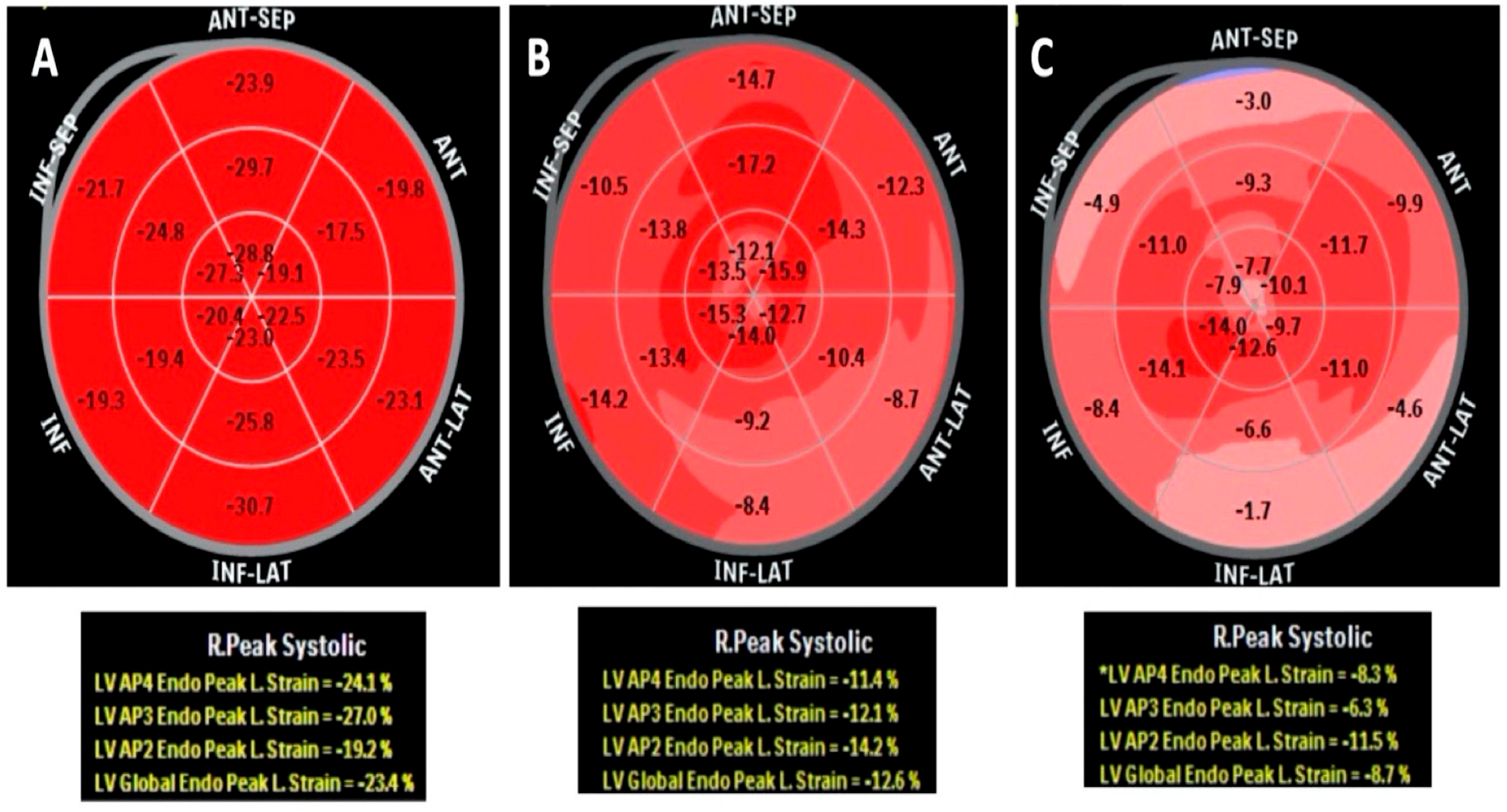
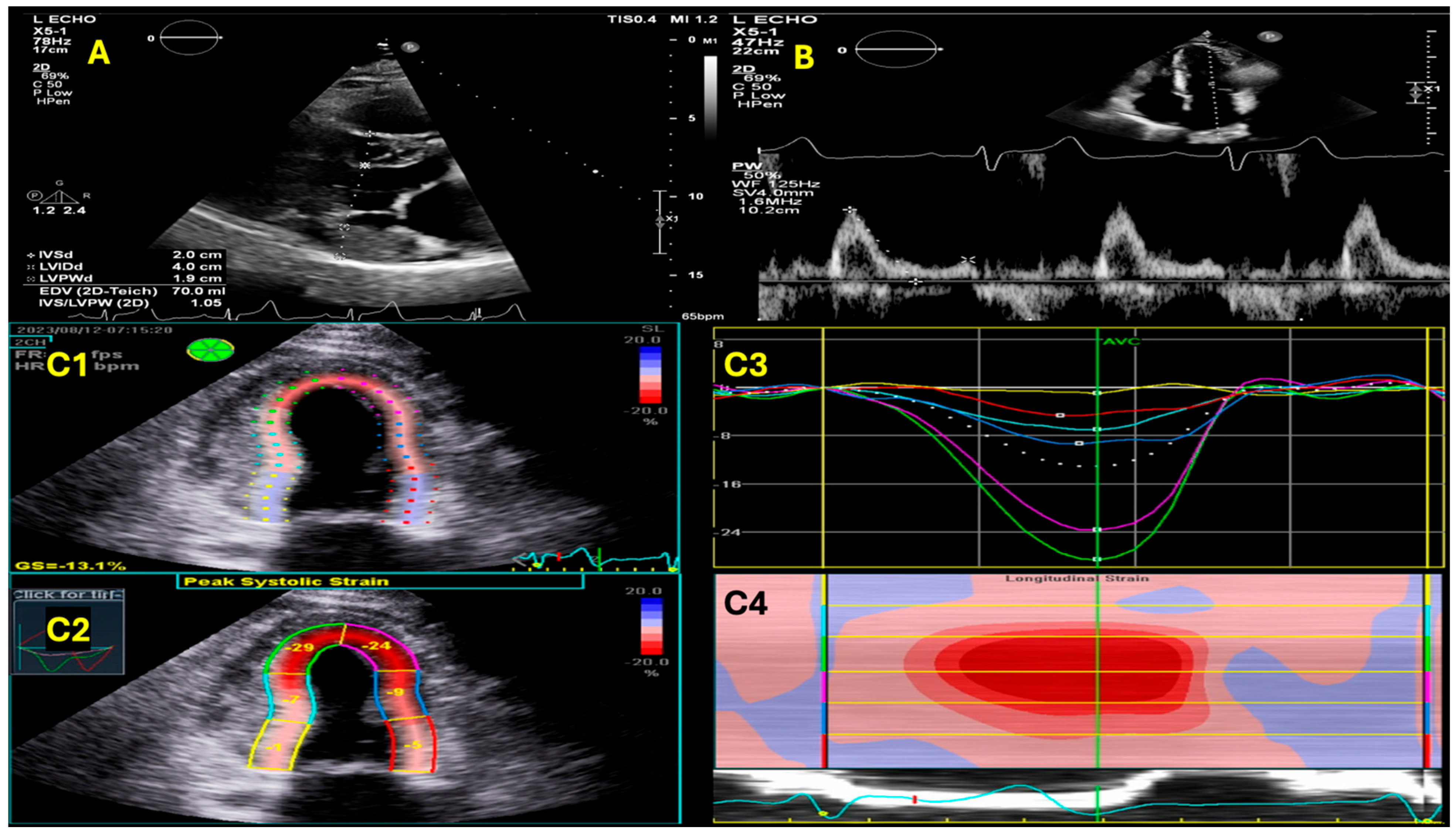
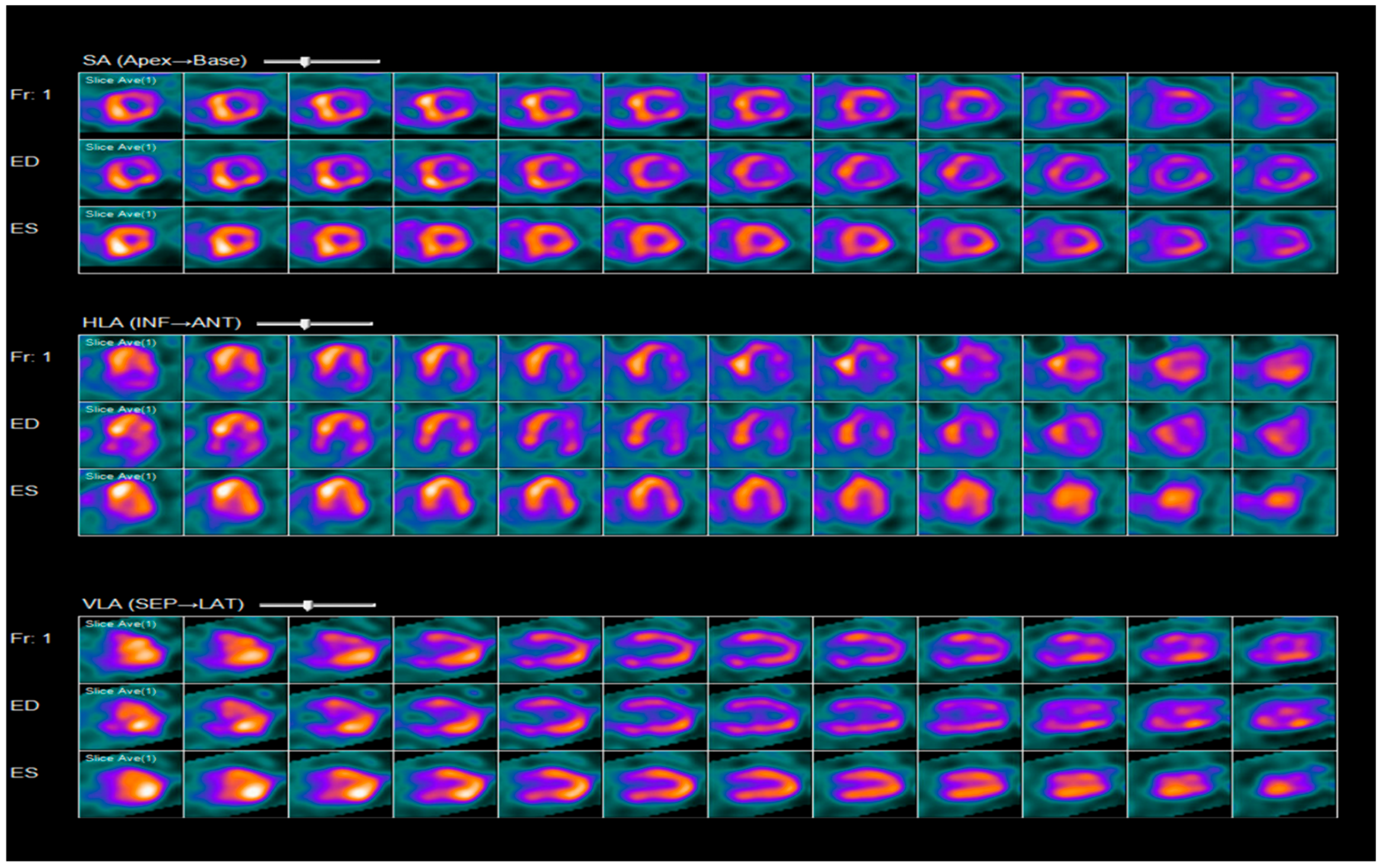
Hereditary TTR-CA has more variability in its phenotypic presentation compared to wtTTR-CA, which can be attributed, at least in part, to the specific genetic mutation involved. Generally, hTTR-CA can manifest as a cardiomyopathy, peripheral or autonomic neuropathy, or a combination of these features [2,15,18]. The distribution of the underlying genetic mutation associated with each hTTR phenotype varies greatly across different geographic locations. Among the mutations observed in hTTR-CA, the most common ones are V122I, Val30Met, and T60A [12].
V122I (Valine to isoleucine at position 122) is the most common mutation responsible for hTTR-CA in the U.S [3]. It is almost exclusively seen in individuals of West African origin and occurs in 3.4–4% of African Americans [3,4]. This may even be underestimated due to healthcare disparities and cardiac amyloidosis underdiagnosis, as these patients are underrepresented in the Southern U.S. despite having larger proportions of self-identified African Americans [29,30].
V122I confers a similar phenotype to that seen with wtTTR-CA, with disease onset typically seen at an earlier age and usually no later than the 7th decade of life [2,5]. Though all V122I carriers over the age of 65 show accumulation of myocardial amyloid to some degree, the severity of accumulation is dependent on unknown factors and varies considerably between individuals. Some experience severe cardiac disease while others’ symptoms are mild or nonexistent [12]. Compared to those with other common mutations, V122I carriers suffer from fewer neurologic complications. In those with neurologic involvement, CTS and spinal stenosis are the most common manifestations [25]. In contrast, compared to patients with wtTTR-CA, V122I hTTR-CA had more neurologic symptoms, which included neuropathic pain and tingling and higher walking disabilities [1,2].
V122I TTR-CA, like other hTTR-CAs, exhibits age-dependent but variable clinical penetrance. In a cross-sectional cohort study carried out by Damrauer et al., 51 out of 116 TTRV122I carriers (44%) had clinical heart failure or cardiac amyloidosis [6]. The prevalence of HF or cardiomyopathy increased to 70% and 100% in patients above the age of 70 and 80 years, respectively. Compared to non-carriers, it was noted that young (<45 years) TTR V122I male carriers had higher rates of left ventricular (LV) hypertrophy and greater interventricular septal wall thickening on transthoracic echocardiography (TTE) prior to overt disease manifestation [6]. In addition to heart failure, mortality has also been reported to be associated with age in V122I carriers. Analysis of the data from the ARIC (Atherosclerosis Risk in Communities) study revealed that TTR V122I carriers with an average age of 50 to 52 were at an increased risk for the development of HF, but no significant difference in mortality was found between carriers and non-carriers [8]. However, when analyzing data from a study population with an average age of 62, the REGARDS (Geographic and Racial Differences in Stroke) study revealed an association between V122I carrier status and increased mortality [9].
Val30Met (Valine to Methionine at position 30) gene mutation is the most common mutation found worldwide [30,31]. This particular gene mutation has been associated with a more rapidly progressive disease [12]. In endemic areas like Portugal, Sweden, and Japan, the age of onset is typically <50 years old and presents with a sensorimotor polyneuropathy with autonomic involvement. If there is cardiac involvement, it will more likely manifest as conduction disturbances. Val30Met has a late-onset variant (>50 years old) in nonendemic areas with a slowly progressive polyneuropathy, more severe motor impairment, and cardiac involvement most commonly manifesting as HfpEF [18,29,32,33,34]. In terms of cardiac manifestations, rhythm abnormalities are more common with late onset compared to early onset, in addition to late onset having significantly more abnormalities on electrocardiogram (ECG) that include pathologic Q waves, increased QRS intervals, and wall thickness [35].
The Thr60Ala (Threonine to Alanine at position 60) hTTR-CA subtype originated in Northern Ireland, where the carrier frequency is about 1%. It is also the most common subtype in the United Kingdom and the second most common type in the U.S., with carriers present in Appalachia, New York, and the Midwest [18,29]. It has a late onset (>60 years old), predominantly affects Caucasians, and has a male predominance, with a phenotypic ratio of 2:1. Cardiomyopathy is the predominant feature of the Thr60Ala subtype, but the conduction system is not commonly affected.1 However, mixed phenotypes with varying degrees of autonomic and peripheral neuropathy are also common [18,29,33,36].

2.3. Diagnosis

In addition to the above clinical manifestations, the initial laboratory tests might be helpful in providing clues towards cardiac amyloidosis, such as proteinuria, which may or may not be accompanied by elevations of serum BUN, creatinine, and serum bilirubin in patients with kidney disease and congestive hepatopathy. On the other hand, Natriuretic peptides and troponins T and I levels have been commonly reported to be elevated in such patients [37,38].
Other initial laboratory tests include Serum and Urine Protein Electrophoresis (SPEP/UPEP) with Immunofixation, which identifies monoclonal light chains in AL amyloidosis; Serum-Free Light Chain Assay, which detects abnormal free light chain production, which is critical for distinguishing AL amyloidosis from ATTR; and Alkaline Phosphatase, which can be elevated in hepatic involvement, particularly in systemic amyloidosis. These initial laboratory tests, in conjunction with advanced imaging and tissue biopsy, when necessary, play a crucial role in the timely diagnosis of amyloidosis.
An electrocardiogram is another non-invasive, inexpensive, useful test that can provide clues for cardiac amyloidosis, especially in the presence of other clinical manifestations. Atrial fibrillation is a common arrhythmia in patients with cardiac amyloidosis, with about 15 percent prevalence, with the highest prevalence in patients with wtATTR amyloidosis (40%) and lower prevalence with hATTR amyloidosis (11%) and AL amyloidosis (9%) [39]. The hallmark of cardiac amyloidosis is discordance between increased left ventricular wall thickness, usually identified by echocardiography, and QRS voltage, which is often reduced [40]. However, it is important to note that this feature has a low sensitivity, and the prevalence of low voltage varies markedly with etiology, with a higher frequency in patients with AL amyloidosis (60%) than in patients with ATTR amyloidosis (20%) [40]. Thus, the absence of low QRS voltage does not exclude cardiac amyloidosis, particularly in patients with wtATTR amyloidosis [40,41].
Among patients with wtATTR amyloidosis, 30 percent have voltage criteria for LV hypertrophy (LVH) or left bundle branch block, and 70 percent have pseudo-infarction patterns; conduction abnormalities affecting the sinus node and His-Purkinje systems are also common [42]. Thus, the presence of atrioventricular block in an older patient with left ventricular hypertrophy should prompt consideration of cardiac amyloidosis [42].
Speckle-tracking echocardiography (STE) to assess peak global longitudinal strain has emerged as a quantitative technique for accurate assessment of myocardial function using routine two-dimensional echocardiography [43,44]. It provides non-Doppler, angle-independent, and objective quantification of myocardial deformation and left ventricular systolic dynamics. Normal strain values has been reported to be between −18 and −22% [43,44]. The utility of STE emerged after a significant reduction in global longitudinal strain was noted to be one of the earliest markers of cardiac amyloidosis and presents with a characteristic pattern of relative apical sparing of longitudinal strain (Figure 1) [43,44].
Other common echocardiographic findings include the presence of unexplained ventricular hypertrophy, which is commonly biventricular with nondilated or small ventricles, biatrial enlargement, thickening of the valves and the interatrial septum, and, rarely, LV outflow obstruction (Figure 2) [43,44,45]. It is worth mentioning that echocardiographic findings have some structural and functional differences between AL and ATTR amyloidosis, such as the symmetrical LVH in AL amyloidosis and the asymmetric LVH with predominantly septal hypertrophy in ATTR amyloidosis, which is usually associated with a sigmoid septum [43,44]. Other recent echocardiographic findings suggest that the inferoseptal segment is highly susceptible to amyloid infiltration, and 2D speckle-tracking echocardiography and CMR may serve as a valuable tool for its early detection [46].
As discussed earlier, cardiac amyloidosis is considered one of the differentials for HF with preserved systolic function presentations [47]. Although its presentation does not fully characterize the functional phenotypic presentation for cardiac amyloidosis, as it typically involves systolic and diastolic impairment with normal range ejection fraction until late stages, which might become mildly reduced [47]. Therefore, stroke volume has been invariably used as a marker for systolic function in this presentation. Other markers used are impaired relaxation manifested by diastolic dysfunction that progresses to restrictive physiology, reduction in peak systolic wall motion velocities, and elevated pulmonary artery systolic pressure [47]. Pericardial and pleural effusions are other common findings as well [47].
Cardiac Magnetic Resonance imaging (CMR) has emerged as a powerful tool in the early detection and diagnosis of cardiac amyloidosis [48]. CMR can distinguish various morphologic phenotypes in cardiac amyloidosis. These phenotypes consist of concentric hypertrophy with diffuse, symmetric left ventricular wall thickening and increased thickness of the atrial wall and interatrial septum, as well as asymmetric septal hypertrophy (sigmoid and reverse septum) (Figure 3 and Figure 4). Unlike AL-CA, CMR shows that asymmetric septal hypertrophy is present in approximately 80% of ATTR-CA cases, while only 20% exhibit symmetric hypertrophy [49]. Furthermore, because of its wide field of view, CMR can detect extracardiac supportive findings such as pleural and pericardial effusions. CMR provides comprehensive myocardial tissue characterization that aids in diagnosis and prognosis. In cases of cardiac amyloidosis, the deposition of myocardial amyloid expands the extracellular space (interstitium). Consequently, when gadolinium is administered intravenously, it accumulates in the interstitium, leading to a shortened myocardial T1 relaxation time. This phenomenon accounts for the abnormal nulling patterns observed in cardiac amyloidosis, indicating abnormal gadolinium kinetics that result in a coincident or reversed nulling pattern on the TI scout (Look-Locker). With the use of gadolinium-based contrast agent (GBCA), it provides a detailed assessment of the cardiac structure, function, and the characteristics of the myocardial tissue [48]. Late gadolinium enhancement (LGE) performance provides highly characteristic findings suggestive of cardiac amyloidosis that correlate with the degree of myocardial function [50]. LGE in CMR visualizes extracellular space expansion due to amyloid fibril deposition, showing various patterns in cardiac amyloidosis: diffuse subendocardial, diffuse transmural, patchy mid-myocardial, atrial, and right ventricular LGE. ATTR-CA is associated with more extensive and transmural LGE compared to AL-CA. CMR using LGE can detect early cardiac abnormalities in amyloidosis patients with normal left ventricular wall thickness [50].
LGE was compared by Zaho et al. with endomyocardial biopsy and/or echocardiographic findings and other clinical features; the pooled sensitivity of LGE for cardiac amyloidosis was 85 percent, and the pooled specificity was 92 percent [51]. Patients with advanced kidney disease with an estimated glomerular filtration rate (eGFR) < 30 mL/min/1.73 m2 are at increased risk of developing nephrogenic systemic fibrosis (NSF) after exposure to group 1GBCA (estimated risk of 1–7%). However, according to the latest clinical evidence, the American College of Radiology (ACR) Committee on Drugs and Contrast Media considers the risk of NSF in patients receiving standard or lower doses of group II GBCAs very low or possibly nonexistent. This allowed patients with advanced kidney disease to receive newer GBCA (group 2 or 3) and undergo cardiac MRI when necessary [52]. However, it is important to note the limitations of LGE usage, as it lacks quantitative results, limiting the ability to track changes over time. To overcome some of these limitations, T1 mapping and extracellular volume fraction measurement may be helpful [53,54]. Nonenhanced (native) T1 mapping reflects myocardial disease and can assess the interstitium without using gadolinium. However, due to variable vendors and native T1-mapping sequences, contrast-enhanced T1 mapping enables the calculation of extracellular volume (ECV) fraction. Cardiac amyloidosis is associated with significantly elevated native T1 and ECV values compared to those observed in other cardiac diseases. Studies have shown that CMR provides good prognostic information for cardiac amyloidosis (both ATTR and AL-CA). Transmural LGE and high ECV are significant and independent predictors of mortality [49,55]. The diagnosis of ATTR-CA historically has been made using invasive methods via endomyocardial biopsy with Congo red staining, which remains the gold standard for diagnosis [15]. Despite being the gold standard for diagnosis, the endomyocardial biopsy presents several challenges, such as a small risk for serious complications, a high financial burden, the need for experienced personnel, and hospital equipment that may not be widely available, all of which can limit or delay diagnosis. Extracardiac screening biopsies such as Congo red staining of abdominal fat pad fine-needle aspiration (FPFNA) are widely used due to simplicity, cost-effectiveness, and low risk associated with their use [36]. FPFNA specificity is unequivocally high for amyloid; however, sensitivity varies vastly and has been reported anywhere from 14 to 90% [36]. Low sensitivity was specifically noted in individuals with a small total body amyloid burden and those with predominantly cardiac TTR-amyloidosis, so its routine use is discouraged because of these limitations [36]. Recent advances in imaging techniques have allowed for the non-invasive diagnosis of TTR-CA.
Cardiac scintigraphy using 99mTc-labeled tracers like 99mTc-3,3-diphosphono-1,2-propanodicarboxylic acid (99mTc-DPD), 99mTc-hydroxymethylene diphosphonate (99mTc-HMDP), and 99mTC-pyrophosphate (99mTc-PYP) can be used for diagnosis [54]. Studies have shown no statistically significant difference in diagnostic performance among these three radiotracers [11]. 99mTc-PYP scintigraphy is used in the United States and has become the primary non-invasive diagnostic imaging modality for TTR-CA [56]. Despite its diagnostic utility, bone scintigraphy may be positive in AL amyloidosis, and, therefore, testing for light chains is still required to differentiate ATTR-CA from AL-CA [19].
Therefore, the diagnostic workup should include serum-free light chain assays and protein immunofixation to rule out light chain amyloidosis, followed by bone scintigraphy to confirm the presence of transthyretin deposition in cardiac tissue [57]. Once light chain amyloidosis has been excluded, a 99mTc-PYP scan can be diagnostic of TTR-CA if there is grade 2/3 cardiac uptake or an H/CL (heart-to-contralateral) ratio > 1.5. To verify that the uptake observed in positive 99mTc-PYP scans accurately corresponds to the retention of the tracer within the myocardium, single-photon emission computed tomography (SPECT) is obtained in all patients (Figure 5). This assessment is crucial to differentiate the myocardial retention signal from potential signals originating from blood pools or rib uptake. Finally, TTR gene sequencing is obtained to differentiate wtTTR from hTTR [57]. If a gene mutation is detected, the patient’s relatives should be made aware of the diagnosis.

2.4. Treatment

Current clinical management is largely guided by expert consensus opinions due to the lack of evidence-based data. Over the recent years, new drugs have expanded the therapeutic options available for the treatment of hTTR, ranging from supportive care to new targeted therapies [22]. Current treatment goals revolve around slowing disease progression and symptom management to help with overall quality of life. Heart failure management in patients with TTR-CA requires a more cautious approach in terms of fluid balance given the restrictive nature of the disease [22,23]. TTR-CA demonstrates a lower stroke-volume index, and thus, diuretics, beta-blockers, angiotensin-converting enzyme inhibitors (ACEis), angiotensin receptor blockers (ARBs), and angiotensin receptor neprilysin inhibitors (ARNIs) can potentially cause harm due to the resultant organ hypoperfusion. There are currently not enough data to support the use of sodium-glucose cotransporter 2 inhibitors (SGLT-2is) [22].
In terms of arrhythmia management, it is recommended that atrial fibrillation be treated with anticoagulation regardless of CHADS2VASC score, as well as transesophageal echocardiography (TEE)-guided direct-current cardioversion (DCCV) independent of anticoagulation status [22]. There are currently no data to support the use of warfarin over direct oral anticoagulants nor superiority of rate versus rhythm control [22]. Caution should be used when using digoxin given the potential risk for digoxin binding amyloid fibrosis [22]. Bradyarrhythmias should be managed as per the current standard of care for the general population [22].
Currently, there are two primary therapy choices for TTR-CA: transthyretin stabilizers and silencers [12,58,59]. The ultimate objective of each of these treatment options is to reduce the quantity of circulating misfolded fibrils that have the potential to accumulate in heart tissue [12,58,59]. Stabilizers work by binding to T4 binding sites and thus blocking the rate-limiting dissociation of TTR and preventing the formation of misfolded proteins [12]. Examples of these medications include Diflunisal and Tafamidis [12]. However, in a recent trial in over 600 patients with ATTR cardiac amyloidosis and HF symptoms, patients randomly assigned to treatment with the ATTR stabilizer Acoramidis had a lower rate of hospitalization over 30 months compared with placebo [58]. Therefore, The U.S. Food and Drug Administration (FDA) has approved Acoramidis as a treatment to help reduce cardiovascular death and cardiovascular-related hospitalization in patients with transthyretin amyloid cardiomyopathy (ATTR-CM). In contrast with Tafamidis, a drug with a similar mechanism of action, Acoramidis did not reduce mortality compared with placebo [58].
Silencers work by the degradation of mRNA, which is achieved through antisense oligonucleotides or small interfering RNA molecules. Examples of these include Inotersen and Patisiran, respectively [12,59]. In a recent trial in over 350 patients with ATTR cardiac amyloidosis, patients randomly assigned to treatment with Patisiran or placebo had similar rates of death, hospitalization, or urgent visits for HF over 12 months [59]. While walk distance and quality of life improved with Patisiran, the lack of an effect on clinically important endpoints and availability of more effective therapies limit its role in ATTR cardiac amyloidosis. Newer drugs being developed include clusters of regularly interspaced short palindromic repeats and Cas9 (CRISPR-Cas9), which work by direct gene-editing by targeting specific DNA segments [12]. Treatment for hTTR is currently being developed using this novel therapeutic option with promising results [12].
Emerging evidence suggests that early initiation of disease-modifying therapies, such as TTR stabilizers (e.g., Tafamidis) or gene-silencing therapies (e.g., Patisiran, Inotersen), may delay or prevent disease progression [12,59]. However, the decision to start therapy must consider the individual’s risk profile, including age, mutation type, and early signs of disease. Some experts recommend initiating therapy in asymptomatic carriers with evidence of early myocardial involvement, such as elevated biomarkers or abnormal imaging findings, even in the absence of clinical symptoms [59]. The decision to start therapy should involve shared decision-making with the patient, considering the potential benefits, risks, and costs of treatment. Patient preferences and quality of life are critical factors in this process.
On a final note, it is very important to take into consideration the following challenges and future directions, the long-term benefits and risks of early intervention in asymptomatic carriers are not yet fully understood, highlighting the need for ongoing research and clinical trials. Disease-modifying therapies are expensive, and access may be limited in some regions. Cost-effectiveness analyses are needed to guide policy decisions and ensure equitable access. Advances in biomarkers, imaging, and genetic profiling will enable more personalized approaches to treatment, tailoring therapy to individual risk profiles and disease trajectories.

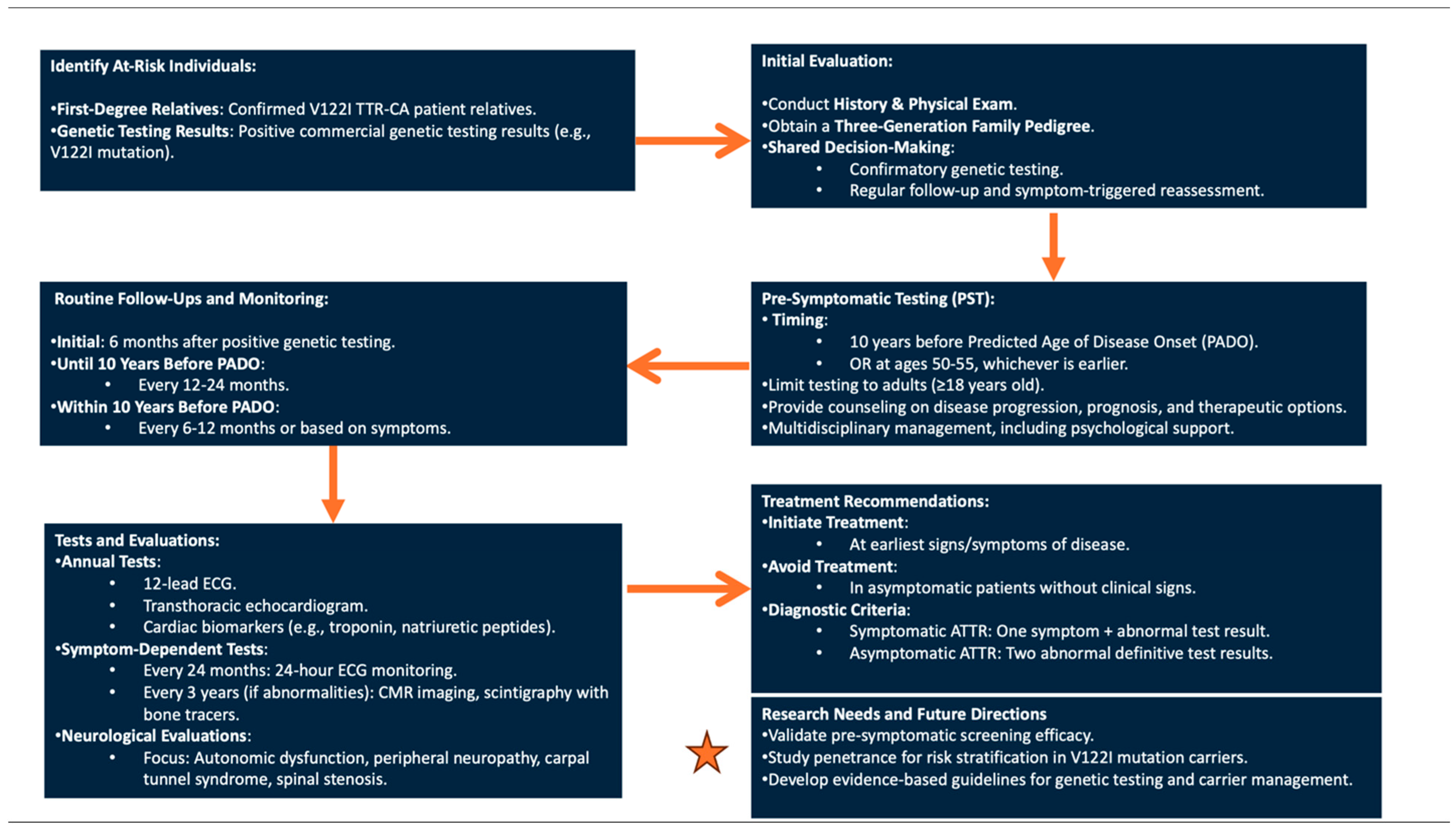
3. V122I TTR-CA: Pre-Symptomatic Genetic Testing Protocol and Management of Asymptomatic Carriers

The current standard of care for genetic testing is to proceed with testing only once a patient has been confirmed with a diagnosis of TTR-CA, to further differentiate wtTTR from hTTR [58]. There are currently no evidence-based data to support recommendations regarding the genetic screening of asymptomatic at-risk individuals [60]. However, recent large population-based cohort studies highlight and add to the growing body of literature the known morbidity and mortality associated with V122I TTR-CA carrier status [7]. The medical community is likely to see a significant rise in the number of asymptomatic individuals undergoing genetic testing for V122I TTR-CA. This trend can be attributed to two key factors. Firstly, there will be heightened awareness and identification of asymptomatic patients who have a high probability of being carriers, specifically those who are first-degree relatives of family members recently diagnosed with V122I TTR-CA. Secondly, the accessibility of commercially available genetic testing kits will become more widespread. The current medical literature lacks definitive guidelines pertaining to pre-symptomatic testing and management of asymptomatic patients carrying the V122I TTR-CA variant. A concerted effort to develop these guidelines is imperative to address the current gap in knowledge and to enhance the quality of care provided to this specific at-risk patient population.
The objective of this final section is to delineate a protocol and present recommendations for pre-symptomatic genetic testing (PST) for V122I TTR-CA, as well as the management of asymptomatic individuals who are carriers of the V122I variant. The recommendations outlined are based on expert opinions on presymptomatic genetic testing for hTTR-CA and the subsequent management of asymptomatic carriers [13,14,61,62,63,64]. The recommendations presented were informed by the current understanding of the natural history and clinical course of V122I-CA to optimize clinical guidance of this unique patient population.
Patients that should be considered at-risk individuals include first-degree relatives of confirmed V122I TTR-CA patients, as well as those who have received a positive result from a commercially available genetic testing kit. The latter group merits discussion given the rise in popularity and financial accessibility of such genetic screening tools. Currently, commercially available genetic testing kits can detect the three most common TTR gene mutations: V122I, Val30Met, and T60A [62].
The initial evaluation of both these patient populations should include a thorough history and physical exam in addition to a thorough family history with three-generation pedigree to identify any potential affected family members. In patients presenting after testing positive on a commercially available test but who do not exhibit symptoms and have a negative or unknown family history, we suggest using shared decision-making to select one of two courses of action. First, a confirmatory genetic screening may be offered to validate the initial test results. Alternatively, patients may undergo routine follow-up with their primary care doctor and return for reevaluation if symptoms arise. This approach ensures a comprehensive assessment of the patient’s condition while taking into account both the potential genetic implications and the clinical presentation. Otherwise, in patients with a confirmed affected first-degree relative, PST should be offered. In both at-risk patient populations, the timing of PST is critical to optimize the clinical benefits for patients while also minimizing unnecessary healthcare resource utilization.
In general, PST should be limited to adult patients over the age of 18 who have received thorough counseling on the disease, which should include natural disease progression, disease prognosis, early signs and symptoms, and treatment options available [14]. It is not recommended to screen patients under 18 as this would not change clinical management given typical disease presentation in later life, and premature screening may have negative effects on the patient’s mental health [13]. Therefore, the management of patients undergoing PST and asymptomatic V122I TTR-CA patients should be carried out by a multidisciplinary team that offers psychological support.
Current expert consensus recommends starting pre-symptomatic monitoring 10 years prior to disease onset in affected family members or predicted age of disease onset (PADO) for the specific gene mutation [11,13,14,64]. Thus, following results from recent studies, PST should be offered to at-risk patients between the ages of 50 to 55 or 10 years prior to disease onset in affected family members, whichever comes earlier [6]. Asymptomatic patients should follow up 6 months after positive genetic testing, then every 12–24 months up until 10 years before PADO if testing was performed earlier than the 10-year window [14]. Follow-ups thereafter can be scheduled every 6-12 months [14,64]. Shorter follow-ups should be scheduled as needed based on the patient’s symptomatology in order to help establish the clinical significance of any abnormalities.
At each visit, the patient should undergo extensive physical and laboratory testing to monitor the disease. Once within 10 years before PADO, annual cardiac evaluation should include at minimum a 12-lead electrocardiogram, transthoracic echocardiogram, and cardiac biomarkers including troponin and natriuretic peptides [11]. Every 24 months, 24 h ECG monitoring may be considered, depending on the patient’s symptoms [11]. Additionally, CMR imaging as well as scintigraphy with bone tracers is recommended every 3 years if any acute abnormalities in the cardiac work-up are encountered [11]. A thorough neurological evaluation is also a crucial part of the routine follow-up of an asymptomatic V122I TTR-CA patient [11,14]. Specifically, the neurologic evaluation should focus on evaluating autonomic dysfunction and peripheral neuropathy, particularly carpal tunnel syndrome and spinal stenosis [25]. Treatment should be offered at the earliest detectable disease sign or symptom, and, currently, it is not recommended to initiate treatment in the absence of any clinical signs or symptoms. It has been proposed that the diagnosis of symptomatic ATTR amyloidosis should be established based on specific criteria. These criteria include the presence of at least one quantified or objective symptom or sign that is definitively associated with the onset of symptomatic ATTR amyloidosis. Alternatively, the diagnosis can be made if there is at least one symptom that is probably related to the condition, along with one abnormal definitive or confirmed test result. Furthermore, in the absence of clinical symptoms, a diagnosis can be made based on the presence of two abnormal definitive or confirmed test results [64].
The 2022 American Heart Association/American College of Cardiology/Heart Failure Society of America heart failure guidelines (2022 AHA/ACC/HFSA Guideline for the Management of Heart Failure) recommend that the following signs and symptoms should increase the level of suspicion for TTR-CA: fatigue, dyspnea, or edema, and a LV wall thickness ≥14 mm, especially if present in a patient with discrepancies between LV wall thickness on TTE and the QRS voltage on ECG, or if present in patients with a history of aortic stenosis, HFpEF, CTS, spinal stenosis, and autonomic or sensory polyneuropathy (Figure 6) [57].
By establishing clear guidelines, healthcare professionals can ensure standardized practices that optimize the evaluation, monitoring, and care of individuals at risk or who are asymptomatic carriers of V122I TTR-CA. Additional clinical trials are necessary to validate the efficacy of pre-symptomatic screening in the current era of accessible therapeutic interventions. Furthermore, it is imperative to conduct studies that investigate the role of penetrance in the risk stratification of individuals affected by or at risk for being carriers of the V122I mutation.

4. Conclusions

Transthyretin cardiac amyloidosis represents a significant and increasingly recognized cause of heart failure and mortality, particularly in populations with genetic predispositions such as the V122I mutation. Advances in non-invasive imaging and genetic testing have enabled earlier detection and the opportunity for pre-symptomatic interventions, yet there remains a substantial gap in standardized protocols for managing asymptomatic carriers of TTR mutations. In this review, we discussed the importance of comprehensive and multidisciplinary approaches to care, integrating genetic counseling, targeted monitoring, and timely therapeutic interventions.
With regards to V122I carriers, the recommendations emphasize the need for structured pre-symptomatic genetic testing and surveillance protocols, informed by family history and individual risk factors. Establishing consistent monitoring strategies, including imaging and biomarker assessments, can improve early disease identification and optimize outcomes. Continued research is essential to refine risk stratification, evaluate emerging therapies, and enhance our understanding of disease penetrance and progression in this at-risk population. Ultimately, bridging these knowledge gaps will significantly impact the management of TTR-CA and improve patient quality of life and survival.

Author Contributions

K.S. and D.A.A. have both equally contributed to this manuscript including writing, editing, and proofreading. All authors have read and agreed to the published version of the manuscript.

Funding

This research received no external funding.

Conflicts of Interest

The authors declare no conflict of interest.

References

  1. Ash, S.; Shorer, E.; Ramgobin, D.; Vo, M.; Gibbons, J.; Golamari, R.; Jain, R.; Jain, R. Cardiac amyloidosis-A review of current literature for the practicing physician. Clin. Cardiol. 2021, 44, 322–331. [Google Scholar] [CrossRef] [PubMed]
  2. Maurer, M.S.; Hanna, M.; Grogan, M.; Dispenzieri, A.; Witteles, R.; Drachman, B.; Judge, D.P.; Lenihan, D.J.; Gottlieb, S.S.; Shah, S.J.; et al. Genotype and Phenotype of Transthyretin Cardiac Amyloidosis: THAOS (Transthyretin Amyloid Outcome Survey). J. Am. Coll. Cardiol. 2016, 68, 161–172. [Google Scholar] [CrossRef] [PubMed]
  3. Shije, J.Z.; Bautista, M.A.B.; Smotherman, C. The Frequency of V122I Transthyretin Mutation in a Cohort of African American Individuals With Bilateral Carpal Tunnel Syndrome. Front. Neurol. 2022, 13, 949401. [Google Scholar] [CrossRef] [PubMed]
  4. Madhani, A.; Sabogal, N.; Massillon, D.; Paul, L.D.; Rodriguez, C.; Fine, D.; Helmke, S.; Winburn, M.; Kurian, D.; Raiszadeh, F.; et al. Clinical Penetrance of the Transthyretin V122I Variant in Older Black Patients With Heart Failure: The SCAN-MP (Screening for Cardiac Amyloidosis With Nuclear Imaging in Minority Populations) Study. J. Am. Heart Assoc. 2023, 12, e028973. [Google Scholar] [CrossRef] [PubMed]
  5. Buxbaum, J.; Alexander, A.; Koziol, J.; Tagoe, C.; Fox, E.; Kitzman, D. Significance of the amyloidogenic transthyretin Val 122 Ile allele in African Americans in the Arteriosclerosis Risk in Communities (ARIC) and Cardiovascular Health (CHS) Studies. Am. Heart J. 2010, 159, 864–870. [Google Scholar] [CrossRef]
  6. Damrauer, S.M.; Chaudhary, K.; Cho, J.H.; Liang, L.W.; Argulian, E.; Chan, L.; Dobbyn, A.; Guerraty, M.A.; Judy, R.; Kay, J.; et al. Association of the V122I Hereditary Transthyretin Amyloidosis Genetic Variant With Heart Failure Among Individuals of African or Hispanic/Latino Ancestry. JAMA 2019, 322, 2191–2202. [Google Scholar] [CrossRef]
  7. Haring, B.; Hunt, R.P.; Shadyab, A.H.; Eaton, C.; Kaplan, R.; Martin, L.W.; Panjrath, G.; Kuller, L.H.; Assimes, T.; Kooperberg, C.; et al. Cardiovascular Disease and Mortality in Black Women Carrying the Amyloidogenic V122I Transthyretin Gene Variant. JACC Heart Fail. 2023, 11, 1189–1199. [Google Scholar] [CrossRef]
  8. Quarta, C.C.; Buxbaum, J.N.; Shah, A.M.; Falk, R.H.; Claggett, B.; Kitzman, D.W.; Mosley, T.H.; Butler, K.R.; Boerwinkle, E.; Solomon, S.D. The Amyloidogenic V122I Transthyretin Variant in Elderly Black Americans. N. Engl. J. Med. 2015, 372, 21–29. [Google Scholar] [CrossRef]
  9. Parcha, V.; Malla, G.; Irvin, M.R.; Armstrong, N.D.; Judd, S.E.; Lange, L.A.; Maurer, M.S.; Levitan, E.B.; Goyal, P.; Arora, G.; et al. Association of Transthyretin Val122Ile Variant with Incident Heart Failure Among Black Individuals. JAMA 2022, 327, 1368–1378. [Google Scholar] [CrossRef]
  10. Gilstrap, L.G.; Dominici, F.; Wang, Y.; El-Sady, M.S.; Singh, A.; Di Carli, M.F.; Falk, R.H.; Dorbala, S. Epidemiology of Cardiac Amyloidosis—Associated Heart Failure Hospitalizations Among Fee-for-Service Medicare Beneficiaries in the United States. Circ. Heart Fail. 2019, 12, e005407. [Google Scholar] [CrossRef]
  11. Garcia-Pavia, P.; Rapezzi, C.; Adler, Y.; Arad, M.; Basso, C.; Brucato, A.; Burazor, I.; Caforio, A.L.; Damy, T.; Eriksson, U.; et al. Diagnosis and treatment of cardiac amyloidosis. A position statement of the European Society of Cardiology Working Group on Myocardial and Pericardial Diseases. Eur. J. Heart Fail. 2021, 23, 512–526. [Google Scholar] [CrossRef] [PubMed]
  12. Ruberg, F.L.; Berk, J.L. Transthyretin (TTR) Cardiac Amyloidosis. Circulation 2012, 126, 1286–1300. [Google Scholar] [CrossRef]
  13. Scirpa, R.; Russo, D.; Tini, G.; Sclafani, M.; Tropea, A.; Cava, F.; Autore, C.; Musumeci, B. Clinical translation of genetic testing in TTR Amyloidosis: Genotype-phenotype correlations, management of asymptomatic carriers and familial screening. Vessel. Plus 2022, 6, 52. [Google Scholar] [CrossRef]
  14. Grandis, M.; Obici, L.; Luigetti, M.; Briani, C.; Benedicenti, F.; Bisogni, G.; Canepa, M.; Cappelli, F.; Danesino, C.; Fabrizi, G.M.; et al. Recommendations for pre-symptomatic genetic testing for hereditary transthyretin amyloidosis in the era of effective therapy: A multicenter Italian consensus. Orphanet J. Rare Dis. 2020, 15, 348. [Google Scholar] [CrossRef] [PubMed]
  15. Jain, A.; Zahra, F. Transthyretin Amyloid Cardiomyopathy (ATTR-CM). In StatPearls; StatPearls Publishing: Treasure Island, FL, USA, 2022. Available online: http://www.ncbi.nlm.nih.gov/books/NBK574531/ (accessed on 3 November 2022).
  16. Buxbaum, J.N.; Ruberg, F.L. Transthyretin V122I (pV142I)* cardiac amyloidosis: An age-dependent autosomal dominant cardiomyopathy too common to be overlooked as a cause of significant heart disease in elderly African Americans. Anesth. Analg. 2017, 19, 733–742. [Google Scholar] [CrossRef] [PubMed]
  17. Grogan, M.; Scott, C.G.; Kyle, R.A.; Zeldenrust, S.R.; Gertz, M.A.; Lin, G.; Klarich, K.W.; Miller, W.L.; Maleszewski, J.J.; Dispenzieri, A. Natural History of Wild-Type Transthyretin Cardiac Amyloidosis and Risk Stratification Using a Novel Staging System. J. Am. Coll. Cardiol. 2016, 68, 1014–1020. [Google Scholar] [CrossRef]
  18. Ruberg, F.L.; Grogan, M.; Hanna, M.; Kelly, J.W.; Maurer, M.S. Transthyretin Amyloid Cardiomyopathy. J. Am. Coll. Cardiol. 2019, 73, 2872–2891. [Google Scholar] [CrossRef]
  19. Kittleson, M.M.; Maurer, M.S.; Ambardekar, A.V.; Bullock-Palmer, R.P.; Chang, P.P.; Eisen, H.J.; Nair, A.P.; Nativi-Nicolau, J.; Ruberg, F.L.; On behalf of the American Heart Association Heart Failure; et al. Cardiac Amyloidosis: Evolving Diagnosis and Management: A Scientific Statement From the American Heart Association. Circulation 2020, 142, E7–E22. [Google Scholar] [CrossRef]
  20. Dispenzieri, A.; Coelho, T.; Conceição, I.; Waddington-Cruz, M.; Wixner, J.; Kristen, A.V.; Rapezzi, C.; Planté-Bordeneuve, V.; Gonzalez-Moreno, J.; Maurer, M.S.; et al. Clinical and genetic profile of patients enrolled in the Transthyretin Amyloidosis Outcomes Survey (THAOS): 14-year update. Orphanet J. Rare Dis. 2022, 17, 236. [Google Scholar] [CrossRef]
  21. Chandrashekar, P.; Alhuneafat, L.; Mannello, M.; Al-Rashdan, L.; Kim, M.M.; Dungu, J.; Alexander, K.; Masri, A. Prevalence and Outcomes of p.Val142Ile TTR Amyloidosis Cardiomyopathy: A Systematic Review. Circ. Genom. Precis. Med. 2021, 14, e003356. [Google Scholar] [CrossRef]
  22. Griffin, J.M.; Rosenthal, J.L.; Grodin, J.L.; Maurer, M.S.; Grogan, M.; Cheng, R.K. ATTR Amyloidosis: Current and Emerging Management Strategies. Cardio Oncol. 2021, 3, 488–505. [Google Scholar] [CrossRef] [PubMed]
  23. Pour-Ghaz, I.; Bath, A.; Kayali, S.; Alkhatib, D.; Yedlapati, N.; Rhea, I.; Khouzam, R.N.; Jefferies, J.L.; Nayyar, M. A Review of Cardiac Amyloidosis: Presentation, Diagnosis, and Treatment. Curr. Probl. Cardiol. 2022, 47, 101366. [Google Scholar] [CrossRef] [PubMed]
  24. Maurer, M.S.; Kale, P.; Fontana, M.; Berk, J.L.; Grogan, M.; Gustafsson, F.; Hung, R.R.; Gottlieb, R.L.; Damy, T.; González-Duarte, A.; et al. Patisiran Treatment in Patients with Transthyretin Cardiac Amyloidosis. N. Engl. J. Med. 2023, 389, 1553–1565. [Google Scholar] [CrossRef] [PubMed]
  25. Milandri, A.; Farioli, A.; Gagliardi, C.; Longhi, S.; Salvi, F.; Curti, S.; Foffi, S.; Caponetti, A.G.; Lorenzini, M.; Ferlini, A.; et al. Carpal tunnel syndrome in cardiac amyloidosis: Implications for early diagnosis and prognostic role across the spectrum of aetiologies. Eur. J. Heart Fail. 2020, 22, 507–515. [Google Scholar] [CrossRef]
  26. Fosbøl, E.L.; Rørth, R.; Leicht, B.P.; Schou, M.; Maurer, M.S.; Kristensen, S.L.; Kober, L.; Gustafsson, F. Association of Carpal Tunnel Syndrome With Amyloidosis, Heart Failure, and Adverse Cardiovascular Outcomes. J. Am. Coll. Cardiol. 2019, 74, 15–23. [Google Scholar] [CrossRef]
  27. Sperry, B.W.; Reyes, B.A.; Ikram, A.; Donnelly, J.P.; Phelan, D.; Jaber, W.A.; Shapiro, D.; Evans, P.J.; Maschke, S.; Kilpatrick, S.E.; et al. Tenosynovial and Cardiac Amyloidosis in Patients Undergoing Carpal Tunnel Release. J. Am. Coll. Cardiol. 2018, 72, 2040–2050. [Google Scholar] [CrossRef]
  28. Westin, O.; Fosbøl, E.L.; Maurer, M.S.; Leicht, B.P.; Hasbak, P.; Mylin, A.K.; Rørvig, S.; Lindkær, T.H.; Johannesen, H.H.; Gustafsson, F. Screening for Cardiac Amyloidosis 5 to 15 Years After Surgery for Bilateral Carpal Tunnel Syndrome. J. Am. Coll. Cardiol. 2022, 80, 967–977. [Google Scholar] [CrossRef]
  29. Arno, S.; Cowger, J. The genetics of cardiac amyloidosis. Heart Fail. Rev. 2021, 27, 1485–1492. [Google Scholar] [CrossRef]
  30. Alexander, K.M.; Orav, J.; Singh, A.; Jacob, S.A.; Menon, A.; Padera, R.F.; Kijewski, M.F.; Liao, R.; Di Carli, M.F.; Laubach, J.P.; et al. Geographic Disparities in Reported US Amyloidosis Mortality From 1979 to 2015: Potential Underdetection of Cardiac Amyloidosis. JAMA Cardiol. 2018, 3, 865. [Google Scholar] [CrossRef]
  31. Ueda, M.; Yamashita, T.; Misumi, Y.; Masuda, T.; Ando, Y. Origin of sporadic late-onset hereditary ATTR Val30Met amyloidosis in Japan. Amyloid 2018, 25, 143–147. [Google Scholar] [CrossRef]
  32. Pinto, M.V.; Pinto, L.F.; Dias, M.; Rosa, R.S.; Mundayat, R.; Pedrosa, R.C.; Waddington-Cruz, M. Late-onset hereditary ATTR V30M amyloidosis with polyneuropathy: Characterization of Brazilian subjects from the THAOS registry. J. Neurol. Sci. 2019, 403, 1–6. [Google Scholar] [CrossRef] [PubMed]
  33. Luigetti, M.; Romano, A.; Di Paolantonio, A.; Bisogni, G.; Sabatelli, M. Diagnosis and Treatment of Hereditary Transthyretin Amyloidosis (hATTR) Polyneuropathy: Current Perspectives on Improving Patient Care. Ther. Clin. Risk Manag. 2020, ume 16, 109–123. [Google Scholar] [CrossRef]
  34. Waddington-Cruz, M.; Wixner, J.; Amass, L.; Kiszko, J.; Chapman, D.; Ando, Y. Characteristics of Patients with Late- vs. Early-Onset Val30Met Transthyretin Amyloidosis from the Transthyretin Amyloidosis Outcomes Survey (THAOS). Neurol. Ther. 2021, 10, 753–766. [Google Scholar] [CrossRef] [PubMed]
  35. Sattianayagam, P.T.; Hahn, A.F.; Whelan, C.J.; Gibbs, S.D.; Pinney, J.H.; Stangou, A.J.; Rowczenio, D.; Pflugfelder, P.W.; Fox, Z.; Lachmann, H.J.; et al. Cardiac phenotype and clinical outcome of familial amyloid polyneuropathy associated with transthyretin alanine 60 variant. Eur. Heart J. 2011, 33, 1120–1127. [Google Scholar] [CrossRef] [PubMed]
  36. Quarta, C.C.; Gonzalez-Lopez, E.; Gilbertson, J.A.; Botcher, N.; Rowczenio, D.; Petrie, A.; Rezk, T.; Youngstein, T.; Mahmood, S.; Sachchithanantham, S.; et al. Diagnostic sensitivity of abdominal fat aspiration in cardiac amyloidosis. Eur. Hear. J. 2017, 38, 1905–1908. [Google Scholar] [CrossRef]
  37. Gillmore, J.D.; Damy, T.; FontAna, M.; Hutchinson, M.; Lachmann, H.J.; Martinez-Naharro, A.; Quarta, C.C.; Rezk, T.; Whelan, C.J.; Gonzalez-Lopez, E.; et al. A new staging system for cardiac transthyretin amyloidosis. Eur. Heart J. 2018, 39, 2799–2806. [Google Scholar] [CrossRef]
  38. Dispenzieri, A.; A Gertz, M.; Kyle, R.A.; Lacy, M.Q.; Burritt, M.F.; Therneau, T.M.; Greipp, P.R.; Witzig, T.E.; Lust, J.A.; Rajkumar, S.V.; et al. Serum Cardiac Troponins and N-Terminal Pro-Brain Natriuretic Peptide: A Staging System for Primary Systemic Amyloidosis. J. Clin. Oncol. 2004, 22, 3751–3757. [Google Scholar] [CrossRef]
  39. Longhi, S.; Quarta, C.C.; Milandri, A.; Lorenzini, M.; Gagliardi, C.; Manuzzi, L.; Bacchi-Reggiani, M.L.; Leone, O.; Ferlini, A.; Russo, A.; et al. Atrial fibrillation in amyloidotic cardiomyopathy: Prevalence, incidence, risk factors and prognostic role. Amyloid 2015, 22, 147–155. [Google Scholar] [CrossRef]
  40. Cyrille, N.B.; Goldsmith, J.; Alvarez, J.; Maurer, M.S. Prevalence and Prognostic Significance of Low QRS Voltage Among the Three Main Types of Cardiac Amyloidosis. Am. J. Cardiol. 2014, 114, 1089–1093. [Google Scholar] [CrossRef]
  41. Mussinelli, R.; Salinaro, F.; Alogna, A.; Boldrini, M.; Raimondi, A.; Musca, F.; Palladini, G.; Merlini, G.; Perlini, S. Diagnostic and Prognostic Value of Low QRS Voltages in Cardiac AL Amyloidosis. Ann. Noninvasive Electrocardiol. 2013, 18, 271–280. [Google Scholar] [CrossRef]
  42. Quarta, C.C.; Zheng, J.; Hutt, D.; Grigore, S.F.; Manwani, R.; Sachchithanantham, S.; Mahmood, S.A.; Whelan, C.J.; Fontana, M.; Martinez-Naharro, A.; et al. 99mTc-DPD scintigraphy in immunoglobulin light chain (AL) cardiac amyloidosis. Eur. Heart J. Cardiovasc. Imaging 2021, 22, 1304–1311. [Google Scholar] [CrossRef] [PubMed]
  43. Maurer, M.S.; Elliott, P.; Comenzo, R.; Semigran, M.; Rapezzi, C. Addressing Common Questions Encountered in the Diagnosis and Management of Cardiac Amyloidosis. Circulation 2017, 135, 1357–1377. [Google Scholar] [CrossRef] [PubMed]
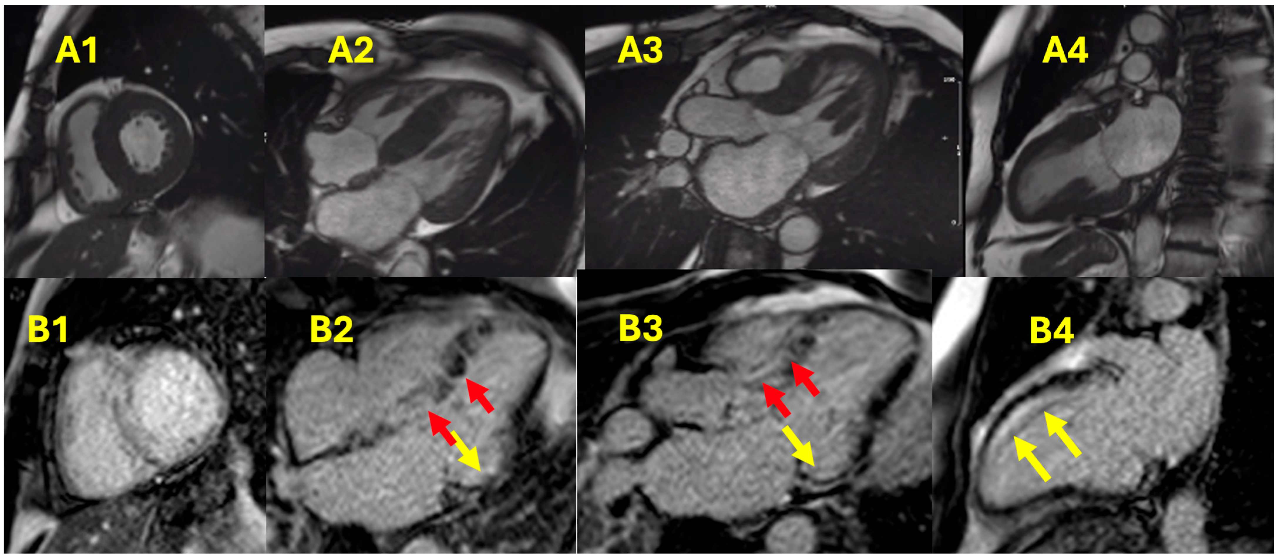
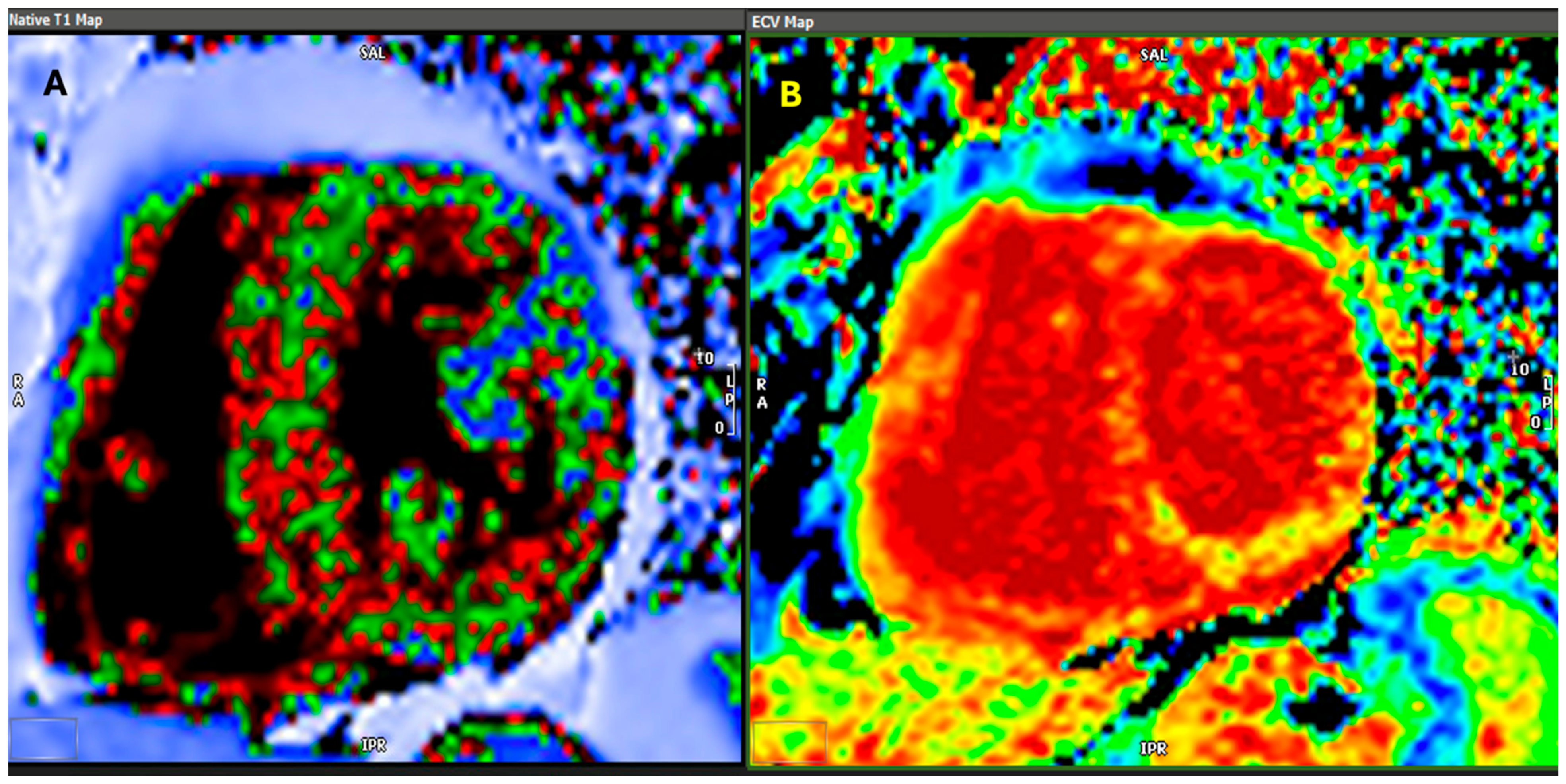
  44. Martinez-Naharro, A.; Treibel, T.A.; Abdel-Gadir, A.; Bulluck, H.; Zumbo, G.; Knight, D.S.; Kotecha, T.; Francis, R.; Hutt, D.F.; Rezk, T.; et al. Magnetic Resonance in Transthyretin Cardiac Amyloidosis. J. Am. Coll. Cardiol. 2017, 70, 466–477. [Google Scholar] [CrossRef] [PubMed]
  45. Boldrini, M.; Cappelli, F.; Chacko, L.; Restrepo-Cordoba, M.A.; Lopez-Sainz, A.; Giannoni, A.; Aimo, A.; Baggiano, A.; Martinez-Naharro, A.; Whelan, C.; et al. Multiparametric Echocardiography Scores for the Diagnosis of Cardiac Amyloidosis. Cardiovasc. Imaging 2019, 13, 909–920. [Google Scholar] [CrossRef] [PubMed]
  46. Tsuruda, T.; Nakada, H.; Yamamura, Y.; Matsuura, Y.; Ogata, M.; Tanaka, M.; Suiko, Y.; Komaki, S.; Tanaka, H.; Moribayashi, K.; et al. Basal inferoseptal segment is highly susceptible to deformation in the clinical spectrum of transthyretin-derived amyloid cardiomyopathy. Eur. Heart J. Open 2024, 4, oeae076. [Google Scholar] [CrossRef]
  47. Knight, D.S.; Zumbo, G.; Barcella, W.; Steeden, J.A.; Muthurangu, V.; Martinez-Naharro, A.; Treibel, T.A.; Abdel-Gadir, A.; Bulluck, H.; Kotecha, T.; et al. Cardiac Structural and Functional Consequences of Amyloid Deposition by Cardiac Magnetic Resonance and Echocardiography and Their Prognostic Roles. JACC Cardiovasc. Imaging 2018, 12, 823–833. [Google Scholar] [CrossRef]
  48. de Carvalho, F.P.; Erthal, F.; Azevedo, C.F. The Role of Cardiac MR Imaging in the Assessment of Patients with Cardiac Amyloidosis. Magn. Reson. Imaging Clin. N.Am. 2019, 27, 453–463. [Google Scholar] [CrossRef]
  49. Fontana, M.; Martinez-Naharro, A.; Hawkins, P.N. Staging Cardiac Amyloidosis With CMR: Understanding the Different Phenotypes. JACC Cardiovasc. Imaging 2016, 9, 1278–1279. [Google Scholar] [CrossRef]
  50. Fontana, M.; Pica, S.; Reant, P.; Abdel-Gadir, A.; Treibel, T.A.; Banypersad, S.M.; Maestrini, V.; Barcella, W.; Rosmini, S.; Bulluck, H.; et al. Prognostic Value of Late Gadolinium Enhancement Cardiovascular Magnetic Resonance in Cardiac Amyloidosis. Circulation 2015, 132, 1570–1579. [Google Scholar] [CrossRef]
  51. Zhao, L.; Tian, Z.; Fang, Q. Diagnostic accuracy of cardiovascular magnetic resonance for patients with suspected cardiac amyloidosis: A systematic review and meta-analysis. BMC Cardiovasc. Disord. 2016, 16, 129. [Google Scholar] [CrossRef]
  52. Starekova, J.; Pirasteh, A.; Reeder, S.B. Update on Gadolinium-Based Contrast Agent Safety, From the AJR Special Series on Contrast Media. Am. J. Roentgenol. 2024, 223, e2330036. [Google Scholar] [CrossRef] [PubMed]
  53. Banypersad, S.M.; Sado, D.M.; Flett, A.S.; Gibbs, S.D.; Pinney, J.H.; Maestrini, V.; Cox, A.T.; Fontana, M.; Whelan, C.J.; Wechalekar, A.D.; et al. Quantification of myocardial extracellular volume fraction in systemic AL amyloidosis: An equilibrium contrast cardiovascular magnetic resonance study. Circ. Cardiovasc. Imaging 2013, 6, 34–39. [Google Scholar] [CrossRef] [PubMed]
  54. Messroghli, D.R.; Moon, J.C.; Ferreira, V.M.; Grosse-Wortmann, L.; He, T.; Kellman, P.; Mascherbauer, J.; Nezafat, R.; Salerno, M.; Schelbert, E.B.; et al. Clinical recommendations for cardiovascular magnetic resonance mapping of T1, T2, T2* and extracellular volume: A consensus statement by the Society for Cardiovascular Magnetic Resonance (SCMR) endorsed by the European Association for Cardiovascular Imaging (EACVI). J. Cardiovasc. Magn. Reson. 2017, 19, 75, Erratum in J. Cardiovasc. Magn. Reson. 2018, 20, 9. [Google Scholar]
  55. Fontana, M.; Chung, R.; Hawkins, P.N.; Moon, J.C. Cardiovascular magnetic resonance for amyloidosis. Heart Fail. Rev. 2015, 20, 133–144. [Google Scholar] [CrossRef]
  56. Tahara, N.; Lairez, O.; Endo, J.; Okada, A.; Ueda, M.; Ishii, T.; Kitano, Y.; Lee, H.; Russo, E.; Kubo, T. 99mTechnetium-pyrophosphate scintigraphy: A practical guide for early diagnosis of transthyretin amyloid cardiomyopathy. ESC Heart Fail. 2021, 9, 251–262. [Google Scholar] [CrossRef]
  57. Heidenreich, P.A.; Bozkurt, B.; Aguilar, D.; Allen, L.A.; Byun, J.J.; Colvin, M.M.; Deswal, A.; Drazner, M.H.; Dunlay, S.M.; Evers, L.R.; et al. 2022 AHA/ACC/HFSA Guideline for the Management of Heart Failure: A Report of the American College of Cardiology/American Heart Association Joint Committee on Clinical Practice Guidelines. Circulation 2022, 145, e895–e1032. [Google Scholar] [CrossRef]
  58. Gillmore, J.D.; Judge, D.P.; Cappelli, F.; Fontana, M.; Garcia-Pavia, P.; Gibbs, S.; Grogan, M.; Hanna, M.; Hoffman, J.; Masri, A.; et al. Efficacy and Safety of Acoramidis in Transthyretin Amyloid Cardiomyopathy. N. Engl. J. Med. 2024, 390, 132–142. [Google Scholar] [CrossRef]
  59. Judge, D.P.; Gillmore, J.D.; Alexander, K.M.; Ambardekar, A.V.; Cappelli, F.; Fontana, M.; García-Pavía, P.; Grodin, J.L.; Grogan, M.; Hanna, M.; et al. Long-Term Efficacy and Safety of Acoramidis in ATTR-CM: Initial Report From the Open-Label Extension of the ATTRibute-CM Trial. Circulation 2024. Advance online publication. [Google Scholar] [CrossRef]
  60. Maurer, M.S.; Bokhari, S.; Damy, T.; Dorbala, S.; Drachman, B.M.; Fontana, M.; Grogan, M.; Kristen, A.V.; Lousada, I.; Nativi-Nicolau, J.; et al. Expert Consensus Recommendations for the Suspicion and Diagnosis of Transthyretin Cardiac Amyloidosis. Circ. Heart Fail. 2019, 12, e006075. [Google Scholar] [CrossRef]
  61. Obici, L.; Kuks, J.B.; Buades, J.; Adams, D.; Suhr, O.B.; Coelho, T.; Kyriakides, T. Recommendations for presymptomatic genetic testing and management of individuals at risk for hereditary transthyretin amyloidosis. Curr. Opin. Neurol. 2016, 29, S27–S35. [Google Scholar] [CrossRef]
  62. Alreshq, R.; Ruberg, F.L. Clinical approach to genetic testing in amyloid cardiomyopathy: From mechanism to effective therapies. Curr. Opin. Cardiol. 2021, 36, 309–317. [Google Scholar] [CrossRef] [PubMed]
  63. Murray, M.F.; Miller, E.J. Can We Manage Presymptomatic TTR V142I Related Risk? Heart Fail. 2022, 10, 139–141. [Google Scholar] [CrossRef] [PubMed]
  64. Conceição, I.; Damy, T.; Romero, M.; Galán, L.; Attarian, S.; Luigetti, M.; Sadeh, M.; Sarafov, S.; Tournev, I.; Ueda, M. Early diagnosis of ATTR amyloidosis through targeted follow-up of identified carriers of TTR gene mutations. Amyloid 2019, 26, 3–9. [Google Scholar] [CrossRef] [PubMed]
Figure 1. Representative Left ventricular peak global longitudinal strain (LV PGLS) polar maps derived from speckle tracking imaging algorithms using GE (General Electric, Medical Systems, Milwaukee, WI, USA) using their automated functional imaging (AFI) method to analyze speckle tracking. The polar display shows all myocardial wall segments, and the three inner circles represent LV basal, mid cavity and apical segments, respectively. The individual values represent strain measurements for each segment encoded in in the LV image acquisition. The legend box below shows mean values for the LV 4-apical chamber, 3-chamber, and 2 chamber image analysis. A final composite value from all three LV views is then reported as LV PGLS, which is the value of interest. (A) demonstrates a representative polar map LV PGLS in patients with normal LV PGLS (−19.2 ± 2.3%, range of −16 to −27%) values. (B) Shows a representative polar in patients with abnormally low normal LV PGLS levels (−15.2 ± 0.6%, range of −14 to −16%) while (C) Shows a representative polar map in patients with a markedly low LV PGLS values (Bull’s eye) (−11.7 ± 1.7%, range −7 to −13%). Image courtesy of Khalid Sawalha, MD.
Hearts 06 00006 g001
Figure 2. Echocardiographic study of a patient with ATTR-CA shows the following: (A) Severe concentric left ventricular hypertrophy. (B) Elevated filling pressures with a restrictive pattern and E to A ratio >2. (C1C4) Reduced longitudinal strain with relative preservation of the apex, resembling a cherry-on-top appearance (C4). ATTR-CA: transthyretin cardiac amyloidosis. Images courtesy of Deya A. Alkhatib, MD, FACC, FASE.
Hearts 06 00006 g002
Figure 3. A cardiac MRI of a patient with ATTR-CA reveals severe concentric left ventricular hypertrophy on SSFP images where images (A1A4) showing Cine MRI heart function and wall motion. (A1) short-axis view of the left ventricle, (A2) four chamber view, (A3) three chamber view, and (A4) sagittal view of the right ventricle and the great vessels. Image B1 shows baseline LGE image with no apparent hyperenhancement, (B2B4) images show two patterns of LGE: mid-myocardial LGE (red arrows) in the hypertrophied basal to mid anteroseptal segments, and subendocardial LGE (yellow arrows) in the basal to mid lateral wall and mid to apical anterior segments. ATTR-CA: transthyretin cardiac amyloidosis; LGE: late gadolinium enhancement; MRI: magnetic resonance imaging; SSFP: steady-state free precession. Images courtesy of Deya A. Alkhatib, MD, FACC, FASE.
Hearts 06 00006 g003
Figure 4. A cardiac MRI of a patient with ATTR-CA reveals elevated native myocardial T1 (A) and severely elevated ECV (B) as depicted on color parametric mapping. ATTR-CA: transthyretin cardiac amyloidosis; ECV: extracellular volume; MRI: magnetic resonance imaging. Images courtesy of Deya A. Alkhatib, MD, FACC, FASE.
Hearts 06 00006 g004
Figure 5. Tc-99mPYP SPECT imaging of a patient with ATTR-CA reveals diffuse myocardial uptake consistent with cardiac amyloidosis. ATTR-CA: transthyretin cardiac amyloidosis; SPECT: single-photon emission computed tomography; Tc-99mPYP: Technetium-99m pyrophosphate. Images courtesy of Deya A. Alkhatib, MD, FACC, FASE.
Hearts 06 00006 g005
Figure 6. Summary of recommendations for pre-symptomatic monitoring and management of V122I TTR-CA.
Hearts 06 00006 g006
Table 1. This table shows different types of amyloidosis based on their precursor proteins, primary organ involvement, and degree of cardiac impact. Author’s own work.
TypePrecursor ProteinPrimary Organ InvolvementCardiac Involvement
AL (Primary) AmyloidosisImmunoglobulin Light ChainsHeart, Kidney,
Nervous System
Frequent and severe
ATTR (Transthyretin) AmyloidosisTransthyretin (TTR)Heart, Nervous SystemCommon, particularly in hereditary forms
AA (Secondary) AmyloidosisSerum Amyloid AKidney, Liver, SpleenRare
Aβ2M (Dialysis-related)β2-microglobulinJoints, BonesRare
Hereditary Non-TTR AmyloidosisFibrinogen
Apolipoproteins
Kidney, Liver, NervesVariable
Disclaimer/Publisher’s Note: The statements, opinions and data contained in all publications are solely those of the individual author(s) and contributor(s) and not of MDPI and/or the editor(s). MDPI and/or the editor(s) disclaim responsibility for any injury to people or property resulting from any ideas, methods, instructions or products referred to in the content.

