Review

Clinical Significance of Endotypes of Asian Chronic Rhinosinusitis: A Review and Expert Commentary

1
Yong Loo Lin School of Medicine, National University of Singapore, Singapore 117597, Singapore
2
Lee Kong Chian School of Medicine, Nanyang Technological University, Singapore 308232, Singapore
3
Department of Otolaryngology-Head and Neck Surgery, University of Insubria, 21100 Varese, Italy
4
Department of Otolaryngology-Head and Neck Surgery, Ng Teng Fong General Hospital, Singapore 609606, Singapore
*
Author to whom correspondence should be addressed.
Sinusitis 2025, 9(2), 19; https://doi.org/10.3390/sinusitis9020019
Submission received: 8 August 2025 / Revised: 12 September 2025 / Accepted: 26 September 2025 / Published: 9 October 2025

Abstract

Chronic rhinosinusitis (CRS) is increasingly recognized as a heterogeneous condition characterized by distinct inflammatory endotypes. In Western populations, CRS is mostly type 2 (T2) eosinophilic. In contrast, Asian cohorts show higher rates of non-T2 endotypes, posing unique challenges and limiting the applicability of Western treatment models. This expert commentary and scoping review explores endotype-driven care in CRS, synthesizes current research on Asian CRS cohorts, and examines existing gaps in our understanding, particularly in non-T2 CRS. Endotype-driven care improves treatment precision and minimizes therapeutic failure. However, most models remain largely Western-centric. Establishing Asia-specific criteria, accessible diagnostics, and therapies for non-T2 disease is essential.

1. Introduction

Chronic rhinosinusitis (CRS) is now widely regarded as not just a single disease entity but rather a heterogeneous clinical syndrome that arises from a complex interplay of multiple environmental and host genetic factors [1]. This diversity of CRS contributes to the challenge of managing a condition that affects so many worldwide. In previous epidemiological studies, the global burden of CRS was found to be 8.71%, and its prevalence has seen a more than a four-fold increase in recent years [2]. The prevalence of such a complex disease has significant impacts on the quality of life of the individuals it affects as well as substantial direct and indirect costs associated with its management [3]. This underscores the need for continued research into the disease. In recent years, there is growing emphasis not just on the different clinical presentations of CRS but also the distinct immunologic pathways that drive its different presentations. As such, we should shift our understanding of the disease from a primarily phenotypic framework towards one that is endotype-focused, creating important refinements in our approach to diagnosing and treating CRS.
This expert commentary explores the case for endotype-driven care in CRS and outlines its clinical benefits. The scoping review synthesizes current research on Asian CRS cohorts, highlighting the limitations of applying a one-size-fits-all approach in CRS and examines existing gaps in our understanding of non-T2 CRS, including diagnostic challenges posed by a lack of biomarker thresholds for endotyping Asian CRS and limitations in therapeutic options.

Physiology of Endotypes in CRS

CRS itself is driven by three primary immunological endotypes: type 1 (T1), type 2 (T2), and type 3 (T3) plus an untypeable/mixed group. Each of these endotypes is defined by a distinct cytokine network that determines histopathology and subsequently clinical presentation and even therapeutic management. Figure 1 describes the different CRS endotypes and their associated inflammatory markers.
In T1 CRS, viral or intracellular pathogens activate TLR3 and TLR9 on epithelial cells, inducing IL-1β, IL-12p70, and IL-18 [4]. These cytokines activate dendritic cells to drive Th1 polarization through the JAK2–TYK2–STAT4 signaling pathway [5], leading to T-bet transcription factor expression that controls IFN-γ production [6]. Group 1 innate lymphoid cells (ILC1s) subsequently provide rapid IFN-γ secretion alongside activated Th1 and CD8+ T cells [7,8]. IFN-γ serves as the central distinct cytokine for T1 endotype CRS, engaging epithelial IFN-γ receptors to activate STAT1 signaling and promote epithelial barrier disruption through degradation of tight junction proteins (occludin, ZO-1) and epithelial-to-mesenchymal transition via multiple pathways including TGF-β, Wnt, and p38/ERK [9,10,11]. Hallmark biomarkers of T1 CRS are the CXCR3 chemokine ligands, CXCL9 (MIG), CXCL10 (IP-10), and CXCL11 (I-TAC), which demonstrate diagnostic performance for endotype identification [12]. In nasal lavage fluid, CXCL9 achieves an area under the curve (AUC) of 0.869 (85% sensitivity, 75% specificity), while CXCL10 achieves an AUC of 0.815 (93% sensitivity, 63% specificity) for T1 endotype detection [12]. These IFN-γ-induced chemokines create gradients that recruit CXCR3-expressing CD8+ T cells, NK cells, and additional Th1 cells. The resulting neutrophil-dominant inflammation; characterized by elevated tissue IFN-γ levels, CD8+ T cell infiltration, and M1 macrophage predominance [13], underlies the non-polypoid CRS phenotype and is associated with steroid resistance and poor treatment outcomes [14].
In the T2 endotype of CRS, epithelial injury induces the release of the alarmin cytokines: long-form thymic stromal lymphopoietin (lf-TSLP), interleukin-25 (IL-25), and interleukin-33 (IL-33) [15,16]. lf-TSLP activates dendritic cells to drive naïve CD4+ T cells toward a Th2 phenotype via OX40 ligand upregulation and directly stimulates group 2 innate lymphoid cells (ILC2s) through TSLPR signaling [17]. IL-25 and IL-33 further amplify ILC2 expansion, which is enriched up to 100-fold in nasal polyp tissue versus non-polyp mucosa [18]. Activated ILC2s and Th2 cells secrete IL-4, IL-5, and IL-13. IL-5 drives eosinophil maturation and survival. IL-4 and IL-13 activate the IL-4Rα/STAT6 pathway, promoting IgE class switching, goblet cell hyperplasia, and periostin expression [19,20]. Periostin, an extracellular matrix protein, serves as a robust tissue and serum biomarker for T2 CRS, with periostin levels > 95 ng/mL distinguishing CRS with nasal polyps (CRSwNP) from CRS without nsal polyps (CRSsNP) and correlating with comorbid asthma and surgical recurrence risk [21]. Ultimately, impaired fibrinolysis, mediated by reduced tissue plasminogen activator expression and excessive fibrin deposition from periostin build-up, contributes to the edematous remodeling characteristic of T2 polyps [22]. Together, these hallmark biomarkers and pathways—the l-TSLP/IL-25/IL-33–ILC2 axis and IL-4/IL-13–STAT6–periostin circuit—define the eosinophilic inflammation and tissue remodeling that distinguishes T2 CRS from other endotypes.
The T3 endotype of CRS, particularly prevalent in many Asian cohorts, centers on a neutrophil-dominant inflammatory circuit orchestrated by IL-17A and IL-22. In nasal polyp tissue, CD68+ M1 macrophages are the principal source of IL-17A. IL-17A binds to the IL-17RA/RC receptor complex on epithelial and stromal cells. This engagement recruits the Act1 adaptor protein and activates NF-κB and MAPK (ERK/STAT3) signaling. Subsequently, this drives the expression of the neutrophil chemoattractants CXCL1, CXCL2, and IL-8 [23]. Parallel secretion of IL-22 by RORγt+ ILC3s engages epithelial IL-22R to activate STAT3, upregulating CCL2 and CXCL10 to recruit monocytes and sustain inflammation [24]. The convergent chemokine gradients result in massive neutrophil infiltration and the release of human neutrophil elastase (HNE), which degrades epithelial junctions and extracellular matrix, driving tissue injury, remodeling, and leading to the characteristic corticosteroid resistance seen in T3 endotypes of CRS [25]. Hallmark biomarkers of the T3 endotype include elevated tissue and lavage IL-17A and IL-22 levels, both correlating with neutrophil markers and polyp burden. Granulocyte colony-stimulating factor (CSF3/G-CSF) is consistently upregulated in T3 sinonasal mucosa via nasal lavage, and testing for it achieves an AUC of 0.662 for endotype prediction [12]. Elevated nasal IL-8 and myeloperoxidase (MPO) further reflect neutrophilic activation, while immunofluorescence-quantified HNE+ cells distinguish T3 inflammation and correlate with refractory disease. This IL-17A/IL-22–chemokine–CSF3–HNE–IL-8/MPO profile defines neutrophil-driven pathology in T3 CRS.
Mixed endotypes in CRS are characterized by simultaneous activation of T1, T2, and T3 pathways, manifesting as concurrent elevations of IFN-γ, IL-13, and IL-17A [26]. This complex immune milieu arises from cross-talk among Th1, Th2, and Th17 cells together with ILC1, ILC2, and ILC3 subsets, resulting in co-existing eosinophilic and neutrophilic inflammation [27]. Mixed-endotype CRS is associated with more severe disease: patients exhibit higher symptom scores, worse computed tomography findings, and reduced olfactory function compared to single-endotype counterparts.

2. Methods

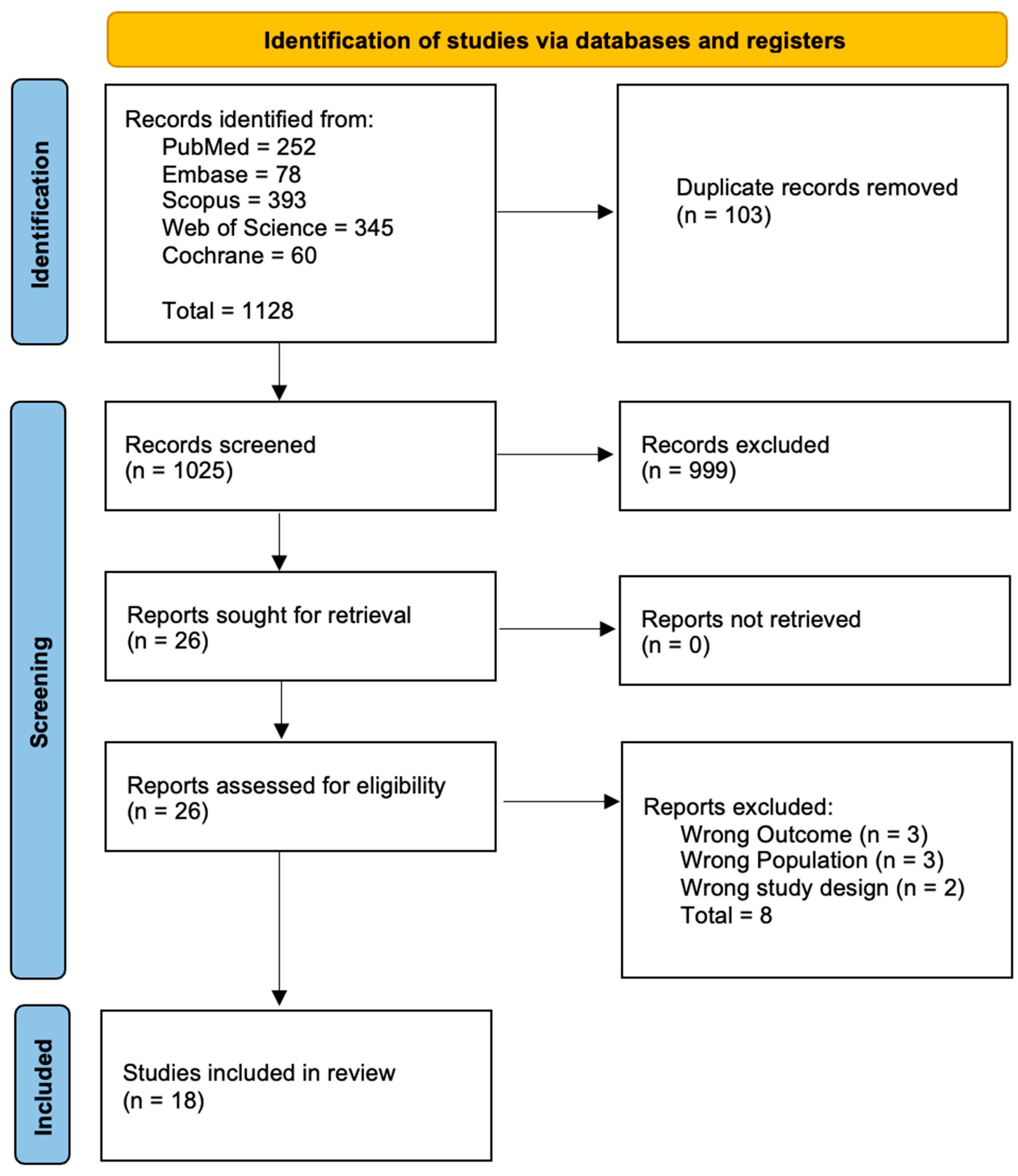
A comprehensive literature search was done on 15 July 2025 to ascertain current research done on CRS endotyping and the gaps in knowledge. The search was conducted on PubMed, Cochrane, Web of Science, Embase, and Scopus from January 2000 to July 2025 using the following keywords: “Chronic Rhinosinusitis”, “Endotypes”, “CRS endotyping”, “Asian populations”, “Precision Medicine”, “personalized treatment”, “Biomarkers”, “Therapeutic targets” and “Research gaps”. Table 1 summarizes the search strategy as well as the inclusion and exclusion criteria used in this review. A total of 1128 studies were obtained from the 5 databases. After removing duplicates and including only the relevant articles, a total of 18 studies were included in our review as shown in Figure 2. The search and screening of relevant articles for discussion were conducted by main authors SLBJ and NCYM. These were later vetted by authors FDP and NCL.
Data extracted from the included studies consisted of key findings on Asian CRS endotypes, endotype directed care, current research gaps, and novel findings associated with endotyping. This was conducted with the main aim of synthesizing existing literature on Asian CRS endotyping and its viability and potential as a therapeutic target moving forward. Novel studies such as in-progress RCTs and unique endotype-based trials were also included in our study. The studies included in our review were further subdivided into whether they were clinical or experimental studies. The risk of bias for each clinical study was evaluated using the Newcastle–Ottawa Scale and presented in Table 2. Experimental studies in Table 3 were evaluated based on their methodology and the strengths and limitations presented within the study.

3. Results

The literature search revealed marked geographic and temporal variations in CRS endotypes with significant clinical implications. Geographic heterogeneity was consistently demonstrated across multiple studies, with Asian populations showing distinctly different inflammatory patterns compared to Western populations [27,31,36]. The multicenter analysis revealed that Asian centers demonstrated significantly lower TH2 cytokine expression and eosinophilia compared to Western sites (p < 0.05) [31]. This geographic pattern was further supported by findings that over 50% of Chinese CRSwNP cases were non-eosinophilic and more than 73% of Korean CRS cases were non-T2 eosinophilic or mixed endotypes, contrasting with the T2 inflammation predominance observed in Western populations [36].
Temporal evolution of CRS endotypes revealed a striking “eosinophilic shift” occurring across multiple Asian populations over recent decades. In Northern China, eosinophil-dominant CRSwNP rose from 12% to 43% between 1993 and 1995 and 2015 and 2019, with increased asthma comorbidity (p < 0.01) [28]. This temporal trend was corroborated by Japanese epidemiological findings, which showed that 97.4% of asthmatic CRS cases had ≥15% tissue eosinophils, suggesting an emerging convergence toward Western inflammatory patterns [35].
These findings collectively demonstrate that CRS endotypes exhibit considerable geographic and temporal heterogeneity, with Asian populations historically showing predominant non-T2 inflammation but experiencing a recent shift toward eosinophilic endotypes. This evolutionary pattern necessitates region-specific and temporally adapted therapeutic approaches, highlighting the importance of endotype-guided precision medicine in CRS management.
Among other included studies, experimental studies have also delineated inflammatory mechanisms underlying Asian CRS [37,42,43], highlighted key immune mediators which serve as predictors of treatment outcomes [40], as well as identified potential non-invasive diagnostic biomarkers for endotyping such as MPO in neutrophilic CRS and ECP in eosinophilic CRS [38]. Several studies have also identified novel therapeutic targets such as the IL-17A/mTOR axis [41] or the IL-36γ/IL-36R pathway in non-T2 CRS [42]. The application of AI technology in CRS has also been investigated, with one study demonstrating its potential to augment CRS endotyping [39].

3.1. Clinical Implications

The investigation of CRS endotypes has the potential for substantial clinical value by enabling precision medicine approaches that optimize treatment selection and patient outcomes. Endotype-guided biomarker identification has demonstrated predictive capacity for therapeutic responses, particularly in T2 inflammation where elevated nasal IL-5 levels predict favorable responses to anti-IL-5 biologics [44]. Tissue eosinophilia, quantified as ≥70 eosinophils per high-power field could serve as both a diagnostic marker for T2 endotype and a prognostic indicator, with eosinophilic CRS patients showing increased recurrence risk following endoscopic sinus surgery [45]. Surgical outcome optimization through endotype classification have further revealed that all inflammatory endotypes demonstrate superior postoperative improvements in patient-reported outcomes, with the T2 endotype having the most significant mean SNOT-22 improvements of −28.3 points and olfactory function improvements of 6.5 TDI points six months after surgery [46].
Risk stratification and treatment optimization are other key clinical applications of endotype identification. Non-T2 endotypes have been found to exhibit steroid resistance and require alternative therapeutic approaches, while T3 endotypes with elevated neutrophil elastase demonstrate poor responses to conventional anti-inflammatory treatments [47]. The recognition of mixed endotypes could aid clinicians to identify patients requiring combined therapeutic approaches to overcome monotherapy limitations [48]. Diagnostic advancement through non-invasive biomarker panels enables point-of-care endotype identification using nasal secretion biomarkers, including CXCL9 and CXCL10 for T1 endotypes (AUC 0.815–0.869), CCL26 for T2 endotypes (AUC 0.778), and CSF3 for T3 endotypes, reducing dependence on invasive tissue sampling [12]. Long-term revision rates [49] for endoscopic sinus surgery have increased to 15.9% overall, with higher rates in CRSwNP patients (30%) compared to CRSsNP patients (9.75%), underscoring the importance of endotype-guided selection of surgical candidates and postoperative management strategies to prevent further recurrence.

3.2. The Case for Endotype-Driven Care

An endotype-driven approach in managing CRS has shown to be associated with positive patient outcomes through the use of endotype-specific treatment, in particular the use of biologics in the management of T2 inflammation CRS [50]. Accurate endotyping not only guides the rational use of biologics but also helps avoid unnecessary surgeries. Thereby optimizing both clinical outcomes and healthcare resource utilization [51,52,53,54,55].
Endotypic testing has also shown significant value in differentiating levels of severity in CRS. The presence of nasal polyps, asthma comorbidity, smell loss, and allergic mucin was significantly associated with the presence of T2 inflammation endotype in all patients with CRS and was associated with more severe disease. On the other hand, non-T2 endotypes of CRS, while often less severe, exhibit steroid resistance and require early surgical intervention [26].
In addition, endotypic testing has provided us with important prognostic value in predicting the associated comorbidities of CRS which share similar immunological pathways. It has been demonstrated in previous studies that allergic rhinitis and asthma are often presented as comorbid conditions in T2 CRS [26,56]. This deeper understanding of the different CRS endotypes and their associated comorbidities have revealed avenues in enabling therapeutic overlaps through the use of biologic treatment [57,58,59,60].
Furthermore, a deeper understanding of CRS endotypes has enhanced our ability to explain both the variability in clinical presentation and the epidemiological differences observed across patient populations. For instance, the T1 endotype was found to be significantly more prevalent among female patients, while the presence of purulent nasal discharge was strongly associated with the T3 endotype [26].

3.3. Endotype-Driven Care in an Asian Population

Despite the significant benefits in an endotype-driven approach to CRS and the growing body of research supporting it, a significant gap remains in the understanding of managing CRS in the context of an Asian population which remains inherently different from Western populations. This presents a distinct clinical challenge in managing Asian CRS characterized by unique inflammatory patterns that defy Western-centric treatment paradigms.
There is significant geographic variability in the distribution of CRS endotypes worldwide [31,36,61,62]. While Western CRS is predominantly driven by T2 inflammation, Asian populations exhibit remarkable heterogeneity, with a significant proportion of patients showing non-T2 endotypes (e.g., neutrophilic, Th1/Th17-mediated) or mixed inflammation [63].
In previous studies, 73–87% of CRSwNP in Western populations demonstrate a T2 endotype, whereas the percentage ranges from 20% to 61% in Asian countries, including China, Korea and Japan [26,42,64,65,66] with most T2 responses having mixed T1 and T3 response, which was significantly different from that of the Western population [63]. For CRSsNP, a similar distribution of endotypes can be observed between Western and Asian populations, with Western countries more consistently displaying a larger proportion of T2 endotypes (33–55%), whereas Asian countries had a smaller and more variable proportion of patients displaying T2 endotypes (5–36%), with mixed features of T1 and T3 inflammation [26,42,64,65,66].
This trend of non-T2 inflammation in the Asian population has also been observed in other sinonasal pathologies. For instance, histopathological studies have revealed a predominantly neutrophilic inflammation in nasal polyps among Chinese patients [67] and other studies in Asian sinonasal inverted papilloma, a condition often presenting concurrently with CRS, has also reported a similar neutrophilic-dominant infiltration where neutrophils accounted for a median of 54.3% of total inflammatory cells [68]. These findings support a broader trend of non-T2 inflammation in Asian sinonasal disease.
Several factors may account for the observed divergence in CRS endotypes between Asian and Western populations. Candidate gene and genome-wide association studies conducted in Western cohorts have identified genetic variations associated with an increase susceptibility to CRS, including increased numbers of classic T2 cytokines such as IL-4 and TSLP [69]. However, Asian studies often fail to replicate such associations [64]. For instance, a study on a Han Chinese cohort was only able to confirm a single variant in acyloxyacyl hydroxylase gene, which is typically involved in non-T2 inflammation [70]. A study of second-generation Asians in Illinois, United States have also revealed a similar predominance of non-T2 inflammation in Asian CRS patients compared to the predominant T2 inflammation in Caucasian CRS patients despite both being raised in the United States. Hence, this further supports the role of genetic factors in influencing CRS endotypic variation. Epigenetics such as environmental factors have also been established to provide an important factor accounting for the variability in CRS endotypes. A multicenter study of CRSwNP patients in China and Belgium have showed that higher long-term air pollution exposure favored non-T2 inflammation, whereas allergen sensitization strongly promoted T2 disease [71]. Despite the differences in CRS endotypes between the Chinese and Belgian cohort, it was reported that both populations experienced a marked “eosinophilic” shift over two decades that coincided with declining levels of pollution, indicating that environmental factors may still play a significant role on a background of genetic differences.
The key differences in CRS endotypes in Asian CRS patients have notable implications on the efficacy of T2-targeted biologics in an Asian population with lower T2 prevalence as well as the frequent presence of mixed inflammatory features. T2-targeted biologics are now increasingly being used and have been found to be efficacious though costs are significant. One important biologic has been shown in recent studies to have an annual cost of approximately 28,000 USD [72]. Studies are required to demonstrate their efficacy and cost-effectiveness in CRS with predominantly T1 and 3 inflammation.
Furthermore, with many biologics costing more per year [72] than the average annual income in developing Asian countries, access to this class of drugs remains severely limited. For instance, the Gross Domestic Product (GDP) per capita is approximately 13,300 USD in China, 12,000 USD in Malaysia, and 5000 USD in Indonesia [73], among other developing Asian countries. Until patents expire and generic versions are available, biologics will remain unaffordable for large portions of the population in these regions.
The divergence in the endotypes of Asian CRS underscores the limitations of a “one-size-fits-all” approach and highlights the critical need for a differentiated approach in managing Asian CRS and the importance of endotype testing to guide precision therapy. Effectiveness does not equate to cost-effectiveness, which is a key factor in the evaluation and acceptance of any treatment. The wide variance of GDP and incomes across Asia [73] implies unequal access to healthcare and that reality must be factored into health policy. While biologics may be inaccessible to many, promotion of and subsidy for other cost-effective standard treatments would be more pragmatic in many Asian countries.

3.4. Current Evidence Base and Limitations

3.4.1. Lack of Research on Treatment for Non-T2 CRS

There exists a lack of research done in testing non-T2 treatment, especially in Asian CRS. Most validated European and American guidelines on managing CRS are catered to the predominantly T2 inflammation seen in Western populations which are not reflective of Asian CRS endotypes. For example in the use of glucocorticoid treatment typically efficacious for T2 inflammation, one study has found that short-term prednisolone treatment was unable to suppress levels of neutrophils and their mediators (IL-8 and interferon-inducible protein 10) seen in T3 neutrophil-dominant CRS; hence, resulting in a significantly diminished improvements in outcomes in T3 CRS patients than in T2 eosinophilic CRS patients [74].
The limited generalizability of managing CRS also extends to other treatment modalities. Although previous RCTs conducted in Europe [75,76] have shown that macrolide therapy is not broadly effective in the treatment of CRS, these studies were limited by their focus on Western populations, which have a significantly different endotypic profile compared to Asian populations. Importantly, they did not account for the distinct immunological pathways associated with different CRS endotypes. Subsequent studies have suggested that macrolide [77] therapy may be effective in patients with non-T2 inflammation, particularly those with elevated local IL-8 levels and non-elevated serum IgE levels.
Given the increasing recognition of a non-T2 endotype predominance in Asian CRS patients, there is a need to explore alternative treatment strategies involving macrolides, JAK inhibitors or IL-17 blockades that may be efficacious in managing non-T2 CRS. However, such novel targeted treatment for non-T2 inflammation in CRS lack evidence for its efficacy in Asian populations; hence, undermining its clinical utility [78]. Therefore, there is a need to systematically compare the effects of each of these therapeutic agents in isolation and in combination to determine the most effective treatment for non-T2 CRS.

3.4.2. Diagnostic Gaps

Currently, there remains no consensus on Asian-specific biomarker thresholds. A wide variability in tissue eosinophil thresholds exists in diagnosing T2 CRS in Asian populations [79,80]. Even within a single patient, eosinophil numbers may differ according to biopsied tissue. As for non-T2 CRS, markers such as IL-17A and IFN-γ are emerging; however, the use of these biomarkers are hindered by a lack of reproducible cut-off values and limits its ability in directing endotype-specific treatment. Evidence-based guidelines and consensus statements will be required.
The diagnostic tools required to endotype CRS and its associated costs have to be recognized in implementing an endotype-driven approach to CRS. In low-resource settings, access to such resource heavy tools for tissue histology or investigating inflammatory markers may be limited; hence, rendering endotyping CRS less feasible.

4. Future Research and Direction

To bridge the critical gaps in understanding and treating Asian chronic rhinosinusitis, the otolaryngology community should take action in a few key areas:

4.1. Establish Asian-Centric Endotype Criteria

Multicenter consortia must be formed to standardize Asian endotypic CRS criteria, as current diagnostic thresholds vary regionally from >15–100 eosinophils/HPF in Asia versus 10/HPF in Western guidelines [79,81,82]. This is critical given Asian CRSwNP’s distinct endotypic profile, with Western patients showing 70–80% T2 inflammation and Asian cohorts demonstrating only 30–40% eosinophilic dominance [83] with greater neutrophilic involvement [83]. Direct comparisons reveal 46.4% of Chinese CRSwNP patients met ≥ 10% eosinophil criteria versus 54% in Belgium using identical thresholds [84], while Japanese studies using ≥70 eosinophils/HPF classified only 32–60% as eosinophilic [85]. These patterns are changing, however, with documented “eosinophilic shift” occurring across Asia—Korean data showing increases from 24% (1993–1994) to 51% (2010–2012). Thailand has demonstrated a seven-fold increase in tissue eosinophil counts over 12 years, and Chinese studies [86] report increases from 15.7% (2000–2001) to 44% (2014–2015). This biological evolution highlights both genetic and environmental influences on Asian CRS pathophysiology, necessitating region-specific diagnostic standards and therapeutic approaches. However, it should be noted that Asia is a large continent with diverse ethnic and genetic make-up, and T1, 2, and 3 inflammation exists on a spectrum across populations. As such, in terms of terminology, calling an endotype a predominantly T1, T2, or T3 inflammation may be more appropriate than generalizing non-T2 inflammation as the Asian endotype.

4.2. Develop Accessible Diagnostics

Accessible diagnostics and the development of point-of-care tools are another future area of development for Asian CRS populations, where endotypic profiles differ from Western cohorts [31,41]. The ECP/MPO ratio represents a clinically validated biomarker that possibly might have value in distinguishing inflammatory endotypes in chronic rhinosinusitis by quantifying the balance between ECP and MPO in nasal secretions. ECP, a cytotoxic ribonuclease released from activated eosinophils, serves as a specific marker for T2 inflammation, while MPO, a heme-containing enzyme from neutrophil granules, indicates T3 neutrophilic inflammation [87,88]. The diagnostic utility of this ratio has been demonstrated across multiple populations, with ECP/MPO > 1 indicating eosinophil-dominant inflammation and ratios < 1 correlating with neutrophil-dominant patterns [38]. Landmark multicenter studies have validated the ratio’s capacity to capture geographic variations in endotype distribution, showing that while 54% of Belgian CRSwNP patients exhibited eosinophilic inflammation (ECP/MPO > 1), only 7.5% of Chinese patients displayed similar patterns [31]. The diagnostic performance has been rigorously evaluated, with the ratio achieving an AUC of 0.8961 for neutrophilic CRS detection and 0.8430 for predicting eosinophilic inflammation in Asian populations [89].
Point-of-care implementation has transformed the clinical utility of ECP/MPO ratio testing, with successful development of rapid colorimetric assays that provide results within 90 min and correlate strongly with traditional laboratory methods [90]. These test papers demonstrate high diagnostic accuracy, with sensitivity ranging from 90.57% to 95.92% and specificity from 85.11% to 89.29% for endotype identification [91]. The ratio shows strong correlations with tissue inflammatory patterns (correlation coefficients 0.5774–0.6613) and clinical outcomes, including its ability to predict postoperative relapse with high AUC values [38]. For Asian CRS populations, where traditional Western diagnostic paradigms may have limited applicability due to lower T2 inflammation prevalence, the ECP/MPO ratio provides particular clinical value by accurately capturing the predominant non-eosinophilic patterns and enabling precision-guided therapeutic decisions [85]. This non-invasive, accessible diagnostic tool represents a significant advancement toward personalized CRS management, offering clinicians evidence-based endotype identification that could guide treatment selection and improve patient outcomes.
AI-based histopathological platforms are a possible diagnostic option. These offer precise, reproducible quantification of inflammatory endotypes in CRS, overcoming the limitations of manual sampling. For example, AICEP 2.0 employs an EfficientNet-B5 convolutional neural network for cytologic analysis across validation cohorts, dramatically reducing the ~37.5% variability seen with manual high-power field counts [39].
Similarly, the CRSAI 1.0 platform integrates a U-Net segmentation model with an EfficientNet-B4 backbone to achieve multicenter mean average precision of 0.88–0.94 for key inflammatory cells, maintaining intercenter consistency and reducing interobserver variability to <2% [92]. Real-world implementation in tertiary centers demonstrates <10-min turnaround per case, seamless integration into pathology workflows, and automated phenotype stratification linked to recurrence risk. The use of such AI-based technology enables robust endotype classification that informs prognosis and guides biologic and surgical decision-making.

4.3. Accelerate Targeted Therapies

The distinct, neutrophil-dominant endotype of Asian CRS requires targeted clinical investigation beyond current T2 focused therapies. Tissue neutrophilia—quantified by HNE-positive cells—has emerged as a strong predictor of treatment refractoriness in Asian CRSwNP, with HNE-high patients exhibiting a 4.38-fold increased risk of poor disease control (95% CI 1.76–10.85) [40]. Concurrent upregulation of IL-36α/γ and IL-36R in neutrophil-rich polyps underscores an inflammatory circuit distinct from T2 pathways, and IL-36γ activation of epithelial STAT3 further amplifies neutrophilic chemokine release (CXCL1/2/8), resisting corticosteroid suppression [42]. These findings highlight IL-36 pathway modulation—already tractable via agents such as spesolimab and imsidolimab in dermatologic indications as a compelling avenue for further CRS trials.
The IL-17 axis represents another possible high-priority target. Secukinumab, an anti-IL-17A antibody, is currently under phase 2 evaluation for non-T2 CRSwNP and has shown promising case–report efficacy in mixed-endotype patients refractory to dupilumab [93]. Preclinical murine models confirm IL-17A neutralization reduces polyp burden and downstream cytokine production, while mTOR inhibition via rapamycin attenuates IL-17A–driven barrier disruption through ERK/STAT3 signaling [23]. The parallel advancement of neutrophil elastase inhibitors is exemplified by brensocatib, a DPP1 inhibitor in phase 3 trials (NCT06013241) that suppresses neutrophil serine protease activation and holds potential to mitigate the dominant neutrophilic inflammation in Asian CRS [94]. Critically, existing studies [95] have promoted the treatable traits approach which enables clinicians to identify and target specific inflammatory signatures while addressing comorbid conditions such as asthma and environmental exposures simultaneously, offering a more comprehensive therapeutic strategy than monotherapy approaches. This is particularly important in the Asian CRS populations, where the “eosinophilic shift” phenomenon over recent decades has created a complex landscape of mixed endotypes requiring precision medicine approaches that extend beyond traditional T2 biologic therapies
Future research must prioritize endotype-matched, multicenter RCTs with robust Asian representation and integrated biomarker stratification. Adaptive trial designs should evaluate monotherapy and combination regimens, such as IL-17 plus DPP1 inhibition or IL-1R antagonism (anakinra), to address mixed inflammatory patterns. Embedding validated diagnostics (ECP/MPO ratio, nasal secretion IL-17A/IL-8) will enable precise enrollment and monitoring, ensuring that novel agents are assessed in the patients most likely to benefit. Only through such focused, mechanism-driven trials can the therapeutic gap for Asian CRS be bridged.

5. Conclusions

The evolving understanding of chronic rhinosinusitis endotypes has enabled precise diagnosis and targeted treatment, particularly with biologics for T2 CRS, while illuminating connections between CRS and comorbidities such as asthma and allergic rhinitis. However, some significant gaps remain, especially in non-T2 endotypes. These remain underrepresented in research despite comprising up to 60% of Asian CRS cases. Future research should prioritize establishing standardized endotypic criteria for routine clinical practice, developing accessible diagnostic biomarkers beyond research settings and advancing precision therapies for non-T2 disease. Addressing these challenges through collaboration and population-specific studies would ensure that endotype-driven CRS management becomes both clinically effective and applicable.

Author Contributions

Conceptualization and methodology, C.L.N., S.B.J.L. and N.Y.M.C.; Acquisition of data and analysis and/or interpretation of data, S.B.J.L. and N.Y.M.C.; Drafting of manuscript, S.B.J.L., N.Y.M.C., L.F.A. and F.D.P.; Revising of manuscript, C.L.N., S.B.J.L., N.Y.M.C., L.F.A. and F.D.P. All authors have read and agreed to the published version of the manuscript.

Funding

This research received no external funding.

Conflicts of Interest

The authors declare no conflict of interest.

References

  1. Fokkens, W.J.; Lund, V.J.; Hopkins, C.; Hellings, P.W.; Kern, R.; Reitsma, S.; Toppila-Salmi, S.; Bernal-Sprekelsen, M.; Mullol, J.; Alobid, I.; et al. European Position Paper on Rhinosinusitis and Nasal Polyps 2020. Rhinology 2020, 50, 1–464. [Google Scholar] [CrossRef]
  2. Min, H.K.; Lee, S.; Kim, S.; Son, Y.; Park, J.; Kim, H.J.; Lee, J.; Lee, H.; Smith, L.; Rahmati, M.; et al. Global Incidence and Prevalence of Chronic Rhinosinusitis: A Systematic Review. Clin. Exp. Allergy 2025, 55, 52–66. [Google Scholar] [CrossRef]
  3. Smith, K.A.; Orlandi, R.R.; Rudmik, L. Cost of Adult Chronic Rhinosinusitis: A Systematic Review. Laryngoscope 2015, 125, 1547–1556. [Google Scholar] [CrossRef]
  4. Suzuki, M.; Cooksley, C.; Suzuki, T.; Ramezanpour, M.; Nakazono, A.; Nakamaru, Y.; Homma, A.; Vreugde, S. TLR Signals in Epithelial Cells in the Nasal Cavity and Paranasal Sinuses. Front. Allergy 2021, 2, 780425. [Google Scholar] [CrossRef]
  5. Abidin, M.R.; Alpan, O.; Plassmeyer, M.; Kozhaya, L.; Loizou, D.; Dogan, M.; Upchurch, Z.; Manes, N.P.; Nita-Lazar, A.; Unutmaz, D.; et al. STAT4 Phosphorylation of T-Helper Cells Predicts Surgical Outcomes in Refractory Chronic Rhinosinusitis. medRxiv 2023. [Google Scholar] [CrossRef]
  6. Soklic, T.K.; Rijavec, M.; Silar, M.; Koren, A.; Kern, I.; Hocevar-Boltezar, I.; Korosec, P. Transcription Factors Gene Expression in Chronic Rhinosinusitis with And Without Nasal Polyps. Radiol. Oncol. 2019, 53, 323–330. [Google Scholar] [CrossRef] [PubMed]
  7. Majak, J. The Role of Innate Lymphoid Cells in Chronic Rhinosinusitis Severity in Children: A Cross-Sectional Study. Alergol. Pol. Pol. J. Allergol. 2023, 10, 100–106. [Google Scholar] [CrossRef]
  8. Drake, L.Y.; Iijima, K.; Kita, H. Group 2 innate lymphoid cells and CD4+ T cells cooperate to mediate type 2 immune response in mice. Allergy 2014, 69, 1300–1307. [Google Scholar] [CrossRef]
  9. Choi, T.; Ryu, S.; Bae, J.-S.; Yoo, S.H.; Mo, J.-H. Epithelial-Mesenchymal Transition in Chronic Rhinosinusitis. J. Rhinol. 2024, 31, 67–77. [Google Scholar] [CrossRef] [PubMed]
  10. Ryu, G.; Mo, J.-H.; Shin, H.-W. Epithelial-to-Mesenchymal Transition in Neutrophilic Chronic Rhinosinusitis. Curr. Opin. Allergy Clin. Immunol. 2021, 21, 30–37. [Google Scholar] [CrossRef]
  11. Youakim, A.; Ahdieh, M. Interferon-Gamma Decreases Barrier Function in T84 Cells by Reducing ZO-1 Levels and Disrupting Apical Actin. Am. J. Physiol. 1999, 276, G1279–G1288. [Google Scholar] [CrossRef] [PubMed]
  12. Klingler, A.I.; Stevens, W.W.; Tan, B.K.; Peters, A.T.; Poposki, J.A.; Grammer, L.C.; Welch, K.C.; Smith, S.S.; Conley, D.B.; Kern, R.C.; et al. Mechanisms and Biomarkers of Inflammatory Endotypes in Chronic Rhinosinusitis without Nasal Polyps. J. Allergy Clin. Immunol. 2021, 147, 1306–1317. [Google Scholar] [CrossRef] [PubMed]
  13. Hao, D.; Wu, Y.; Li, P.; Li, C.; Jiang, T.; Zhang, Q.; Liu, S.; Shi, L. An Integrated Analysis of Inflammatory Endotypes and Clinical Characteristics in Chronic Rhinosinusitis with Nasal Polyps. J. Inflamm. Res. 2022, 15, 5557–5565. [Google Scholar] [CrossRef]
  14. Van Bruaene, N.; Pérez-Novo, C.A.; Basinski, T.M.; Van Zele, T.; Holtappels, G.; De Ruyck, N.; Schmidt-Weber, C.; Akdis, C.; Van Cauwenberge, P.; Bachert, C.; et al. T-Cell Regulation in Chronic Paranasal Sinus Disease. J. Allergy Clin. Immunol. 2008, 121, 1435–1441.e3. [Google Scholar] [CrossRef]
  15. Nagarkar, D.R.; Poposki, J.A.; Tan, B.K.; Comeau, M.R.; Peters, A.T.; Hulse, K.E.; Suh, L.A.; Norton, J.; Harris, K.E.; Grammer, L.C.; et al. Thymic Stromal Lymphopoietin Activity Is Increased in Nasal Polyps of Chronic Rhinosinusitis. J. Allergy Clin. Immunol. 2013, 132, 593–600.e12. [Google Scholar] [CrossRef]
  16. Lam, M.; Hull, L.; Imrie, A.; Snidvongs, K.; Chin, D.; Pratt, E.; Kalish, L.; Sacks, R.; Earls, P.; Sewell, W.; et al. Interleukin-25 and Interleukin-33 as Mediators of Eosinophilic Inflammation in Chronic Rhinosinusitis. Am. J. Rhinol. Allergy 2015, 29, 175–181. [Google Scholar] [CrossRef]
  17. Laidlaw, T.M.; Menzies-Gow, A.; Caveney, S.; Han, J.K.; Martin, N.; Israel, E.; Lee, J.K.; Llanos, J.-P.; Martin, N.; Megally, A.; et al. Tezepelumab Efficacy in Patients with Severe, Uncontrolled Asthma with Comorbid Nasal Polyps in NAVIGATOR. J. Asthma Allergy 2023, 16, 915. [Google Scholar] [CrossRef]
  18. Walford, H.H.; Lund, S.J.; Baum, R.E.; White, A.A.; Bergeron, C.M.; Husseman, J.; Bethel, K.J.; Scott, D.R.; Khorram, N.; Miller, M.; et al. Increased ILC2s in the Eosinophilic Nasal Polyp Endotype Are Associated with Corticosteroid Responsiveness. Clin. Immunol. 2014, 155, 126–135. [Google Scholar] [CrossRef] [PubMed]
  19. Zielińska-Bliźniewska, H.; Paprocka-Zjawiona, M.; Merecz-Sadowska, A.; Zajdel, R.; Bliźniewska-Kowalska, K.; Malinowska, K. Serum IL-5, POSTN and IL-33 Levels in Chronic Rhinosinusitis with Nasal Polyposis Correlate with Clinical Severity. BMC Immunol. 2022, 23, 33. [Google Scholar] [CrossRef]
  20. Danielides, G.; Lygeros, S.; Kanakis, M.; Naxakis, S. Periostin as a Biomarker in Chronic Rhinosinusitis: A Contemporary Systematic Review. Int. Forum Allergy Rhinol. 2022, 12, 1535–1550. [Google Scholar] [CrossRef]
  21. Asano, T.; Kanemitsu, Y.; Takemura, M.; Yokota, M.; Fukumitsu, K.; Takeda, N.; Ichikawa, H.; Uemura, T.; Takakuwa, O.; Ohkubo, H.; et al. Serum Periostin as a Biomarker for Comorbid Chronic Rhinosinusitis in Patients with Asthma. Ann. Am. Thorac. Soc. 2017, 14, 667–675. [Google Scholar] [CrossRef] [PubMed]
  22. Lu, P.-C.; Lee, T.-J.; Huang, C.-C.; Chang, P.-H.; Chen, Y.-W.; Fu, C.-H. Serum Eosinophil Cationic Protein: A Prognostic Factor for Early Postoperative Recurrence of Nasal Polyps. Int. Forum Allergy Rhinol. 2021, 11, 766–772. [Google Scholar] [CrossRef] [PubMed]
  23. Ryu, G.; Bae, J.-S.; Kim, J.H.; Kim, E.H.; Lyu, L.; Chung, Y.-J.; Mo, J.-H. Role of IL-17A in Chronic Rhinosinusitis with Nasal Polyp. Allergy Asthma Immunol. Res. 2020, 12, 507–522. [Google Scholar] [CrossRef] [PubMed]
  24. Wang, X.; Gao, M.; Xu, Y.; Guo, H.; Zhao, C. Expression of Interleukin-22 and Its Significance in the Pathogenesis of Chronic Rhinosinusitis. Int. J. Clin. Exp. Pathol. 2014, 7, 5709–5716. [Google Scholar]
  25. Sun, Z.; Yang, P. Role of Imbalance between Neutrophil Elastase and Alpha 1-Antitrypsin in Cancer Development and Progression. Lancet Oncol. 2004, 5, 182–190. [Google Scholar] [CrossRef]
  26. Stevens, W.W.; Peters, A.T.; Tan, B.K.; Klingler, A.I.; Poposki, J.A.; Hulse, K.E.; Grammer, L.C.; Welch, K.C.; Smith, S.S.; Conley, D.B.; et al. Associations between Inflammatory Endotypes and Clinical Presentations in Chronic Rhinosinusitis. J. Allergy Clin. Immunol. Pract. 2019, 7, 2812–2820.e3. [Google Scholar] [CrossRef]
  27. Min, J.-Y.; Kim, J.Y.; Sung, C.M.; Kim, S.T.; Cho, H.-J.; Mun, S.J.; Cho, S.-W.; Hong, S.D.; Ryu, G.; Cho, K.R.; et al. Inflammatory Endotypes of Chronic Rhinosinusitis in the Korean Population: Distinct Expression of Type 3 Inflammation. Allergy Asthma Immunol. Res. 2023, 15, 437–450. [Google Scholar] [CrossRef]
  28. Yu, J.; Xian, M.; Piao, Y.; Zhang, L.; Wang, C. Changes in Clinical and Histological Characteristics of Nasal Polyps in Northern China over the Past 2-3 Decades. Int. Arch. Allergy Immunol. 2021, 182, 615–624. [Google Scholar] [CrossRef]
  29. Pan, L.; Liao, B.; Guo, C.-L.; Liu, J.-X.; Wang, H.; Long, X.-B.; Liu, Z. Inflammatory Features and Predictors for Postsurgical Outcomes in Patients with Nasal Polyps Stratified by Local and Systemic Eosinophilia. Int. Forum Allergy Rhinol. 2021, 11, 846–856. [Google Scholar] [CrossRef]
  30. Liao, B.; Liu, J.-X.; Li, Z.-Y.; Zhen, Z.; Cao, P.-P.; Yao, Y.; Long, X.-B.; Wang, H.; Wang, Y.; Schleimer, R.; et al. Multidimensional Endotypes of Chronic Rhinosinusitis and Their Association with Treatment Outcomes. Allergy 2018, 73, 1459–1469. [Google Scholar] [CrossRef]
  31. Wang, X.; Zhang, N.; Bo, M.; Holtappels, G.; Zheng, M.; Lou, H.; Wang, H.; Zhang, L.; Bachert, C. Diversity of TH Cytokine Profiles in Patients with Chronic Rhinosinusitis: A Multicenter Study in Europe, Asia, and Oceania. J. Allergy Clin. Immunol. 2016, 138, 1344–1353. [Google Scholar] [CrossRef]
  32. Lou, H.; Meng, Y.; Piao, Y.; Zhang, N.; Bachert, C.; Wang, C.; Zhang, L. Cellular Phenotyping of Chronic Rhinosinusitis with Nasal Polyps. Rhinology 2016, 54, 150–159. [Google Scholar] [CrossRef]
  33. Ikeda, K.; Shiozawa, A.; Ono, N.; Kusunoki, T.; Hirotsu, M.; Homma, H.; Saitoh, T.; Murata, J. Subclassification of Chronic Rhinosinusitis with Nasal Polyp Based on Eosinophil and Neutrophil. Laryngoscope 2013, 123, E1–E9. [Google Scholar] [CrossRef]
  34. Nakayama, T.; Yoshikawa, M.; Asaka, D.; Okushi, T.; Matsuwaki, Y.; Otori, N.; Hama, T.; Moriyama, H. Mucosal Eosinophilia and Recurrence of Nasal Polyps—New Classification of Chronic Rhinosinusitis. Rhinology 2011, 49, 392–396. [Google Scholar] [CrossRef]
  35. Yoshimura, K.; Kawata, R.; Haruna, S.; Moriyama, H.; Hirakawa, K.; Fujieda, S.; Masuyama, K.; Takenaka, H. Clinical Epidemiological Study of 553 Patients with Chronic Rhinosinusitis in Japan. Allergol. Int. 2011, 60, 491–496. [Google Scholar] [CrossRef] [PubMed]
  36. Cao, P.-P.; Li, H.-B.; Wang, B.-F.; Wang, S.-B.; You, X.-J.; Cui, Y.-H.; Wang, D.-Y.; Desrosiers, M.; Liu, Z. Distinct Immunopathologic Characteristics of Various Types of Chronic Rhinosinusitis in Adult Chinese. J. Allergy Clin. Immunol. 2009, 124, 478–484.e1–2. [Google Scholar] [CrossRef]
  37. Jin, Y.; Liang, Y.; Wang, Z.; Jiang, Y.; Yuan, F.; Zhang, T. Single Cell Transcriptomic Analysis Reveals Transcriptome Differences of Different Cells between Eosinophilic Chronic Rhinosinusitis with Nasal Polyps and Non-Eosinophilic Chronic Rhinosinusitis with Nasal Polyps. PLoS ONE 2025, 20, e0328241. [Google Scholar] [CrossRef]
  38. Jin, J.; Guo, B.; Zhang, W.; Chen, J.-J.; Deng, Y.-Q.; Xiang, R.; Tan, L.; Qin, D.-X.; Zheng, L.; Chen, Z.; et al. Diagnostic Value of Myeloperoxidase and Eosinophil Cationic Protein in Nasal Secretions for Endotypes of Chronic Rhinosinusitis. Eur. Arch. Otorhinolaryngol. 2023, 280, 3707–3720. [Google Scholar] [CrossRef] [PubMed]
  39. Wu, Q.; Chen, J.; Ren, Y.; Qiu, H.; Yuan, L.; Deng, H.; Zhang, Y.; Zheng, R.; Hong, H.; Sun, Y.; et al. Artificial Intelligence for Cellular Phenotyping Diagnosis of Nasal Polyps by Whole-Slide Imaging. EBioMedicine 2021, 66, 103336. [Google Scholar] [CrossRef] [PubMed]
  40. Kim, D.K.; Kim, J.Y.; Han, Y.E.; Kim, J.K.; Lim, H.S.; Eun, K.M.; Yang, S.K.; Kim, D.W. Elastase-Positive Neutrophils Are Associated with Refractoriness of Chronic Rhinosinusitis with Nasal Polyps in an Asian Population. Allergy Asthma Immunol. Res. 2020, 12, 42–55. [Google Scholar] [CrossRef]
  41. Kim, D.W.; Eun, K.M.; Roh, E.Y.; Shin, S.; Kim, D.-K. Chronic Rhinosinusitis without Nasal Polyps in Asian Patients Shows Mixed Inflammatory Patterns and Neutrophil-Related Disease Severity. Mediat. Inflamm. 2019, 2019, 7138643. [Google Scholar] [CrossRef]
  42. Wang, H.; Li, Z.-Y.; Jiang, W.-X.; Liao, B.; Zhai, G.-T.; Wang, N.; Zhen, Z.; Ruan, J.-W.; Long, X.-B.; Wang, H.; et al. The Activation and Function of IL-36γ in Neutrophilic Inflammation in Chronic Rhinosinusitis. J. Allergy Clin. Immunol. 2018, 141, 1646–1658. [Google Scholar] [CrossRef]
  43. Xia, W.; Bai, J.; Wu, X.; Wei, Y.; Feng, S.; Li, L.; Zhang, J.; Xiong, G.; Fan, Y.; Shi, J.; et al. Interleukin-17A Promotes MUC5AC Expression and Goblet Cell Hyperplasia in Nasal Polyps via the Act1-Mediated Pathway. PLoS ONE 2014, 9, e98915. [Google Scholar] [CrossRef]
  44. Bachert, C.; Zhang, N.; Cavaliere, C.; Weiping, W.; Gevaert, E.; Krysko, O. Biologics for Chronic Rhinosinusitis with Nasal Polyps. J. Allergy Clin. Immunol. 2020, 145, 725–739. [Google Scholar] [CrossRef] [PubMed]
  45. Alaraifi, A.K.; Alanizy, B.; Alsalamah, S.; Alraddadi, J.; Alhedaithy, R. Predictors and Time Interval of Chronic Rhinosinusitis Recurrence After Endoscopic Sinus Surgery. Turk. Arch. Otorhinolaryngol. 2023, 61, 160–165. [Google Scholar] [CrossRef] [PubMed]
  46. Chapurin, N.; Schlosser, R.J.; Gutierrez, J.; Mace, J.C.; Smith, T.L.; Bodner, T.E.; Khan, S.; Mulligan, J.K.; Mattos, J.L.; Alt, J.A.; et al. All Chronic Rhinosinusitis Endotype Clusters Demonstrate Improvement in Patient-Reported and Clinical Outcome Measures after Endoscopic Sinus Surgery. Int. Forum Allergy Rhinol. 2024, 14, 765–774. [Google Scholar] [CrossRef] [PubMed]
  47. Delemarre, T.; Bachert, C. Neutrophilic Inflammation in Chronic Rhinosinusitis. Curr. Opin. Allergy Clin. Immunol. 2023, 23, 14–21. [Google Scholar] [CrossRef]
  48. Kato, A.; Peters, A.T.; Stevens, W.W.; Schleimer, R.P.; Tan, B.K.; Kern, R.C. Endotypes of Chronic Rhinosinusitis: Relationships to Disease Phenotypes, Pathogenesis, Clinical Findings, and Treatment Approaches. Allergy 2022, 77, 812–826. [Google Scholar] [CrossRef]
  49. Smith, K.A.; Orlandi, R.R.; Oakley, G.; Meeks, H.; Curtin, K.; Alt, J.A. Long-Term Revision Rates for Endoscopic Sinus Surgery. Int. Forum Allergy Rhinol. 2019, 9, 402–408. [Google Scholar] [CrossRef]
  50. Bachert, C.; Han, J.K.; Desrosiers, M.; Hellings, P.W.; Amin, N.; Lee, S.E.; Mullol, J.; Greos, L.S.; Bosso, J.V.; Laidlaw, T.M.; et al. Efficacy and Safety of Dupilumab in Patients with Severe Chronic Rhinosinusitis with Nasal Polyps (LIBERTY NP SINUS-24 and LIBERTY NP SINUS-52): Results from Two Multicentre, Randomised, Double-Blind, Placebo-Controlled, Parallel-Group Phase 3 Trials. Lancet 2019, 394, 1638–1650. [Google Scholar] [CrossRef]
  51. Russo, P.; Bassano, E.; Menichetti, M.; Lucidi, D.; Minniti, R.M.; Cigarini, E.; Menabue, S.; Marchioni, D.; Perano, D.; Ghidini, A. Long-Term Effectiveness of Dupilumab in Severe Uncontrolled Chronic Rhinosinusitis with Nasal Polyps: A Multicenter Retrospective Study. Am. J. Rhinol. Allergy 2025, 39, 175–180. [Google Scholar] [CrossRef] [PubMed]
  52. Desrosiers, M.; Mannent, L.P.; Amin, N.; Canonica, G.W.; Hellings, P.W.; Gevaert, P.; Mullol, J.; Lee, S.E.; Fujieda, S.; Han, J.K.; et al. Dupilumab Reduces Systemic Corticosteroid Use and Sinonasal Surgery Rate in CRSwNP. Rhinology 2021, 59, 301–311. [Google Scholar] [CrossRef]
  53. Fokkens, W.J.; Mullol, J.; Kennedy, D.; Philpott, C.; Seccia, V.; Kern, R.C.; Coste, A.; Sousa, A.R.; Howarth, P.H.; Benson, V.S.; et al. Mepolizumab for Chronic Rhinosinusitis with Nasal Polyps (SYNAPSE): In-Depth Sinus Surgery Analysis. Allergy 2023, 78, 812–821. [Google Scholar] [CrossRef]
  54. Zhang, Y.; Yan, B.; Shen, S.; Song, X.; Jiang, Y.; Shi, L.; Zhao, C.; Yang, Y.; Jiang, L.; Li, J.; et al. Efficacy and Safety of CM310 in Severe Eosinophilic Chronic Rhinosinusitis with Nasal Polyps (CROWNS-1): A Multicentre, Randomised, Double-Blind, Placebo-Controlled Phase 2 Clinical Trial. eClinicalMedicine 2023, 61, 102076. [Google Scholar] [CrossRef] [PubMed]
  55. Wautlet, A.; Bachert, C.; Desrosiers, M.; Hellings, P.W.; Peters, A.T. The Management of Chronic Rhinosinusitis with Nasal Polyps (CRSwNP) With Biologics. J. Allergy Clin. Immunol. Pract. 2023, 11, 2642–2651. [Google Scholar] [CrossRef]
  56. Tomassen, P.; Vandeplas, G.; Van Zele, T.; Cardell, L.-O.; Arebro, J.; Olze, H.; Förster-Ruhrmann, U.; Kowalski, M.L.; Olszewska-Ziąber, A.; Holtappels, G.; et al. Inflammatory Endotypes of Chronic Rhinosinusitis Based on Cluster Analysis of Biomarkers. J. Allergy Clin. Immunol. 2016, 137, 1449–1456.e4. [Google Scholar] [CrossRef]
  57. Gevaert, P.; Calus, L.; Van Zele, T.; Blomme, K.; De Ruyck, N.; Bauters, W.; Hellings, P.; Brusselle, G.; De Bacquer, D.; van Cauwenberge, P.; et al. Omalizumab Is Effective in Allergic and Nonallergic Patients with Nasal Polyps and Asthma. J. Allergy Clin. Immunol. 2013, 131, 110–116.e1. [Google Scholar] [CrossRef]
  58. Bachert, C.; Sousa, A.R.; Lund, V.J.; Scadding, G.K.; Gevaert, P.; Nasser, S.; Durham, S.R.; Cornet, M.E.; Kariyawasam, H.H.; Gilbert, J.; et al. Reduced Need for Surgery in Severe Nasal Polyposis with Mepolizumab: Randomized Trial. J. Allergy Clin. Immunol. 2017, 140, 1024–1031.e14. [Google Scholar] [CrossRef]
  59. Grimm, D.; Hwang, P.H.; Lin, Y.-T. The Link between Allergic Rhinitis and Chronic Rhinosinusitis. Curr. Opin. Otolaryngol. Head Neck Surg. 2023, 31, 3–10. [Google Scholar] [CrossRef]
  60. Bachert, C.; Mannent, L.; Naclerio, R.M.; Mullol, J.; Ferguson, B.J.; Gevaert, P.; Hellings, P.; Jiao, L.; Wang, L.; Evans, R.R.; et al. Effect of Subcutaneous Dupilumab on Nasal Polyp Burden in Patients with Chronic Sinusitis and Nasal Polyposis: A Randomized Clinical Trial. JAMA 2016, 315, 469–479. [Google Scholar] [CrossRef] [PubMed]
  61. Tan, B.K.; Klingler, A.I.; Poposki, J.A.; Stevens, W.W.; Peters, A.T.; Suh, L.A.; Norton, J.; Carter, R.G.; Hulse, K.E.; Harris, K.E.; et al. Heterogeneous Inflammatory Patterns in Chronic Rhinosinusitis without Nasal Polyps in Chicago, Illinois. J. Allergy Clin. Immunol. 2017, 139, 699–703.e7. [Google Scholar] [CrossRef]
  62. Zhang, N.; Van Zele, T.; Perez-Novo, C.; Van Bruaene, N.; Holtappels, G.; DeRuyck, N.; Van Cauwenberge, P.; Bachert, C. Different Types of T-Effector Cells Orchestrate Mucosal Inflammation in Chronic Sinus Disease. J. Allergy Clin. Immunol. 2008, 122, 961–968. [Google Scholar] [CrossRef]
  63. Yao, Y.; Zeng, M.; Liu, Z. Revisiting Asian Chronic Rhinosinusitis in the Era of Type 2 Biologics. Clin. Exp. Allergy 2022, 52, 231–243. [Google Scholar] [CrossRef] [PubMed]
  64. Zhang, Y.; Gevaert, E.; Lou, H.; Wang, X.; Zhang, L.; Bachert, C.; Zhang, N. Chronic Rhinosinusitis in Asia. J. Allergy Clin. Immunol. 2017, 140, 1230–1239. [Google Scholar] [CrossRef]
  65. Chen, C.-L.; Yao, Y.; Pan, L.; Hu, S.-T.; Ma, J.; Wang, Z.-C.; Kern, R.C.; Schleimer, R.P.; Liu, Z. Common Fibrin Deposition and Tissue Plasminogen Activator Downregulation in Nasal Polyps with Distinct Inflammatory Endotypes. J. Allergy Clin. Immunol. 2020, 146, 677–681. [Google Scholar] [CrossRef]
  66. Staudacher, A.G.; Peters, A.T.; Kato, A.; Stevens, W.W. Use of Endotypes, Phenotypes, and Inflammatory Markers to Guide Treatment Decisions in Chronic Rhinosinusitis. Ann. Allergy Asthma Immunol. 2020, 124, 318–325. [Google Scholar] [CrossRef] [PubMed]
  67. Gao, T.; Ng, C.L.; Li, C.; Li, Y.Y.; Duan, C.; Shen, L.; Jiao, Y.F.; Liu, M.; Wang, D.Y. Smoking Is an Independent Association of Squamous Metaplasia in Chinese Nasal Polyps. Int. Forum Allergy Rhinol. 2016, 6, 66–74. [Google Scholar] [CrossRef] [PubMed]
  68. Zhao, L.; Li, C.W.; Jin, P.; Ng, C.L.; Lin, Z.B.; Li, Y.Y.; Li, T.Y.; Petersson, B.F.; Shi, L.; Wang, D.Y. Histopathological Features of Sinonasal Inverted Papillomas in Chinese Patients. Laryngoscope 2016, 126, E141. [Google Scholar] [CrossRef]
  69. Bossé, Y.; Bacot, F.; Montpetit, A.; Rung, J.; Qu, H.-Q.; Engert, J.C.; Polychronakos, C.; Hudson, T.J.; Froguel, P.; Sladek, R.; et al. Identification of Susceptibility Genes for Complex Diseases Using Pooling-Based Genome-Wide Association Scans. Hum. Genet. 2009, 125, 305–318. [Google Scholar] [CrossRef]
  70. Zhang, Y.; Endam, L.M.; Filali-Mouhim, A.; Zhao, L.; Desrosiers, M.; Han, D.; Zhang, L. Polymorphisms in RYBP and AOAH Genes Are Associated with Chronic Rhinosinusitis in a Chinese Population: A Replication Study. PLoS ONE 2012, 7, e39247. [Google Scholar] [CrossRef]
  71. Chen, M.; Xu, Z.; Fu, Y.; Zhang, N.; Liu, W.; Shi, L.; Lu, T.; Li, Z.; Tu, Z.; Li, J.; et al. Association of Long-Term Air Pollution and Allergen Exposure with Endotype Shift in Chronic Rhinosinusitis with Nasal Polyps. Ann. Allergy Asthma Immunol. 2025, 135, 171–179.e2. [Google Scholar] [CrossRef] [PubMed]
  72. Bhat, A.M.; Soler, Z.M.; Rathi, V.K.; Schlosser, R.J. Spending, Utilization, and Coverage for Chronic Rhinosinusitis with Nasal Polyposis Therapies among Medicare Advantage Beneficiaries. Int. Forum Allergy Rhinol. 2024, 14, 1643–1646. [Google Scholar] [CrossRef]
  73. World Bank Open Data. Available online: https://data.worldbank.org (accessed on 13 September 2025).
  74. Wen, W.; Liu, W.; Zhang, L.; Bai, J.; Fan, Y.; Xia, W.; Luo, Q.; Zheng, J.; Wang, H.; Li, Z.; et al. Increased Neutrophilia in Nasal Polyps Reduces the Response to Oral Corticosteroid Therapy. J. Allergy Clin. Immunol. 2012, 129, 1522–1528.e5. [Google Scholar] [CrossRef]
  75. Videler, W.J.; Badia, L.; Harvey, R.J.; Gane, S.; Georgalas, C.; van der Meulen, F.W.; Menger, D.J.; Lehtonen, M.T.; Toppila-Salmi, S.K.; Vento, S.I.; et al. Lack of Efficacy of Long-Term, Low-Dose Azithromycin in Chronic Rhinosinusitis: A Randomized Controlled Trial. Allergy 2011, 66, 1457–1468. [Google Scholar] [CrossRef]
  76. Haxel, B.R.; Clemens, M.; Karaiskaki, N.; Dippold, U.; Kettern, L.; Mann, W.J. Controlled Trial for Long-Term Low-Dose Erythromycin after Sinus Surgery for Chronic Rhinosinusitis. Laryngoscope 2015, 125, 1048–1055. [Google Scholar] [CrossRef]
  77. Wallwork, B.; Coman, W.; Mackay-Sim, A.; Greiff, L.; Cervin, A. A Double-Blind, Randomized, Placebo-Controlled Trial of Macrolide in the Treatment of Chronic Rhinosinusitis. Laryngoscope 2006, 116, 189–193. [Google Scholar] [CrossRef]
  78. Wang, M.; Li, Y.; Li, J.; Yan, B.; Wang, C.; Zhang, L.; Lan, F. New Insights into the Endotypes of Chronic Rhinosinusitis in the Biologic Era. J. Allergy Clin. Immunol. 2025, 156, 51–60. [Google Scholar] [CrossRef]
  79. Toro, M.D.C.; Antonio, M.A.; Alves Dos Reis, M.G.; de Assumpcao, M.S.; Sakano, E. Achieving the Best Method to Classify Eosinophilic Chronic Rhinosinusitis: A Systematic Review. Rhinology 2021, 59, 330–339. [Google Scholar] [CrossRef]
  80. Soler, Z.M.; Sauer, D.; Mace, J.; Smith, T.L. Impact of Mucosal Eosinophilia and Nasal Polyposis on Quality-of-Life Outcomes after Sinus Surgery. Otolaryngol. –Head Neck Surg. 2010, 142, 64–71. [Google Scholar] [CrossRef] [PubMed]
  81. Karpischenko, S.; Jung, Y.G.; Kim, D.-W.; Spriggs, K.; Tsang, R.K.-Y.; Yeh, T.-H. Management of Chronic Rhinosinusitis with Nasal Polyps in the Asia-Pacific Region and Russia: Recommendations from an Expert Working Group. Asia Pac. Allergy 2024, 14, 77–83. [Google Scholar] [CrossRef] [PubMed]
  82. Lou, H.; Zhang, N.; Bachert, C.; Zhang, L. Highlights of Eosinophilic Chronic Rhinosinusitis with Nasal Polyps in Definition, Prognosis, and Advancement. Int. Forum Allergy Rhinol. 2018, 8, 1218–1225. [Google Scholar] [CrossRef]
  83. Yoo, S.H.; Mo, J.-H. The Different Features of Inflammatory Endotypes in Korean Patients with Chronic Rhinosinusitis. Allergy Asthma Immunol. Res. 2023, 15, 413–415. [Google Scholar] [CrossRef]
  84. Mahdavinia, M.; Suh, L.A.; Carter, R.G.; Stevens, W.W.; Norton, J.E.; Kato, A.; Tan, B.K.; Kern, R.C.; Conley, D.B.; Chandra, R.; et al. Increased Non-Eosinophilic Nasal Polyps in Chronic Rhinosinusitis in U.S. Second-Generation Asians Suggests Genetic Regulation of Eosinophilia. J. Allergy Clin. Immunol. 2015, 135, 576–579. [Google Scholar] [CrossRef]
  85. Ryu, G.; Kim, H.Y.; Jung, Y.G.; Hong, S.D. Endotypes of Asian Chronic Rhinosinusitis with Nasal Polyps: A Narrative Review. Precis. Future Med. 2022, 6, 170–176. [Google Scholar] [CrossRef]
  86. Xu, S.; Vallei, M.; Hwang Siok Gek, J.; Tze Choong, C.; Wei Yang Teo, N. Endotyping of Nasal Polyps in a Multiracial Asian Population. Rhinol. Online 2022, 5, 142–148. [Google Scholar] [CrossRef]
  87. Bystrom, J.; Patel, S.Y.; Amin, K.; Bishop-Bailey, D. Dissecting the Role of Eosinophil Cationic Protein in Upper Airway Disease. Curr. Opin. Allergy Clin. Immunol. 2012, 12, 18–23. [Google Scholar] [CrossRef] [PubMed]
  88. Linder, A.; Venge, P.; Deuschl, H. Eosinophil Cationic Protein and Myeloperoxidase in Nasal Secretion as Markers of Inflammation in Allergic Rhinitis. Allergy 1987, 42, 583–590. [Google Scholar] [CrossRef] [PubMed]
  89. Wei, B.; Liu, F.; Zhang, J.; Liu, Y.; Du, J.; Liu, S.; Zhang, N.; Bachert, C.; Meng, J. Multivariate Analysis of Inflammatory Endotypes in Recurrent Nasal Polyposis in a Chinese Population. Rhinology 2018, 56, 216–226. [Google Scholar] [CrossRef] [PubMed]
  90. Xi, Y.; Deng, Y.-Q.; Li, H.-D.; Jiao, W.-E.; Chen, J.; Chen, J.-J.; Tao, Z.-Z. Diagnostic Value of a Novel Eosinophil Cationic Protein-Myeloperoxidase Test Paper Before and After Treatment for Allergic Rhinitis. J. Asthma Allergy 2022, 15, 1005–1019. [Google Scholar] [CrossRef]
  91. Xi, Y.; Deng, Y.-Q.; Li, H.-D.; Jiao, W.-E.; Chen, J.; Chen, J.-J.; Tao, Z.-Z. Evaluation of the Correlation Between Nasal Secretion ECP-MPO Test Papers and Immune Markers in Subcutaneous Immunotherapy of Dust Mites. J. Asthma Allergy 2024, 17, 847–862. [Google Scholar] [CrossRef]
  92. Ding, J.; Yue, C.; Wang, C.; Liu, W.; Zhang, L.; Chen, B.; Shen, S.; Piao, Y.; Zhang, L. Machine Learning Method for the Cellular Phenotyping of Nasal Polyps from Multicentre Tissue Scans. Expert Rev. Clin. Immunol. 2023, 19, 1023–1028. [Google Scholar] [CrossRef] [PubMed]
  93. Plaas, M.; Brazovskaja, A.; Kisand, K.; Tserel, L.; Peterson, P. Secukinumab (Anti-IL-17A Therapeutic Antibody) Improves Clinical Outcome for a Mixed Endotype CRS. Clin. Case Rep. 2024, 12, e9692. [Google Scholar] [CrossRef] [PubMed]
  94. Chalmers, J.D.; Burgel, P.-R.; Daley, C.L.; De Soyza, A.; Haworth, C.S.; Mauger, D.; Loebinger, M.R.; McShane, P.J.; Ringshausen, F.C.; Blasi, F.; et al. Phase 3 Trial of the DPP-1 Inhibitor Brensocatib in Bronchiectasis. N. Engl. J. Med. 2025, 392, 1569–1581. [Google Scholar] [CrossRef]
  95. Okano, M.; Yamada, M.; Oka, A. Personalized Medicine in Chronic Rhinosinusitis: Treatable Traits Using Biologics for Unmet Needs. Allergy Asthma Immunol. Res. 2025, 17, 8–21. [Google Scholar] [CrossRef] [PubMed]
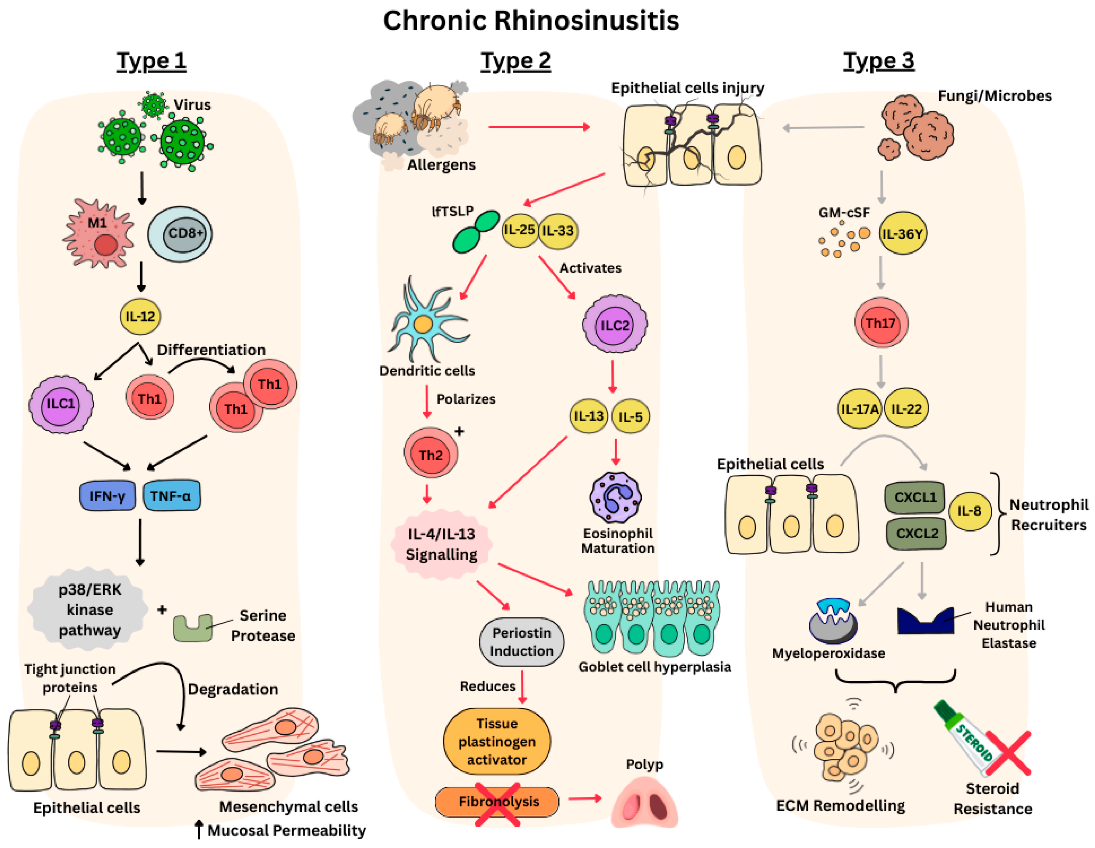
Figure 1. Pathophysiology pathways of different CRS endotypes.
Sinusitis 09 00019 g001
Figure 2. PRISMA 2020 diagram of studies included in the review.
Sinusitis 09 00019 g002
Table 1. Search strategy utilized.
Search ParametersSpecifications
Date of search15 July 2025
Databases searchedPubMed, Cochrane, Web of Science, Embase, Scopus
Search terms utilized“Chronic Rhinosinusitis”, “Endotypes”, “CRS endotyping”, “Asian populations”, “Precision Medicine”, “personalized treatment”, “Biomarkers”, “Biologics” and “Research gaps”
TimeframeJanuary 2000 to July 2025
Inclusion criteriaClinical and experimental studies investigating CRS endotypes, studies exploring current barriers to implementation, studies exploring inflammatory patterns or biomarkers related to CRS in Asian populations
Excusion criteriaNon-Asian studies, case reports, conference abstracts
Table 2. Clinical studies of CRS endotypes.
Author, YearTitle of StudyStudy DesignSample Size (n)Key FindingsStudy StrengthsStudy LimitationsStudy Quality (NOS)
Min et al., 2023 [27]Inflammatory endotypes of chronic rhinosinusitis in the Korean population: Distinct expression of type 3 inflammationProspective cohort study244 CRS patients
  • Asian CRS heterogeneity confirmed: 3 distinct endotypes identified
  • Endotypes are mild-mixed (42.2%), T3 neutrophilic (31.1%), and T2 eosinophilic (26.6%). T3 endotype showed high CRSwNP proportion and severe disease, challenging Western T2-predominant paradigms.
  • First multicenter Korean endotype study
  • Comprehensive 16-biomarker panel
  • Statistical validation
  • Single country limits broader Asian generalizability
  • Limited follow-up for treatment outcomes
High (8/9)
Jiang W et al., 2021 [28]Changes in clinical and histological characteristics of nasal polyps in Northern ChinaRetrospective cohort study300 Chinese CRS patients
  • Temporal endotype shift shows eosinophil-dominant CRSwNP increased from 12% to 43% over 22-year period, with increased asthma comorbidity.
  • Demonstrates “eosinophilic shift” phenomenon in modernizing Asian populations.
  • Unique temporal study
  • Consistent and large population evaluated over time
  • Standardized methodology across different eras
  • Single geographic region analysis
  • Era-specific confounding factors not accounted for
  • Limited biomarker analysis
Medium (6/9)
Pan L et al., 2021 [29]Inflammatory features and predictors for postsurgical outcomes in patients with nasal polyps stratified by local and systemic eosinophiliaProspective cohort study535 Asian CRS patients
  • Stratification by blood/tissue eosinophilia revealed distinct inflammatory patterns. Group 1 (concordant eosinophilia) showed marked T2 inflammation; Group 4 (no eosinophilia) demonstrated neutrophilic inflammation.
  • Blood eosinophilia, asthma, prior surgery were top predictors of uncontrolled disease.
  • Large prospective cohort
  • Long-term follow-up assessment
  • Comprehensive biomarker panel
  • Single-center Chinese study
  • Hospital-based population
  • Limited ethnic diversity within Chinese cohort
High (8/9)
Liao B et al., 2018 [30]Multidimensional endotypes of chronic rhinosinusitis and their association with treatment outcomesProspective cohort study246 Chinese CRS patients
  • 7 clusters identified with distinct treatment outcomes. Cluster 1 (13.01%), classic eosinophilic CRSwNP had highest difficult-to-treat rate.
  • Clusters 3 and 6 (28.45%), neutrophil-dominant with elevated IL-8 also showed poor outcomes, supporting IL-6/IL-1β/G-CSF targeting for neutrophilic CRS.
  • Largest prospective Chinese cohort
  • Multidimensional analysis
  • Assessment of treatment response
  • Discovery of novel biomarkers
  • Non-standardized treatment across patients
  • Limited to Chinese population only
  • Complex clustering is not clinically applicable
High (8/9)
Wang XD et al., 2016 [31]Diversity of TH cytokine profiles in patients with chronic rhinosinusitisCross-sectional study435 Chinese CRS patients; 138 Western controls
  • TH1/TH2/TH17 signatures varied significantly by region. Asian centers (Beijing, Chengdu) showed lower TH2 and eosinophilia than Western sites.
  • <30% of Beijing/Chengdu patients had eosinophilic endotype vs. >50% in Western centers.
  • Unique multicenter global endotype comparison
  • Large sample size accounting for geographic diversity
  • High statistical power for regional comparisons
  • Cross-sectional design limits causal inference
  • Selection criteria not identical across clusters
  • Limited Southeast Asian representation
High (7/9)
Lou H et al., 2016 [32]Cellular phenotyping of chronic rhinosinusitis with nasal polypsRetrospective cohort study366 Chinese CRS patients
  • 5 inflammatory phenotypes identified with distinct recurrence patterns.
  • Eosinophil-dominant group showed 98.5% polyp recurrence rate vs. significantly lower rates in other phenotypes.
  • Long-term follow-up data
  • Clear recurrence definition
  • Single-center Chinese study
  • Phenotyping not standardized across centers
  • Limited biomarker analysis
Medium (6/9)
Ikeda et al., 2013 [33]Subclassification of CRSwNP based on eosinophil and neutrophil infiltrationCross-sectional Study130 Japanese CRS patients
  • Eosinophilic subtype showed highest serum IgE, CT scores, and recurrence risk compared to neutrophilic and pauci-cellular subtypes.
  • Validates endotype–clinical severity relationships in Japanese population.
  • Multiple outcome measures
  • Clinical correlation assessment
  • Small sample size
  • Cross-sectional design
  • Limited follow-up data
Medium (5/9)
Nakayama T et al., 2011 [34]Mucosal eosinophilia and recurrence of nasal polyps—new classification of CRSProspective cohort study175 Japanese CRS patients
  • ≥70 eosinophils/high power field (HPF) threshold tripled postoperative recurrence risk (HR 3.0, p = 0.001).
  • Mucosal eosinophilia more predictive than nasal polyp presence alone for Japanese patients.
  • Kaplan–Meier survival analysis
  • Clear histological threshold definition
  • Moderate sample size
  • Limited biomarker analysis beyond eosinophils
  • Short follow-up period
High (7/9)
Yoshimura K et al., 2011 [35]Clinical epidemiological study of 553 patients with chronic rhinosinusitis in JapanCross-sectional study553 Japanese CRS patients
  • 97.4% of asthmatic CRS patients had ≥15% tissue eosinophils, demonstrating strong eosinophilia–asthma link in Japanese population.
  • 23.1% overall asthma prevalence provides Asian epidemiological baseline.
  • Clear statistical associations
  • Standardized tissue assessment
  • Clinical relevance for comorbidity management
  • Cross-sectional design
  • Hospital-based sampling bias
  • Limited endotype characterization beyond eosinophils
Medium (6/9)
Cao PP et al., 2009 [36]Distinct immunopathologic characteristics of chronic rhinosinusitis in adult ChineseCross-sectional study245 Chinee CRS patients, 50 Chinese controls
  • >50% of Chinese CRSwNP were non-eosinophilic, contrasting with Western patterns.
  • Established T1/T2/T17 cytokine profiles differ between Chinese populations and Western cohorts.
  • Only eosinophilic CRSwNP showed enhanced GATA-3 and IL-5 expression.
  • First major Asian endotype characterization
  • Multiple CRS subtypes included
  • Foundational study that established Asian CRS endotypes
  • Early study with limited biomarker panel
  • Cross-sectional design
  • Limited clinical outcome assessment
Medium (6/9)
Table 3. Experimental studies of CRS endotypes.
Author, YearTitle of StudyMethodologyKey FindingsStudy StrengthsStudy Limitations
Jin et al., 2025 [37]Single cell transcriptomic analysis reveals transcriptome differences of different cells between eosinophilic chronic rhinosinusitis with nasal polyps and non-eosinophilic chronic rhinosinusitis with nasal polyps
  • Single-cell RNA sequencing of 77,697 cells from healthy controls (n = 3), eosinophilic CRSwNP (eCRSwNP) (n = 3), and non-eosinophilic CRSwNP (neCRSwNP) (n = 3).
  • Unbiased clustering identified 14 cell types. Differential gene expression analysis and cellular communication pathway analysis performed.
  • eCRSwNP showed increased glandular cells, reduced fibroblasts vs. neCRSwNP. CD4+ Th2 cells significantly enriched in eCRSwNP.
  • ICAM1-CD226 pathway from cDC2 to CD4+ Th2 upregulated in eCRSwNP.
  • IL2-STAT5 signaling activated while apoptotic pathways inhibited.
  • Most up to date and comprehensive molecular analysis of CRS endotypes
  • Pathway-level mechanistic insights into CRS endotypes
  • Novel biomarker discovery and analysis
  • Limited validation of studies
  • Only transcriptomic analysis, no protein validation carried out
  • Requires larger cohort validation
Jin et al., 2023 [38]Diagnostic Value of Myeloperoxidase and Eosinophil Cationic Protein in Nasal Secretions for Endotypes of Chronic Rhinosinusitis
  • ELISA measurement of MPO and Eosinophil Cationic Protein (ECP) in nasal secretions. Tissue samples were analyzed for neutrophil/eosinophil counts.
  • Subsequently, ROC curve analysis was conducted for diagnostic performance along with correlation analysis between secretion and tissue markers.
  • MPO in secretions achieved highest AUC (0.8961) for neutrophil-dominant CRS diagnosis.
  • ECP achieved AUC (0.8430) for eosinophilic CRS. Strong correlations: tissue neutrophils vs. secretion MPO (r = 0.6613), tissue eosinophils vs. secretion ECP (r = 0.5774).
  • Strong statistical correlation between tissue and secretions
  • High diagnostic accuracy
  • Non-invasive biomarker approach
  • Requires validation in other Asian populations
  • Limited to two biomarkers only
  • Single-center Chinese study
Wu et al., 2021 [39]Artificial Intelligence for Cellular Phenotyping Diagnosis of Nasal Polyps by Whole-Slide Imaging
  • Developed AICEP 2.0 using EfficientNet-B5 neural network. Analyzed 453 total patients: 179 for development, 158 prospective cohort, 116 retrospective cohort.
  • 24,625 patches extracted from whole-slide images. Manual cell counting by pathologists for training.
  • AI analysis identified mean absolute errors of inflammatory cell ratios: eosinophils (1.64%), neutrophils (1.06%), lymphocytes (2.13%), plasma cells (1.22%).
  • Four distinct phenotypes identified with different clinical characteristics.
  • Strong correlation between peripheral and tissue eosinophilia (r = 0.560, p < 0.001).
  • Largest AI-based CRS study (n = 453)
  • Multi-cohort validation
  • High accuracy (<2.13% error)
  • Clinical outcome correlation
  • Requires high-quality histology analysis
  • High computational requirements
  • Limited external validation
Kim et al., 2020 [40]Elastase-Positive Neutrophils Are Associated With Refractoriness of Chronic Rhinosinusitis With Nasal Polyps in an Asian Population
  • Multiplex immunoassay of 17 neutrophil-related mediators in nasal polyps from 160 Korean CRSwNP patients.
  • Immunofluorescence for HNE and MPO.
  • Neutrophil elastase predicts treatment failure: Tissue neutrophilia strongest risk factor for CRSwNP refractoriness (HR 4.38, 95% CI 1.76–10.85, p < 0.05).
  • HNE-positive cells and IL-36α significantly upregulated in refractory group.
  • IL-36R + HNE + double-positive cells increased in treatment-resistant cases. HNE/α1-antitrypsin ratio imbalance associated with neutrophilic pathophysiology.
  • First Asian study linking neutrophil elastase to outcomes
  • Large Korean sample of patients
  • Limited to CRSwNP patients only
  • Short follow-up period
Ryu et al., 2020 [23] Role of IL-17A in Chronic Rhinosinusitis With Nasal Polyp
  • Korean tissue study with CRSwNP patients (n = 47), CRSsNP (n = 20), controls (n = 15).
  • Immunohistochemistry and immunofluorescence for IL-17A. Primary cell culture experiments.
  • Murine nasal polyp model with anti-IL-17A and anti-TNF-α treatment. mTOR pathway studies with rapamycin.
  • CD68+ M1 macrophages identified as major IL-17A source in Korean CRSwNP, followed by neutrophils and T helper cells.
  • Anti-IL-17A and anti-TNF-α treatment reduced nasal polyp formation in murine model.
  • mTOR pathway inhibition (rapamycin) decreased IL-17A expression and polyp burden. IL-17A/mTOR axis represents therapeutic target.
  • Combined human tissue and animal validation
  • Therapeutic intervention proof-of-concept
  • Novel mTOR pathway identification
  • Animal model limitations
  • Single-center study
  • Therapeutic dosing optimization needed
Kim et al., 2019 [41]Chronic Rhinosinusitis without Nasal Polyps in Asian Patients Shows Mixed Inflammatory Patterns and Neutrophil-Related Disease Severity
  • Experimental analysis of uncinate process tissues from Korean patients.
  • -CRSsNP (n = 57), CRSwNP (n = 13), controls (n = 10).
  • Comprehensive cytokine analysis (32 inflammatory mediators) using multiplex immunoassay.
  • Correlation analysis between inflammatory markers and disease severity.
  • Korean CRSsNP showed mixed Th2/Th17 response with prominent neutrophilic inflammation.
  • IL-17A highest in CRSsNP vs. controls and CRSwNP. Strong correlations between neutrophil markers (CXCL2, IL-8, MMP-9/TIMP-1) and disease extent (r = 0.317–0.424, p < 0.05).
  • Eosinophil markers showed no disease correlation.
  • Large inflammatory mediator panel (32 markers)
  • Strong disease correlation data
  • Novel mixed inflammatory findings
  • Small CRSwNP comparison group (n = 13)
  • Single-center study
  • Limited treatment outcome correlation
Wang et al., 2018 [42]The activation and function of IL-36γ in neutrophilic inflammation in chronic rhinosinusitis
  • Tissue samples from CRS patients (n = 145) and controls (n = 40).
  • IL-36 family cytokine expression by qPCR and immunofluorescence.
  • Primary nasal epithelial cell culture experiments. Neutrophil functional assays. IL-36γ cleavage and activation studies with neutrophil elastase.
  • IL-36γ most abundant IL-36 isoform in CRS tissue, mainly produced by epithelial cells.
  • IL-36γ induced CXCL1, CXCL2, CXCL8 production from neutrophils. Positive feedback loop: IL-17A→IL-36γ→neutrophil recruitment→IL-17A production.
  • Neutrophil elastase cleaved IL-36γ into active form with 100-fold increased activity.
  • Novel mechanistic pathway discovery
  • Comprehensive in vitro validation
  • Functional studies with primary cells
  • Therapeutic target identification
  • In vitro studies need in vivo validation
  • Mechanistic focus limits clinical application
  • Long-term pathway effects unknown
Xia et al., 2014 [43]Interleukin-17A promotes MUC5AC expression and goblet cell hyperplasia in nasal polyps via the Act1-mediated pathway
  • Chinese nasal polyp patients (n = 25) and controls (n = 22).
  • Immunohistochemical staining for IL-17A, MUC5AC, and Act1.
  • Primary polyp epithelial cell culture. DNA microarray analysis of IL-17A-induced genes. Western blot and qPCR validation.
  • IL-17A significantly correlated with MUC5AC expression and goblet cell hyperplasia in Chinese polyps (p < 0.05).
  • IL-17A stimulated IL-17RA, IL-17RC, Act1, and MUC5AC expression via MAPK pathway (ERK, p38, JNK).
  • Act1 siRNA blocked IL-17A-induced MUC5AC production. Chinese CRS shows distinct IL-17A-driven pathophysiology.
  • Comprehensive pathway analysis
  • Multiple experimental validation techniques
  • Mechanistic depth with siRNA studies
  • Limited to in vitro studies
  • Clinical translation uncertain
  • Pathway interactions unexplored
Disclaimer/Publisher’s Note: The statements, opinions and data contained in all publications are solely those of the individual author(s) and contributor(s) and not of MDPI and/or the editor(s). MDPI and/or the editor(s) disclaim responsibility for any injury to people or property resulting from any ideas, methods, instructions or products referred to in the content.