Case Report

Cholesterol Granuloma of the Frontal Sinus Complicated by Mycetoma: A Rare Case Report

by
Chiara Rustichelli
1,
Alessandro Serrone
1,
Giovanni Cavallo
1,
Antonino Maniaci
2 and
Gian Luca Fadda
1,*
1
Department of Otorhinolaryngology, University of Turin, “San Luigi Gonzaga” Hospital, Regione Gonzole 10, Orbassano, 10043 Turin, Italy
2
Department of Medical and Surgical Sciences, Faculty of Medicine, University of Enna Kore, 94100 Enna, Italy
*
Author to whom correspondence should be addressed.
Sinusitis 2025, 9(2), 15; https://doi.org/10.3390/sinusitis9020015
Submission received: 24 July 2025 / Revised: 14 August 2025 / Accepted: 21 August 2025 / Published: 25 August 2025

Abstract

Cholesterol granuloma is an uncommon inflammatory lesion whose etiology is still unclear. It is thought to originate from blood accumulation in a pneumatized space, probably after bleeding. The most frequent site is the petrous apex of the temporal bone, whilst it is very uncommon in the paranasal sinuses. We present a clinical case of an 80-year-old female patient with intense frontal headache and diplopia who underwent a neuro-navigated endoscopic sinus surgery. The histological diagnosis was a cholesterol granuloma of the frontal sinus, contaminated by Aspergillus Fumigatus hyphae. Cholesterol granuloma of the frontal sinus is a rare finding in clinical practice, but it should be taken into account in the diagnostic path in presence of rapidly increasing sight alterations and headache, even without rhinological symptoms.

1. Introduction

Cholesterol granuloma is a benign lesion characterized by the progressive accumulation of blood-derived products within a pneumatized structure, leading to gradual expansion [1]. It is hypothesized that the lesion arises from blood extravasation into a pneumatized cavity, potentially facilitated by a bony dehiscence between the bone marrow and the cavity itself, which could account for a non-traumatic source of hemorrhage [1,2]. Subsequent anaerobic degradation of blood components, including cholesterol crystals, triggers a foreign-body granulomatous reaction that progressively enlarges over time.
Cholesterol granulomas are uncommon lesions, most frequently occurring in the petrous apex, with an incidence of approximately 0.6 cases per million for this specific location [1,3]. Involvement of the paranasal sinuses is uncommon and predominantly affects males over 40 years of age, often following trauma-related hemorrhage. Other proposed etiologies include bone malformations and impaired nasosinusal ventilation [4].
We report the case of a female patient who presented to the Emergency Department with severe headache, progressive diplopia, and left-sided exophthalmos. Endoscopic sinus surgery (ESS) revealed a cholesterol granuloma associated with a concurrent mycetoma in the left frontal sinus.

2. Case Report

In April 2023, an 80-year-old woman presented to our Emergency Department with a debilitating frontal headache and left-sided diplopia. Headache had been persistent for the previous six months, with progressive worsening, while ocular symptoms had appeared over the preceding three weeks. The patient had been referred to the Emergency Department by the ophthalmologist she had consulted for diplopia, who had excluded primary ocular disease.
The patient’s medical history was notable for gastroesophageal reflux disease and hypertension. No drug allergies, nasal obstruction, or recent craniofacial trauma were reported.
Endoscopic examination of the nasal cavities revealed no mucosal inflammation or nasal discharge.
Contrast-enhanced computed tomography (CT) demonstrated inflammatory tissue involving the left frontal sinus and multiple ipsilateral ethmoidal air cells. With erosion of both the anterior and posterior frontal sinus walls as well as the lamina papyracea, with proximity to the left orbital cavity. Also, rectus medialis and inferior rectus muscles seemed displaced from their normal anatomical position (Figure 1).
Magnetic resonance imaging (MRI) revealed T2-weighted hyperintensity and mixed hyper-/hypointensity on T1-weighted sequences involving the left ethmoidal–orbital region, with extension to the frontal sinuses. The lesion, resembling two adjacent mucoceles, demonstrated extraconal spread, with displacement of the medial rectus muscle and, to a lesser extent, the superior oblique muscle (Figure 2).
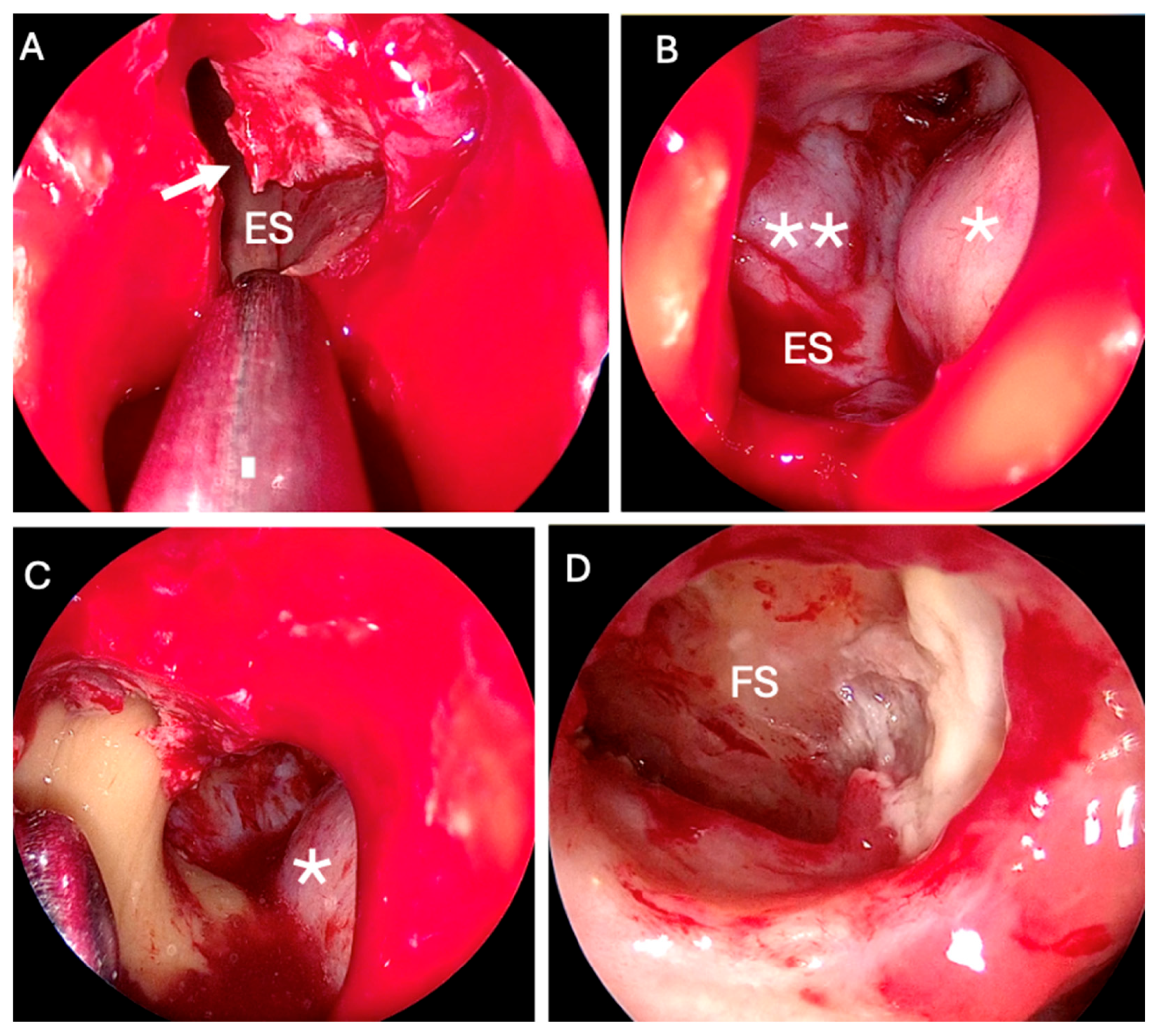
The patient underwent bilateral endoscopic sinus surgery (ESS) and endoscopic frontal sinusotomy via a Draf IIb approach. Neuronavigation was used to facilitate access to the left ethmoid and frontal sinuses, where a double mucocelic sac filled with thick, clay-like material occupying the entire sinus and extending close to orbital lamina papyracea (Figure 3). No intraoperative complications occurred. Histopathological analysis on specimens confirmed the presence of a cholesterol granuloma in the left frontal sinus. In addition, fungal material was identified, excised, and submitted for histological and bacteriological examination. The findings were consistent with a mycetoma (hyphae compatible with Aspergillus fumigatus) without evidence of invasion into the frontal sinus mucosa, thereby ruling out the invasive form.
The patient was discharged two days postoperatively without complications and was prescribed a seven-day course of oral antibiotics (Cefditoren 400 mg). Additionally, the patient performed multiple daily nasal irrigations with saline solution at home. Scheduled outpatient follow-up visits included crust removal under endoscopic guidance. Throughout the follow-up period, there were no signs of cerebrospinal fluid leakage, infection, or epistaxis. A computed tomography scan conducted six months postoperatively demonstrated favorable surgical outcomes, with satisfactory pneumatization of the frontal sinuses and no evidence of disease recurrence (Figure 4).

3. Discussion

Our patient presented with two distinct conditions: cholesterol granuloma and isolated mycetoma of the frontal sinus. Cholesterol granuloma results from a foreign body reaction to the deposition of cholesterol crystals and membrane phospholipids following hemorrhage. Several hypotheses have been proposed to explain the origin of this hemorrhage, including prior trauma, bone malformations, and impaired ventilation in the setting of nasal polyposis [4].
In a review by Durgam et al. [5], 135 cases were reported between 1970 and 2010. The frontal sinus was the most frequently involved site (60%), followed by the maxillary sinus (34.1%) and the ethmoid sinus (3.7%). A more recent study by Wershoven et al. [6], which updated and extended Durgam’s dataset to include the period from 2011 to 2021, reported similar findings, with an additional 18 cases involving the frontal sinus alone.
Reports of cholesterol granuloma in other anatomical locations are also found in the literature (Table 1).
The clinical and radiological presentation of our patient could resemble that of an orbitofrontal cholesterol granuloma, a benign lesion involving the diploe of the frontal bone, typically located in the superolateral quadrant. This condition predominantly affects males over 40 years of age [25] and may present with proptosis, periorbital pain, reduced ocular motility, diplopia, and decreased visual acuity [26]. Radiologically, it demonstrates a characteristic pattern, appearing hyperintense on both T1- and T2-weighted MRI sequences and hypodense on CT [1,26,27]. Histologically, it is composed of aggregates of cholesterol crystals and phospholipids, without epithelial lining [28].
One possible hypothesis in our case is that the lesion initially developed as an orbitofrontal cholesterol granuloma and progressively extended into the frontal sinus cavity, as suggested by the areas of bone erosion evident on CT and similarly reported by Shrirao et al. [29]. Alternatively, obstruction of the frontal sinus ostium by coagulation products—secondary to mucosal bleeding—may have impaired ventilation, leading to the accumulation of cholesterol crystals. This mechanism was also proposed by Ochiai et al. [4], who described a 59-year-old patient with a post-traumatic left frontal sinus cholesterol granuloma exhibiting a radiological appearance similar to that of a mucocele, given that the sinus mucosa may produce mucus-like secretions in response to non-specific inflammatory stimuli. This presentation closely resembles the cases reported by Li et al. [30] and our own, both of which exhibited a mucocele-like appearance on CT imaging.
Beyond mucoceles, the differential diagnosis of orbitofrontal cholesterol granuloma includes cholesteatoma, dermoid and epidermoid cysts, and lacrimal gland tumors [31].
The treatment of cholesterol granuloma is surgical [32,33], with the approach varying according to lesion characteristics, ranging from simple cyst drainage to frontal craniotomy or lateral orbitotomy [4]. Endoscopic sinus surgery (ESS) is preferred in cases with intracranial extension [34,35], as was the case for our patient. The most common complication associated with this procedure is cerebrospinal fluid (CSF) leakage [36], which was successfully avoided in our case.
Similarly, an isolated fungus ball (FB), or mycetoma, of the frontal sinus is an uncommon fungal infection that rarely invades the mucosa, except under predisposing conditions such as immunosuppression [37,38]. It is believed to result from the inhalation of fungal spores—most commonly Aspergillus fumigatus—which subsequently colonize the frontal sinus. Involvement of the frontal sinus is rare (approximately 2% of all FB cases) due to its poor accessibility to inhaled spores [39]. According to the diagnostic criteria proposed by deShazo et al. [40], radiological evaluation is fundamental and highly pathognomonic. Fadda et al. [41] reported that CT scans typically demonstrate near-complete opacification of the affected sinus, with multiple hyperdense foci representing microcalcifications from matted fungal hyphae and metabolic deposits. Bony wall alterations—such as sclerosis or thickening from chronic reactive osteitis, or focal thinning, dehiscence, and sinus expansion—may also be present. On gadolinium-enhanced MRI, FB usually appears iso- or hypointense on T1-weighted images and hypointense on T2-weighted images, reflecting high iron and manganese content as well as calcium in the form of microcalcifications.
In the case presented, radiological findings were more suggestive of a cholesterol granuloma, with no clear evidence of FB. CT scans revealed homogeneous isodense inflammatory tissue without microcalcifications, while MRI demonstrated T2-weighted hyperintensity and mixed hyper-/hypointensity on T1-weighted sequences. These MRI patterns, together with possible bone erosion, may occur in both FB and cholesterol granuloma. Therefore, in addition to imaging, intraoperative identification of the characteristic cheesy, clay-like material and subsequent histopathological confirmation were essential for diagnosis.
Regarding the unusual coexistence of fungal colonization within a cholesterol granuloma, we hypothesize that FB development may have been facilitated by obstruction and impaired ventilation of the sinus caused by the granuloma itself. Furthermore, both cholesterol granuloma and FB may mimic mycetoma [39], underscoring the need for an accurate differential diagnosis, particularly in the presence of bone erosion and sinus involvement. If left untreated, both conditions tend to enlarge, causing bone erosion and subsequent extension to adjacent structures, which may result in complications such as debilitating headache, proptosis, visual impairment, and displacement of the ocular globe, as observed in the present case.

4. Conclusions

Cholesterol granuloma of the frontal sinus is an uncommon lesion that should be considered during the diagnostic workup when a frontal or supraorbital mass associated with ocular symptoms is encountered. It is important to differentiate it from the more common frontal mucocele, as well as from other less frequent lesions. An accurate clinical and radiological assessment is crucial as well as a rapid treatment, which is always surgical with an endoscopic approach if possible. Histological and bacteriological examination of the samples is essential for a definitive diagnosis.
Our findings suggest that in challenging cases, neuronavigation, in addition to adequate instrumentation, can assist the surgeon during ESS and reduce the risk of complications.

Author Contributions

Conceptualization: G.L.F. and C.R.; Methodology: G.L.F. and G.C.; Resources: G.L.F., C.R., and A.S.; Writing—Original Draft Preparation: C.R. and A.S.; Writing—Review and Editing: G.L.F. and A.M.; Supervision: G.L.F. and G.C. All authors have read and agreed to the published version of the manuscript.

Funding

This research received no external funding.

Institutional Review Board Statement

Not applicable.

Informed Consent Statement

Written informed consent has been obtained from the patient to publish this paper.

Data Availability Statement

The original contributions presented in this study are included in the article. Further inquiries can be directed to the corresponding author.

Conflicts of Interest

The authors declare no conflicts of interest.

Abbreviations

The following abbreviations are used in this manuscript:
ESSEndoscopic Sinus Surgery
CTComputed Tomography
MRIMagnetic Resonance Imaging
CSFCerebrospinal Fluid
FBFungus Ball

References

  1. Isaacson, B. Cholesterol Granuloma and Other Petrous Apex Lesions. Otolaryngol. Clin. N. Am. 2015, 48, 361–373. [Google Scholar] [CrossRef]
  2. Jackler, R.K.; Cho, M. A new theory to explain the genesis of petrous apex cholesterol granuloma. Otol. Neurotol. 2003, 24, 96–106. [Google Scholar] [CrossRef]
  3. Li, K.L.; Agarwal, V.; Moskowitz, H.S.; Abuzeid, W.M. Surgical approaches to the petrous apex. World J. Otorhinolaryngol. Head Neck Surg. 2020, 6, 106–114. [Google Scholar] [CrossRef] [PubMed] [PubMed Central]
  4. Ochiai, H.; Yamakawa, Y.; Fukushima, T.; Nakano, S.; Wakisaka, S. Large cholesterol granuloma arising from the frontal sinus-case report. Neurol. Med. Chir. 2001, 41, 283–287. [Google Scholar] [CrossRef] [PubMed]
  5. Durgam, A.; Batra, P.S. Paranasal sinus cholesterol granuloma: Systematic review of diagnostic and management aspects. Int. Forum Allergy Rhinol. 2013, 3, 242–247. [Google Scholar] [CrossRef] [PubMed]
  6. Wershoven, N.; Miller, C.; Gibson, P.; Ciolino, A.; Rimash, T. Bilateral cholesterol granulomas of the maxillary sinus with review of the literature. SAGE Open Med. Case Rep. 2022, 10, 2050313X221116711. [Google Scholar] [CrossRef] [PubMed] [PubMed Central]
  7. Lin, J.I.; Tseng, C.H.; Marsidi, P.J.; Bais, V.C. Cholesterol granuloma of right testis. Urology 1979, 14, 522–523. [Google Scholar] [CrossRef] [PubMed]
  8. Grignon, D.J.; Kirk, M.E.; Haines, D.S. Cholesterol granulomas in lymph nodes draining a benign ovarian neoplasm. Arch. Pathol. Lab Med. 1985, 109, 1124–1126. [Google Scholar] [PubMed]
  9. Amer, A.F.; Walia, H.S.; Madda, J.P. Cholesterol granuloma of the peritoneum. Can. J. Surg. 1990, 33, 410–413. [Google Scholar] [PubMed]
  10. Mafee, M.F.; Kumar, A.; Heffner, D.K. Epidermoid cyst (cholesteatoma) and cholesterol granuloma of the temporal bone and epidermoid cysts affecting the brain. Neuroimaging Clin. N. Am. 1994, 4, 561–578. [Google Scholar] [CrossRef] [PubMed]
  11. Nativ, O.; Mor, Y.; Nass, D.; Leibovitch, I.; Goldwasser, B. Cholesterol granuloma of the tunica vaginalis mimicking a neoplasm. Isr. J. Med. Sci. 1995, 31, 235–236. [Google Scholar] [PubMed]
  12. Fariña Pérez, L.A.; Menéndez, P.; Macho, V. Hydrocele and cholesterol granuloma of the tunica vaginalis simulating a tumor in echography. Actas Urol. Esp. 1998, 22, 70–73. (In Spanish) [Google Scholar] [PubMed]
  13. Schultz, R.; Mattila, J.; Gappa, M.; Verronen, P. Development of progressive pulmonary interstitial and intra-alveolar cholesterol granulomas (PICG) associated with therapy-resistant chronic systemic juvenile arthritis (CJA). Pediatr. Pulmonol. 2001, 32, 397–402. [Google Scholar] [CrossRef] [PubMed]
  14. Mendonça, R.; Peron, C.S.; Stefani, M.A.; Gallo, P. Cerebral cholesterol granuloma: Case report. Arq. Neuropsiquiatr. 2007, 65, 540–541. [Google Scholar] [CrossRef] [PubMed]
  15. Paluzzi, A.; Gardner, P.; Fernandez-Miranda, J.C.; Pinheiro-Neto, C.D.; Scopel, T.F.; Koutourousiou, M.; Snyderman, C.H. Endoscopic endonasal approach to cholesterol granulomas of the petrous apex: A series of 17 patients: Clinical article. J. Neurosurg. 2012, 116, 792–798. [Google Scholar] [CrossRef] [PubMed]
  16. Krishnan, T.R.; Sinha, S.K.; Kejriwal, N.K. A rare case of cholesterol granuloma in the anterior mediastinum. Heart Lung Circ. 2013, 22, 303–304. [Google Scholar] [CrossRef] [PubMed]
  17. Young Sa, Y.J.; Hwang, S.J.; Sim, S.B.; Lee, S.H.; Moon, S.W.; Park, C.B. Cholesterol granuloma: A rare benign rib tumor. Ann. Thorac. Surg. 2013, 95, 1801–1803. [Google Scholar] [CrossRef]
  18. Unal, D.; Kilic, M.; Erkinuresin, T.; Demirbas, M.; Coban, S.; Aydos, M.M. Cholesterol granuloma of the paratesticular tissue: A case report. Can. Urol. Assoc. J. 2015, 9, 390–392. [Google Scholar] [CrossRef] [PubMed] [PubMed Central]
  19. Raghavan, D.; Lee, T.C.; Curtin, H.D. Cholesterol Granuloma of the Petrous Apex: A 5-Year Review of Radiology Reports with Follow-Up of Progression and Treatment. J. Neurol. Surg. B Skull Base 2015, 76, 266–271. [Google Scholar] [CrossRef] [PubMed] [PubMed Central]
  20. Zhang, M.; Tie, H.T.; Wang, C.L.; Wu, Q.C. Pulmonary interstitial cholesterol crystals associated with diffuse lung cysts in adult: A case report and literature review. J. Cardiothorac. Surg. 2016, 19, 11. [Google Scholar] [CrossRef] [PubMed] [PubMed Central]
  21. Nam, G.; Singer, T.M.; Lourenco, A.P.; Wang, Y. Cholesteroloma of the breast: A 10 year retrospective review of 79 cases with radiology correlation. Breast J. 2019, 25, 1177–1181. [Google Scholar] [CrossRef] [PubMed]
  22. Eba, S.; Tanaka, R.; Watanabe, T.; Watanabe, Y.; Notsuda, H.; Suzuki, T.; Hirama, T.; Oishi, H.; Niikawa, H.; Noda, M.; et al. Anterior Mediastinal Cholesterin Granuloma Associated with Bilateral Pleurisy and Pericarditis. Kyobu Geka 2021, 74, 1059–1062. (In Japanese) [Google Scholar] [PubMed]
  23. Hassen, S.S.; Ali, G.A.; Ahmed, A.O.; Goravey, W. Cholesterol Granuloma: An Underrecognized Cause of Benign Lung Granuloma. Eur. J. Case Rep. Intern. Med. 2022, 9, 003264. [Google Scholar] [CrossRef] [PubMed] [PubMed Central]
  24. Vinciguerra, A.; Turri-Zanoni, M.; Verillaud, B.; Guichard, J.P.; Spirito, L.; Karligkiotis, A.; Castelnuovo, P.; Herman, P. Typical and Atypical Symptoms of Petrous Apex Cholesterol Granuloma: Association with Radiological Findings. J. Clin. Med. 2022, 11, 4297. [Google Scholar] [CrossRef] [PubMed] [PubMed Central]
  25. Yan, J.; Cai, Y.; Liu, R.; Lin, J.; Li, J. Cholesterol Granuloma of the Orbit. J. Craniofacial Surg. 2015, 26, 124–126. [Google Scholar] [CrossRef]
  26. Sia, D.I.T.; Davis, G.; Selva, D. Recurrent orbitofrontal cholesterol granuloma: A case report. Orbit 2012, 31, 184–186. [Google Scholar] [CrossRef]
  27. Arat, Y.O.; Chaudhry, I.A.; Boniuk, M. Orbitofrontal cholesterol granuloma: Distinct diagnostic features and management. Ophthalmic Plast Reconstr. Surg. 2003, 19, 382–387. [Google Scholar] [CrossRef] [PubMed]
  28. Khalatbari, M.R.; Moharamzad, Y. Recurrent orbitofrontal cholesterol granuloma in pediatric patient: Case report and review of the literature. Childs Nerv. Syst. 2012, 28, 291–296. [Google Scholar] [CrossRef]
  29. Shrirao, N.; Mukherjee, B.; Krishnakumar, S.; Biswas, J. Cholesterol granuloma: A case series & review of literature. Graefes Arch. Clin. Exp. Ophthalmol. 2016, 254, 185–188. [Google Scholar] [CrossRef] [PubMed]
  30. Li, R.; Ren, M.; Wang, W.; Li, R.; Zhang, L.; Liu, L. Orbitofrontal cholesterol granuloma masquerading as frontal sinus mucoceles: Report of two cases. BMC Ophthalmol. 2023, 23, 98. [Google Scholar] [CrossRef] [PubMed] [PubMed Central]
  31. Ugga, L.; Stilo, S.; Napolitano, P.; Guadagno, E.; Iuliano, A.; Del Basso de Caro, M.L.; Elefante, A. Orbitofrontal cholesterol granuloma: Case report and review of the literature. Quant. Imaging Med. Surg. 2017, 7, 373–377. [Google Scholar] [CrossRef] [PubMed] [PubMed Central]
  32. Deep, N.L.; Chaaban, M.R.; Chaudhry, A.L.; Woodworth, B.A. Frontal sinus cholesterol granuloma: Case report. Allergy Rhinol. 2014, 5, 36–38. [Google Scholar] [CrossRef] [PubMed] [PubMed Central]
  33. Yokoi, H.; Yamanaka, H.; Matsumoto, Y.; Kawada, M.; Fujiwara, M.; Ohara, A.; Saito, K. Modified Lothrop (Draf III) procedure for the treatment of a recurrent orbitofrontal cholesterol granuloma: A case report. SAGE Open Med. Case Rep. 2020, 8, 2050313X20907809. [Google Scholar] [CrossRef] [PubMed] [PubMed Central]
  34. Selva, D.; Lai, T.; Krishnan, S. Orbitofrontal cholesterol granuloma: Percutaneous endoscopic-assisted curettage. J. Laryngol. Otol. 2003, 117, 892–894. [Google Scholar] [CrossRef]
  35. Selva, D.; Phipps, S.E.; O’Connell, J.X.; White, V.A.; Rootman, J. Pathogenesis of orbital cholesterol granuloma. Clin. Exp. Ophthalmol. 2003, 31, 78–82. [Google Scholar] [CrossRef]
  36. Ong, L.Y.; McNab, A.A. Recurrent orbital cholesterol granuloma. Orbit 2008, 27, 119–121. [Google Scholar] [CrossRef]
  37. Kim, Y.K.; Kim, H.J.; Kim, H.Y.; Cha, J.; Lee, J.Y.; Chung, S.K.; Dhong, H.J.; Song, M.; Kim, S.T. Extrasinonasal infiltrative process associated with a sinonasal fungus ball: Does it mean invasive fungal sinusitis? Diagn. Interv. Radiol. 2016, 22, 347–353. [Google Scholar] [CrossRef]
  38. Fadda, G.L.; Allevi, F.; Rosso, C.; Martino, F.; Pipolo, C.; Cavallo, G.; Felisati, G.; Saibene, A.M. Treatment of Paranasal Sinus Fungus Ball: A Systematic Review and Meta-Analysis. Ann. Otol. Rhinol. Laryngol. 2021, 130, 1302–1310. [Google Scholar] [CrossRef] [PubMed]
  39. Popko, M.; Broglie, M.A.; Holzmann, D. Isolated fungus ball mimicking mucocele or frontal sinus tumour: A diagnostic pitfall. J. Laryngol. Otol. 2010, 124, 1111–1115. [Google Scholar] [CrossRef] [PubMed]
  40. DeShazo, R.D.; O’Brien, M.; Chapin, K.; Soto-Aguilar, M.; Swain, R.; Lyons, M.; Bryars, W.C., Jr.; Alsip, S. Criteria for the diagnosis of sinus mycetoma. J. Allergy Clin. Immunol. 1997, 99, 475–485. [Google Scholar] [CrossRef] [PubMed]
  41. Fadda, G.L.; Succo, G.; Moretto, P.; Veltri, A.; Castelnuovo, P.; Bignami, M.; Cavallo, G. Endoscopic Endonasal Surgery for Sinus Fungus Balls: Clinical, Radiological, Histopathological, and Microbiological Analysis of 40 Cases and Review of the Literature. Iran. J. Otorhinolaryngol. 2019, 31, 35–44. [Google Scholar] [PubMed] [PubMed Central]
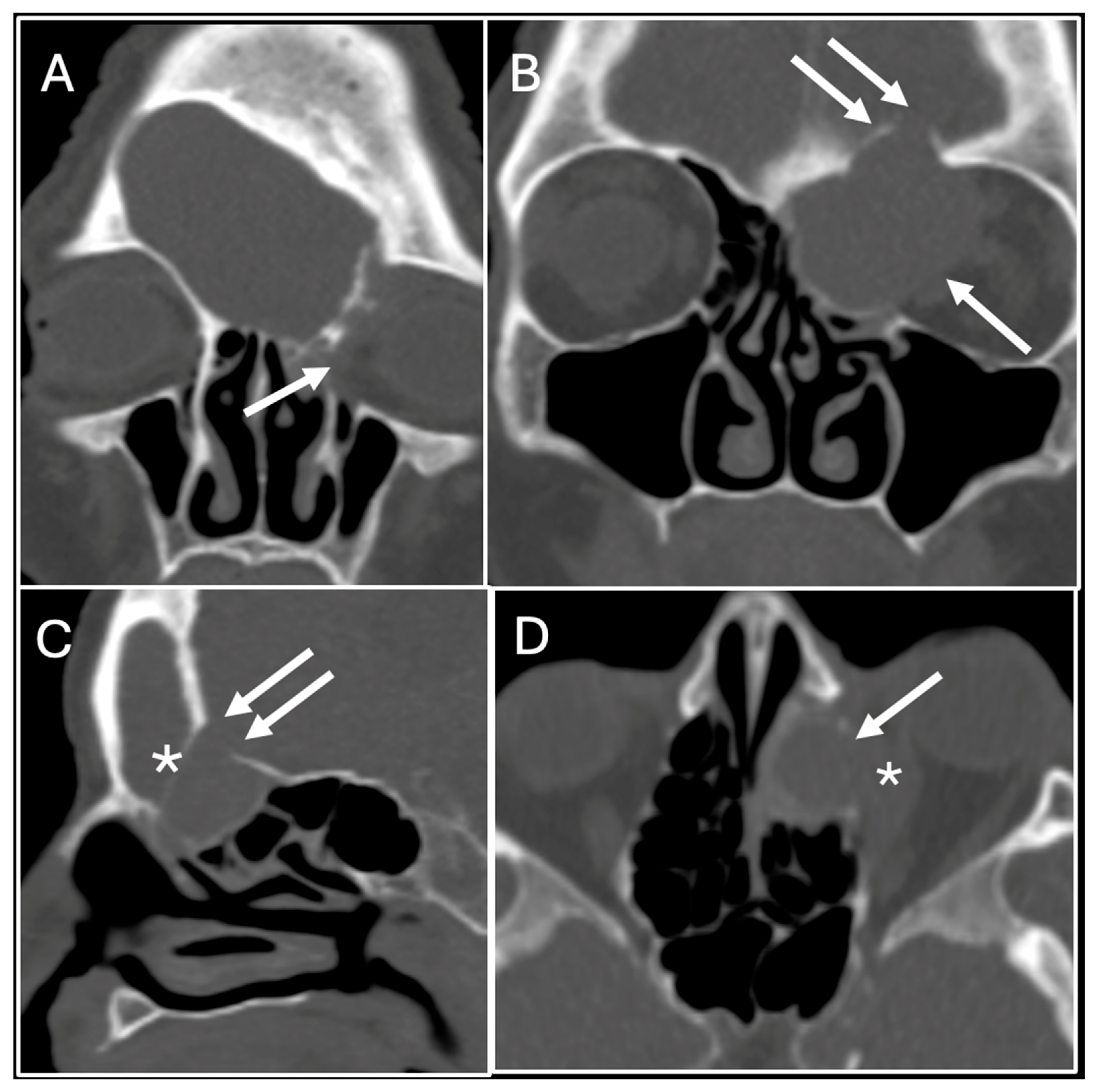
Figure 1. Pre-operative CT images. Coronal (A,B) images revealed inflammatory involvement of the left frontal sinus with compression and dislocation of the papyracea (white arrow) and erosion of the posterior wall of the frontal sinus (double white arrow). (C) Sagittal CT scan shows erosion of the posterior wall of the frontal sinus (double white arrow) and the wall of the lesion’s sac (asterisk): (D) axial CT scan show erosion of the left papyracea (white arrow) and dislocation of the rectus medialis muscle (asterisk).
Sinusitis 09 00015 g001
Figure 2. T2-weighted coronal (A) and T1-weighted axial (B) MRI images without contrast shows lesion suggesting for two adjacent mucoceles (asterisk) involving the left ethmoid–orbital lesion with extension to the frontal sinuses, extraconal extension, and displacement of the rectus medialis muscle (white arrow) (R = right; L = left).
Sinusitis 09 00015 g002
Figure 3. Intraoperative image during endoscopic sinus surgery. (A) Marsupialization of the first mucocelic pouch and its anterior wall (white arrow). (B) Anterior wall of the frontal mucocele (double asterisk). Medial wall of the left orbit (asterisk). (C) Marsupialization of the second mucocelic pouch. (D) Fungal material on the left frontal sinus. (ES = ethmoid sinus; FS = frontal sinus).
Sinusitis 09 00015 g003
Figure 4. Intraoperative view (A) during endoscopic sinus surgery shows left Draf IIB frontal sinusotomy. (B) Postoperative CT scan on (B) coronal and sagittal (C) planes show good surgical outcomes with satisfactory pneumatization of the frontal sinuses (asterisk) and no signs of recurrence (FS = frontal sinus).
Sinusitis 09 00015 g004
Table 1. Cases of cholesterol granuloma reported in other anatomical sites. Both case reports and cases series studies are reported in this table.
AuthorsSiteNo. Cases
Lin J. I. et al. [7] (1979)Testis1
Grignon D. J. et al. [8] (1985)Lymph nodes1
Amer A. F. et al. [9] (1990)Peritoneum1
Mafee M. F. et al. [10] (1994)Brain7
Nativ O. et al. [11] (1995)Testis1
Fariña Pérez L. A. et al. [12] (1998)Testis1
Schultz R. et al. [13] (2001)Lung1
Mendonça R. et al. [14] (2007)Brain1
Paluzzi A. et al. [15] (2012)Petrous apex17
Krishnan T. R. et al. [16] (2013)Mediastinum1
Young Sa Y. J. et al. [17] (2013)Bone (rib)1
Unal D. et al. [18] (2015)Testis1
Raghavan D. et al. [19] (2015)Petrous apex18
Zhang M. et al. [20] (2016)Lung1
Nam G. et al. [21] (2019)Breast79
Eba S. et al. [22] (2021)Mediastinum1
Hassen S. S. et al. [23] (2022)Lung1
Vinciguerra A. et al. [24] (2022)Petrous apex29
Disclaimer/Publisher’s Note: The statements, opinions and data contained in all publications are solely those of the individual author(s) and contributor(s) and not of MDPI and/or the editor(s). MDPI and/or the editor(s) disclaim responsibility for any injury to people or property resulting from any ideas, methods, instructions or products referred to in the content.