Case Report

Unmasking the Fungal Menace: A Case Report of Chronic Granulomatous Invasive Fungal Sinusitis of Maxilla

1
Department of Otolaryngology and Head and Neck Surgery, Karuna Medical College, Palakkad 678103, Kerala, India
2
Department of Pathology, Karuna Medical College, Palakkad 678103, Kerala, India
*
Author to whom correspondence should be addressed.
Sinusitis 2025, 9(1), 4; https://doi.org/10.3390/sinusitis9010004
Submission received: 14 December 2024 / Revised: 7 February 2025 / Accepted: 11 February 2025 / Published: 12 February 2025

Abstract

:
Chronic granulomatous invasive fungal sinusitis (CGIFS) is an uncommon type of invasive sinusitis that is characteristically seen in immunocompetent individuals. Common clinical manifestations of this condition include proptosis, cheek swelling, and headache. The pathogenic organism is Aspergillus in the majority of reported cases. Diagnosis is made by histopathological and microbiological examination of tissue specimens. Due to its expansible nature, bone erosion is also associated with this condition. Treatment is surgical clearance/debulking followed by long-term antifungal therapy. Here, we report the case of a 31-year-old male presented with right-side cheek swelling. The patient underwent a medial maxillectomy and was diagnosed with CGIFS. The patient was put on oral voriconazole for 3 months. To conclude, CGIFS is a rare variant of invasive fungal sinusitis that may mimic malignancy or granulomatous diseases such as tuberculosis, rhinoscleroma, and syphilis. Accurate diagnosis is of utmost importance in providing management for CGIFS.

1. Introduction

Fungal sinusitis involving the sinonasal cavities can be classified broadly into invasive and non-invasive types. To diagnose fungal sinusitis as invasive, it is important to establish the presence of fungal hyphae in deeper mucosa or submucosa or involving neurovascular bundle or cortical bone or in systemic circulation [1]. The invasive type is again divided into three types, namely, (i) acute invasive fungal sinusitis, (ii) chronic invasive fungal sinusitis, and (iii) chronic granulomatous variants. Unlike the acute fulminant and chronic invasive types, which are seen in immunosuppressed patients, chronic granulomatous invasive fungal sinusitis (CGIFS) is seen in immunocompetent individuals [2].
CGIFS is characterized by a granulomatous reaction, which can lead to significant proptosis and craniofacial deformity if not detected earlier. CGIFS presents with non-specific symptoms such as headache, swelling in the maxillary area/infra orbital area, ocular symptoms such as pain around the eye, proptosis, or swelling in the extra conal region of the eyeball. Because of these non-specific varied clinical presentations, delay in diagnosis is common [3]. The disease can sometimes progress to focally expansive lesions resulting in pressure symptoms. The diagnosis is made with the help of histopathological and microbiological examination [4]. Preoperatively imaging may show the characteristic features of fungal sinusitis, such as hypo dense areas within a hyper dense sinus filling lesion, called a double density sign, or bone erosions, which may lead to the involvement of pre-maxillary soft tissues or extension of sinus pathology into the pterygopalatine fossa. The differential diagnosis includes malignancy and other soft tissue tumors of the maxilla [5]. Management of CGIFS is still a topic of debate as no proper guidelines are available. Surgical debridement and systemic antifungals are the core stone modalities in the management of CGIFS [6]. Here, we report a case of a patient who presented with facial swelling.

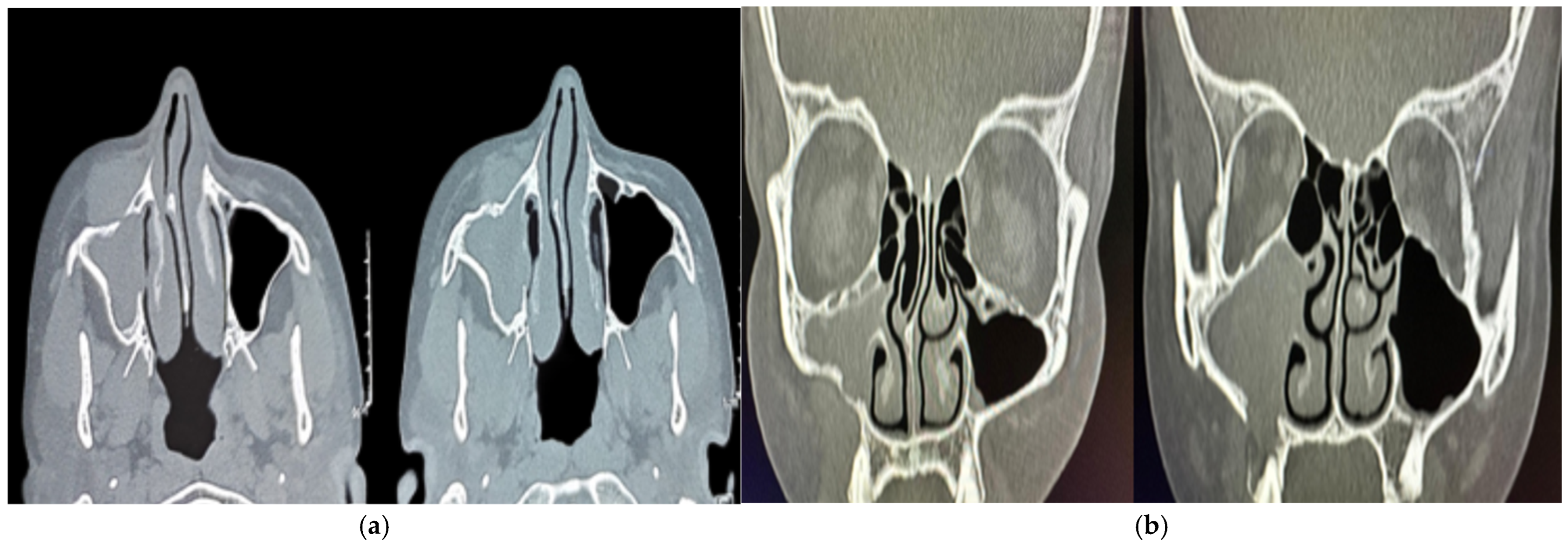
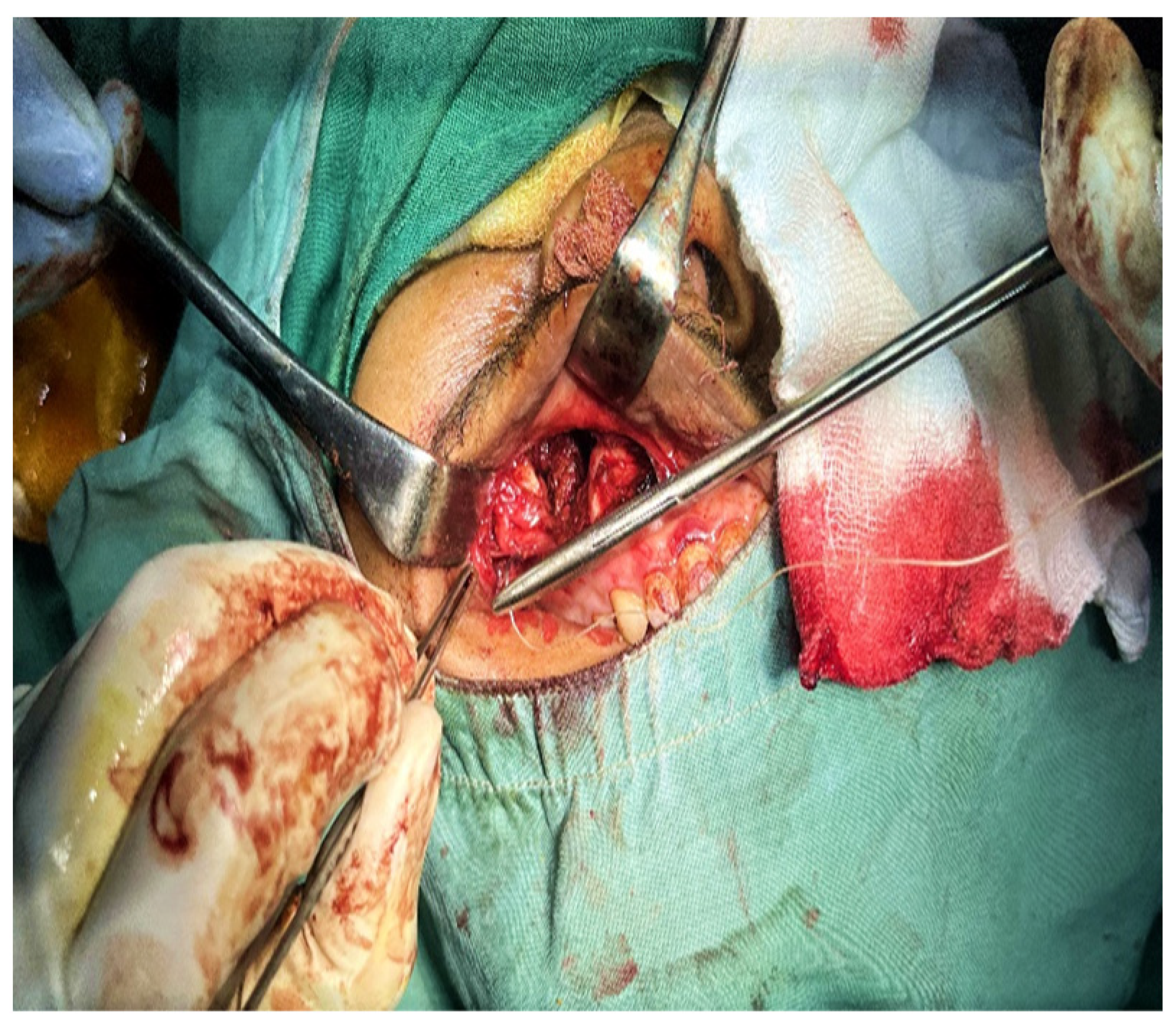
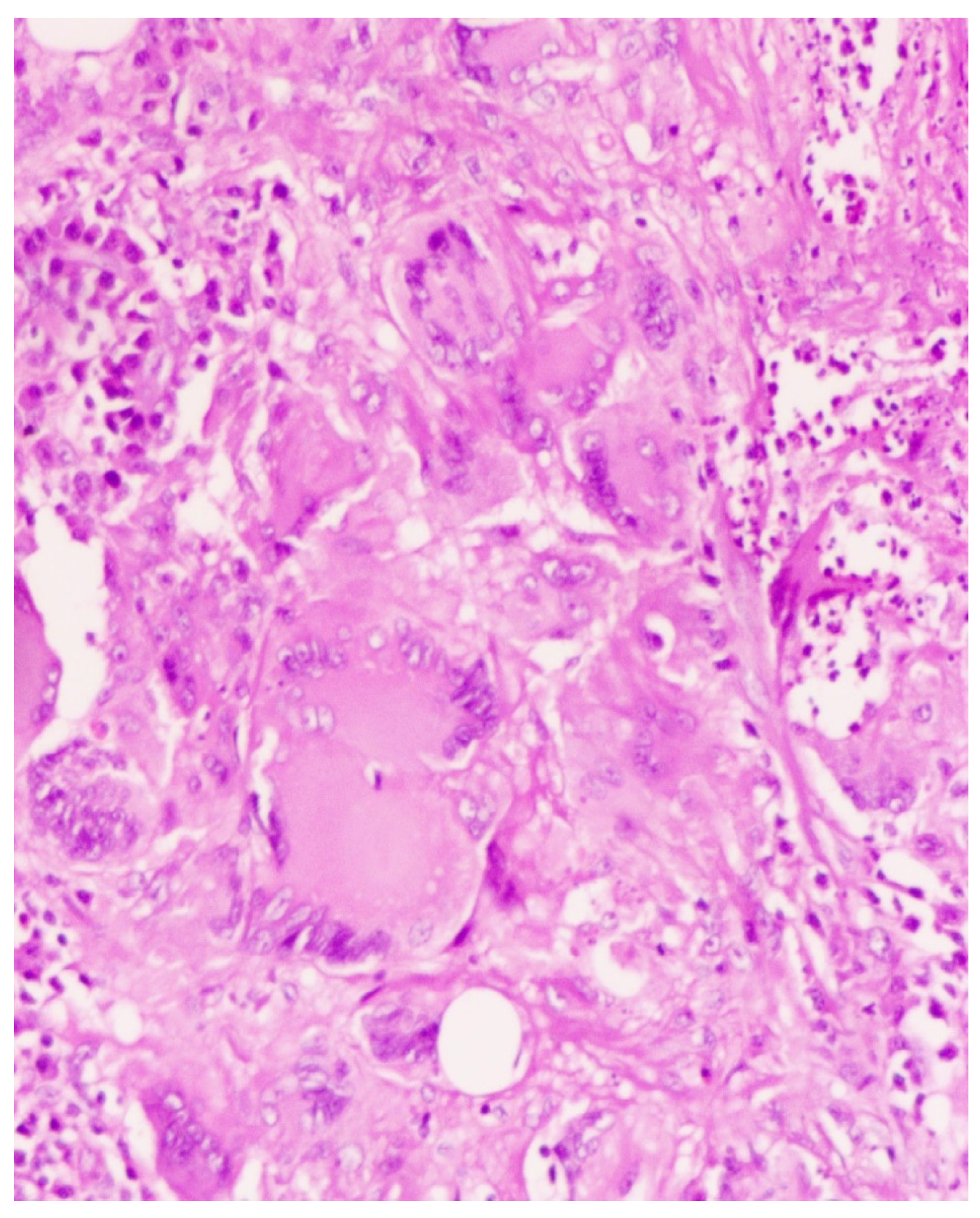
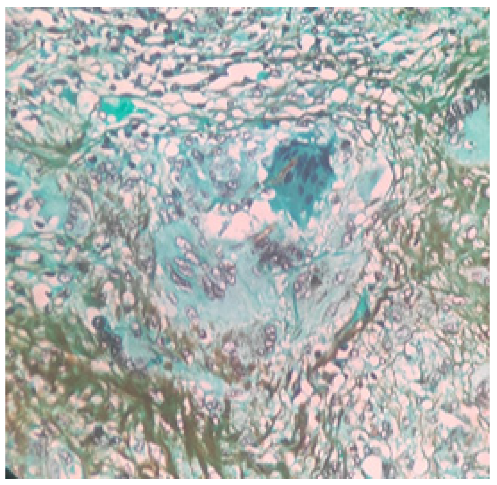
2. Case Presentation

A 31-year-old male from Meenakshipuram village of the southern Indian state Kerala, with no known comorbidities, a carpenter who follows a strict vegetarian diet, with no history of immunodeficiency disorders and no family history of immunodeficiency diseases presented to the otorhinolaryngology outpatient department with complaints of right-side cheek swelling of 18 months duration. He noticed a gradual increase in the size of the swelling over the past 6 months. Initially, he consulted a nearby hospital and was treated for chronic rhinosinusitis. As the facial swelling gradually increased in spite of treatment, he visited our hospital. On thorough history taking, he informed that the swelling was not associated with any pain, loosening of teeth, weight loss, bleeding through the nose, nasal obstruction, visual disturbances, swelling in the neck or ear symptoms.
On clinical evaluation, the swelling was soft to firm consistency of approximately 3 × 3 cm, non-tender, lying approximately 1 cm inferior to the infraorbital margin, with ill-defined borders and without any signs of inflammation. The skin over the swelling was pinchable, no intra oral extension was seen, and there were normal sensations over the trigeminal nerve region bilaterally. On ophthalmology evaluation, visual acuity was normal, no spontaneous or evoked nystagmus was noted, and there was no restriction of extra ocular movements bilaterally.
A diagnostic nasal endoscopy was performed, and it showed a deviation of the anterior nasal septum to the right with a bony spur not touching the lateral wall. Non contrast computerized tomography of the nose and paranasal sinuses revealed partial opacification of the right maxillary sinus with partial erosion of the anterior maxillary wall. The lesion was found to be extending through a defect into the soft tissue over the premaxilla area without involving the subcutaneous fat planes. Post-intravenous contrast administration, the lesion showed mild enhancement with bony scalloping and erosion of the right maxillary sinus and part of the alveolar process of the maxilla (Figure 1a,b). As there was no involvement of orbit or intracranial extension magnetic resonance imaging was not advised.
The patient was counseled regarding the possible nature of the condition and also regarding the need to undergo a surgical procedure to reach a proper diagnosis. The case was presented in the institutional tumor board meeting, and a consensus was reached regarding the treatment plan. An open medial maxillectomy was performed with complete removal of the tumor (Figure 2). The histopathological examination showed numerous multinucleated giant cells in the background of chronic inflammation with signs of non-caseating granulomatous changes (Figure 3). On Grocott’s Methenamine Silver staining (GMS staining), dark brown colored septate fungal hyphae were seen (Figure 4). On a fungal culture with malt extract agar as culture media, the presence of Aspergillus flavus was confirmed. Cartridge-based nucleic acid amplification tests for mycobacteria species showed a negative result. With the histopathological and microbiological evidence, a diagnosis of CGIFS was made, and the patient was put on voriconazole 200 mg twice daily for 12 weeks under liver function monitoring fortnightly. The patient showed no signs of recurrence till three months of follow up and the patient lost to follow up thereafter.

3. Discussion

Fungal sinusitis can be classified into invasive and non-invasive, with the former being again sub classified as acute fulminant, chronic, and chronic granulomatous [1,7,8]. When the duration of the disease is less than 4 weeks, it is termed acute, and for a duration of more than 4 weeks, it is defined as the chronic form [9]. The invasiveness of the disease is strictly defined in terms of the histopathological presence of fungal hyphae in the sinonasal mucosa, not limited to surface mucosa, submucosa, or tissues beyond submucosa, which includes neurovascular structures, bone, muscles, and orbit [10].
Chronic granulomatous invasive fungal rhinosinusitis, also known as indolent fungal sinusitis and primary nasal granuloma, is distinct from the other two types of invasive types. CGIFS is usually seen in immunocompetent individuals with a peculiar geographic distribution. The condition is more commonly reported in dry, humid tropical countries. The majority of reported cases are from Sudan, Western Asia, Pakistan, and India. This can be attributed to the wide presence of Aspergillus fungal species [11].
The pathogenesis of CGIFS is different from chronic invasive fungal sinusitis (CIFS). The underlying immune mechanism associated with invasive fungal sinusitis is not well understood. Th-17 T cells, which are a part of innate immune mechanisms against fungal pathogens, are found to be more in CGIFS individuals. This increased Th 17 cell population may lead to an unchecked Th 1 response, which further results in the chemo attraction of cells involved in chronic inflammation. This cascade ends with a cycle of chronic tissue inflammation with failure to effectively remove the fungal pathogen. Thus, in tissue specimens, CIFS shows a similar picture of chronic inflammation with an abscess-like reaction in the background of low-grade mixed inflammatory cells. This nonspecific histopathology picture often fails the pathologist and the surgeon to alert the possibility of fungal etiology, leading to a delay in diagnosis and subsequent proper management [12]. The most common differential diagnoses at this point are malignancy, an extensive Para sellar tumor, and common granulomatous conditions, which include tuberculosis, syphilis, rhinoscleroma, and granulomatosis with polyangiitis (previously known as Wegner’s granulomatosis). A strong suspicion of fungal pathology should be raised in the clinical history of immunocompromised status, with histological examination showing chronic inflammation changes with non-caseating granulomas [13].
The majority of the documented cases of CGIFS belong to the younger age group. In a case series of 10 cases reported by Asoegwu et al. in Nigeria [14], the average age was 33.9 years. A similar age group was also seen in a series of seven cases from Saudi Arabia [15]. Other reported cases in the last 15 years are a 40-year-old male in Nigeria and a 64-year-old Type II Diabetes mellitus patient with SARS-CoV-2 infection [16]. The most common presenting symptom is proptosis, followed by nasal mass, cheek swelling, and CRS not responding to conventional treatments. The disease is locally aggressive with the involvement of orbit, intracranial extension, or pterygopalatine fossa [14,15,16]. In our case, a 31-year-old male presented with a long-standing cheek swelling, not responding to CRS management.
Radiological imaging gives hints that point towards the possibility of fungal etiology. Computed tomography is essential in identifying bone changes, whereas MRI may give much more detailed information about soft tissue extensions. This is particularly important in cases where orbit, sellar, or intracranial extension is suspected. CT findings supporting the CFRS diagnosis are disease limited to a few sinuses, bone erosion rather than necrosis or sequestrum formation, and heterogeneous sinus mucosal thickening [17]. Because of this bone-erosion-causing property, disease extension to adjacent regions is not uncommon. A total of 7 out of 10 patients of Asoegwu et al. [14] had an orbital extension, with 5 patients having intracranial and one patient with pterygopalatine fossa extension. A similar aggressive nature of the disease was noted by Alarifi et al. [15], where five out of seven patients had orbital involvement, and two patients had intracranial extension. The SARS-CoV-2 co-existed case reported by Gonzalez et al. [16] also had an extension into orbit as an intraconal abscess. However, our patient had a localized involvement with minimal erosion of the anterior maxillary wall and pre-antral tissue infiltration without orbital involvement. This may be due to previous repeated treatments taken by the patient from outside, where the underlying inflammation process is of less intensity. The bone erosion could be attributed to long-standing pressure changes as no lytic or periostitis changes were seen.
The treatment primarily focuses on removing or debulking the source of inflammation/infection [18]. The open approach has an added advantage in extensive disease. Whenever possible, endoscopic resection or debulking is preferred, considering post-operative morbidity associated with open approaches. In the present case report, during the pre-operative counselling, we explained the need to shift to an open approach in case of a failed endoscopic approach, and the patient requested to perform an open medial maxillectomy without attempting an endoscopic approach. Intraoperatively, the anterior wall of the maxilla was eroded with the lesion involving the pre-antral soft tissue. The orbital floor and posterior wall of the maxilla were found intact. The endoscopic approach was the preferred treatment choice wherever possible in the studies by Asoegwu et al. [14] and Alarifi et al. [15].
Once the histopathological diagnosis is suspicious of fungal granulomatosis, it is essential to provide evidence of fungal elements. A preliminary diagnosis of fungus etiology can be obtained either by KOH mount or calcofluor staining [19]. These tests are not useful in cases where fungal elements are sparse. In such a scenario, either a GMS stain or fungal culture helps in establishing the diagnosis of invasive mycosis. The GMS staining or culture has the added advantage of differentiating viable and non-viable fungi elements [20]. This is of treatment importance as the allergic fungal noninvasive variant is primarily because of non-viable fungi elements evoking an allergic response, and treatment is corticosteroids in AFRS. At the same time, the use of steroid formulations can aggravate the disease in invasive fungal diseases.
The newer diagnostic tools for the detection of fungal pathogens include Matrix-Assisted Laser Desorption/Ionization Time-of-Flight (MALDI-TOF) mass spectrometry, Next generation sequencing (NGS), PCR based molecular assays, Loop-Mediated Isothermal amplification (LAMP), β-D-Glucan and Galactomannan Assays, Fluorescence in situ hybridization techniques (FISH) [21]. MALDI-TOF is a rapid and accurate tool for diagnosing Aspergillus infections. It enables the identification of Aspergillus species by analyzing unique protein spectra from fungal isolates, reducing the time required for conventional culture-based methods. MALDI-TOF also shows potential for direct detection from clinical samples, improving early diagnosis and guiding appropriate antifungal treatment [22]. NGS, PCR, and LAMP are molecular diagnostic methods for the diagnosis of fungal pathology, which can be performed in clinical specimens [23]. NGS is highly specific in cases of mixed fungal species infections [24]. LAMP is particularly useful for early diagnosis of invasive aspergillosis in immunocompromised patients, where timely antifungal treatment is critical. The advantage of LAMP over PCR is that the former is a rapid test (results are available in 30–60 min), and has no need for thermal cycling, whereas PCR takes 2–3 h and requires a constant working temperature [25]. Techniques like real-time PCR and multiplex PCR enable the direct detection of Aspergillus DNA from clinical samples, enhancing early diagnosis [26].
The most common organism causing CGIFS is Aspergillus, especially Aspergillus flavus, and other organisms include Aspergillus nidulans, Aspergillus Niger, and Candida species [14,15,16,27]. Once tissue diagnosis of invasive fungal sinusitis is reached, systemic anti-fungal therapy is recommended. The duration of therapy has not been clearly suggested or defined anywhere, but literature evidence shows that antifungal therapy of at least three months is required to prevent progression and recurrence [28]. The choice of antifungal agent to be used depends on patient factors and the organism isolated. The various agents used in invasive types of fungal diseases are intravenous amphotericin B, Liposomal amphotericin B, and the azole group comprising itraconazole, voriconazole, and posaconazole. Other newer agents, such as Micafungin and anidulfungin, also have shown response to aspergillosis. According to Patterson et al. [29] and Walsh et al. [30], whenever the diagnosis of invasive aspergillosis is made, the preferred drug of choice should be voriconazole. In our patient, as the disease was localized and less evidence of an invasive nature, we opted for oral voriconazole for a period of three months. The duration of therapy can be continued till clinical resolution of the symptoms or till a negative fungal culture is obtained. Such long-term treatment has the risk of hepatic or kidney damage as most of the antifungals are either hepatotoxic or nephrotoxic.
The lack of a proper treatment algorithm makes clinicians unable to manage this condition. A classification system proposed by Rupah et al. [31], which integrates clinical and radiological parameters, has been found useful in developing a treatment plan. They categorized the disease into three stages. Stage 1 is where the disease is confined to the nasal cavity and paranasal sinus and can be resected completely by an endoscopic approach alone. Extension to the orbit, palate, or oral cavity is defined as stage 2, and a combined open and endoscopic approach is needed for complete clearance. Stage 3 is when the disease spreads intracranially, with either intradural or extradural, pterygopalatine fossa, cavernous sinus, or periorbital tissues. In stage 3, more destructive procedures such as craniotomy, craniofacial resection, or midfacial degloving are needed.
Although rare, CGIFS progressing to or co-existing with other fungal variants should also be considered. Alarifi et al. [15] reported a case of CGIFS on post-operative treatment, becoming a case of AFRS. This may be due to inadequate surgical clearance initiating allergic reactions around a focus of non-viable fungal elements.
The authors acknowledge several limitations of this study, such as the short follow-up period, the lack of genetic testing to assess any predisposition for the patient to develop CGIFS, and the unavailability of molecular microbiological tests to confirm or identify the Aspergillus species implicated in this case.

4. Conclusions

Differentiating fungal granulomatosis from other granulomatosis conditions is of paramount importance in providing disease-specific management. Granulomatosis with chronic inflammatory findings in an immunocompetent patient should raise the suspicion of CGIFS, and a fungal culture or stain has to be advised. The management includes complete resection whenever possible, followed by long-term systemic antifungal therapy.

Author Contributions

Conceptualization S.K. and V.A.; methodology S.K., V.A. and S.A., data curation, S.K., V.A., S.A. and R.J., writing—original draft preparation, S.K., V.A. and R.J.; writing—review and editing, S.A. and P.R.; supervision, P.R. All authors have read and agreed to the published version of the manuscript.

Funding

This research received no external funding.

Institutional Review Board Statement

The study was conducted in accordance with the Declaration of Helsinki, and as per the Institutional Ethical Committee rules, case reports have been waived off committee Approval for publications. According to the Institution Human Ethical Committee for publishing case reports, patient consent should be obtained in the language known to the patient.

Informed Consent Statement

Written informed consent has been obtained from the patient to publish this paper.

Data Availability Statement

The original contributions presented in this study are included in the article. Further inquiries can be directed to the corresponding author.

Conflicts of Interest

The authors declare no conflicts of interest.

References

  1. Jariwal, R.; Heidari, A.; Sandhu, A.; Patel, J.; Shoaepour, K.; Natarajan, P.; Cobos, E. Granulomatous Invasive Aspergillus flavus Infection Involving the Nasal Sinuses and Brain. J. Investig. Med. High Impact Case Rep. 2018, 6, 2324709618770473. [Google Scholar] [CrossRef]
  2. Bahethi, R.; Talmor, G.; Choudhry, H.; Lemdani, M.; Singh, P.; Patel, R.; Hsueh, W. Chronic invasive Fungal Rhinosinusitis and Granulomatous Invasive Fungal Sinusitis: A systemic review of symptomatology and Outcomes. Am. J. Otolaryngol. 2024, 45, 104064. [Google Scholar] [CrossRef] [PubMed]
  3. Kawaji-Kanayama, Y.; Nishimura, A.; Yasuda, M.; Sakiyama, E.; Shimura, Y.; Tsukamoto, T.; Kuroda, J. Chronic Invasive Fungal Rhinosinusitis with Atypical Clinical Presentation in an Immunocompromised Patient. Infect. Drug Resist. 2020, 13, 3225–3232. [Google Scholar] [CrossRef] [PubMed]
  4. Kim, T.H.; Jang, H.U.; Jung, Y.Y.; Kim, J.S. Granulomatous invasive fungal rhinosinusitis extending into the pterygopalatine fossa and orbital floor: A case report. Med. Mycol. Case Rep. 2012, 1, 107–111. [Google Scholar] [CrossRef] [PubMed]
  5. Bulut, H.T.; Kaplan, E.; Çoraplı, M. Acute and chronic invasive fungal sinusitis and imaging features: A review. J. Surg. Med. 2021, 5, 1214–1217. [Google Scholar] [CrossRef]
  6. Lee, D.H.; Yoon, T.M.; Lee, J.K.; Joo, Y.E.; Park, K.H.; Lim, S.C. Invasive Fungal Sinusitis of the Sphenoid Sinus. Clin. Exp. Otorhinolaryngol. 2014, 7, 181–187. [Google Scholar] [CrossRef]
  7. Lisac, R.R.; Garber, M.; Mirza, A.; Shah, C.C. Allergic fungal rhinosinusitis presenting with intracranial spread along large sphenoidal emissary foramen. Med. Mycol. Case Rep. 2021, 32, 10–13. [Google Scholar] [CrossRef]
  8. Kim, K.S.; Kim, B.K.; Chung, M.J.; Cho, H.B.; Cho, B.H.; Jung, Y.G. Detection of maxillary sinus fungal ball via 3-D CNN-based artificial intelligence: Fully automated system and clinical validation. PLoS ONE 2022, 17, e0263125. [Google Scholar] [CrossRef]
  9. Ferguson, B.J. Definitions of fungal sinusitis. Otolaryngol. Clin. N. Am. 2000, 33, 227–235. [Google Scholar] [CrossRef]
  10. Deshazo, R.D. Syndromes of invasive fungal sinusitis. Med Mycol. 2009, 47, S309–S314. [Google Scholar] [CrossRef]
  11. Alrajhi, A.A.; Enani, M.; Mahasin, Z.; Al-Omran, K. Chronic invasive aspergillosis of the paranasal sinuses in immunocompetent hosts from Saudi Arabia. Am. J. Trop. Med. Hyg. 2001, 65, 83–86. [Google Scholar] [CrossRef] [PubMed]
  12. Rae, W.; Doffinger, R.; Shelton, F.; Sproson, E.; Ismail-Koch, H.; Lund, V.J.; Harries, P.G.; Eren, E.; Salib, R.J. A Novel Insight into the Immunologic Basis of Chronic Granulomatous Invasive Fungal Rhinosinusitis. Allergy Rhinol. 2016, 7, 102–106. [Google Scholar] [CrossRef] [PubMed]
  13. Montone, K.T. Pathology of Fungal Rhinosinusitis: A Review. Head Neck Pathol. 2016, 10, 40–46. [Google Scholar] [CrossRef] [PubMed]
  14. Asoegwu, C.N.; Oladele, R.O.; Kanu, O.O.; Nwawolo, C.C. Chronic granulomatous invasive fungal rhinosinusitis in Nigeria: Challenges of management. Int. J. Otorhinolaryngol. Head Neck Surg. 2020, 6, 1417–1424. [Google Scholar] [CrossRef]
  15. Alarifi, I.; Alsaleh, S.; Alqaryan, S.; Assiri, H.; Alsukayt, M.; Alswayyed, M.; Sumaily, I. Chronic Granulomatous Invasive Fungal Sinusitis: A Case Series and Literature Review. Ear Nose Throat J. 2021, 100 (Suppl. S5), 720S–727S. [Google Scholar] [CrossRef]
  16. Treviño-Gonzalez, J.L.; Santos-Santillana, K.M.; Maldonado-Chapa, F.; Morales-Del Angel, J.A.; Gomez-Castillo, P.; Cortes-Ponce, J.R. Chronic granulomatous invasive fungal rhinosinusitis associated with SARS-CoV-2 infection: A case report. Ann. Med. Surg 2021, 72, 103129. [Google Scholar] [CrossRef]
  17. Khan, M.A.; Rasheed, A.; Awan, M.R.; Hameed, A. Aspergillus Infection of Paranasal Sinuses. J. Taibah Univ. Med. Sci. 2010, 5, 60–65. [Google Scholar] [CrossRef]
  18. Halderman, A.; Shrestha, R.; Sindwani, R. Chronic granulomatous invasive fungal sinusitis: An evolving approach to management. Int. Forum Allergy Rhinol. 2014, 4, 280–283. [Google Scholar] [CrossRef]
  19. Pauw, D.B.; Walsh, T.J.; Donnelly, J.P.; Stevens, D.A.; Edwards, J.E.; Calandra, T.; Pappas, P.G.; Maertens, J.; Lortholary, O.; Kauffman, C.A.; et al. Revised Definitions of Invasive Fungal Disease from the European Organization for Research and Treatment of Cancer/Invasive Fungal Infections Cooperative Group and the National Institute of Allergy and Infectious Diseases Mycoses Study Group (EORTC/MSG) Consensus Group. Clin. Infect. Dis. 2008, 46, 1813–1821. [Google Scholar] [CrossRef]
  20. Adhya, A.K. Grocott methenamine silver positivity in Neutrophils. J. Cytol. 2019, 36, 184. [Google Scholar] [CrossRef]
  21. Pham, D.; Sivalingam, V.; Tang, H.M.; Montgomery, J.M.; Chen, S.C.-A.; Halliday, C.L. Molecular Diagnostics for Invasive Fungal Diseases: Current and Future Approaches. J. Fungi 2024, 10, 447. [Google Scholar] [CrossRef] [PubMed]
  22. Jeican, I.I.; Barbu Tudoran, L.; Florea, A.; Flonta, M.; Trombitas, V.; Apostol, A.; Dumitru, M.; Aluaș, M.; Junie, L.M.; Albu, S. Chronic Rhinosinusitis: MALDI-TOF Mass Spectrometry Microbiological Diagnosis and Electron Microscopy Analysis; Experience of the 2nd Otorhinolaryngology Clinic of Cluj-Napoca, Romania. J. Clin. Med. 2020, 9, 3973. [Google Scholar] [CrossRef] [PubMed]
  23. Gudisa, R.; Harchand, R.; Rudramurthy, S.M. Nucleic-Acid-Based Molecular Fungal Diagnostics: A Way to a Better Future. Diagnostics 2024, 14, 520. [Google Scholar] [CrossRef] [PubMed]
  24. Jiang, S.; Chen, Y.; Han, S.; Lv, L.; Li, L. Next-Generation Sequencing Applications for the Study of Fungal Pathogens. Microorganisms 2022, 10, 1882. [Google Scholar] [CrossRef]
  25. Bumbrah, G.S.; Jain, S.; Singh, S.; Fatima, Z.; Hameed, S. Diagnostic Efficacy of LAMP Assay for Human Fungal Pathogens: A Systematic Review and Meta-analysis. Curr. Fungal Infect. Rep. 2023, 17, 239–249. [Google Scholar] [CrossRef]
  26. Carvalho-Pereira, J.; Fernandes, F.; Araújo, R.; Springer, J.; Loeffler, J.; Buitrago, M.J.; Pais, C.; Sampaio, P. Multiplex PCR Based Strategy for Detection of Fungal Pathogen DNA in Patients with Suspected Invasive Fungal Infections. J. Fungi 2020, 6, 308. [Google Scholar] [CrossRef]
  27. Chatterjee, S.S.; Chakrabarti, A. Epidemiology and medical mycology of fungal rhinosinusitis. Otorhinolaryngol. Clin. Int. J. 2009, 1, 1–3. [Google Scholar] [CrossRef]
  28. Rupa, V.; Peter, J.; Michael, J.S.; Thomas, M.; Irodi, A.; Rajshekhar, V. Chronic Granulomatous Invasive Fungal Sinusitis in Patients With Immunocompetence: A Review. Otolaryngol.–Head Neck Surg. 2023, 168, 669–680. [Google Scholar] [CrossRef]
  29. Patterson, T.F.; Thompson, G.R.; Denning, D.W.; Fishman, J.A.; Hadley, S.; Herbrecht, R.; Bennett, J.E. Practice Guidelines for the Diagnosis and Management of Aspergillosis: 2016 Update by the Infectious Diseases Society of America. Clin. Infect. Dis. 2016, 63, e1–e60. [Google Scholar] [CrossRef]
  30. Walsh, T.J.; Anaissie, E.J.; Denning, D.W.; Herbrecht, R.; Kontoyiannis, D.P.; Marr, K.A.; Morrison, V.A.; Segal, B.H.; Steinbach, W.J.; Stevens, D.A.; et al. Treatment of Aspergillosis: Clinical Practice Guidelines of the Infectious Diseases Society of America. Clin. Infect. Dis. 2008, 46, 327–360. [Google Scholar] [CrossRef]
  31. Rupa, V.; Maheswaran, S.; Ebenezer, J.; Mathews, S. Current therapeutic protocols for chronic granulomatous fungal sinusitis. Rhinol. J. 2015, 53, 181–187. [Google Scholar] [CrossRef]
Figure 1. (a) Axial plain computed tomography of paranasal sinuses showing opacification of the right maxillary sinus with erosion of the anterior wall of the maxilla. The presence of soft tissue density in the pre-maxillary region can also be seen. (b) Contrast-enhanced computed tomography of paranasal sinuses (coronal section) showing heterogeneous soft tissue opacification of the right maxillary sinus with bony erosion of the anterior maxillary wall and extension of the lesion into the anterior aspect of the maxilla.
Sinusitis 09 00004 g001
Figure 2. Intraoperative image showing opening of maxilla via sub labial approach.
Sinusitis 09 00004 g002
Figure 3. Tissue specimen with Hematoxylin and Eosin staining (100× magnification) showing non caseating granuloma formation in the background of chronic inflammatory changes (presence of multinucleated giant cells can be seen).
Sinusitis 09 00004 g003
Figure 4. Grocott’s Methenamine Silver staining of tissue specimen (40× magnification) showing brownish slender septate hyphae suggestive of tissue invasion of Aspergillosis species.
Sinusitis 09 00004 g004
Disclaimer/Publisher’s Note: The statements, opinions and data contained in all publications are solely those of the individual author(s) and contributor(s) and not of MDPI and/or the editor(s). MDPI and/or the editor(s) disclaim responsibility for any injury to people or property resulting from any ideas, methods, instructions or products referred to in the content.

