Article

Antioxidant Activity and Cytotoxicity of Baru Nut Oil (Dipteryx alata Vogel) Nanoemulsion in Human Cells

by
José Queiroz
1,2,
Arthur Figueredo
2,
Bruno Silva Sá
1,
Daniel Carneiro Moreira
1,
João Bueno Nunes
3,
Peter Eaton
4,
José Roberto Souza de Almeida Leite
1,5,* and
Andreanne Gomes Vasconcelos
1,2,5,*
1
Research Center in Morphology and Applied Immunology (NuPMIA), Area of Morphology, Faculty of Medicine (FM), University of Brasilia (UnB), Brasilia 70910-900, Brazil
2
Unified Education Center of the Federal District (UDF), Brasilia 70390045, Brazil
3
Laboratory for the Synthesis and Analysis of Biomolecules, Institute of Chemistry, University of Brasilia (UnB), Brasília 70910900, Brazil
4
The Bridge and School of Natural Sciences, University of Lincoln, Lincoln LN6 7EL, UK
5
People & Science Pesquisa Desenvolvimento e Inovação Ltda., Centro de Apoio ao Desenvolvimento Tecnológico (CDT), University of Brasília (UnB), Brasília 70910000, Brazil
*
Authors to whom correspondence should be addressed.
Appl. Nano 2025, 6(1), 3; https://doi.org/10.3390/applnano6010003
Submission received: 23 December 2024 / Revised: 22 January 2025 / Accepted: 26 January 2025 / Published: 1 February 2025

Abstract

:
Baru nut oil (Dipteryx alata Vogel) is a lipidic extract from a species endemic to the Cerrado biome, renowned for its antioxidant potential. This study aimed to develop a nanoemulsion containing baru nut oil (BNON) using lecithin and polysorbate 80, and to evaluate its antioxidant activity and cytotoxicity. The physicochemical properties of BNON were characterized, and its cytotoxicity was assessed in human erythrocytes and keratinocytes. Antioxidant activity was evaluated using the DPPH method and inhibition of AAPH-induced hemolysis. BNON exhibited a droplet size of 530.1 ± 20.48 nm, a polydispersity index of 0.496 ± 0.057, and a zeta potential of −35.7 ± 2.19 mV. Free baru nut oil showed no cytotoxicity to keratinocytes or erythrocytes within the concentration ranges tested (1.0–0.031 mg/mL and 0.8–0.006 mg/mL, respectively). In contrast, BNON displayed cytotoxic effects on keratinocytes and erythrocytes only at the highest tested concentration. Atomic force microscopy analysis of erythrocytes from the hemolysis assay revealed normal morphology for cells treated with free oil at 0.8 mg/mL, whereas cells treated with BNON at the same concentration exhibited a slightly widened concave center. Free oil at 0.8 mg/mL significantly protected erythrocytes from AAPH-induced hemolysis, while BNON did not. However, BNON (5 mg/mL) demonstrated free radical scavenging activity, quantified at 0.0074 mg Trolox equivalents/mg via the DPPH assay. These findings suggest that baru nut oil has potential as an antioxidant product, although further optimization of the nanoformulation is required.

1. Introduction

Dipteryx alata Vogel, commonly known as Baru, is a species native to the Brazilian Cerrado biome belonging to the Fabaceae family, popularly used as food or for medicinal purposes, and it has garnered attention for its high economic exploitation potential [1,2,3]. From its nut, an oil-rich fraction is extracted, offering abundant therapeutic potential. This oil is particularly rich in oleic acid, linoleic acid, ⍺-tocopherol, and phenolic compounds with antioxidant properties [3,4,5,6]. The search for natural products with antioxidant activity has become a priority, as disruptions in free radical production and regulation by endogenous antioxidant systems—collectively termed oxidative stress—are implicated in the pathogenesis of numerous diseases, including neurodegenerative disorders, cardiovascular conditions, and diabetes. The antioxidant activity of baru nut oil may therefore have the potential to mitigate oxidative stress and associated processes, including inflammatory and metabolic disorders [6,7,8,9].
Baru oil’s low bioavailability, immiscibility in aqueous solutions, and susceptibility to thermoxidation significantly limit its industrial application in pharmaceutical formulations [4]. To overcome these limitations, nanocarriers such as nanoemulsions could be used as effective delivery systems. These nanostructures enhance the solubility, bioavailability, absorption, stability, and controlled release of lipophilic bioactive compounds, thereby improving their functional potential [10]. In particular, a nanoemulsion is a thermodynamically and kinetically stable drug delivery system formed by a mixture of immiscible liquids that, with appropriate surfactant and cosurfactant, result in a homogeneous phase [10,11]. One of the main advantages of a nanoemulsion in comparison to other lipid-based nanoformulations and polymeric nanoparticle formulations is the ease of preparation with simple components and the effective delivery of bioactive lipophilic compounds [11,12]. Moreover, spontaneous nanoemulsions stand out due to their ability to form without the need for high-energy input methods such as sonication or high-pressure homogenization, making them more cost-effective and scalable for industrial applications. Their small droplet size also contributes to enhanced stability, improved solubility of poorly water-soluble drugs, and better bioavailability compared to conventional systems. Additionally, the use of biocompatible and biodegradable components in spontaneous nanoemulsions aligns with the growing demand for sustainable and safe delivery systems. These features make them a promising platform for applications in pharmaceutical, food, and cosmetic industries [13].
Studies exploring the potential of baru nut oil, free or in nanoformulations, are still rare in the scientific literature [3]. Thus, research is important to establish methodologies for the nanoformulation of this oil, aiming at the development of new products combining natural compounds and nanotechnology. This could have the capacity for scaling, unlocking the bioeconomic potential of the Brazilian Cerrado for uses in the food, cosmetic, or health industries. This is the first report of the development of a nanoformulation containing baru nut oil by the spontaneous emulsification method. In this study we describe the development of a nanobiotechnological formulation of baru oil, evaluate its cytotoxic and antioxidant activities, and assess its efficacy and safety for future applications.

2. Material and Methods

2.1. Baru Nut Oil

Baru vegetable oil was purchased from a commercial supplier, Mundo dos Óleos (Brasilia, Brazil) located in Brasília, DF, Brazil. According to the technical data sheet provided by the manufacturer for the batch used in this study (No. 056060110/22), the oil was extracted through cold pressing and filtration. The technical data sheet specifies a density of 0.920 g/mL and an acidity index of 0.056 g/100 g for this batch.

2.2. Baru Nut Oil Nanoemulsion (BNON)

The nanoemulsion encapsulating baru nut oil was prepared using the spontaneous emulsification method, following a previously described protocol with modifications [14]. Baru nut oil (50 mg) was mixed with polysorbate 80 (Sigma-Aldrich) and soy lecithin (Dinamica) in a 1:1:2 ratio (w:w:w), respectively, in a total volume of 10 mL phosphate-buffered saline (PBS) solution (pH 7.1). The mixture was homogenized using a magnetic stirrer for 10 min and then subjected to ultrasonic treatment (SSDu—SolidSteel) at 40 kHz for 10 min, both performed at room temperature (25 °C). The resulting sample was transferred to a plastic container with a lid and was stored at room temperature, protected from light. For the subsequent experiments, the concentration of oil used in the nanoformulation was considered in comparison with the free oil. For the following biological tests, BNO was diluted in culture medium or PBS with 0.5% DMSO at the same common concentration as the oil in the nanoemulsion immediately before use.

2.3. Physicochemical Characterization

The nanoemulsion was characterized using Dynamic Light Scattering (DLS) at a 90° angle to determine the droplet diameter (Z-average) and polydispersity index (PDI). Zeta potential (ZP) was measured by the electrophoretic mobility method. All measurements were performed using a polystyrene cuvette in a Zetasizer Nano-ZS90 (Malvern) after diluting the formulation in ultrapure water (1:10, 1 mL) and equilibrating the sample to 25°C for 60 s. Results are expressed as mean ± standard deviation from three replicates of each sample.

2.4. Cytotoxicity to Human Keratinocytes

Human keratinocytes (HaCat—code 0341) were obtained from the Rio de Janeiro Cell Bank (BCRJ, BR). The cytotoxicity of the nanoemulsion was evaluated using the MTT (3-[4,5-dimethyl-2-thiazolyl]-2,5-diphenyl-2H-tetrazolium bromide) reduction assay. Cells (1 × 10⁴ cells/well) were cultured in Dulbecco’s Modified Eagle Medium (DMEM) supplemented with 10% fetal bovine serum in a 96-well plate and incubated at 37°C, 5% CO2, in a humidified atmosphere for 24 h.
After incubation, BNON or BNO diluted in DMEM was added to the cells at concentrations ranging from 1.0 to 0.031 mg/mL. Controls included DMEM (viability control), 20% DMSO (cell death control), and 0.5% DMSO (vehicle control for baru oil). Following a 24-h incubation under the same conditions, cell viability was assessed by measuring the absorbance of purple formazan crystals produced by mitochondrial activity at 595 nm using a microplate reader (SpectraMax® Plus 384, Molecular Devices, San Jose, CA, USA). All experiments were performed as technical triplicates.

2.5. Hemolytic Activity Assay

The hemolytic activity assay was performed following the protocol described by Vasconcelos et al. (2020) [15]. Blood was collected from a single human volunteer using EDTA (1.8 mg/mL) as an anticoagulant. The blood was centrifuged at 292× g for 10 min, and the erythrocytes were washed three times with 1× PBS at 37 °C. The erythrocyte suspension was then adjusted to achieve a 5% cell concentration in 1× PBS. Subsequently, 80 µL of this erythrocyte suspension was mixed with 20 µL of BNON diluted in 1× PBS at concentrations ranging from 0.8 to 0.006 mg/mL. The mixture was incubated for 1 h at 37 °C, followed by the addition of 200 µL of 1× PBS. The samples were centrifuged at 1000× g for 10 min, and the absorbance of the supernatant was measured at 550 nm using a SpectraMax® Plus 384 microplate reader (Molecular Devices, San Jose, CA, USA). Negative and positive controls were included to represent the absence of hemolysis (1× PBS) and complete hemolysis (Triton X-100), respectively. The experiment was performed in triplicate, using samples stored for no longer than one month. The project was carried out in accordance with the Ethics Committee for Research with Human Subjects (CEP number 69148723.6.0000.5558, Faculty of Medicine, University of Brasília, UnB, Brazil).

2.6. Human Erythrocyte Morphology by Atomic Force Microscopy (AFM)

For erythrocyte morphology analysis, control and treated cells (0.8 mg/mL) were fixed in a solution of 2.5% glutaraldehyde in 0.1 M cacodylate buffer (1:100) for 1 h. Morphological imaging was performed using an AFMWorkshop HR-AFM atomic force microscope operating in vibrating mode (AC-AFM). Cantilevers with an approximate resonance frequency of 300 kHz were used for image acquisition.

2.7. Free Radical Scavenging Activity

A working solution of DPPH was prepared in chromatographic-grade ethanol, and its absorbance was adjusted to 0.7 at 517 nm (1 cm pathlength) using a spectrophotometer (UV-1280, Shimadzu, Kyoto, Japan) after dilution. Nanoemulsion samples were tested at concentrations of 5, 2.5, 1.25, and 0.625 mg/mL (25 µL), diluted in buffered saline solution, and added to 225 µL of DPPH solution in microtubes. The microtubes were incubated at room temperature in the dark for 30 min, followed by centrifugation (2k15, Sigma, Kanagawa, Japan) at 5000× g for 5 min.
After centrifugation, 200 µL of the supernatant was transferred to a black, opaque, flat-bottom 96-well plate (Corning, Corning, NY, USA) for spectrophotometric reading at 517 nm using a microplate reader (SpectraMax Plus 384, Molecular Devices, San Jose, CA, USA). The scavenging ability of BNON against the DPPH radical was compared to Trolox, a water-soluble vitamin E analog, prepared in buffered saline solution. Results were analyzed in technical triplicates and expressed in mg Trolox equivalents per milliliter (mgEq/mL).

2.8. Activity of BNON Against AAPH-Induced Oxidative Stress

An erythrocyte suspension was prepared to achieve a 3% cell concentration in 1× PBS and distributed into a 96-well plate. To each well, 20 µL of BNON diluted in 1× PBS (0.8–0.006 mg/mL) was added to 80 µL of the erythrocyte suspension. The plate was incubated for 20 min at 37 °C, after which 100 µL of 120 mM AAPH (2,2′-azobis(2-methylpropionamidine) dihydrochloride), a free radical generator, was added to each well. The plate was incubated for 3 h at 37 °C, followed by centrifugation at 1000× g for 10 min. The absorbance of the supernatant was measured at 550 nm using a SpectraMax® Plus 384 microplate reader (Molecular Devices, USA). Negative and positive controls were included to represent the absence (1× PBS) and complete hemolysis (AAPH), respectively. The experiment was conducted in triplicate using samples stored for no longer than one month.

2.9. Statistical Analysis

Statistical differences between groups in biological and antioxidant tests were analyzed using one-way ANOVA followed by Bonferroni’s post hoc multiple comparisons test. For physicochemical parameters of nanoemulsion, an unpaired t-test was used. All analyses were conducted using GraphPad Prism 6.0 software (GraphPad Software, La Jolla, CA, USA). Differences were considered statistically significant at p < 0.05. Results are presented as mean ± standard deviation (SD) or mean ± standard error of the mean (SEM) for triplicate experiments.

3. Results

3.1. Characterization of Baru Nut Oil Nanoemulsion (BNON)

Macroscopically, BNON appeared as a homogeneous, opaque liquid with a milky appearance and whitish color (Figure 1A), showing no phase separation initially. However, a pellet formation was observed during storage, but the formulation regained homogeneity upon gentle homogenization. Table 1 summarizes the physicochemical characterization of BNON at two different time points. Over the storage period of 3 months at room temperature, no statistically significant differences were observed in droplet size or zeta potential (ZP) parameters. However, a statistically significant increase in the polydispersity index (t = 3.733, p < 0.05) was detected (Table 1).

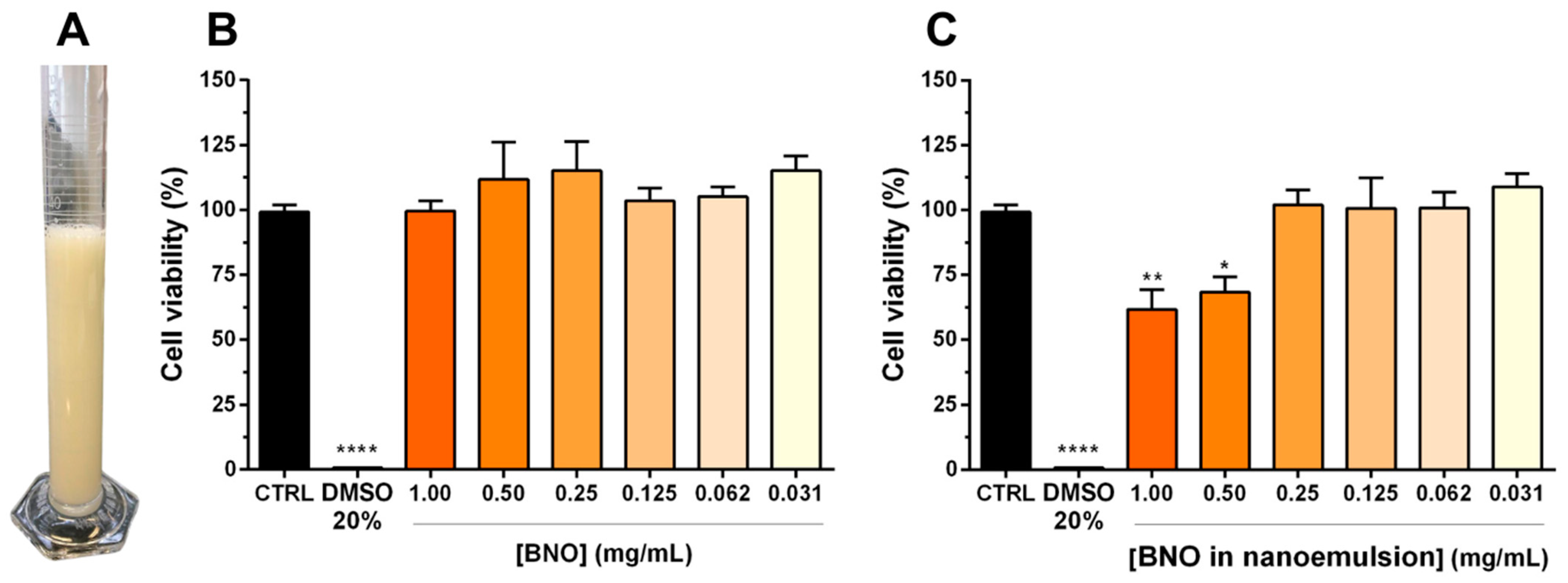
3.2. Cytotoxicity on Human Keratinocytes

The viability of human keratinocytes (HaCaT cell line), assessed using the MTT assay, demonstrated that the nanoemulsion (BNON) was not cytotoxic at concentrations up to 0.25 mg/mL (Figure 1C). However, a significant reduction in cell viability was observed at higher concentrations of 1.0 mg/mL (38.20 ± 0.09%) and 0.5 mg/mL (31.70 ± 0.07%) of BNON (Figure 1C). In contrast, free baru nut oil (BNO) showed no cytotoxicity across the tested concentration range (Figure 1B).

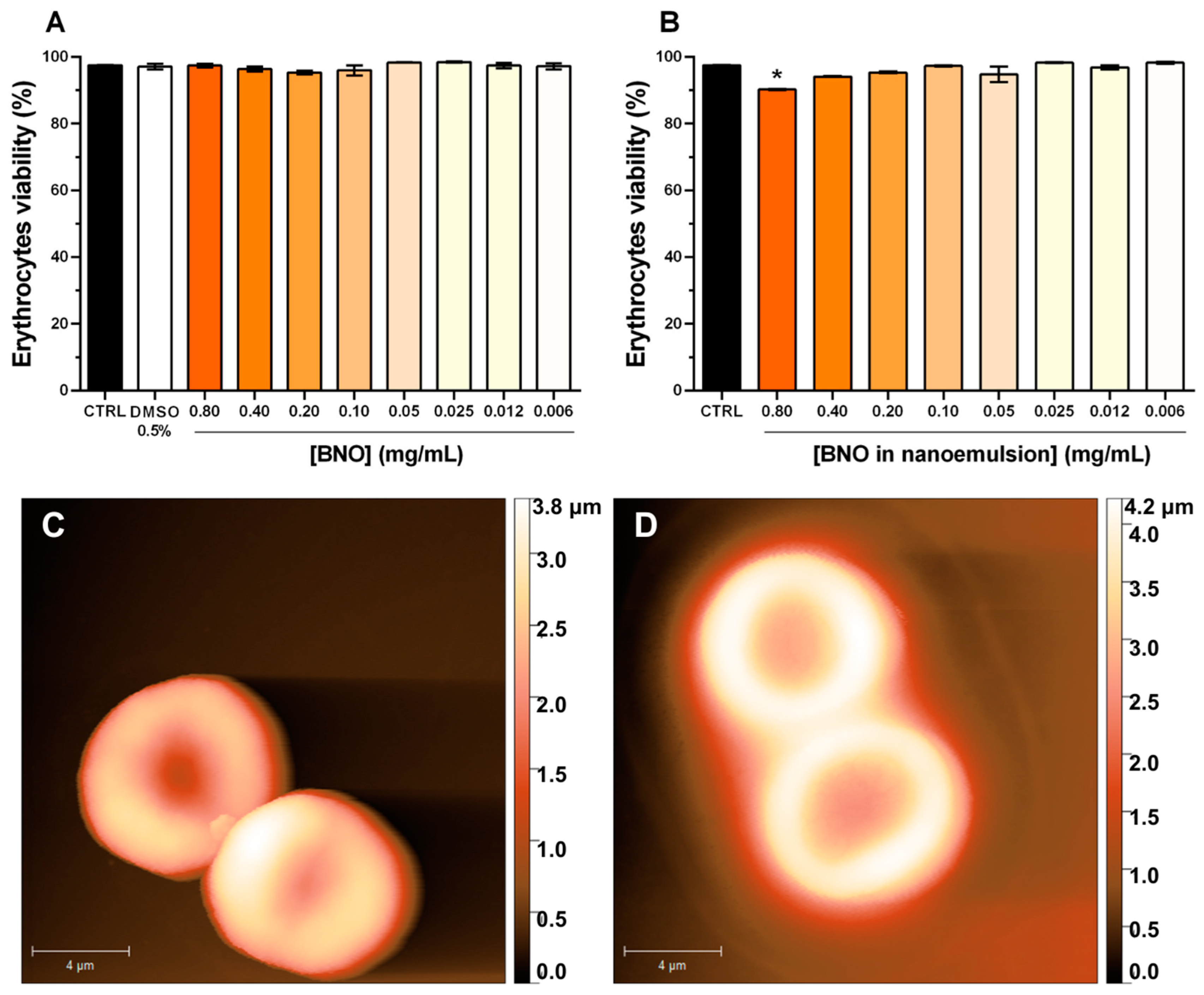
3.3. Hemolytic Activity and Erythrocyte Morphology

The free baru oil did not exhibit any hemolytic effect at the tested concentrations (Figure 2A), while BNON showed statistically significant cytotoxicity at the highest tested concentration (0.8 mg/mL), resulting in a reduction in erythrocyte viability by 9.816 ± 0.167% (Figure 2B). Furthermore, AFM analysis revealed no morphological changes in red blood cells treated with the free oil, which maintained their classic biconcave disc shape (Figure 2C). In contrast, red blood cells treated with 0.8 mg/mL of BNON displayed a slightly wider concave center, potentially indicative of lower cytoplasmic hemoglobin content (Figure 2D). However, no quantitative measurements were performed to confirm this observation. The erythrocytes treated by BNON also display an amorphous halo around the cells, perhaps indicating association of the emulsion with the cells. These findings align with the hemolytic activity observed for BNON.

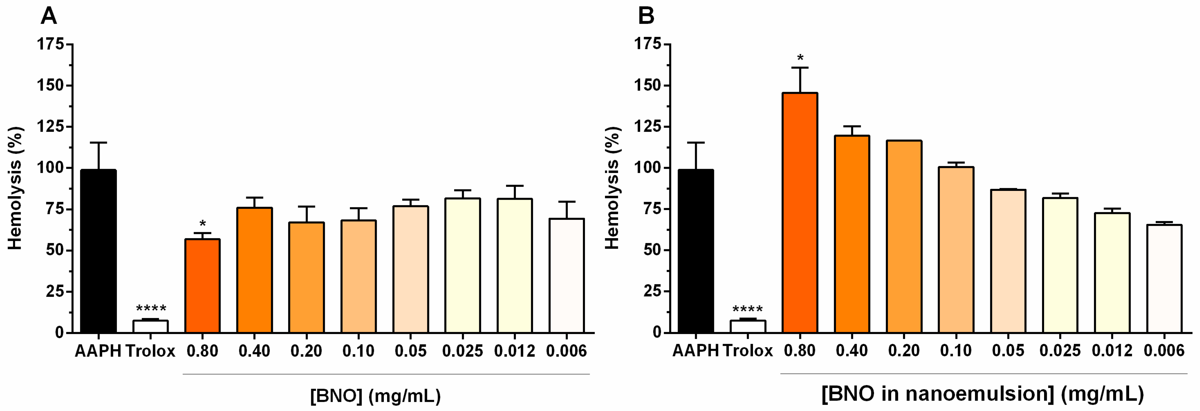
3.4. Antioxidant Activity

The antioxidant activity of BNON was evaluated using the DPPH (2,2-diphenyl-1-picrylhydrazyl) free radical assay, a widely used method for measuring the free radical scavenging ability of antioxidants. DPPH is a stable, hydrophobic free radical that readily reacts with antioxidant compounds, making it particularly suitable for assessing the activity of hydrophobic substances such as baru nut oil. The in vitro antioxidant activity assay demonstrated that BNON at a concentration of 5 mg/mL exhibited DPPH radical scavenging activity, equivalent to 0.0074 mg Trolox equivalents per milligram of BNON (Figure 3).
In the antioxidant activity assay against AAPH-induced oxidative stress, free baru oil at a concentration of 0.8 mg/mL significantly (p < 0.05) protected erythrocytes from the hemolytic effects of AAPH, with a hemolysis inhibition rate of 43.18 ± 0.003% (Figure 4A). In contrast, BNON not only failed to inhibit the hemolytic activity of AAPH but also induced a statistically significant (p < 0.05) increase in hemolysis compared to AAPH alone (Figure 4B), consistent with the hemolytic activity results reported previously.

4. Discussion

The nanoformulation of baru nut oil reported here represents an oil-in-water (O/W) dispersion system stabilized using lecithin and polysorbate 80 as surfactant and co-surfactant, respectively. Nanoemulsion production involves mixing two immiscible phases, such as oil and water, to form nanodroplets. The application of kinetic and thermal energy, combined with surfactants, provides the shear tension required for droplet breakage and stabilization at nanometric dimensions, enabling effective emulsification [16].
The prioritization of the system’s functionality over its optimization was a deliberate choice to establish a proof of concept for the nanoemulsion’s efficacy and biological application. Initial experiments aimed to confirm whether the formulation could effectively deliver bioactive compounds and exhibit the desired biological effects. This approach allowed us to evaluate the potential of the system under real-world conditions, laying the groundwork for subsequent optimization steps to enhance its stability, scalability, and performance. Such a strategy is common in exploratory stages of nanotechnology research, where the focus is on validating the system’s core functionality before refining its parameters.
Physicochemical characteristics such as droplet size, polydispersity index (PDI), and zeta potential (ZP) are important factors influencing the functionality, toxicity, and potential applications of nanoformulations [17]. Based on the mean droplet size measured by DLS (Table 1), no statistically significant size changes were observed over the 3-month storage period, suggesting the stability of the formulation under the analyzed conditions. The PDI reflects the homogeneity or heterogeneity of the droplet size distribution. According to Vasconcelos et al. (2020) [15], PDI values between 0.1 and 0.3 indicate a narrow size distribution, while values greater than 0.5 signify a higher heterogeneity in size distribution.
The PDI of BNON suggests a heterogeneous size distribution, which increased over time. Furthermore, pellet formation was observed at the bottom of the tubes during the storage period, but the formulation regained homogeneity upon manual agitation or vortexing. The formation of precipitates in nanoemulsions can occur by phenomena such as aggregation, flocculation, creaming, sedimentation, and coalescence [18,19]. Among them, creaming is likely to have occurred because the initial droplet size may result in different phase densities, and larger droplets are more prone to gravitational forces. In addition, creaming is a reversible aspect in that the number and size of droplets remain unchanged, but the droplet distribution becomes heterogeneous, as observed in this study.
It is worth noting that, although high PDI values are generally undesirable in applications that require high uniformity, such as intracellular drug delivery, they can be advantageous in specific contexts, such as catalysis or agriculture. This is because heterogeneity offers varied active surfaces, optimizing reactions; they allow the prolonged release of nutrients or bioactive substances on surfaces; and different sizes can favor different properties, such as absorption or cellular interactions [20].
Zeta potential reflects the surface electric charge of nanodroplets. The negative zeta potential observed for BNON will result in electrostatic repulsion, which helps prevent droplet aggregation and flocculation, contributing to some stability of the nanoemulsion [18,21]. Soy lecithin was chosen due to its well-established properties as an emulsifier and its good compatibility with the pH used in this study. In future optimization studies, pH variations could be explored to potentially impact the stability or functionality of the system, as pH can influence the charge of lecithin and, consequently, alter the physicochemical parameters of nanoemulsion.
Although our nanoemulsion has become functional, the system requires optimization, as in the case of precipitation. Precipitation can be caused by insolubility or phase restructuring, processes that affect the system’s stability. Phase restructuring, such as under dilution conditions, can alter interactions between components, leading to phase separation or particle aggregation. Although not addressed in this study, these phenomena may impact the release of bioactive compounds. Future studies could investigate stability under different dilution conditions to better understand the system’s behavior in practical applications.
Cytotoxicity assessment of the samples was performed in a human keratinocyte cell line (HaCat) and erythrocytes. These cells were chosen as a model considering that they are the most abundant cells in the human body and are susceptible to contact with exogenous substances through different routes of product administration (topical, oral, or intravenous). The cytotoxic effects observed for the nanoformulation at higher concentrations may be explained by hyperosmolar stress in the cellular environment. Alterations in extracellular osmolarity are closely linked to both physiological and pathophysiological processes [22]. This explanation is supported by the turbid appearance of the nanoformulation at these concentrations, although further investigations are necessary to confirm this hypothesis. In contrast, baru oil demonstrated no cytotoxic effects on either keratinocytes or erythrocytes, highlighting its potential for diverse biological and biotechnological health and cosmetic-related applications. The nanoemulsion appearance may be related to the size and uniformity of the droplets, which increases the scattering of light, as well as the type and concentration of oil and surfactant [23]. In this study, the droplet size, PDI, and components corroborate the appearance of BNON.
Polysorbate 80, widely used as a surfactant in pharmaceutical formulations, is generally well-tolerated at low concentrations but can exhibit cytotoxicity at higher levels or with prolonged exposure. Studies using cell models, such as Caco-2, show that its biocompatibility depends on the concentration and formulation, emphasizing the need for safety assessments to ensure its efficacy and safety in therapeutic applications [24].
AFM analysis provided valuable insights into the effects of oil and nanoemulsion on erythrocyte morphology. The normal morphology observed in erythrocytes treated with the maximum tested concentration of oil supports the biocompatibility of the product. In contrast, the slightly wider concave center in cells exposed to BNON suggests a small loss of cytoplasmic content, aligning with the mild hemolytic activity of the formulation. Notably, this effect does not appear to involve abrupt membrane rupture. The hemocompatibility of nanoparticles is a critical consideration for healthcare applications, given the significant proportion of erythrocytes in the blood. Furthermore, previous studies have demonstrated the hemocompatibility of polysorbate 80 and lecithin, the key components used in our nanoemulsion formulation [25].
Baru oil is recognized in the scientific literature as a potent antioxidant, largely due to the presence of phenolic compounds [26]. In this study, baru oil at a concentration of 0.8 mg/mL effectively protected erythrocytes from the hemolytic effects of AAPH, highlighting its potential for health and cosmetic-related applications. While the nanostructured oil (BNON) did not exhibit the same protective antioxidant effect on erythrocytes under the tested experimental conditions, data from the DPPH assay suggests some antioxidant activity at a higher concentration (5 mg/mL).
However, it is crucial to determine whether this concentration, which demonstrated a scavenging effect on the DPPH radical, represents a viable dose for practical applications. Considering that the DPPH radical acts as a hydrogen acceptor, converting into a stable molecule [27], it can be inferred that the antioxidant activity of BNON involves a hydrogen donation mechanism. Nevertheless, despite its measurable antioxidant activity in the DPPH assay, the inability of BNON to protect erythrocytes from hemolysis may be attributed to differences in bioavailability, interaction with cell membranes, or potential changes in osmolarity and medium occlusion compared to free oil.

5. Conclusions

The results demonstrate that baru nut oil was non-cytotoxic to human keratinocytes and erythrocytes under the experimental conditions tested, caused no morphological changes in erythrocytes as assessed by AFM, and exhibited antioxidant activity in an AAPH-induced hemolysis model. The nanotechnological formulation containing baru oil (BNON) showed cytotoxicity only at the highest concentrations tested, and the morphological changes in erythrocytes observed were consistent with its hemolytic activity. Despite these findings, nanoemulsion exhibited antioxidant activity, suggesting its potential utility in future optimized formulations. Future optimization of the formulation composition may enhance its biosafety and biological activity. These data provide valuable insights for the development of safe and effective nanotechnological products with antioxidant properties for health-related applications.

Author Contributions

Conceptualization, A.G.V.; methodology, J.Q., A.F., D.C.M., P.E., and A.G.V.; investigation, J.Q., A.F., D.C.M., P.E., B.S.S. and A.G.V.; resources, A.G.V. and J.R.S.d.A.L.; data curation, A.G.V. and J.R.S.d.A.L.; writing—original draft preparation, B.S.S. and J.Q.; writing—review and editing, J.B.N., A.G.V., D.C.M., and P.E.; supervision, A.G.V.; project administration, A.G.V.; funding acquisition, A.G.V. and J.R.S.d.A.L. All authors have read and agreed to the published version of the manuscript.

Funding

This research received no external funding.

Data Availability Statement

The raw data supporting the conclusions of this article will be made available by the authors on request.

Acknowledgments

The authors thank the Laboratory of Food, Drug and Cosmetics (LTMAC) at the University of Brasília for providing access to the Zetasizer Nano-ZS90 (Malvern, UK). A.F. expresses gratitude to the Fundação de Apoio à Pesquisa do Distrito Federal (FAPDF) for supporting the Institutional Scholarship Program for Scientific Initiation (PIBIC) at the University Center of the Federal District (UDF), which provided a scholarship.

Conflicts of Interest

Authors Jose Roberto de Souza de Almeida Leite and Andreanne Gomes Vasconcelos were employed by the company People & Science Pesquisa Desenvolvimento e Inovação Ltda. The remaining authors declare that the research was conducted in the absence of any commercial or financial relationships that could be construed as a potential conflict of interest.

References

  1. Silva, F.B.; Costa, A.C.; Müller, C.; Nascimento, K.T.; Batista, P.F.; Vital, R.G.; Megguer, C.A.; Jakelaitis, A.; Domingos, M. Dipteryx Alata, a Tree Native to the Brazilian Cerrado, Is Sensitive to the Herbicide Nicosulfuron. Ecotoxicology 2020, 29, 217–225. [Google Scholar] [CrossRef] [PubMed]
  2. Silva, R.W.L.D.; Machado, S.S.; Faria, K.D.C.; Oliveira, F.A.D.; Souza, A.P.D.; Menezes, I.P.P.D.; Silva, J.M.D. Molecular Insight for Baru Dipteryx Alata (Fabaceae) Populations Based on Novel SSRs. Acta Bot. Bras. 2023, 37, e20220168. [Google Scholar] [CrossRef]
  3. Lima, D.C.; Alves, M.D.R.; Noguera, N.H.; Nascimento, R.D.P.D. A Review on Brazilian Baru Plant (Dipteryx alata Vogel): Morphology, Chemical Composition, Health Effects, and Technological Potential. Future Foods 2022, 5, 100146. [Google Scholar] [CrossRef]
  4. Paulo, L.; Fernandes, R.; Gandra, K.; Minim, V.; Minim, L.; Grimaldi, R.; Vidigal, M. Baru Seed Extracted Oil (Dipteryx alata Vog.): Chemical Composition and Thermal and Oxidative Stability. J. Braz. Chem. Soc. 2023, 34, 664–672. [Google Scholar] [CrossRef]
  5. Scapin, E.; Sarri, D.R.A.; Augusco, M.A.C.; Rodrigues, M.A.M.; Fernandes, R.M.N.; Silva, J.F.M.; Cardoso, C.A.L.; Rambo, M.K.D. Phytochemical Analysis, Toxicity and Evaluation of Antioxidant and Antimicrobial Activities of Leaves of Dipteryx Alata Vogel. Braz. J. Biol. 2024, 84, e278004. [Google Scholar] [CrossRef] [PubMed]
  6. Dos Santos, J.M.; Marangoni Faoro, J.A.; Fava De Souza, M.; De Matos Balsalobre, N.; Leite Kassuya, C.A.; Kappel Trichez, V.D.; Mussury Franco Da Silva, R.M.; Formagio, A.S.N. Anti-Arthritic Potential and Antioxidant Properties of Infusion, Fractions and Flavonoid Glycosides from Dipteryx alata (Baru) Leaves. J. Ethnopharmacol. 2025, 338, 118973. [Google Scholar] [CrossRef]
  7. Leite, N.R.; Araújo, L.C.A.D.; Rocha, P.D.S.D.; Agarrayua, D.A.; Ávila, D.S.; Carollo, C.A.; Silva, D.B.; Estevinho, L.M.; De Picoli Souza, K.; Dos Santos, E.L. Baru Pulp (Dipteryx alata Vogel): Fruit from the Brazilian Savanna Protects against Oxidative Stress and Increases the Life Expectancy of Caenorhabditis Elegans via SOD-3 and DAF-16. Biomolecules 2020, 10, 1106. [Google Scholar] [CrossRef] [PubMed]
  8. Silva-Luis, C.C.; De Brito Alves, J.L.; De Oliveira, J.C.P.L.; De Sousa Luis, J.A.; Araújo, I.G.A.; Tavares, J.F.; Do Nascimento, Y.M.; Bezerra, L.S.; Araújo De Azevedo, F.D.L.A.; Sobral, M.V.; et al. Effects of Baru Almond Oil (Dipteryx alata Vog.) Treatment on Thrombotic Processes, Platelet Aggregation, and Vascular Function in Aorta Arteries. Nutrients 2022, 14, 2098. [Google Scholar] [CrossRef]
  9. Reis, M.Á.; Novaes, R.D.; Baggio, S.R.; Viana, A.L.M.; Salles, B.C.C.; Duarte, S.M.D.S.; Rodrigues, M.R.; Paula, F.B.D.A. Hepatoprotective and Antioxidant Activities of Oil from Baru Almonds (Dipteryx alata Vog.) in a Preclinical Model of Lipotoxicity and Dyslipidemia. Evid.-Based Complement. Altern. Med. 2018, 2018, 8376081. [Google Scholar] [CrossRef] [PubMed]
  10. Preeti; Sambhakar, S.; Malik, R.; Bhatia, S.; Al Harrasi, A.; Rani, C.; Saharan, R.; Kumar, S.; Geeta; Sehrawat, R. Nanoemulsion: An Emerging Novel Technology for Improving the Bioavailability of Drugs. Scientifica 2023, 2023, 6640103. [Google Scholar] [CrossRef]
  11. Algahtani, M.S.; Ahmad, M.Z.; Ahmad, J. Investigation of Factors Influencing Formation of Nanoemulsion by Spontaneous Emulsification: Impact on Droplet Size, Polydispersity Index, and Stability. Bioengineering 2022, 9, 384. [Google Scholar] [CrossRef]
  12. Malode, M.G.P.; Chauhan, S.A.; Bartare, S.A.; Malode, L.M.; Manwar, J.V.; Bakal, R.L. A Critical Review on Nanoemulsion: Advantages, Techniques and Characterization. J. App. Pharm. Sci. Res. 2022, 4, 6–12. [Google Scholar] [CrossRef]
  13. Mushtaq, A.; Mohd Wani, S.; Malik, A.R.; Gull, A.; Ramniwas, S.; Ahmad Nayik, G.; Ercisli, S.; Alina Marc, R.; Ullah, R.; Bari, A. Recent Insights into Nanoemulsions: Their Preparation, Properties and Applications. Food Chem. X 2023, 18, 100684. [Google Scholar] [CrossRef] [PubMed]
  14. Himanath, G.; Shruthy, R.; Preetha, R.; Sreejit, V. Nanoemulsion with Coconut Oil and Soy Lecithin as a Stable Delivery System for Lycopene and Its Incorporation into Yogurt to Enhance Antioxidant Properties and Maintain Quality. ACS Food Sci. Technol. 2021, 1, 1538–1549. [Google Scholar] [CrossRef]
  15. Vasconcelos, A.G.; Valim, M.O.; Amorim, A.G.; do Amaral, C.P.; de Almeida, M.P.; Borges, T.K.; Socodato, R.; Portugal, C.C.; Brand, G.D.; Mattos, J.S.C. Cytotoxic Activity of Poly-ɛ-Caprolactone Lipid-Core Nanocapsules Loaded with Lycopene-Rich Extract from Red Guava (Psidium guajava L.) on Breast Cancer Cells. Food Res. Int. 2020, 136, 109548. [Google Scholar] [CrossRef] [PubMed]
  16. Hassanshahian, M.; Saadatfar, A.; Masoumipour, F. Formulation and Characterization of Nanoemulsion from Alhagi Maurorum Essential Oil and Study of Its Antimicrobial, Antibiofilm, and Plasmid Curing Activity against Antibiotic-Resistant Pathogenic Bacteria. J. Environ. Health Sci. Eng. 2020, 18, 1015–1027. [Google Scholar] [CrossRef] [PubMed]
  17. Azarnezhad, A.; Samadian, H.; Jaymand, M.; Sobhani, M.; Ahmadi, A. Toxicological Profile of Lipid-Based Nanostructures: Are They Considered as Completely Safe Nanocarriers? Crit. Rev. Toxicol. 2020, 50, 148–176. [Google Scholar] [CrossRef] [PubMed]
  18. Klang, V.; Valenta, C. Lecithin-Based Nanoemulsions. J. Drug Deliv. Sci. Technol. 2011, 21, 55–76. [Google Scholar] [CrossRef]
  19. Fernandez, P.; André, V.; Rieger, J.; Kühnle, A. Nano-Emulsion Formation by Emulsion Phase Inversion. Colloids Surf. A Physicochem. Eng. Asp. 2004, 251, 53–58. [Google Scholar] [CrossRef]
  20. Sneha, K.; Kumar, A. Nanoemulsions: Techniques for the Preparation and the Recent Advances in Their Food Applications. Innov. Food Sci. Emerg. Technol. 2022, 76, 102914. [Google Scholar] [CrossRef]
  21. Sharifi, F.; Jahangiri, M.; Nazir, I.; Asim, M.H.; Ebrahimnejad, P.; Hupfauf, A.; Gust, R.; Bernkop-Schnürch, A. Zeta Potential Changing Nanoemulsions Based on a Simple Zwitterion. J. Colloid Interface Sci. 2021, 585, 126–137. [Google Scholar] [CrossRef] [PubMed]
  22. Thiemicke, A.; Neuert, G. Kinetics of Osmotic Stress Regulate a Cell Fate Switch of Cell Survival. Sci. Adv. 2021, 7, eabe1122. [Google Scholar] [CrossRef]
  23. Gupta, A.; Eral, H.B.; Hatton, T.A.; Doyle, P.S. Nanoemulsions: Formation, Properties and Applications. Soft Matter 2016, 12, 2826–2841. [Google Scholar] [CrossRef] [PubMed]
  24. Bu, P.; Narayanan, S.; Dalrymple, D.; Cheng, X.; Serajuddin, A.T.M. Cytotoxicity Assessment of Lipid-Based Self-Emulsifying Drug Delivery System with Caco-2 Cell Model: Cremophor EL as the Surfactant. Eur. J. Pharm. Sci. 2016, 91, 162–171. [Google Scholar] [CrossRef] [PubMed]
  25. Bender, E.A.; Adorne, M.D.; Colomé, L.M.; Abdalla, D.S.P.; Guterres, S.S.; Pohlmann, A.R. Hemocompatibility of Poly(ɛ-Caprolactone) Lipid-Core Nanocapsules Stabilized with Polysorbate 80-Lecithin and Uncoated or Coated with Chitosan. Int. J. Pharm. 2012, 426, 271–279. [Google Scholar] [CrossRef] [PubMed]
  26. Oliveira-Alves, S.C.; Pereira, R.S.; Pereira, A.B.; Ferreira, A.; Mecha, E.; Silva, A.B.; Serra, A.T.; Bronze, M.R. Identification of Functional Compounds in Baru (Dipteryx alata Vog.) Nuts: Nutritional Value, Volatile and Phenolic Composition, Antioxidant Activity and Antiproliferative Effect. Food Res. Int. 2020, 131, 109026. [Google Scholar] [CrossRef] [PubMed]
  27. Marsup, P.; Yeerong, K.; Neimkhum, W.; Sirithunyalug, J.; Anuchapreeda, S.; To-anun, C.; Chaiyana, W. Enhancement of Chemical Stability and Dermal Delivery of Cordyceps Militaris Extracts by Nanoemulsion. Nanomaterials 2020, 10, 1565. [Google Scholar] [CrossRef] [PubMed]
Figure 1. Photograph of the baru nut oil nanoemulsion (BNON, (A)). Effect of free baru nut oil (BNO, (B)) and BNO in nanoemulsion (C) on human keratinocyte cells (HaCaT) viability after 24 h of exposure. Values were expressed as means ± SEM. * p < 0.05 and ** p < 0.01 versus DMEM control group (CTRL). **** p < 0.001.
Applnano 06 00003 g001
Figure 2. Effect of free baru nut oil (BNO, (A)) and baru nut oil in nanoemulsion (B) on the human erythrocytes viability. Atomic force microscopy micrographs of erythrocytes treated with BNO (C) and BNON 0.8 mg/mL (D). The values are expressed as mean ± SEM. * p < 0.05. Scale bar = 4 µm.
Applnano 06 00003 g002
Figure 3. Antioxidant activity of BNON by the DPPH radical scavenging assay compared to Trolox. Data expressed as mean ± SD.
Applnano 06 00003 g003
Figure 4. Antioxidant effect of free baru nut oil (BNO, (A)) and baru nut oil in nanoemulsion (B) on human erythrocytes in a model of hemolysis induced by oxidative stress using AAPH (2,2′-azobis(2-methylpropionamidine) dihydrochloride). Data expressed as mean ± SEM. * p < 0.05. **** p < 0.001.
Applnano 06 00003 g004
Table 1. Physicochemical characteristics of BNON on the day of synthesis and after 3 months of storage.
MonthSize (nm)PDIZP (mV)
0530.1 ± 20.480.496 ± 0.057−35.7 ± 2.19
3467.1 ± 38.080.726 ± 0.235 *−33.5 ± 1.99
PDI: Polydispersity Index. ZP: Zeta Potential. * p < 0.05.
Disclaimer/Publisher’s Note: The statements, opinions and data contained in all publications are solely those of the individual author(s) and contributor(s) and not of MDPI and/or the editor(s). MDPI and/or the editor(s) disclaim responsibility for any injury to people or property resulting from any ideas, methods, instructions or products referred to in the content.

