Review

Emerging Trends in Nanomaterial-Based Biomedical Aspects

by
Mansi Gandhi
1,2,3,* and
Khairunnisa Amreen
4
1
Department of Chemistry, School of Advanced Sciences, Vellore Institute of Technology, Vellore 632014, India
2
Institute of Chemistry, Hebrew University of Jerusalem, Jerusalem 9190401, Israel
3
School of Biodesign and Engineering, Translational Health Science and Technology Institute, Faridabad 120001, India
4
Department of Chemistry, St Anns College for Women, Mehdipatnam, Hyderabad 500028, India
*
Author to whom correspondence should be addressed.
Electrochem 2023, 4(3), 365-388; https://doi.org/10.3390/electrochem4030024
Submission received: 12 June 2023 / Revised: 17 July 2023 / Accepted: 21 July 2023 / Published: 4 August 2023

Abstract

:
Comprehending the interfacial interaction of nanomaterials (NMs) and biological systems is a significant research interest. NMs comprise various nanoparticles (NPs) like carbon nanotubes, graphene oxides, carbon dots, graphite nanopowders, etc. These NPs show a variety of interactions with biological interfaces via organic layers, therapeutic molecules, proteins, DNA, and cellular matrices. A number of biophysical and colloidal forces act at the morphological surface to regulate the biological responses of bio-nanoconjugates, imparting distinct physical properties to the NMs. The design of future-generation nano-tools is primarily based on the basic properties of NMs, such as shape, size, compositional, functionality, etc., with studies being carried out extensively. Understanding their properties promotes research in the medical and biological sciences and improves their applicability in the health management sector. In this review article, in-depth and critical analysis of the theoretical and experimental aspects involving nanoscale material, which have inspired various biological systems, is the area of focus. The main analysis involves different self-assembled synthetic materials, bio-functionalized NMs, and their probing techniques. The present review article focuses on recent emerging trends in the synthesis and applications of nanomaterials with respect to various biomedical applications. This article provides value to the literature as it summarizes the state-of-the-art nanomaterials reported, especially within the health sector. It has been observed that nanomaterial applications in drug design, diagnosis, testing, and in the research arena, as well as many fatal disease conditions like cancer and sepsis, have explored alongwith drug therapies and other options for the delivery of nanomaterials. Even the day-to-day life of the synthesis and purification of these materials is changing to provide us with a simplified process. This review article can be useful in the research sector as a single platform wherein all types of nanomaterials for biomedical aspects can be understood in detail.

1. Introduction

Nanotechnology refers to the use of nanoscale-level materials for various applications. The nanoscale is a billionth of a meter, and the most common example is single-stranded DNA (~2.5 nm). The exclusive size-inspired properties of nanomaterials are indispensable in various domains of human sustenance, such as the DNA (2 nm diameter), while the protein is nearly 10 nm in diameter. Proper tailoring of NM properties, i.e., biological components, can be studied with a new field approach that has become the center of interactions in the scientific community, with a worldwide market of over USD 1.5 trillion since 2015. This was started by the early visions that were put forward by Nobel Laureate Richard Feynman in 1960 [1]. In addition, the discovery of fullerene C60 in 1985 by Kroto et al. further broadened/pushed the stream/sight of nanotechnology [1].
After almost two decades, a number of different NMs varying in shapes, sizes, and structures are now being globally produced, like nanowires, nanotubes, nanoclusters, nanocrystals, nano-coatings, nanocomposites, quantum dots, etc. These have led to enhancement in properties compared to their bulkier counterparts [2,3]. They have a huge scope of applications, including electronics, optics, photonics, displays, magnetics, dot-circuits, cantilevers, QD-cellular automata, self-assembly, lithography, aerospace, tooling, DNA-based molecular switches, biosensing, coating, robotics, and renewable energy resources [4]. The study of bio-mimicking naturally occurring materials has inspired nanoscale synthesis. A major domain is to build a nano-biological interface that bio-mimics the naturally occurring phenomenon for a better understanding. In this respect, the International Complex Adaptive Matter (ICAM) workshop was convened by an interdisciplinary group of scientists in November 2005 on “Biologically inspired Nanomaterials” [5]. The whole idea was to aid the research of nanoscale materials in biological sciences. In this regard, a series of materials (e.g., polymers) are being developed on a wide scale that behave like antibodies [6,7]. In addition, engineered surfaces possessing the properties of cells [8,9] and synthetic work harnessed by local power [10] via mimicking nano-architectures are being developed. This emerging frontier can produce environmentally benign systems that are essential for minimizing poor impacts, wherein natural systems provide inspiration for the ability to operate using renewable resources. Other relevant queries—such as how to mimic their electronic, optical, and architectural components—are yet to be unveiled. We have begun to view nanoscale aspects and novel theoretical models to describe nature. The best example is the invention of the light microscope, which changed the view altogether. Although it was designed for observational purposes around five centuries ago, alterations in the technological landscape are still being seen now. This view of cells, diatoms, and bacteria has influenced architects, engineers, and scientists. The well-established bottom–up approach to supermolecular organization has led to the incorporation of selective properties of biologically derived materials (e.g., NPs such as nanowires) having different scales (i.e., nanometer to micrometer); their properties, functionality, and complexity have changed, as shown in Figure 1.
Nature has also shown evidence for synthetic self-assembly using the molecular machinery approach. Emerging research in different areas of bioactive, self-assembling peptide nanofiber hydrogels for tissue regeneration has been reported [11]. Sleyter and co-workers have reported S-layer protein assembly as building blocks similar to the bottom–up approach using periodic crystalline proteins (from B. Sphaericus) [12,13,14,15]. These building blocks have shown promising approaches for future molecular, electronic, optical, and magnetic devices. On the other hand, Allar’s group has recently reported the use of self-assembly to form well-organized materials relying on covalent and non-covalent interactions [6,16].
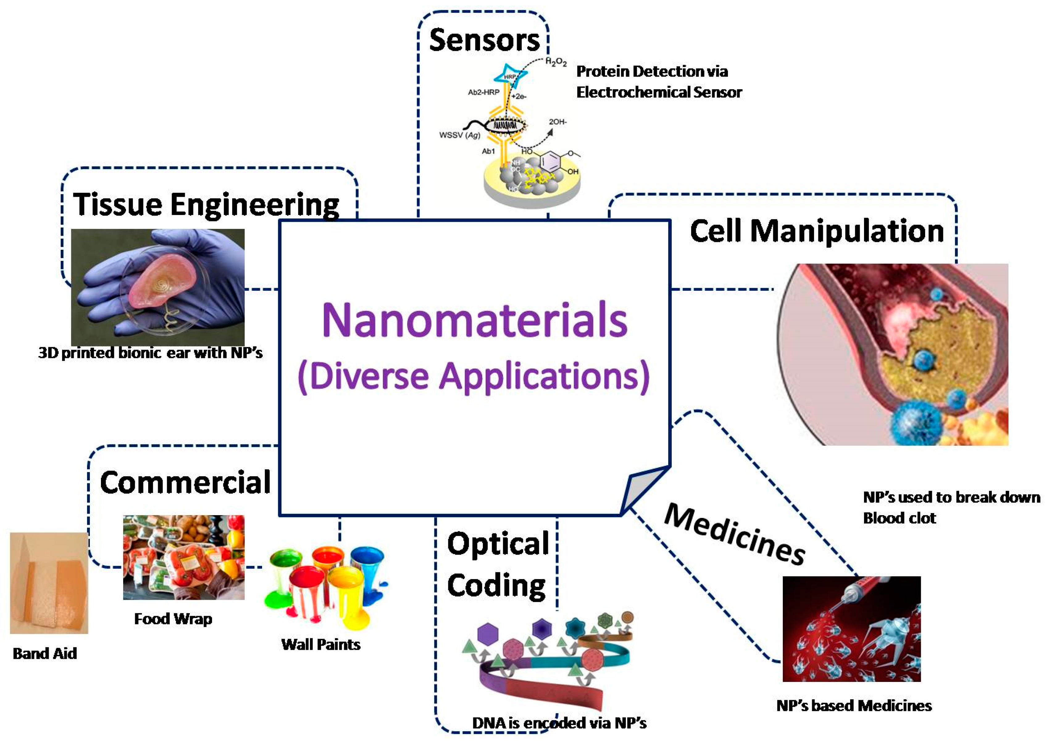
The nano-bio interface is one of the fastest emerging frontiers in the area of nanotechnology. Since NMs have distinct biological and chemical aspects compared to their counterparts, an understanding of their physiochemical characterization and biological media is essential. This interface comprises dynamic physiochemical interactions, kinetics, and thermodynamic exchanges. They have a variety of biomedical applications, such as drug delivery, bio-imaging, tissue engineering, bio-sensors, etc., as shown in Figure 2. This helps in developing unique functionalities required by biomedical systems. An equal understanding of forces and molecular counterparts is needed to govern their interactions. Functionalized NPs are being developed for various medical alternatives to electronics [7,17,18,19,20] and healthcare sector needs. Willner et al. have demonstrated electrical contact between redox enzymes and the macroscopic environment via the reconstruction of apoenzyme with FAD (Au-NP conjugate). Their (redox enzyme) inherent properties of controlled shape, size, and structure making biomolecules a potentially attractive field for functional circulatory devices [21]. In this context, we introduce various biomaterials, their applications, and their limitations. Furthermore, their future prospects have been stated in the conclusion of this article. The main aim of this review is to primarily provide the reader with a historical perspective of NMs, and their applications to the interdisciplinary field of biology, chemistry, electronics, and medicine. Secondly, it is to give an overview of the most recent developments in this field. And finally, we discuss the hard road to commercialization. This holistic approach makes this review article special and a standout as compared to other articles, as other articles are majorly short-windowed and do not provide a complete outlook towards the involvement of nanomaterials for biomedical applications and their prerequisites. Many articles have been written on the topic of the biomedical use of specific nanosystems, but none of the articles provide a complete outlook from its origin to various types, applications, characterization types, and their usage in the distinct fields of biomedicine. The growing interest in the future of medical applications leads to the emergence of an amalgamation of nano-biomedicines. They have come out as an essential game player in modern medicine with clinical applications. The focus of the current review paper is on recently developing trends in the production and utilization of nanomaterials in relation to diverse biomedical applications. This article will provide value to the literature as it summarizes the state-of-the-art nanomaterials reported, especially for the health sector. Research on numerous grave medical situations, including cancer and sepsis, has been exploring medication therapy and other methods for the distribution of nanomaterials. These applications include drug creation, diagnostics, testing, and research. Even the laborious process for the synthesis and purification of these materials is changing to provide us with a simplified process. This review article can be useful in the research sector for a single platform wherein all the types of nanomaterials for biomedical aspects could be understood in detail. The manuscript provides an overall review of the various nanoparticles synthesized which has the potential to be utilized in diagnostic monitoring as one of many applications—especially the applications pertaining to biosensing in the case of disease biomarkers.

2. Combination of Biology and Nanomaterials

A well-known fact is that living organisms are made up of smaller sub-units called cells (size approx. 10 mm). However, the cell organelles present inside cells are even smaller, which are quite comparable to man-made nanoparticles (NPs). Hence, these NPs can act as small probes that allow us to spy on cellular machinery without any obstruction. Synthetic Biology, a new field that integrates science, engineering, and technology, is the result of the precise control of particle size, their inner core (composition, size, shape), stability, and functionalization of molecules [22]. Several NPs also showed excellent interfacial interactions with biological components like de-oxy ribonucleic acid (DNA) and ribonucleic acid (RNA). The following are some of the applications of NPs at the cellular level. Kam et al. reported that single-walled carbon nanotubes (SWNTs) facilitate DNA delivery into multi-vascular endothelial cells. In continuation, it was suggested that CNTs act therapeutically as potential drug components and can be used as gene-delivery vehicles [23,24,25,26,27,28]. Furthermore, the NPs showed anti-microbial activities, e.g., Rudramurthy et al. demonstrated that SWNTs possess antibacterial properties against Salmonella Enterica and E. coli, indicating CNTs are effective antibiotics with drug-resistant and multi-resistant bacterial strains [29,30,31]. S. Anuchapreeda et al. developed a new formulation of a curcumin lipid nano-emulsion of size 47–55 nm, which was employed for cancer chemotherapy [32]. Furthermore, in 2009, Delong et al. characterized nucleic acid conjugates with protamine and AuNPs as nanorods and explained their detailed features [33]. In addition, reports accounted for the use of different magnetic nanocomposites for the extraction of nucleic acids [34,35,36]. The other important approach is based on signaling networks in the field of biomedicine and biomedical engineering. These networks are critical for the production of natural biological NMs resulting in synthetic biology holding tremendous potential for a generation of new NMs.
Scientists can even alter the size of biogenic particles by varying metal ion concentrations in the cellular environment. One of the apt examples is the engineered synthetic circuit domain wherein several molecular biological control components are required, which are encoded in the cell’s DNA. This DNA blueprint can be read by several proteins having enzymatic activity, while other proteins can bind to metallic ions and result in the formation of NP, thereby inheriting their genetic information. Another perfect example is by Lutz and Bujard [37,38], as they produced engineered promoters with strong and reliable ON and OFF outputs in bacteria. The promoters were compatible with the repressors and hence were quite critical in designing networks like toggle switches.

3. Involvement of Nanomaterials in the Domain of Healthcare

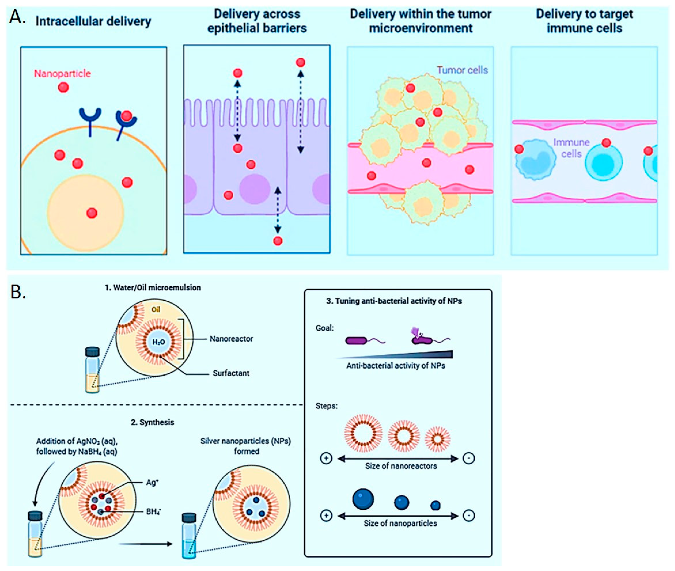
According to the World Health Organization (WHO), 33.4 million people were diagnosed with HIV/AIDS in 2008. Approximately 5.5 million people are suffering from Alzheimer’s disease in the USA, with a medical expense of USD 183 billion (according to 2011 statistics), which will further increase to six-fold that number, i.e., USD 1.1 trillion by 2050. Another report portrays that about 25% of deaths are caused due to cancer, out of which only 0.5% are diagnosed. Cancer has become the second leading cause of death after cardiac disease. All this has a huge impact on human health, leading to a staggering of society. Indeed, scientists, researchers, and medical experts are trying to find out approximate nanotechnological medications to curb the issue. The application of nanotechnology in the fields of medicine and pharmaceuticals has generated considerable optimism due to its potential to drive substantial advancements in disease diagnosis, treatment, and prevention. This transition from a realm of fantasy to a tangible reality signifies a noteworthy development [39]. Figure 3 has been given in this respect to portray the silver nanoparticles overcoming the biological barriers.
The medical applications of nanostructured biomaterials are in the form of polypeptides, proteins, nucleic acid, DNA-based self-assemblies, biopolymers, cells, glycans, etc., which are involved in studying the bio-compatibility and interaction between nanoscale biomaterials like cells, tissues, and organs, as well as their application in a variety of medical and health sciences, including nanocarriers for drug, gene and vaccine delivery, orthopedics, cardiovascular, dentistry, etc.
This scientific development is quite a promising approach. These aspects of nanotechnology are explored via the use of nanoscale biochips that will transmit information and monitor the biological changes in humans. Major advances are emerging in the form of DNA chips, carbohydrate chips, protein chips, and MEMS/NEMS, where a single biochip processes more than a million features with supersensitive detection, diagnostics, and analytical monitoring abilities. Few basic nanomaterials have adsorption properties for different medicines, such as ibuprofen, naproxen, and paclitaxel, as reported by Golbabaei et al. in 2016 [40]. The CNTs are common, having an efficient adsorption matrix, as concluded in the report.
PEGylated MWCNTs are equally effective, with their particle size and zeta potential being similar to MWCNTs. This concept is quite common for the integration and commercialization of nanotech in the bioinformatics field and pharmaceuticals. As reported by Thakor et al. in 2013 [41], emerging development in the area of cancer was summarized for diagnostic, prevention, and treatment of bone, colon, and prostate cancer and even their usage as potent nano-biological markers, immunotherapy, photodynamic therapy, and as anticancer nanodrugs and nano-oncology.
These NPs play a significant role in drug delivery as nanoscale drugs bind to targeted receptors and induce a receptor’s signaling and activity. Apart from these, NPs have gained great impetus in the field of pharmaceuticals and medicine. However, new protocols are being implemented against conventional medical procedures with the use of these nanomedicines. Various novel nanoformulations and nanocarriers for therapeutic treatments help in reducing the associated problems (emulsions, polymers, vitamins, inorganic composites, liposomes, polymers, metal nanoparticles, etc.) [42,43,44,45,46,47,48,49,50]. The nano drug delivery system includes lipids, lysosomes, gene NPs, nano-micelles, etc. Nanomedicines are about 10−9 m in size, can efficiently cross the blood–brain barrier, and support cancer treatment. They can be used for the diagnosis, prevention, and treatment of human diseases. The planned use of nanotechnology enables ground-breaking ideas for drug practices that have been delayed due to numerous hindrances. The precise choice of nanomaterials as a promising drug delivery platform can help in altering the hydrophobicity, thereby enhancing the water-solubility and upgrading their bio-availability characteristics [40,41]. Drug delivery systems that are specifically designed to respond to stimuli such as changes in optical, thermal, and hydrogen ion concentrations can be acquired through passive or active targeting methods. Mittal et al. described the involvement of magnetic nanoparticles (magnetofection), gold nanoparticles, and quantum dots for oligonucleotide delivery and gene therapy based on changing the “hydrophobic and hydrophilic properties” [28]. Owing to minute size exclusion, they infiltrate through the capillaries to the targeted tumor sites where they accumulate and help in decreasing/reduction in the after-effects, i.e., mainly side effects and toxicity issues, along with accelerating their efficacy. Representing features of controlled release, prolonged action of work, and balancing in drug levels in the therapeutic window, thereby avoiding hazardous drug concentration in the organs. The choice of co-delivery of more than one for simultaneous therapy and diagnosis, [51,52] cumulatively incorporating traits of synergistic effects, tuning relative therapeutic regime, drug resistance, etc., is possible using nano-based systems.
The established lipid nanoparticulate methods lead to numerous distinct ways for the delivery of micelles, nanoformulations, emulsions, solid lipid NPs, etc. Their characteristics, such as eco-friendly nature, biodegradability, ability for tailored water-loving/hating features, biocompatibility, economical set-up, easy manufacturing, and dispersity constraints, play a major role when compared with polymers. These characteristics enable better status and are one of the most sought platforms with easy scale-up options. Additionally, the involvement of such lipid-nanoparticular platforms for drug delivery apart from food and medicines has been recommended by GRAS (Generally Recognized as Safe). Traditional liposomes incorporate a hydrated lipid lamellar phase that encompasses a lipid bilayer with an aqueous core. This propensity of liposomes for transportation (of different molecules such as medicines, antigens, nucleotides, etc.), tailorable size and structure, charge alteration characteristics, simplicity, and other properties have ensured the delivery of therapeutics. Interestingly, the first sought drug delivery-based nanosystem was initiated in the 1960s and involved none other than lipid vesicles and/or liposomes. These systems have been in vast use, especially for clinical procedures. Secondly, these drug particulates are subjected to competitive advantages (specificity and selectivity) in comparison with unbound or free systems, thus adding to the permittivity and retention advantages, thereby deteriorating the tumor activities. Even though these techniques/modes of medication are prevalent in clinics, they do not ensure any improved avenues for the desired level of drug. Some of the major issues encountered in the biological milieu are that nanoparticles accumulate in the tumor due to slow lymphatic drainage, while high osmotic pressure creates a strong outflow due to the enhanced permittivity effect. Different ways to enhance the enviable uptake of targeted drug delivery is via the interaction with serum components. The water-loving and flexibility inbuilt attributes of different polymers and surface polymers (polyethylene glycol and poloxamers) have been selected to overcome the possible hindrances. Unfortunately, they constitute the drawbacks and limitations that disqualify their widespread applications. Different complications of polydispersity within the organ system and speedy excretion limit its usage. Through new advancements with purified PEGs, better dispersity comprises their molecular weights (<1000 Da only). The better availability of enhanced molecular weights can be a viable future prospect in this domain. Furthermore, PEG moieties of liposomes have an immunogenic ability and evoke antibodies in response to secondary administration. Thus, the anticipated PEGylated liposomal systems show/display unpredictable pharmacological features, creating issues of uncertainty and utility loopholes [53,54,55]. To add to this, apt synthesis techniques for nanoparticulate formulations provide an opportunity for achieved drug delivery. Ironically, these liposomes passively accumulate in the body organs/tissues and later exploit the body functions [55]. Furthermore, poly(vinylpyrrolidone-codimethyl maleic anhydride) co-polymer formulations viz. Poly(VP-co-DMMAn) is an interesting tailored superoxide dismutase that has issues of accumulation in kidneys after intravenous administration [56]. It further compels new advances in improving the efficacy of drug delivery based on harnessing their advantages [57]. Moreover, unveiling the opportunities for harnessing their properties remains a challenge for nanotechnologists. With developments in the fields of medicine and material synthesis, novel platforms for drug delivery are becoming better at overcoming the en route barriers and coming to a more rationalistic voyage for development. Figure 4 is a comparative description of the use of various nanoparticles in drug delivery.
The initial phase of polymer-based drug delivery was initiated with macromolecules in early 1976. These systems had varied properties of biodegradability and biocompatibility, with the most promising example of poly(lactic-co-glycolic acid) (PLGA), which is one of the most sought materials as per FDA approval. This can be attributed to their degradation products that are easily metabolized and finally excreted by the body, making them less toxic compared to others. The stoichiometric ratio of glycolic: lactic acid is variable, which directionalizes/triggers their degradation properties, i.e., a 50:50 ratio makes the degradation faster [58]. These methodologies have exploited the opportunity for fruitful chemotherapeutic drug delivery-based reservoirs. One such example is Taxol, a commercialized PGA—paclitaxel nanoformulation involved in the treatment of prostate-specific cancer. Another example in Korea, Genexol-PM−paclitaxel, is a well-accepted polymeric micelle-based system for the treatment of breast and lung cancer. It consists of block co-polymers of PEG and PLGA [59] in the size formulation of 20–50 nm, which completely disperse in the solvent systems. As tested in Phase I clinical trials, the maximum tolerated dosage (MTD) is 390 mg/m2. Genexol-PM−paclitaxel showed a selective response rate in subsequent trials. These nanoformulations help in enriching the efficacy of chemotherapeutics along with improving the potency of biologically active molecular entities which were considered fallow. A suitable example is a monoterpene, i.e., an essential phytonutrient obtained from the oils of plants having potent anti-cancerous properties. Perillyl alcohol (POH) possesses properties against different types of cancer, but its use is limited due to the various challenges encountered. Similarly, the PLGA-POH microparticle system has been extended for the treatment of tumors in Swiss albino mice induced due to skin epidermoid cancer cell line (A253) and di-methyl benzo anthracene (DMBA). The microparticle system showed an enhanced survival rate of PLGA-POH ~80%, while free POH~40%. The increment in survival efficiency can be attributed to circumventing the associated stumbling blocks of POH along with bestowing other desirable features [60].
The use of inorganic nanoparticle-based systems has spiked developments, especially for designing nanostructured materials with cohesive features of physical properties to enhance delivery both in the case of diagnosis and medication modalities [61]. Their tailored features include functionalization options, safer carrier capabilities, high surface area, and amphiphilicity, with examples of AuNP-based enticing scaffolds for drug delivery [62,63,64,65]. The prerequisite for the spatio-temporal release of AuNPs is triggered by internal and extra stimuli-based systems (glutathione (GSH), pH, light, temperature, etc.) [66,67,68]. Different attributes of their complexation, distribution, and delivery can be monitored using surface plasmon resonance (SPR). Integral properties have made it a potential resource to be harnessed in the near future. Ironically, these nanoformulations of Au have been extended for phase I and II clinical trials for cancer therapy due to their embedded variable traits of synthesis protocol offering distinct morphological features and functionalities like nanostars, nanospheres, nanocages, nanorods, etc. [69]. Different methods of synthesis involve the generation of distinct morphology and properties as chemical reduction leads to spherical (monodispersity), biomimetic and physical reduction leads to hollow Au, photochemical reduction to cubic shape, solvent evaporation to 2D Au superlattice, and so on [70]. There have been tremendous efforts to revolutionize cancer therapeutics and support recent scenario systems. In 2009, Eric Dreaden et al. suggested tamoxifen-PEG-thiol-AuNP conjugates for medication against breast cancer, wherein 2.7 times enhanced potency was accounted for in vivo selective targeting of estrogen receptor alpha [71]. Later, in 2011, Duncan et al. initiated nanoAu-drug formulations as water-hating agglomerates for effective cancer therapy. Functionalization of agglomerates using a water-hating alkenethiol interior and water-loving tetra (ethylene glycol) (TEG) shell makes the whole as a zwitter-ion system that reduces non-specific bindings with other macromolecular entities [72]. In addition, the size constraints enhanced facile circulation and accumulation in targeted organs using the EPR spin-trapping method. Later, in 2009, Asmaa Elbakry et al. suggested monodisperse AuNPs scaffolds used as layer-by-layer into siRNA-AuNP conjugates, comprising (polyethylene-imine) PEI/siRNA/PEI-AuNPs [73]. The PEI renders opportunities for well-defined and homogenously distributed nanocarriers for mediation of endosomal escape with a net negative charge of siRNA-AuNP formulation that evokes cellular uptake due to decreased repulsion from the cell membrane. Similarly, in 2010, Song et al. reported a small interfering RNA (siRNA) for clinical applications of PEI-capped gold nanoparticles with invariable shapes and sizes, used as an efficient and safe PEI (reductant and stabilizer). Complications related to cytotoxicity, cell apoptosis, and systematic knockdown of oncogene (PLK-1) were not accounted for. Hence, a suitable carrier was later involved for intracellular siRNA delivery [74]. But the major downside of using PEI is its toxicity, which has evoked a search for other excipients (mainly cationic) for modulation of the platform with other functionalities and lower toxicity. In 2012, Han et al. initiated an AuNP-CS core (gold nanoparticle chitosan stabilized) for electro-deposition of (cis-aconitic anhydride-functionalized poly(allylamine) PAH-Cit/PEI and siRNA. The well-defined fabricated platform owed negligible cytotoxicity against the HeLa cell line contemporary to efficient protection after nuclease degradation and triggered siRNA release [75]. In continuation, in 2011, Ghosh et al. introduced a robust method for delivering the microRNA using cysteamine-functionalized AuNPs as the best formulation [66]. These nanoformulations led to the targeted delivery of chemically unmodified miRNAs in living cells after inculcating the optical properties due to strong SPR absorption wavelengths, parallel to photo-thermal effects that endeavors myriads of biological manifestations.
The versatile features of gold nanoparticles enable several advantages. The spherical AuNP is an ideal delivery system, while nanorods and spheres provide strong SPR tenability, etc. Braun and the group exploited this shape-characteristic in the formulation of spatio-temporal siRNA cargo release using a NIR laser. Upon excitation, AuNS (gold nanospheres) liberated the siRNAs, which eliminated the chances of toxicity and introduced power-dependent bond cleavage (at low-power excitation) while escaping from the endosome (high-power irradiation). Han et. al., in 2012, initiated a AuNS-NAs (nucleic acid)-based spatio-temporal system for poly-Lysine peptide (PLL) epilayer delivery platform using PLL inclusion for electrostatic nucleic acid capture and NIR excitation-induced delivery using NA-PLL for down-regulation of the targeted GFP expression in H1299 lung cancer cells by 50% [75]. These nanoformulations have great potential for drug delivery, bio-imaging, and biosensing based on their anatomical structures, interaction counterparts with biological media, and many physiological functions/behaviors. The functionalization of these nanoformulations leads to association/interaction with blood components, mainly albumin, insulin, fibrinogen proteins, etc. As serum albumin transfers other components from one part of the body to another, they mediate the trafficking of such nanoparticles across the endothelium membrane along with steroids, peptides, drugs, and so on [76]. Papasani et al., in 2012, reported that serum albumin-AuNP conjugates internalization by transcytosis and fluid-phase endocytosis by 90% and 10%, respectively [62]. Obozina et al. reported genetically engineered self-assembled protein nanoparticles for in vivo and in vitro targeted delivery [77]. Further research is ongoing for drug delivery implications based on the specific interaction of AuNP on the surface of serum albumin and other proteins. Moreover, complications regarding the permeability of such exogenous NPs through the astrocytes (blood–brain barrier) are observed due to the high selectivity of such moieties as they have to breach the barrier junction. In 2009, Bonoiu et al. suggested siRNA-AuNPs complex (termed nanoplexes) delivery to the brain for modulation of the dopaminergic neuron signaling pathway in an in vitro model [78]. In this regard, later, they were decorated with different homing ligands, such as folic acid, cholesterol, epidermal growth factor, transferrin, etc., that ferry them to selective destinations [79,80]. In addition, AuNRs and AuNSs have distinct NIR absorbing characteristics, and elevated temperature upon irradiation helps to cause enhanced apoptosis of cancerous cells [78]. Different inorganic-NP composites (CNTs, graphene, CB, CNF, etc.) consist of physicochemical and optoelectronic properties with variable options of structures and size that bestow attributes ensuring multiple bindings of drugs and effective conjugation to cells. But constraints of insolubility and toxicity concerns limit their therapeutic applicability [81]. While proper functionalization using covalent and non-covalent interactions facilitates their solubility and prevents them from non-specific interactions ensuring biocompatibility [82]. The exploration of this aspect of functionalization holds potential in the fields of gene therapy and biodistribution. A new domain of calcium carbonate nanoparticles for biomedical applications has been elaborately studied by Zhao et al. in 2022 and Popola et al. in 2023 [83,84]. Magnetic nanoparticles are a promising tool with a whole set of unique features providing widespread applications. They have attributes of target-specific delivery with customization of biocompatible versions for in vivo applications with high solubility and low toxicity. The good magnetic core surface protection and low interaction with solvent make them a good match. One of them is calcium carbonate, which is one of the cheapest inorganic coatings [84]. One review has been presented for the study of labile nanomaterials and their development in the synthesis and applications in biomedicine [85]. Moreover, a mind map has been given in Figure 5 to illustrate the reasons why nanoparticles are best suited for drug delivery applications.
The dimensions of NPs help in directionalizing the traits of nanosystems, such as SWCNTs cylinders that have strong absorbance in the NIR domain, which is opposite to the case of biological mediums and further accounts for optical imaging and PTT. Liu et al., in 2007, highlighted CNT-drug-based-nanoconjugate for effective tumor therapy based on the EPR effect. Especially, the SWCNT–paclitaxel-based nanoconjugate helped in reducing tumor growth in murine 4T1 breast cancer cells [86]. In continuation, McCarroll et al. reported SWCNTs functionalized with lysine-based dendrimers covalently attached to lipid chains to mask the water-loving characteristic of siRNAs for specific cell-binding while the dendrimer (positively charged) condenses the siRNA into discrete particles [63]. These SWCNTs functionalized with lysine-based dendrimer conjugate have been extended to anti-ApoB siRNA delivery. But their fibrillary structures lead to cytotoxic manifestations, inflammation, and DNA damage [63]. On the contrary, CNTs lead to platelet aggregation, ROS-free radical generation, lipid peroxidation, mitochondrial dysfunction, and oxidative stress leading to cell lysis. Complications of chronic lung inflammation, toxicity, interstitial fibrosis, and granuloma formation, that limit their applications [64]. A specific route of administration is considered with minimal after-effects. Even though these novel carriers are undoubtedly attractive, it would be too early to initiate clinical settings without further investigations. Hence, combination protocol therapy is one such alternative for such treatments with ensured efficacy directed against different targets. Further investigations can be worked on altering the therapeutic modalities, simultaneous administration of nanodrugs via synergism, decreasing toxicity variables, free drug and subduing drug resistance, etc., to initiate other benefits to make them promising agents for the future. On this account, these systems have been deployed for higher efficacy for in vivo cells. Eldar et al., in 2013, suggested a book on nanomaterials for various drug delivery systems and combinational therapy [3]. Although combination therapy is quite complicated compared to monotherapy, nevertheless, traits of reduced case-fatality, decreasing drug resistance, and economic factors favor it. Thus, the appropriate nanoformulations help to ensure combinational therapy.
NIH (National Institute of Health) and the National Cancer Institute have contributed about USD 144.3 million to the Nanotech in Cancer program initiated in 2005 [87]. These small initiatives have led to nanotechnological application at a faster pace [88] with a promising outlook in the medical field.
Nanotechnological characterizations have also developed over the years. These new imaging technologies play a significant role in breast cancer detection with better contrast for biological imaging. Magnetic Resonance Imaging (MRI) is usually used to produce efficient and excellent anatomical images. Gadofullerenes and Gadonanotubes have been shown to increase efficiency 100 times more than usual clinical contrast agents (CAs) [89,90]. CAs are chemical contrast enhancement products used for better disease diagnostic sensitivity. These gadofullerenes and gadonanotubes have superior features compared to chelate-based agents, being biologically stable and less toxic with longer retention time in vivo and in case of blood-pool imaging. NPs are much more hemocompatible and enzymatically mobilized with Gd ions relative to their bulk.

4. Nanomaterial Characterization Techniques

With the growing demand for the application of NPs, the synthesis of new nanoformulations is the need of the hour. The synthesized systems are characterized using various technologies that demand the requirement of the progress of more reliable and precise protocols. Nevertheless, such techniques do not account for complete characterization. This is related to the inherent glitches of nanoscale substances to be appropriate for analysis, compared to the bulkier versions (such as minute quantities, nano-size structures, etc.). The multidisciplinary credentials of nanoscience do not permit all individuals in the research domain to have easy access to all the characterization facilities. In fact, a wider window for the characterization of NPs after combining techniques helps to account for the limitations and strengths of the different techniques with reliable information. Figure 6 mentions different analysis parameters for studying particular and combined properties that are analyzed in depth for information about structure, optical, physical property, elemental composition, etc. There are microscopy-based techniques (e.g., TEM (Transmission Electron Microscope), HRTEM (High Resolution-TEM), and AFM (Atomic Force Microscope), which provide information on the size, morphology, and crystal structure of the nanomaterials [91]. Οther techniques are specialized for certain groups of materials, such as magnetic ones. Examples of these techniques are SQUID (Superconducting Quantum Interference Device), VSM (Vibrating Sample Magnetometer), and FMR (Ferromagnetic Resonance). These techniques are based on scattering, X-rays, or spectroscopy, for instance. Au-nanorods have attracted the attention of biological imaging due to their plasmon-resistant adsorption and scattering properties at visible and near-IR wavelengths. These nanorods can be further functionalized with a variety of biological molecules such as biotin, DNA, FA, etc.
An essential parameter in the context of nanomaterial characterization is to ensure its ‘stability’ and ‘purity’. The quality of nanoparticles is quite an important aspect of basic research and also in the case of industrial applications, wherein quality is inspected using stability and purity measurements. Figure 6 shows the various characterization techniques for their specification studies. Purity is defined as its content ratio to the impurity, while stability refers to the preservation of a particular nanostructure technology ranging from aggregation, composition, crystallinity, shape, size, and surface chemistry [92,93]. Stability has huge concerns in terms of the safety and efficacy of nanosystems. Intravenously administered nanosuspensions can lead to the formation of larger particles (>5 μm), leading to capillary blockade and embolism. Hence, the storage of these nanoparticles needs to be administered, and issues might emerge due to storage and shipping, ruining their stability. For instance, high temperature or pressure can cause crystallinity change and also bring sedimentation issues following crystal growth. Hence, a protocol has to be followed to maintain stability. Similarly, impure nanoparticles can deviate from their actual course of action in therapeutics and can be very dangerous for humans.
A new tool in healthcare is fluorescent NPs using the MRI technique. Suriamoorthy et al. reported preparation, luminescence, and cancer cell-targeting properties of folic acid CdTe QD conjugates [94]. CdTe QD are water-soluble, which helped in the easy investigation of efficiency via incubating them with a human nasopharyngeal epidermal carcinoma cell line. Their high uptake by cancer cells can be regarded as an effective targeting molecule for tumor cells with over-expressed folate receptors. Similarly, near-IR frequency imaging tools can be worked in parallel with NP labeling and their bio-distribution, clearance, and biocompatibility for in vivo biomedical imaging. Anti-microbial NPs kill or inhibit the growth of microorganisms.

5. Nanotechnological Applications

Nanotechnology is more frequently used in surgery, medicine, and real-life applications as developers introduce novel ways to employ these nanoparticle systems for their profits. Even though there are huge developments in the field of science, researchers are not confident enough as to what to expect from this new technology or what it is capable of. A few of the most interesting developments in this field with significant implications are in the domains of medical care and disease prevention.
The NPs are artificially developed and later attach themselves to living matter and cells in case of drug delivery or diagnostic system. In the case of dyes, direct drug transportation is seen wherein the main particle is protected with materials such as silica to keep them intact until delivery. Once the outermost layers are adsorbed, particles do their job. One such application is protein detection using NPs, dyes, etc. Proteins are an important part of a cell’s language, machinery, and structure, understanding their functionalities. AuNPs are used in immuno-histo-chemistry to identify protein−protein interactions. One such recognized technique for dye molecule identification is Surface-enhanced Raman Scattering spectroscopy (SERS). Combining the NPs and SERS, the multiplexing capabilities of protein probes can be drastically enhanced. These together bind to offending cells and are later viewed in the scans. They are extended to determine the purity of the concerned nanomaterials too. Nanotechnology can increase the efficiency of using nanorobots or probes that ensures accurate targeting of amino acid chains, dispensing NPs, dyes, and, in some cases, both. Yet, new achievements are yet to be made in this domain. Dr. Mirkin has come up with a refined multifunctional probe with 13 nm gold nanoparticles [95,96].
New developments are in the screening for treatment options for cancers or cancer therapy. Photodynamic cancer therapy leads to the destruction of cells via atomic oxygen, which is obtained using a laser and is cytotoxic. Quite a considerable amount of dye is needed to generate atomic oxygen when compared to healthy tissue. The leftover excess dye migrates to the skin and the eye, thereby creating side effects. The purpose is to boost the potential of photodynamic cancer therapy by highlighting the target cells using a laser. The more accurate the detection rate, the more effective the treatment using a hydrophobic version of dye enclosed inside a porous nanoparticle [97]. Using nanotechnology, we can increase the chances of NP reaching targeting cells by greater detection rates, making it easier to establish a diagnosis and foster treatment, thereby lowering cancer mortality rates.
Artificial implants are currently being used in tissue engineering with wide options for joint, bone, and other body structures. Recent developments are with regard to natural bone surfaces which often contain features that are about 100 nm across. If the surface of an artificial bone is left smooth, the body will reject it. The smooth surface produces fibrous tissue wrapped outside the implant. It causes a reduction in the contact between the bone and the implant leading to inflammation. This decreases the chances of acceptance and produces osteoblasts. At present, titanium is used due to its defined features of ductility, high fracture resistance, weight-to-strength ratio, and superb inherent features. Yet, it suffers from a lack of bioactivity, thickness, non-uniformity, and poor adhesion leading to its easy rejection by the body as a foreign object, which is a major drawback. Hence, new NMs are included in the list with a porous structure to support nutrient transport through the cell. NP tissue engineering could create a new tissue surface that would allow new bone or tissue to fuse [98,99,100]. This shows the biomimetic approach to stimulate body fluid with strong adhesion and uniform nanoporous layer.
Multicolor optical coding is an important part of genetics in order to determine a particular sequence. It provides a usual map of genes and proteins that aid in the identification of sequences, defects, and abnormalities. Old conventional systems require dyes and incorporate numerous colors in a series. Over the years, new technology has brought a series of compound semiconductors to manipulate better and associate themselves to form patterns, allowing fluorescent options with different tones and visibility. In total, more than a million coding combinations with 99.9% identification accuracy have been observed [101].
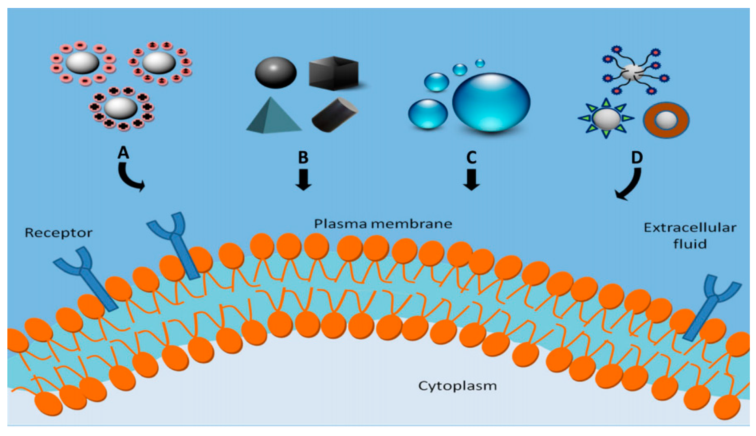
The beauty of human-made or cell-manipulated nanoparticles is that they are crafted from the best material with the right properties [102]. Some use capsules to transport and dispense items to a particular area, while others use magnetism to use these particles and manipulate the shape of cells. This impact of particles, their shape, surface chemistry, and magnetic forces can be fine-tuned using different ranges and metal thicknesses which are adaptive and mobile. It allows dealing differently with all cases, rather than a one-size-fits-all approach to healthcare, as shown in the case of liposomal targeted drug delivery (Figure 7).
The next generation of nanotechnology will focus on commercial exploration. It accounts for more than basic medicines, drug delivery, and biomaterials which are the major focus areas of developers. One of the latest creations is Band-Aid and dressing with NPs of colloidal silver and anti-microbial elements. Several companies have been involved in this commercialization, which has been listed in Table 1. These companies utilize their quantum size effects in semiconductor nanocrystals for labeling bio-molecules and use bio-conjugated nanoparticles for tagging. Enhanced activity to destroy dangerous toxins and other organic constituents can be accomplished via nanoceramics and noble metals like platinum.

Application in Sensor Technology

Water filtration systems have potential applications using NPs involving nanoceramic sand metals that are being tried at experimental levels due to effective anti-microbial efficiency [104].
Proteomics and genomics generate an escalating number of sequences through screening. Various array technologies need multicolor coding for biological assays based on “bar coding” of polymer particles in solution, limited by the number of unique tags that are reliable to detect. Single quantum dots of compound semiconductors are effectively involved as a replacement for organic dyes via bio-tagging [105].
Another very interesting field is the manipulation of cells and bio-molecules, allowing the use of functionalized magnetic nanoparticles for cell separation and probing. The magnetic nanoparticles are usually spherical, which limits their functions. Cylindrical-shaped nanoparticles are a better choice allowing metal electro-deposition with tuned properties. Different ligands can be linked to different segments, like in the case of gold and nickel, used to produce spatially segregated fluorescent parts.
To conclude the application section, numerous and diverse applications have been observed for nanoparticles and are used as fluorescent biological labels [106], drug/ gene delivery [51], bio-detection of pathogens [107,108], protein detection [109], probing of DNA structure [110,111], tissue engineering [99], tumor detection via heating or hyperthermia, separation, and purification of biological molecules [112], MRI contrast enhancement, phago-kinetic studies [113]. All these technological innovations evolve towards integrating new synthetic patterning and material synthesis into a collection of synthetic biological parts and tools.

6. Drawbacks/Limitations

Currently, nano-research has a wide range of applications that offer numerous advantages for daily use in various fields, such as food, packaging, accessories, storage, transportation, and more. Even electronics, medical surgery, lighting, clothes, etc., all have uses for these nano-based systems. Despite the potential good uses accompanying these technologies, the potential for evil is tremendously high. A crucial system of checks and balances should be maintained to prevent the mishandling of scientific research and capabilities. Regardless of their growth and advancement, several critical considerations on the prevalence of nanotechnology in the world form the basis of ongoing debates. There are anticipated risks and issues that are concerning with several disadvantages. It has evolved the standard of living parallel to increasing the pollution levels (air, soil, water, food), commonly termed nano-pollution. It is quite hazardous for the biotic components [114]. A few bottlenecks of nanoworld have been listed:
  • A major shortcoming is the lack of work opportunities in the arenas of traditional farming with the manufacturing and engineering sector. These nanorobots and sophisticated machines have lessened the prominence of manpower but guarantee fast and accurate output;
  • This technology has invoked an opportunity for atomic-scale evaluation devices, which would have been virtually impossible. Moreover, since they are very minute, they can be weaponized in due course. These nano-sized weapons would be comparatively easier to create, and therefore novel systems can be initiated. One of the likely prospective is a ‘smart-bullet’, a computerized bullet to facilitate the aim in a controlled and accurate manner. Such kinds of innovations might be a boon for the military, but if not produced thoughtfully and carefully, the consequences could be dire. These kinds of atomic weapon systems are much more powerful and destructive. The potential toxic effect due to industrial-scale manufacturing on human health is unforeseen;
  • Mass production of nano-size formulations might not be possible. Such nanotechnological-based procurement for honest and true molecular-sized manufacturing might not be possible;
  • There is the probable scope of mass poisoning over the time frame that could eventually lead to health complications for those who consume them, i.e., health deterioration. Inhaled nanoparticulates settle in the brain and lungs, which will lead to an increase in biomarkers causing inflammation and stress;
  • Roughly more extravagant negative future scenarios are debunked by experts in nanotechnology. One such example is the so-called ‘gray go’ scenario. This concept was a hypothetical catastrophic scenario that involves molecular nanotechnology wherein self-replicating nanobots consume all the biomass on the Earth while replicating themselves. It was assumed that machines might somehow gain the capability of eating the habitation and self-replicating by accident. It might be due to potential new toxins and pollutants;
  • The absence of complete understanding regarding nanotechnology makes it a rather challenging subject for manufacturers. The proper and essential impact of these products needs to be considered equally;
  • This technological innovation is quite uneconomical for the generation and assembly of particles in various models and needs expertise. Moreover, technological advances prior to it had caused alterations in many ways with the trend of luxury products, thereby making the system obsolete and expensive. Change in the industrial sector had resulted in huge job losses;
  • With the advancements and performance initiatives, certain markets like diamond and oil have fallen due to the introduction of various alternatives. These alternatives are better off with faster production, robustness and do not require fossil fuels as starting materials. This is one of the major reasons for the fall in the market value of diamonds, as quite massive amounts can be used as potential alternates;
  • The risk factor involved in manufacturing accounts for huge money investments with upscaling of nanoplants and customer satisfaction risk could make them bear surplus losses. Maintaining such products is equally costly;
  • Issues of practicality, including production from masses such as fossil fuels (like coal and petroleum), have led to the crashing of small-scale industrial sectors as nanotechnology does not leave a single particle unfurnished.
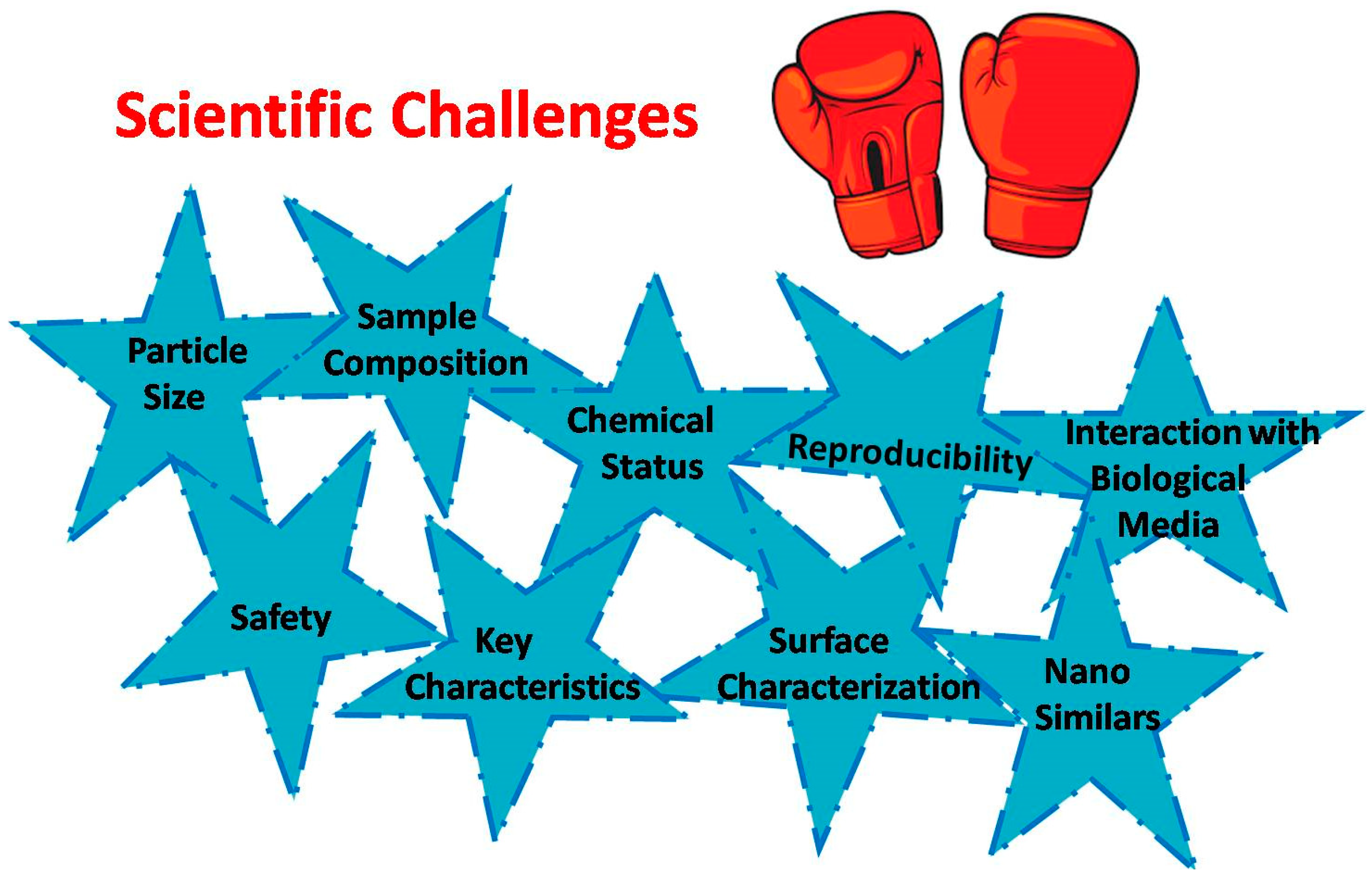
Continuing on the mentioned downsides, many researchers have documented the adversities of toxicological responses over the decades. However, various changes in the behavior of nanoparticulate entities can help us in building a safe environment, and it is foreseen that even minute deviations in the physicochemical features lead to substantial changes in the behavioral patterns, compelling predictivity on toxicity concerns [85]. Our lab has initiated some low-cost carbon-based systems for the electrochemical immunosensor fabrications further used for biomimetic catalysis of bioanalytes [107,115,116,117,118,119,120]. The rationale behind such advancements could be a pioneering platform that could be highly beneficial. The basic scientific challenges encountered during their authorization are described in Figure 8.
In continuing the discussion on low-cost nanosystems for biomimetic applications, we are working towards the exploration of opportunities such as novel systems against acute diseases. Astonishingly, global efforts for computational modeling and screening are essential to explore the requisites of such atomic scale materials viz a viz, dimensions, homing material characteristics, water-loving/hating properties, stability, and surface characteristics for precise and effective therapeutic applications.

7. Future of Nanomaterials

Nanotech opens a wonderful window into the upcoming era of bio-mechanics and bio-medicine. All the applications mentioned in this review are functional and in continuation of developmental processes. Further, there is plenty of room to be unveiled for prospective ventures with drug delivery systems and cell targeting, which makes them even more competent and effective [121]. A few of the reported nanoparticles used in company-manufactured products are listed in Table 1. The real future of nanomaterials will be determined by their rationale design, new instrumentations, and medical purposes expansion.
Nanomaterials are the cornerstone of the rapidly advancing field of nanotechnology, having the potential to revolutionize diagnostics and therapeutics. The majority of commercially available nanoparticles are geared towards basic medications. Nanoparticles replacing organic dyes require photo-stability and high multiplexing capabilities. Developments in directing and remotely controlling the functions of nanoprobes help drive magnetic nanoparticles to tumor cells, making them release the drug load or heat them in order to destroy affected cells or cure the diseased cells. Major developments of nonmaterial are to make them multifunctional and controllable by external signals for turning them into nano-devices. There are yet new advancements to be worked upon, especially focused devices with multifunctional operations that can provide medical benefits.

8. Conclusions and Outlook

As the physical parameters change, their properties are influenced drastically. The most promising platforms, like carbon nanotubes composed of pure carbon (sp2 hybridized carbons), that has the same composition as that of pencil graphite and have been used as pencil graphite electrodes (PGE) [122]. Later on, as the carbon is arranged in a layered tubular structure, the material obtains better mechanical and thermal properties, which makes the system stiffer compared to steel [123]. AuNPs have the same material composition as Au but contribute to various colors similar to that of old jewelry, but this is due to the various shapes of AuNP, which allows various colors compared to yellow for bulk gold, the root cause being the interaction of light and further reflectance with AuNPs. These different colors can be employed as biomarkers and fluorescent inducers for the diagnosis of health-related conditions. Similarly, as we go towards the nano levels, the basic characteristics of the material change. For example, many metal-based systems, such as copper, which is extremely rigid at the nano level, can be ductile wire in bulk metal. In continuation, the use of magnetic nanoparticles is equally promising, with a unique set of features ensuring target-specific delivery with customizations. They have been extended for cell isolation, immunoassays, diagnostic testing, and drug delivery approaches. Moreover, many inorganic and polymer-based systems have been extended for real-time biomedical applications in real life, as described in Table 1.
This nano-atomic-scale study involves a vivid imagination that incorporates diverse streams of engineering, science and technology, biomedical, manufacturing, etc. In the near future, their effective usage can be broadened from energy storage and biofuel cell applications to other modes of computing, wearable sensors, storage systems, packaging of food and consumables, lightweight accessories, and to road and railway transportation vehicles, thereby creating a safer, satisfactory and effective consumer-oriented end-products. Many more arenas of exploration are yet to be initiated by the researchers.
Innovative and brainstorming systems for the detection and cure of foreign diseases are being established for the first counter alert. These are based on the determination of changes in the vitals, changes in biomarker levels, hormonal misbalance, etc., to enhance the ability for defined diagnosis and early treatment. With the technological upgradation in the designing of nanostructures, targeted delivery and healing acceleration are a few alterations being included.
Scientists are improving diagnostic and therapeutic devices in line with modern health needs [124,125]. Medicine nanoformulations (coated nanoparticles) for treatment, nanorobots for visualization of inbody elements, nanoprobes for monitoring of fatty plaques, nanofibers for tooth−decay-based detection, and nanoelectrodes being employed for regaining brain function are being researched. By understanding the current development and process of functionalized nanoparticle application in theranostics, they can directionalize to the most promising study in the future.
Health and safety concerns hold much promise for future prospects; challenging concerns related to issues of safe and sustainable cofactors are on the agenda list of NIH. They carry out essential research evaluations and add transformations for safety and material concerns. The national nanotechnology initiative has participated in the national nanotechnology initiative (NNI), which co-ordinates the federal government’s implementation of the 21st-century nanotechnology research and development act. These activities account for about 1.3 billion annual total government expenditure.
In extension to the drug delivery mediated system, the important roles of the new armamentarium of therapeutics are pipelines to existing drugs, later improving their efficacy. Various efforts are being put forth to revisit the status of suboptimal but biologically active molecular systems that follow through conventional sources at a prompt pace. Fascinatingly, the attributes of these nano-objects are bestowed to their therapeutic properties, which might be polluting. Hence, fine-tuning of adversities that might occur needs to be lessened. So, more elaborate studies in such sectors for a smarter, faster, eco-friendly, and sustainable system can trounce daunting challenges associated with various platforms.

Author Contributions

M.G.—Conceptualization, methodology, writing, project administration, validation, investigation, supervision, and writing, validation, investigation and supervision; K.A.—validation, investigation, and supervision. All authors have read and agreed to the published version of the manuscript.

Funding

This research received no external funding.

Data Availability Statement

Not applicable.

Acknowledgments

The authors would like to acknowledge Vellore Institute of Technology for giving them an opportunity to pursue higher education. Mansi Gandhi would like to thank ICMR for the support of SRF (2019-4952). Mansi Gandhi would like to thank Hebrew University of Jerusalem for her postdoc research. ICMR K. Amreen acknowledges St. Anns’ College for Women, Mehdipatnam-Hyderabad, for the support.

Conflicts of Interest

The authors declare no conflict of interest.

References

  1. Bayda, S.; Adeel, M.; Tuccinardi, T.; Cordani, M.; Rizzolio, F. The history of nanoscience and nanotechnology: From chemical-physical applications to nanomedicine. Molecules 2020, 25, 112. [Google Scholar] [CrossRef] [Green Version]
  2. Zhang, W. Application of Fe3O4 nanoparticles functionalized carbon nanotubes for electrochemical sensing of DNA hybridization. J. Appl. Electrochem. 2016, 46, 559–566. [Google Scholar] [CrossRef]
  3. Eldar-Boock, A.; Polyak, D.; Scomparin, A.; Satchi-Fainaro, R. Nano-sized polymers and liposomes designed to deliver combination therapy for cancer. Curr. Opin. Biotechnol. 2013, 24, 682–689. [Google Scholar] [CrossRef]
  4. Shivenduuranjan, N.; Bhartenduunathhmishraaeditors, E. (Eds.) Environmental Chemistry for a Sustainable World 37 Environmental Nanotechnology; Springer: Berlin/Heidelberg, Germany, 2021; Volume 5. [Google Scholar]
  5. Parikh, A.N. Biologically Inspired Nanomaterials: A Conference Report Hybrid opto-plasmonic integration for nanoscale light & heat management View project. arXiv 2007, arXiv:0708.0164. [Google Scholar]
  6. Allara, D.L. A perspective on surfaces and interfaces. Nature 2005, 437, 638–639. [Google Scholar] [CrossRef]
  7. Yan, M.; Ren, J. Covalent immobilization of polypropylene thin films. J. Mater. Chem. 2005, 15, 523–527. [Google Scholar] [CrossRef]
  8. Pursel, S.; Horn, M.W.; Demirel, M.C.; Lakhtakia, A. Growth of sculptured polymer submicronwire assemblies by vapor deposition. Polymer 2005, 46, 9544–9548. [Google Scholar] [CrossRef]
  9. Vegting, I.L.; Nanayakkara, P.W.B.; van Dongen, A.E.; Vandewalle, E.; van Galen, J.; Kramer, M.H.H.; Bonjer, J.; Koole, G.M.; Visser, M.C. Analysing completion times in an academic emergency department: Coordination of care is the weakest link. Neth. J. Med. 2011, 69, 392–398. [Google Scholar]
  10. Muthukrishnan, G.; Roberts, C.A.; Chen, Y.C.; Zahn, J.D.; Hancock, W.O. Patterning surface-bound microtubules through reversible DNA hybridization. Nano Lett. 2004, 4, 2127–2132. [Google Scholar] [CrossRef]
  11. Lu, C.; Wang, Y.; Yang, S.; Wang, C.; Sun, X.; Lu, J.; Yin, H.; Jiang, W.; Meng, H.; Rao, F. Bioactive Self-Assembling Peptide Hydrogels Functionalized with Brain-Derived Neurotrophic Factor and Nerve Growth Factor Mimicking Peptides Synergistically Promote Peripheral Nerve Regeneration. ACS Biomater. Sci. Eng. 2018, 4, 2994–3005. [Google Scholar] [CrossRef]
  12. Hynonen, U.; Palva, A. Lactobacillus surface layer proteins: Structure, function and applications. Appl. Microbiol. Biotechnol. 2013, 97, 5225–5243. [Google Scholar] [CrossRef] [Green Version]
  13. Pum, D.; Neubauer, A.; Gyoervary, E.; Sára, M.; Sleytr, U.B. S-layer proteins as basic biomolecular building blocks S-layer proteins as basic biomolecular building blocks. Adv. Packag. 2008, 100, 2–8. [Google Scholar]
  14. Sleytr, U.B.; Schuster, B.; Egelseer, E.M.; Pum, D. S-layers: Principles and applications. FEMS Microbiol. Rev. 2014, 38, 823–864. [Google Scholar] [CrossRef] [Green Version]
  15. Messner, P.; Pum, D.; Sara, M.; Stetter, K.O.; Sleytr, U.B. Ultrastructure of the cell envelope of the archaebacteria Thermoproteus tenax and Thermoproteus neutrophilus. J. Bacteriol. 1986, 166, 1046–1054. [Google Scholar] [CrossRef] [Green Version]
  16. Allara, D.L.; Nuzzo, R.G. Spontaneously Organized Molecular Assemblies. 1. Formation, Dynamics, and Physical Properties of n-alkanoic acids adsorbed from solution on an oxidized aluminum surface. Langmuir 1985, 1, 45–52. [Google Scholar] [CrossRef]
  17. Yun, C.S.; Javier, A.; Jennings, T.; Fisher, M.; Hira, S.; Peterson, S.; Hopskin, B.; Reich, N.O.; Strouse, G.F. Nanometal surface energy transfer in optical rulers, breaking the FRET barrier. J. Am. Chem. Soc. 2005, 127, 3115–3119. [Google Scholar] [CrossRef]
  18. Shim, M.; Kam, N.W.S.; Chen, R.J.; Li, Y.; Dai, H. Functionalization of Carbon Nanotubes for Biocompatibility and Biomolecular Recognition. Nano Lett. 2002, 2, 285–288. [Google Scholar] [CrossRef]
  19. Reed, S.M.; Clegg, R.S.; Hutchison, J.E. Tuning the Electrical Properties of Monolayers by Using Internal Peptide Bonds. ACS Symp. Ser. 2003, 844, 36–50. [Google Scholar]
  20. You, C.C.; De, M.; Rotello, V.M. Monolayer-protected nanoparticle-protein interactions. Curr. Opin. Chem. Biol. 2005, 9, 639–646. [Google Scholar] [CrossRef]
  21. Xiao, Y.; Patolsky, F.; Katz, E.; Hainfeld, J.F.; Willner, I. “Plugging into Enzymes”: Nanowiring of redox enzymes by a gold nanoparticle. Science 2003, 299, 1877–1882. [Google Scholar] [CrossRef]
  22. Rice, M.K.; Ruder, W.C. Creating biological nanomaterials using synthetic biology. Sci. Technol. Adv. Mater. 2014, 15, 014401. [Google Scholar] [CrossRef] [Green Version]
  23. Rastogi, V.; Yadav, P.; Bhattacharya, S.S.; Mishra, A.K.; Verma, N.; Verma, A.; Pandit, J.K. Carbon Nanotubes: An Emerging Drug Carrier for Targeting Cancer Cells. J. Drug Deliv. 2014, 2014, 1–23. [Google Scholar] [CrossRef] [Green Version]
  24. Press, D. IJN-16923-new-generation-of-cancer-treatment--carbon-nanotubes-with-fl. Dovepress 2011, 2011, 2963–2979. [Google Scholar] [CrossRef] [Green Version]
  25. Chen, Z.; Zhang, A.; Wang, X.; Zhu, J.; Fan, Y.; Yu, H.; Yang, Z. The Advances of Carbon Nanotubes in Cancer Diagnostics and Therapeutics. J. Nanomater. 2017, 2017, 3418932. [Google Scholar] [CrossRef] [Green Version]
  26. Kam, N.W.S.; Dai, H. Single walled carbon nanotubes for transport and delivery of biological cargos. Phys Status Solidi B Basic Res. 2006, 243, 3561–3566. [Google Scholar] [CrossRef] [Green Version]
  27. da Silva Bruckmann, F.; Nunes, F.B.; da Rosa Salles, T.; Franco, C.; Cadoná, F.C.; Bohn Rhoden, C.R. Biological Applications of Silica-Based Nanoparticles. Magnetochemistry 2022, 8, 131. [Google Scholar] [CrossRef]
  28. Mittal, A.; Roy, I.; Gandhi, S. Magnetic Nanoparticles: An Overview for Biomedical Applications. Magnetochemistry 2022, 8, 107. [Google Scholar] [CrossRef]
  29. Baptista, P.V.; McCusker, M.P.; Carvalho, A.; Ferreira, D.A.; Mohan, N.M.; Martins, M.; Fernandes, A.R. Nano-strategies to fight multidrug resistant bacteria–“A Battle of the Titans”. Front. Microbiol. 2018, 9, 1–26. [Google Scholar] [CrossRef] [Green Version]
  30. Rudramurthy, G.R.; Swamy, M.K.; Sinniah, U.R.; Ghasemzadeh, A. Nanoparticles: Alternatives against drug-resistant pathogenic microbes. Molecules 2016, 21, 1–30. [Google Scholar] [CrossRef] [Green Version]
  31. Guru, E.; Chatterjee, S. Study of Synthetic Biomolecular Network in Escherichia Coli. Int. J. Biophys. 2013, 3, 38–50. [Google Scholar]
  32. Anuchapreeda, S.; Fukumori, Y.; Okonogi, S.; Ichikawa, H. Preparation of lipid nanoemulsions incorporating curcumin for cancer therapy. J. Nanotechnol. 2012, 2012, 270383. [Google Scholar] [CrossRef] [Green Version]
  33. DeLong, R.K.; Akhtar, U.; Sallee, M.; Parker, B.; Barber, S.; Zhang, J.; Caig, M.; Garrad, R.; Hickey, A.J.; Engstrom, E. Characterization and performance of nucleic acid nanoparticles combined with protamine and gold. Biomaterials 2009, 30, 6451–6459. [Google Scholar] [CrossRef] [Green Version]
  34. Popova, V.; Dmitrienko, E.; Chubarov, A. Magnetic Nanocomposites and Imprinted Polymers for Biomedical Applications of Nucleic Acids. Magnetochemistry 2023, 9, 12. [Google Scholar] [CrossRef]
  35. Bulgakova, A.; Chubarov, A.; Dmitrienko, E. Magnetic Nylon 6 Nanocomposites for the Microextraction of Nucleic Acids from Biological Samples. Magnetochemistry 2022, 8, 85. [Google Scholar] [CrossRef]
  36. Irkham, I.; Ibrahim, A.U.; Pwavodi, P.C.; Al-Turjman, F.; Hartati, Y.W. Smart Graphene-Based Electrochemical Nanobiosensor for Clinical Diagnosis: Review. Sensors 2023, 23, 2240. [Google Scholar] [CrossRef]
  37. Chen, Y.; Ho, J.; Shis, D.; Gupta, C.; Long, J.; Wagner, D.; Ott, W.; Josic, K.; Bennett, M. Tuning the dynamic range of bacteria; promoters regulated by ligand-inducible transcription factors. Nat. Comm. 2018, 9, 64. [Google Scholar] [CrossRef] [Green Version]
  38. Rudge, T.J.; Brown, J.R.; Federici, F.; Dalchau, N.; Phillips, A.; Ajioka, J.W.; Haseloff, J. Characterization of Intrinsic Properties of Promoters. ACS Synth. Biol. 2016, 5, 89–98. [Google Scholar] [CrossRef] [Green Version]
  39. Joseph, T.M.; Mahapatra, D.K.; Esmaeili, A.; Piszczyk, L.; Hasanin, M.S.; Kattali, M.; Haponiuk, J.; Thomas, S. Nanoparticles: Taking a Unique Position in Medicine. Nanomaterials 2023, 13, 574. [Google Scholar] [CrossRef]
  40. Golbabaei, F.; Ebrahimi, A.; Shirkhanloo, H.; Koohpaei, A.; Faghihi-zarandi, A. Single-Walled Carbon Nanotubes (SWCNTs), as a Novel Sorbent for Determination of Mercury in Air. Glob. J. Heal. Sci. 2016, 8, 273–280. [Google Scholar] [CrossRef] [Green Version]
  41. Thakor, A.S.; Gambhir, S.S. Nanooncology: The Future of Cancer Diagnosis and Therapy. CA A Cancer J. Clin. 2013, 63, 395–418. [Google Scholar] [CrossRef]
  42. Walia, N.; Dasgupta, N.; Ranjan, S.; Ramalingam, C.; Gandhi, M. Methods for nanoemulsion and nanoencapsulation of food bioactives. Environ. Chem. Lett. 2019, 17, 1471–1483. [Google Scholar] [CrossRef]
  43. Dasgupta, N.; Ranjan, S.; Gandhi, M. Nanoemulsions in food: Market demand. Environ. Chem. Lett. 2019, 17, 1003–1009. [Google Scholar] [CrossRef]
  44. Walia, N.; Dasgupta, N.; Ranjan, S.; Ramalingam, C.; Gandhi, M. Food-grade nanoencapsulation of vitamins. Environ. Chem. Lett. 2019, 17, 991–1002. [Google Scholar] [CrossRef]
  45. Dasgupta, N.; Ranjan, S.; Ramalingam, C.; Gandhi, M. Silver nanoparticles engineered by thermal co-reduction approach induces liver damage in Wistar rats: Acute and sub-chronic toxicity analysis. 3 Biotech 2019, 9, 1–12. [Google Scholar] [CrossRef]
  46. Ranjan, S.; Dasgupta, N.; Singh, S.; Gandhi, M. Toxicity and regulations of food nanomaterials. Environ. Chem. Lett. 2018, 17, 929–944. [Google Scholar] [CrossRef]
  47. Sharma, A.K.; Keservani, R.K.; Kesharwani, R.K. Nanobiomaterials Applications in Drug Delivery; Apple Academic Press: Waretown, NJ, USA, 2021; ISBN 9781774636442. [Google Scholar]
  48. Sharmila, G.; Muthukumaran, C.; Sangeetha, E.; Saraswathi, H.; Soundarya, S.; Kumar, N.M. Green fabrication, characterization of Pisonia alba leaf extract derived MgO nanoparticles and its biological applications. Nano-Struct. Nano-Objects 2019, 20, 100380. [Google Scholar] [CrossRef]
  49. Amos-Tautua, B.M.; Fakayode, O.J.; Songca, S.P.; Oluwafemi, O.S. Evolution of gluconic acid capped paramagnetic iron oxide nanoparticles. Nano-Struct. Nano-Objects 2019, 20, 100389. [Google Scholar] [CrossRef]
  50. Bharti, D.K.; Gupta, M.K.; Srivastava, A.K. Temperature dependent dielectric and electric properties of zinc silicate nanorods. Nano-Struct. Nano-Objects 2019, 17, 123–128. [Google Scholar] [CrossRef]
  51. Knipe, J.; Peters, J.; Peppas, N. Theranostic agents for intracellular gene delivery with spatiotemporal imaging. Nano Today 2013, 8, 21–38. [Google Scholar] [CrossRef] [Green Version]
  52. Gao, Q.; Feng, J.; Liu, W.; Wen, C.; Wu, Y.; Liao, Q.; Zou, L.; Sui, X.; Xie, T.; Zhang, J.; et al. Opportunities and challenges for co-delivery nanomedicines based on combination of phytochemicals with chemotherapeutic drugs in cancer treatment. Adv. Drug Deliv. Rev. 2022, 188, 114445. [Google Scholar] [CrossRef]
  53. Veronese, F.M.; Pasut, G. PEGylation, successful approach to drug delivery. Drug Discov. Today 2005, 10, 1451–1458. [Google Scholar] [CrossRef]
  54. Ganson, N.J.; Kelly, S.J.; Scarlett, E.; Sundy, J.S.; Hershfield, M.S. Research article Control of hyperuricemia in subjects with refractory gout, and induction of antibody against poly (ethylene glycol) (PEG), in a phase I trial of subcutaneous PEGylated urate oxidase. Thromb. Haemost. 2006, 8, R12. [Google Scholar] [CrossRef] [Green Version]
  55. Schroeder, A.; Heller, D.A.; Winslow, M.M.; Dahlman, J.E.; Pratt, G.W.; Langer, R.; Jacks, T.; Anderson, D.G. Treating metastatic cancer with nanotechnology. Nat. Rev. Cancer 2012, 12, 39–50. [Google Scholar] [CrossRef]
  56. Kamada, H.; Tsutsumi, Y.; Sato-Kamada, K.; Yamamoto, Y.; Yoshioka, Y.; Okamoto, T.; Nakagawa, S.; Nagata, S.; Mayumi, T. Synthesis of a poly (vinylpyrrolidone-co-dimethyl maleic anhydride) co-polymer and its application for renal drug targeting. Nat. Biotechnol. 2003, 21, 399–404. [Google Scholar] [CrossRef]
  57. Eslami, P.; Albino, M.; Scavone, F.; Chiellini, F.; Morelli, A.; Baldi, G.; Cappiello, L.; Doumett, S.; Lorenzi, G.; Ravagli, C.; et al. Smart Magnetic Nanocarriers for Multi-Stimuli On-Demand Drug Delivery. Nanomaterials 2022, 12, 303. [Google Scholar] [CrossRef]
  58. Jain, R.A. The manufacturing techniques of various drug loaded biodegradable poly (lactide-co-glycolide) (PLGA) devices. Biomaterials 2000, 21, 2475–2490. [Google Scholar] [CrossRef]
  59. Tian, J.; Min, Y.; Rodgers, Z.; Au, K.M.; Hagan, C.; Zhang, M.; Roche, K.; Yang, F.; Wagner, K.; Wang, A. Co-delivery of paclitaxel and cisplatin with biocompatible PLGA-PEG nanoparticles enhances chemoradiotherapy in non-small cell lung cancel models. J. Mat. Chem. B 2017, 5, 6045–6057. [Google Scholar] [CrossRef]
  60. Farazuddin, M.; Sharma, B.; Khan, A.A.; Joshi, B.; Owais, M. Anticancer efficacy of perillyl alcohol-bearing PLGA microparticles. Int. J. Nanomed. 2012, 7, 35–47. [Google Scholar]
  61. Li, S.; Lin, M.M.; Toprak, M.S.; Kim, D.K.; Muhammed, M. Nanocomposites of polymer and inorganic nanoparticles for optical and magnetic applications. Nano Rev. 2010, 1, 1–19. [Google Scholar] [CrossRef]
  62. Papasani, M.R.; Wang, G.; Hill, R.A. Gold nanoparticles: The importance of physiological principles to devise strategies for targeted drug delivery. Nanomedicine 2012, 8, 804–814. [Google Scholar] [CrossRef]
  63. McCarroll, J.; Baigude, H.; Yang, C.; Rana, T. Nanotubes functionalized with lipids and natural amino acid dendrimers: A new strategy to create nanomaterials for delivering systemic RNAi. Bioconjug. Chem. 2010, 1, 56–63. [Google Scholar] [CrossRef] [Green Version]
  64. Connor, E.E.; Mwamuka, J.; Gole, A.; Murphy, C.J.; Wyatt, M.D. Toxicity of nanoparticles Gold Nanoparticles Are Taken Up by Human Cells but Do Not Cause Acute Cytotoxicity. Small 2005, 1, 325–327. [Google Scholar] [CrossRef]
  65. Shukla, R.; Bansal, V.; Chaudhary, M.; Basu, A.; Bhonde, R.R.; Sastry, M. Biocompatibility of Gold Nanoparticles and Their Endocytotic Fate Inside the Cellular Compartment: A Microscopic Overview. Langmuir 2005, 21, 10644–10654. [Google Scholar] [CrossRef]
  66. Ghosh, P.; Han, G.; De, M.; Kim, C.K.; Rotello, V.M. Gold nanoparticles in delivery applications. Adv. Drug Deliv. Rev. 2008, 60, 1307–1315. [Google Scholar] [CrossRef]
  67. Hong, R.; Han, G.; Ferna, J.M.; Kim, B.; Forbes, N.S.; Rotello, V.M. Glutathione-Mediated Delivery and Release Using Monolayer Protected Nanoparticle Carriers. J. Am. Chem. Soc. 2006, 128, 1078–1079. [Google Scholar] [CrossRef]
  68. Polizzi, M.A.; Stasko, N.A.; Schoenfisch, M.H. Water-Soluble Nitric Oxide-Releasing Gold Nanoparticles. Langmuir 2007, 23, 4938–4943. [Google Scholar] [CrossRef]
  69. Chen, C.-C.; Lin, Y.-P.; Wang, C.-W.; Tzeng, H.-C.; Wu, C.-H.; Chen, Y.-C.; Chen, C.-P.; Chen, L.-C.; Wu⊥, Y.-C. DNA−Gold Nanorod Conjugates for Remote Control of Localized Gene Expression by near Infrared Irradiation. J. Am. Chem. Soc. 2006, 128, 3709–3715. [Google Scholar] [CrossRef]
  70. Chauhan, A.; Zubair, S.; Tufail, S.; Sherwani, A.; Sajid, M.; Raman, S.C.; Azam, A.; Owais, M. Fungus-mediated biological synthesis of gold nanoparticles: Potential in detection of liver cancer. Int. J. Nanomed. 2011, 6, 2305–2319. [Google Scholar]
  71. Dreaden, E.; Mwakwari, S.; Sodji, Q.; Oyelere, A.; Sayed, M. Tamoxifen-Poly(ethylene glycol)-thiol gold nanoparticle conjugates: Enhanced potency and selective delivery for breast cancer treatment. Bioconjug Chem. 2010, 20, 2247–2253. [Google Scholar] [CrossRef] [Green Version]
  72. Ducan, B.; Kim, C.; Rotello, V.M. Gold nanoparticle platforms as drug and biomacromolecule delivery systems. J. Control. Release 2011, 148, 122–127. [Google Scholar] [CrossRef] [Green Version]
  73. Elbakry, A.; Zaky, A.; Liebl, R.; Rachel, R.; Goepferich, A.; Breunig, M. Layer-by-Layer Assembled Gold Nanoparticles for siRNA Delivery. Nano Lett. 2009, 9, 2059–2064. [Google Scholar] [CrossRef]
  74. Song, W.; Du, J.; Sun, T.; Zhang, P.; Wang, J. Gold Nanoparticles Capped with Polyethyleneimine for Enhanced siRNA Delivery. Small 2010, 6, 239–246. [Google Scholar] [CrossRef]
  75. Biocompatibility, G.; Han, L.; Zhao, J.; Zhang, X.; Cao, W.; Hu, X.; Zou, G.; Duan, X.; Liang, X.-J. Enhanced siRNA Delivery and Silencing Gold À Chitosan Nanosystem with Surface Charge-Reversal Polymer. ACS Nano 2012, 6, 7340–7351. [Google Scholar]
  76. Fraguas-Sánchez, A.I.; Lozza, I.; Torres-Suárez, A.I. Actively Targeted Nanomedicines in Breast Cancer: From Pre-Clinal Investigation to Clinic. Cancers 2022, 14, 1198. [Google Scholar] [CrossRef]
  77. Obozina, A.S.; Komedchikova, E.N.; Kolesnikova, O.A.; Iureva, A.M.; Kovalenko, V.L.; Zavalko, F.A.; Rozhnikova, T.V.; Tereshina, E.D.; Mochalova, E.N.; Shipunova, V.O. Genetically Encoded Self-Assembling Protein Nanoparticles for the Targeted Delivery In Vitro and In Vivo. Pharmaceutics 2023, 15, 231. [Google Scholar] [CrossRef]
  78. Bonoiu, A.C.; Mahajan, S.D.; Ding, H.; Roy, I.; Yong, K.; Kumar, R.; Hu, R.; Bergey, E.L.; Schwartz, S.A.; Prasad, P.N. Nanotechnology approach for drug addiction therapy: Gene silencing using delivery of gold nanorod-siRNA nanoplex in dopaminergic neurons. Proc. Nalt. Acad. Sci. USA 2009, 106, 5546–5550. [Google Scholar] [CrossRef]
  79. Massich, M.D.; Giljohann, D.A.; Seferos, D.S.; Ludlow, L.E.; Horvath, C.M.; Mirkin, C.A. Regulating immune response using polyvalent nucleic acid-gold nanoparticle conjugates. Mol. Pharmaceutics 2009, 6, 1934–1940. [Google Scholar] [CrossRef] [Green Version]
  80. Meade, B.R.; Dowdy, S.F. Enhancing the cellular uptake of siRNA duplexes following noncovalent packaging with protein transduction domain peptides. Adv. Drug Deliv. Rev. 2008, 60, 530–536. [Google Scholar] [CrossRef] [Green Version]
  81. Salvador-morales, C.; Flahaut, E.; Sim, E.; Sloan, J.; Green, M.L.H.; Sim, R.B. Complement activation and protein adsorption by carbon nanotubes. Mol. Immunol. 2006, 43, 193–201. [Google Scholar] [CrossRef] [Green Version]
  82. Gandhi, M.; Chen, S.; Ray, S.S.; Jaiswal, N.; Ranjan, S. Phyto-nanosensors: Advancement of phytochemicals as an electrochemical platform for various biomedical applications. Environ. Nanotechnol. 2021, 5, 311–338. [Google Scholar] [CrossRef]
  83. Zhao, P.; Tian, Y.; You, J.; Hu, X.; Liu, Y. Recent Advances of Calcium Carbonate Nanoparticles for Biomedical Applications. Bioengineering 2022, 9, 691. [Google Scholar] [CrossRef]
  84. Popova, V.; Poletaeva, Y.; Chubarov, A.; Dmitrienko, E. pH-Responsible Doxorubicin-Loaded Fe3O4@CaCO3 Nanocomposites for Cancer Treatment. Pharmaceutics 2023, 15, 771. [Google Scholar] [CrossRef] [PubMed]
  85. Chevallet, M.; Veronesi, G.; Fuchs, A.; Mintz, E.; Michaud-Soret, I.; Deniaud, A. Impact of labile metal nanoparticles on cellular homeostasis. Current developments in imaging, synthesis and applications. Biochim. Biophys. Acta (BBA)-Gen. Subj. 2017, 1861, 1566–1577. [Google Scholar] [CrossRef] [PubMed]
  86. Liu, Z.; Sun, X.; Nakayama-ratchford, N.; Dai, H. Supramolecular Chemistry on Water- Soluble Carbon Nanotubes for Drug Loading and Delivery. ACS Nano 2007, 1, 50–56. [Google Scholar] [CrossRef] [PubMed]
  87. Kim, K.Y. Nanotechnology platforms and physiological challenges for cancer therapeutics. Nanomed. Nanotechnol. Biol. Med. 2007, 3, 103–110. [Google Scholar] [CrossRef] [PubMed]
  88. Mohapatra, D.P.; Gassara, F.; Brar, S.K. Nanoparticles—Production and Role in Biotransformation Copyright: American Scientific Publishers. J. Nanosci. Nanotechnol. 2011, 11, 899–918. [Google Scholar] [CrossRef]
  89. Sitharaman, B.; Wilson, L.J. Gadofullerenes and gadonanotubes: A new paradigm for high-performance magnetic resonance imaging contrast agent probes. J. Biomed. Nanotechnol. 2007, 3, 342–352. [Google Scholar] [CrossRef] [Green Version]
  90. Sitharaman, B.; Bolskar, R.D.; Rusakova, I.; Wilson, L.J. Gd@C 60 [C(COOH)2 ]10 and Gd@C60(OH)x: Nanoscale Aggregation Studies of Two Metallofullerene MRI Contrast Agents in Aqueous Solution. Nano Lett. 2004, 4, 2373–2378. [Google Scholar] [CrossRef]
  91. Galanis, A.; Rauch, E.; Arenal, R.; Portillo, J.; Casablanca, J.; Margiolaki, I. A novelty for cultural heritage material analysis: Transmission Electron Microscope (TEM) 3D electron diffraction tomography applied to Roman glass tesserae. Microchem. J. 2017, 138, 19–25. [Google Scholar] [CrossRef]
  92. Wu, L.; Zhang, J.; Watanabe, W. Physical and chemical stability of drug nanoparticles. Adv. Drug Deliv. Rev. 2011, 63, 456–469. [Google Scholar] [CrossRef]
  93. Hwang, Y.; Lee, J.-K.; Lee, J.-K.; Jeong, Y.-M.; Cheong, S.-I.; Ahn, Y.-C.; Kim, S.H. Production and dispersion stability of nanoparticles in nanofluids. Powder Technol. 2008, 186, 145–153. [Google Scholar] [CrossRef]
  94. Suriamoorthy, P.; Zhang, X.; Hao, G. Folic acid-CdTe quantum dot conjugates and their applications for cancer cell targeting. Cancer Nanotechnol. 2010, 1, 19–28. [Google Scholar] [CrossRef] [PubMed] [Green Version]
  95. Cao, Y.C.; Jin, R.; Nam, J.; Thaxton, C.S.; Mirkin, C.A. Raman Dye-Labeled Nanoparticle Probes for Proteins. J. Am. Chem. Soc. 2003, 125, 14676–14677. [Google Scholar] [CrossRef]
  96. Mallapragada, S.K.; Brenza, T.M.; McMillan, J.M.; Narasimhan, B.; Sakaguchi, D.S.; Sharma, A.D.; Zbarska, S.; Gendelman, H.E. Enabling nanomaterial, nanofabrication and cellular technologies for nanoneuromedicines. Nanomed. Nanotechnol. Biol. Med. 2015, 11, 715–729. [Google Scholar] [CrossRef] [Green Version]
  97. Roy, I.; Ohulchanskyy, T.Y.; Pudavar, H.E.; Bergey, E.J.; Oseroff, A.R.; Morgan, J.; Dougherty, T.J.; Prasad, P.N. Ceramic-Based Nanoparticles Entrapping Water-Insoluble Photosensitizing Anticancer Drugs: A Novel Drug—Carrier System for Photodynamic Therapy. J. Am. Chem. Soc. 2003, 125, 7860–7865. [Google Scholar] [CrossRef] [PubMed]
  98. Piszczek, P.; Radtke, A.; Tomasz, J. Biocompatibility of Titania Nanotube Coatings Enriched with Silver Nanograins by Chemical Vapor Deposition. Nanomaterials 2017, 7, 274. [Google Scholar] [CrossRef] [Green Version]
  99. Claro, A.P.R.A.; Konatu, R.T.; Escada, A.L.D.A.; Nunes, M.C.D.S.; Maurer-Morelli, C.V.; Dias-Netipanyj, M.F.; Popat, K.C.; Mantovani, D. Incorporation of Silver Nanoparticles on Ti7.5Mo Alloy Surface Containing TiO2 Nanotubes Arrays for Promoting Antibacterial Coating—In Vitro And In Vivo Study. Appl. Surf. Sci. 2018, 455, 780–788. [Google Scholar] [CrossRef]
  100. Bark, D.; Vahabi, H.; Bui, H.; Movafaghi, H.; Kota, A.; Popat, K.; Dasi, L.P. Hemodynamic performance and thrombogenic properties of superhydrophobic bileaflet mechanical heart valve. Annala Biomed. Eng. 2018, 45, 452–463. [Google Scholar] [CrossRef] [Green Version]
  101. Chan, W.C.W. Quantum-dot nanocrystals for ultrasensitive biological labeling and multicolor optical encoding. J. Biomed. Opt. 2002, 7, 532–537. [Google Scholar] [CrossRef]
  102. Gertz, F.; Khitun, A.; Gertz, F.; Khitun, A. Biological cell manipulation by magnetic nanoparticles Biological cell manipulation by magnetic nanoparticles. AIP Adv. 2016, 6, 025308. [Google Scholar] [CrossRef] [Green Version]
  103. Foroozandeh, P.; Aziz, A.A. Insight into Cellular Uptake and Intracellular Trafficking of Nanoparticles. Nanoscale Res. Lett. 2018, 13, 339. [Google Scholar] [CrossRef] [Green Version]
  104. Ray, S.S.; Gandhi, M.; Chen, S.-S.; Chang, H.-M.; Cao, D.-T.; Huy, L.Q. Anti-wetting behaviour of Superhydrophobic Octadecyltrimethoxysilane blended PVDF/Recycled Carbon Black Composite Membrane for enhanced desalination. Environ. Sci. Water Res. Technol. 2018, 4, 1612–1623. [Google Scholar] [CrossRef]
  105. Ma, W.; Aboagye-Mensah, D.; Soloviev, M.; Davletov, B.; Ferrari, E. ScienceDirect Symposium on Flexible Organic Electronics Protein Conjugation to Nanoparticles by Designer Affinity Tags. Mater. Today Proc. 2017, 4, 6923–6929. [Google Scholar] [CrossRef]
  106. Bruchez, M.P.; Weiss, S.; Angeles, L. Semiconductor Nanocrystals as Fluorescent Biological Labels Semiconductor Nanocrystals as Fluorescent Biological Labels. Science 2013, 281, 2013–2016. [Google Scholar] [CrossRef] [Green Version]
  107. Gandhi, M.; Rajagopal, D.; Parthasarathy, S.; Raja, S.; Huang, S.; Kumar, A.S. In Situ Immobilized Sesamol-Quinone / Carbon Nanoblack-Based Electrochemical Redox Platform for Efficient Bioelectrocatalytic and Immunosensor Applications. ACS Omega 2018, 3, 10823–10835. [Google Scholar] [CrossRef] [PubMed]
  108. Sharifi, S.; Vahed, S.Z.; Ahmadian, E.; Dizaj, S.M.; Eftekhari, A.; Khalilov, R.; Ahmadi, M.; Hamidi-Asl, E.; Labib, M. Detection of pathogenic bacteria via nanomaterials-modified aptasensors. Biosens. Bioelectron. 2020, 150, 111933. [Google Scholar] [CrossRef]
  109. Retout, M.; Valkenier, H.; Triffaux, E.; Doneux, T.; Bartik, K.; Bruylants, G. Rapid and Selective Detection of Proteins by Dual Trapping using Gold Nanoparticles functionalized with Peptide Aptamers. ACS Sensors 2016, 1, 929–933. [Google Scholar] [CrossRef] [Green Version]
  110. Ouyang, L.; Yao, R.; Mao, S.; Chen, X.; Na, J.; Sun, W. Three-dimensional bioprinting of embryonic stem cells directs highly uniform embryoid body formation. Biofabrication 2015, 7, 044101. [Google Scholar] [CrossRef]
  111. Mahtab, R.; Murphy, C.J. Probing DNA Structure With Nanoparticles. Method Mol. Biol. 2005, 303, 179–190. [Google Scholar]
  112. Chatterjee, S.; Basu, S.; Paul, T.; Bandyopadhyay, A.; Chattopadhyay, D.; Sarkar, K. A superparamagnetic nanoparticle based approach towards the purification of biomolecules. In Proceedings of the 4th International Conference on Advanced Nano Materials, IIT-Madras, Chennai, India, 17–19 October 2012. [Google Scholar]
  113. Parak, W.J.; Boudreau, R.; Le Gros, M.; Gerion, D.; Zanchet, D.; Micheel, C.M.; Willaims, S.C.; Alivisatos, A.P.; Larabell, C. Cell Motility and Metastatic Potential Studies Based on Quantum Dot Imaging of Phagokinetic Tracks. Adv. Mater. 2002, 4095, 10–14. [Google Scholar]
  114. Mishra, S.; Sundaram, B. Fate, transport and toxicity of nanoparticles: An emerging pollutant on biotic factors. Process Saf. Environ. 2023, 174, 595–607. [Google Scholar] [CrossRef]
  115. Gandhi, M.; Rajagopal, D.; Senthil Kumar, A. Facile Electrochemical Demethylation of 2-Methoxyphenol to Surface-Confined Catechol on the MWCNT and Its Efficient Electrocatalytic Hydrazine Oxidation and Sensing Applications. ACS Omega 2020, 5, 16208–16219. [Google Scholar] [CrossRef] [PubMed]
  116. Gandhi, M. RSC Advances In situ electro-organic synthesis of hydroquinone using anisole on MWCNT / Na fi on modi fi ed electrode surface and its heterogeneous. RSC Adv. 2021, 11, 4062–4076. [Google Scholar] [CrossRef] [PubMed]
  117. Gandhi, M.; Rajagopal, D.; Senthil Kumar, A. Molecularly wiring of Cytochrome c with carboxylic acid functionalized hydroquinone on MWCNT surface and its bioelectrocatalytic reduction of H2O2 relevance to biomimetic electron-transport and redox signalling. Electrochimica Acta 2021, 368, 137596. [Google Scholar] [CrossRef]
  118. Gandhi, M.; Rajagopal, D.; Senthil Kumar, A. In-situ Electro-organic Conversion of Lignocellulosic-Biomass Product-Syringaldehyde to a MWCNT Surface-Confined Hydroquinone Electrocatalyst for Biofuel Cell and Sensing of Ascorbic acid Applications. Appl. Surf. Sci. 2021, 562, 150158. [Google Scholar] [CrossRef]
  119. Vishnu, N.; Gandhi, M.; Badhulika, S.; Kumar, A.S. Tea quality testing using 6B pencil lead as an electrochemical sensor. Anal. Methods 2018, 10, 2327–2336. [Google Scholar] [CrossRef]
  120. Pujalte, I.; Passagne, I.; Daculsi, R.; Portal, C.; Courtes, C.; Azous, B. Cytotoxic effects and cellular oxidative mechanisms of metallic nanoparticles on renal tubular cells: Impact of particle solubility. Toxicol. Res. 2014, 4, 409–422. [Google Scholar] [CrossRef]
  121. Badrealam, K.F.; Owais, M. Nanoscale Drug Delivery Systems: An Updated View. 2014, pp. 180–204. Available online: https://api.semanticscholar.org/CorpusID:33201981 (accessed on 12 June 2023).
  122. Nirmala, A.; Sonar, G.P.; Bhardwaj, P.; Chakravorty, A. Materials Horizons: From Nature to Nanomaterials Handbook of Porous Carbon Materials, 1st ed.; Springer: Berlin/Heidelberg, Germany, 2023; ISBN-10:9811971870. [Google Scholar]
  123. Nebu, J.; Anu, K.S.; Devi, J.A.; Aparna, R.; Aswathy, A.; Lekha, G.; Sony, G. Pottasium triiodide enhanced turn-off sensing of tyrosine in carbon dot platform. Microchem. J. 2018, 146, 12–19. [Google Scholar] [CrossRef]
  124. Gandhi, M.; Amreen, K.; Tiwari, B.K. Recent Advances in the Electro-Active Therapeutic Phytochemical-Based Sensors. Electrochem 2022, 3, 613–632. [Google Scholar] [CrossRef]
  125. Gandhi, M.; Indiramma, J.; Jayaprakash, N.S.; Kumar, A.S. An efficient electrochemical sandwich ELISA for urinary human serum albumin-biomarker based on highly redox-active thionine surface-confined MWCNT/PEDOT.PSS platform. J. Electroanal. Chem. 2022, 906, 116018. [Google Scholar] [CrossRef]
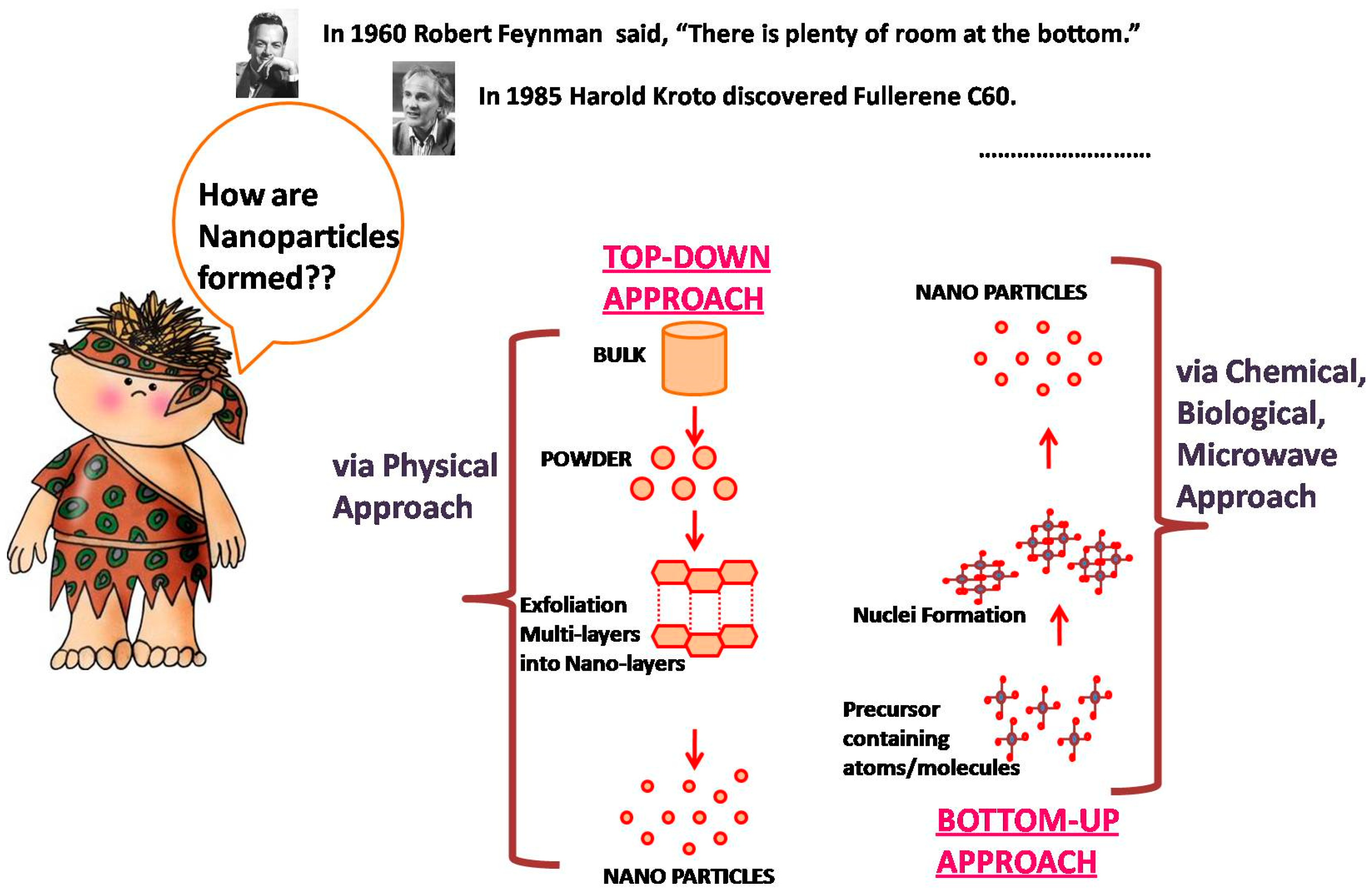
Figure 1. Accounts for the initiating protocols for nanoparticle synthesis.
Electrochem 04 00024 g001
Figure 2. Accounts for the broad applications of nanomaterials in diverse fields.
Electrochem 04 00024 g002
Figure 3. (A) Involvement of nanoparticles in overcoming the biological barriers for the broad applications of nanomaterials in diverse fields. (B) Synthesis of silver nanoparticles using microemulsions via adjustment of water-to-surfactant ratio for a controllable size and composition of NMs. Copyright and Permissions, Nanomaterials, 2023 [39].
Electrochem 04 00024 g003
Figure 4. Illustration for the comparative description of the use of nanoparticles in drug delivery approach.
Electrochem 04 00024 g004
Figure 5. Illustration of the reasons behind the usage of nanoformulations.
Electrochem 04 00024 g005
Figure 6. Pictorial representation of the information derived via the use of various nanomaterial characterization techniques.
Electrochem 04 00024 g006
Figure 7. Physicochemical factors that affect the cellular uptake of nanoparticles (a. surface charge, b. shape, c. size, and d. surface chemistry). Copyright [103].
Electrochem 04 00024 g007
Figure 8. Scientific challenges encountered during the development and authorization of nanoparticles.
Electrochem 04 00024 g008
Table 1. Tabulation of the various companies employing the use of specific nanomaterials and their work interests.
CompaniesWork of InterestNanoparticles UsedTechnological B
Advectus Life Science, Inc., West Vancouver, BC, CanadaDrug Delivery Approachpoly (D,L-lactide-co-glycolide) (PLGA)Polymeric nanoparticles engineered to carry anti-tumor drugs across the blood–brain barrier
Alnis Biosciences, Inc., Bancroft, Berkeley, CA, USABio-Pharmaceutical-Biodegradable nanoparticles for drug delivery
Argonide, Sanford, FL, USAMembrane Filtration-Nanoporous ceramic material for endotoxin filtration, orthopedics and dental implants, DNA and protein separation
Biophan Technologies, Inc., NY, USAMRI shielding-Nanomagnetic/carbon composite material to shield medical devices from RF field
BASF, Ludwigshafen, GermanyToothpasteHydroxyapatite NPsHydroxyapatite NPs improve dental surface
Capsulation Nanoscience AG, Berlin, GermanyImprove drug solubility-Layer-by-layer poly-electrolyte coating, 8–50 nm
Eiffel Biotech, Taiwan, ROCDrug delivery-Size reduction of drug particles to 50–100 nm
Enviro Systems, Inc., OK, USASurface disinfection-Nano-emulsions
Dynal Biotech, Carlsbad, CA, USA -Magnetic beads
Immunicon, PA, USATracking and separation of different cell types-Magnetic core surrounded by a polymeric layer coated with antibody for capturing cells
KES Science and Technology, Inc., USAAirocide FiltersNano-TiO2To destroy airborne pathogens
Evident Technologies, NY, USALuminescent biomarkers-Semiconductor QDs with amine, carboxyl functional groups having emissions from 350–2500 nm
Nanobio corporation, USAPharmaceutical-Antimicrobial nano-emulsion
Nanocarrier Co., Ltd., Tokyo, JapanDrug delivery-Micellar NPs for encapsulation of drugs, proteins, and DNA
NanoPharma AG, Czech RepublicDrug deliveryPolybutylcyanoacrylate NPsNP coated with drugs and later with surfactant to go across blood–brain barrier
Nanoplex Technologies, Inc., CA, USANanobar codes for bioanalysis-
Nanoprobes, Inc., Yaphank, NY, USABiological MarkersAuNPsBio-conjugates for TEM and fluorescent microscopy
Nanosphere, Inc., Northbrook, IL, USAGold biomarkersAu and Ag NPsDNA barcode attached to nanoprobe for identification purposes; PCR to amplify the signal; catalytic silver deposition to amplify signal using surface plasmon resonance
Nanomed Pharmaceutical, Inc., MI, USADrug delivery-NP for drug delivery
Oxonica Ltd., UKSunscreens-Doped transparent NPs to absorb harmful UV and convert to heat
PSi Vida Ltd., MA, USATissue engineering, implants, drug and gene delivery, bio-filtration-Exploiting material properties of nanostructures porous silicone
Smith & Nephew, Watford, UKActicoatbandagesAg nanocrystalNanocrystal silver is highly toxic to pathogens
Quantum Dot corporation, CA, USALuminescent biomarkers-Bio-conjugated semiconductor QD
Disclaimer/Publisher’s Note: The statements, opinions and data contained in all publications are solely those of the individual author(s) and contributor(s) and not of MDPI and/or the editor(s). MDPI and/or the editor(s) disclaim responsibility for any injury to people or property resulting from any ideas, methods, instructions or products referred to in the content.