
Automated Clinical Decision Support for Coronary Plaques Characterization from Optical Coherence Tomography Imaging with Fused Neural Networks


Abstract
1. Introduction
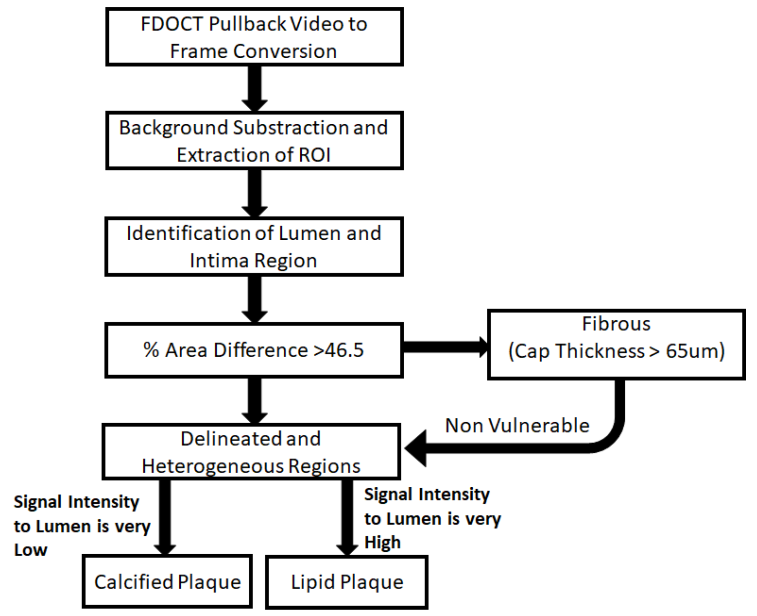
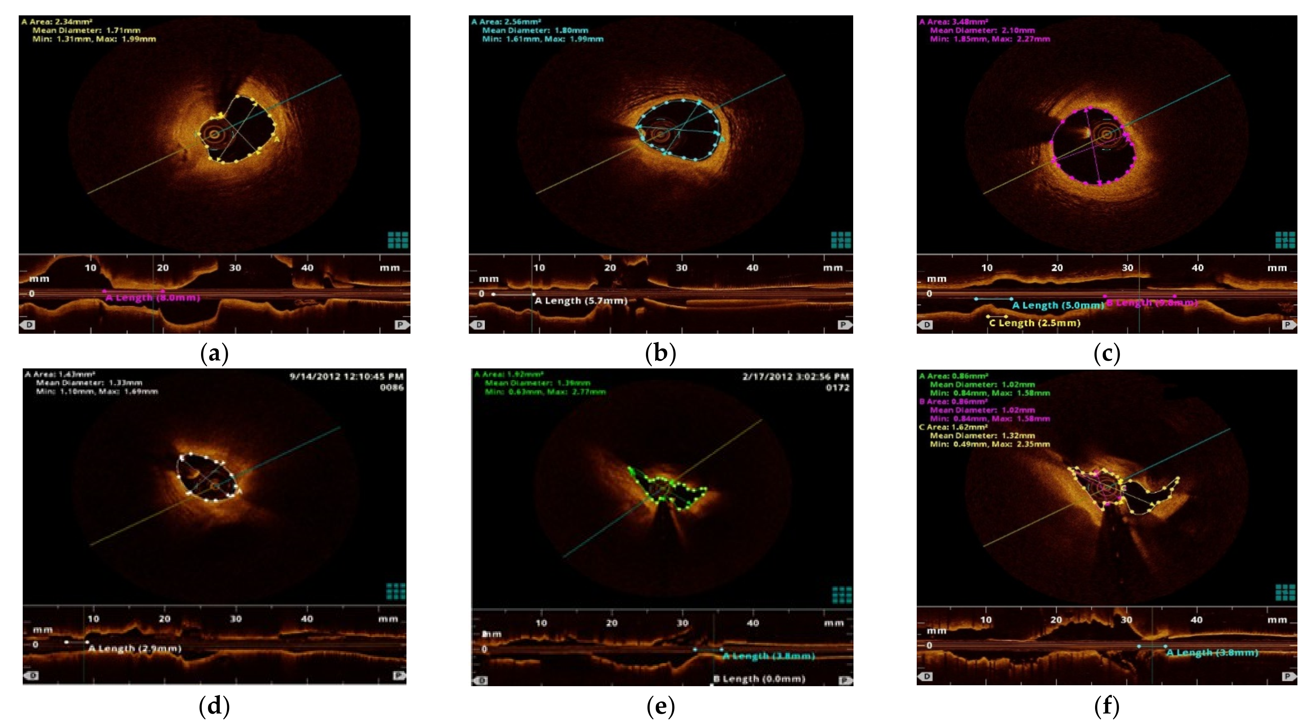
2. Methods
2.1. Data Augmentation Using GANs
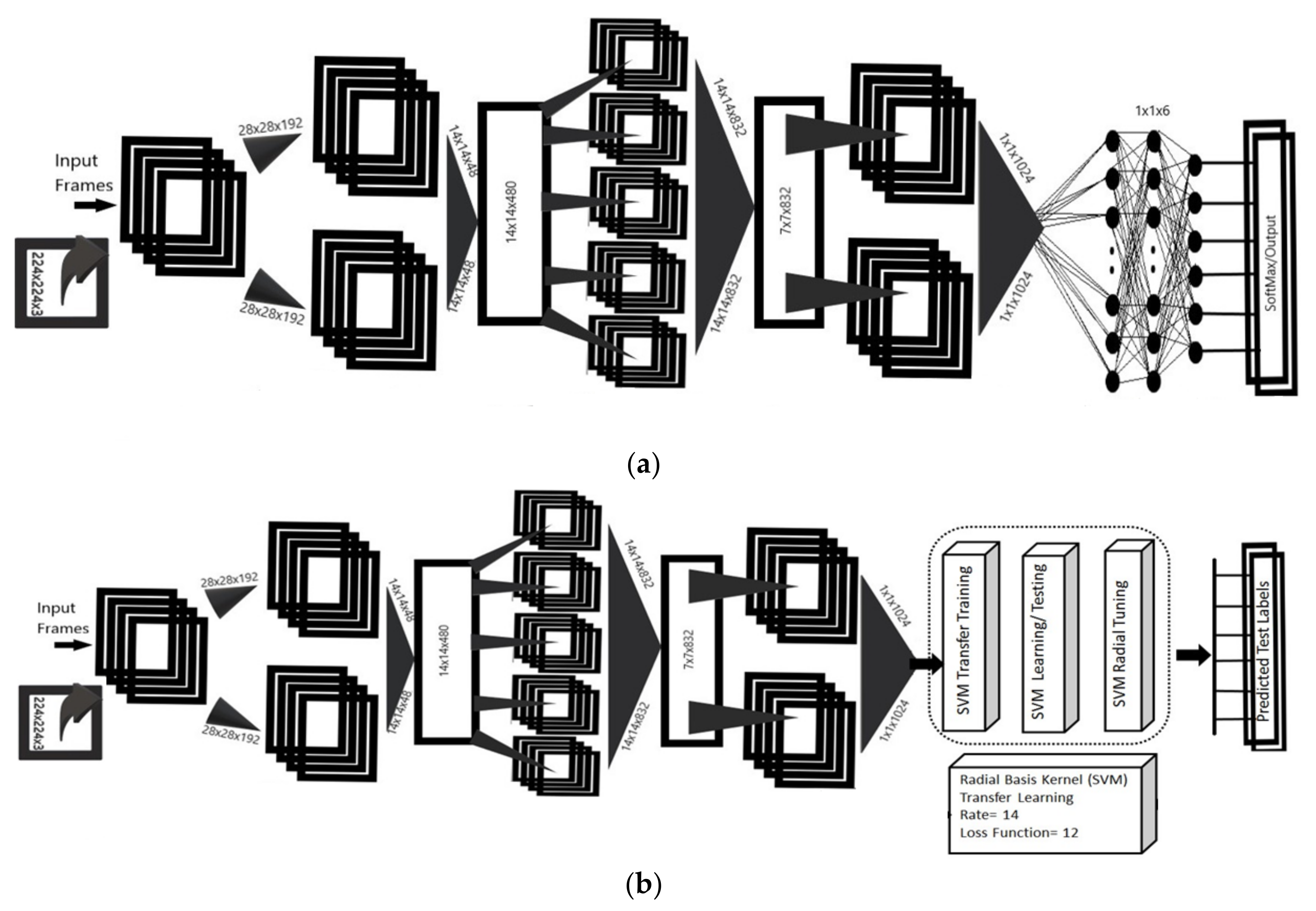
2.2. Proposed Fused Deep Learning Classifier
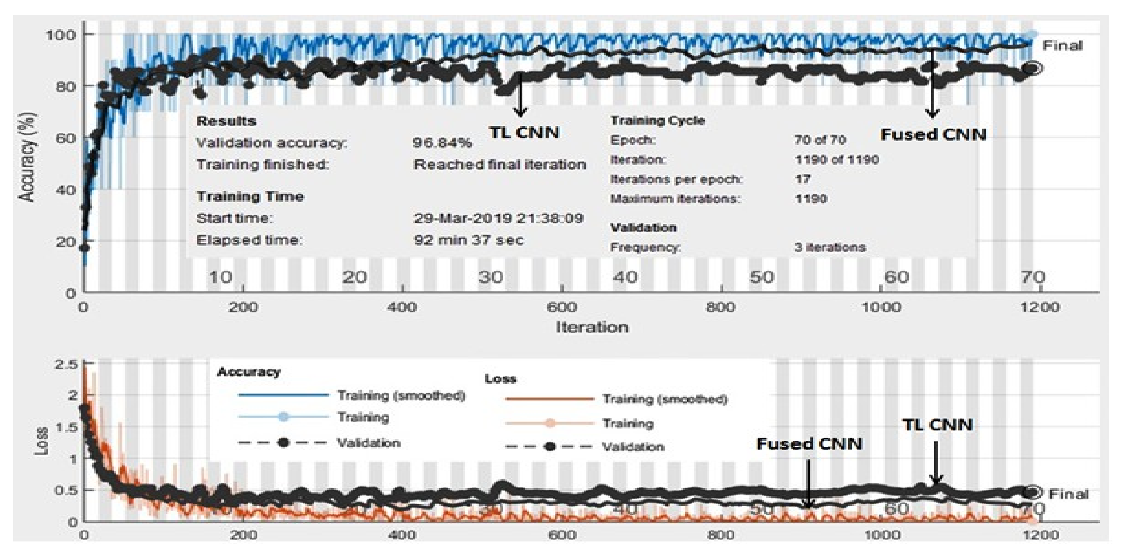
3. Results and Discussion
4. Conclusions
Author Contributions
Funding
Institutional Review Board Statement
Informed Consent Statement
Acknowledgments
Conflicts of Interest
References
- Abdolmanafi, A.; Duong, L.; Dahdah, N.; Adib, I.; Cheriet, F. Characterization of coronary artery pathological formations from OCT imaging using deep learning. Biomed. Opt. Express. 2018, 9, 4936–4960. [Google Scholar] [CrossRef] [PubMed]
- Retson, T.A.; Besser, A.H.; Sall, S.; Golden, D.; Hsiao, A. Machine Learning and Deep Neural Networks in Thoracic and Cardiovascular Imaging. J. Thorac. Imaging 2017, 34, 192–201. [Google Scholar] [CrossRef] [PubMed]
- Henglin, M.; Stein, G.; Hushcha, P.V.; Snoek, J.; Wiltschko, A.B.; Cheng, S. Machine Learning Approaches in Cardiovascular Imaging. Circ. Cardiovasc. Imaging 2017, 10, 005614. [Google Scholar] [CrossRef] [PubMed]
- Su, S.; Hu, Z.; Lin, Q.; Hau, W.K.; Gao, Z.; Zhang, H. An artificial neural network method for lumen and media-adventitia border detection in IVUS. Comput. Med. Imaging Graph. 2017, 57, 29–39. [Google Scholar] [CrossRef]
- Gao, Z.; Chung, J.; Abdelrazek, M.; Leung, S.; Hau, W.K.; Xian, Z.; Li, S. Privileged Modality Distillation for Vessel Border Detection in Intracoronary Imaging. IEEE Trans. Med. Imaging 2020, 39, 1524–1534. [Google Scholar] [CrossRef]
- Tearney, G.J.; Regar, E.; Akasaka, T.; Adriaenssens, T.; Barlis, P.; Bezerra, H.; Bouma, B.; Bruining, N.; Cho, J.; Chowdhary, S.; et al. Consensus standards for acquisition, measurement, and reporting of intravascular optical coherence tomography studies: A report from the International Working Group for Intravascular Optical Coherence Tomography Standardization and Validation. J. Am. Coll. Cardiol. 2012, 59, 58–72. [Google Scholar] [CrossRef]
- Lee, J.; Prabhu, D.; Vladislav, N.; Zimin, H.G.; Wilson, D.L. Automated plaque characterization using deep learning on coronary intravascular optical coherence tomographic images. Biomed. Opt. Express. 2019, 10, 6497–6515. [Google Scholar] [CrossRef]
- Zafar, H.; Sharif, F.; Leahy, M. Assessment of coronary artery stenosis with FD-OCT derived blood flow measurements: Relationship with FFR. J. Am. Coll. Cardiol. 2014, 64, 307–311. [Google Scholar] [CrossRef][Green Version]
- He, S.; Zheng, J.; Maehara, A. Convolutional neural network based automatic plaque characterization for intracoronary optical coherence tomography images. Med. Imaging 2018, 32, 10574. [Google Scholar]
- Abdolmanafi, A.; Duong, L.; Dahdah, N.; Adib, I.; Cheriet, F. Deep feature learning for automatic tissue classification of coronary artery using optical coherence tomography. Biomed. Opt. Express. 2017, 8, 1203–1220. [Google Scholar] [CrossRef]
- Aref, S.; Anchouche, K.; Singh, G.; Slomka, P.; Kolli, K.; Kumar, A. Clinical applications of machine learning in cardiovascular disease and its relevance to cardiac imaging. Eur. Heart J. 2019, 40, 1975–1986. [Google Scholar] [CrossRef]
- Johnson, K.W.; Soto, J.T.; Glicksberg, B.S.; Shameer, K.; Miotto, R.; Ali, M. Artificial Intelligence in Cardiology. J. Am. Coll. Cardiol. 2018, 71, 2668–2679. [Google Scholar] [CrossRef]
- Khened, M.; Alex, V.; Krishnamurthi, Fully convolutional multi-scale residual DenseNets for cardiac segmentation and automated cardiac diagnosis using ensemble of classifiers. G. Med Image Anal. 2019, 51, 21–45. [Google Scholar] [CrossRef]
- He, C.; Li, Z.; Wang, J.; Huang, Y.; Yin, Y.; Zhiyong, L. Atherosclerotic Plaque Tissue Characterization: An OCT-Based Machine Learning Algorithm With ex vivo Validation. Front Bioeng. Biotechnol. 2020, 8, 749. [Google Scholar] [CrossRef]
- Fedewa, R.; Puri, R.; Fleischman, E.; Lee, J.; Prabhu, D.; Wilson, D.L.; Fleischman, A. Artificial Intelligence in Intracoronary Imaging. Curr. Cardiol. Rep. 2020, 22, 46. [Google Scholar] [CrossRef]
- Prabhu, D.; Bezerra, H.; Kolluru, C.; Gharaibeh, Y.; Mehanna, E.; Wu, H.; Wilson, D.L. Automated A-line coronary plaque classification of intravascular optical coherence tomography images using handcrafted features and large datasets. J. Biomed. Opt. 2019, 24, 106002. [Google Scholar] [CrossRef]
- Zeng, X.; Cui, S.; Qian, J.; Cheng, X.; Dong, J.; Zhou, J.; Xu, Z.; Feng, Y. 10 W low-noise green laser generation by the single-pass frequency doubling of a single-frequency fiber amplifier. Laser Phys. 2020, 30, 075001. [Google Scholar] [CrossRef]
- Macedo, M.M.; Oliveira, D.A.; Gutierrez, M.A. Atherosclerotic Plaques Recognition in Intracoronary Optical Images Using Neural Networks. In Proceedings of the 2019 Computing in Cardiology (CinC), Singapore, 8–11 September 2019; pp. 1–4. [Google Scholar]
- Liu, X.; Du, J.; Yang, J.; Xiong, P.; Liu, J.; Lin, F.J. Recent progress of chatter prediction, detection and suppression in milling. Signal. Process. Syst. 2020, 92, 325–333. [Google Scholar] [CrossRef]
- Gessert, N.; Lutz, M.; Heyder, M.; Latus, S.; Leistner, D.M.; Abdelwahed, Y.S.; Schlaefer, A. Automatic Plaque Detection in IVOCT Pullbacks Using Convolutional Neural Networks. IEEE Trans. Med. Imaging 2019, 38, 426–434. [Google Scholar] [CrossRef]
- Athanasiou, L.S. Three-dimensional reconstruction of coronary arteries and plaque morphology using CT angiography-comparison and registration using IVUS. IEEE Eng. Med. Biol. Soc. 2015, 2015, 5638–5641. [Google Scholar]
- Taqi, A.M.; Awad, A.; Al-Azzo, F.; Milanova, M. The Impact of Multi-Optimizers and Data Augmentation on TensorFlow Convolutional Neural Network Performance. In Proceedings of the IEEE Conference on Multimedia Information Processing and Retrieval, Miami, FL, USA, 10–12 April 2018; pp. 140–145. [Google Scholar]
- Miyagawa, M.; Costa, M.G.; Gutierrez, M.A.; Costa, J.P.; Costa, F. Detecting vascular bifurcation in IVOCT images using convolutional neural networks with transfer learning. IEEE Access 2019, 7, 66167–66175. [Google Scholar] [CrossRef]
- Cheplygina, V.; Bruijne, M.; Pluim, J. Not-so-supervised: A survey of semi-supervised, multi-instance, and transfer learning in medical image analysis. Med. Image Analy. 2019, 54, 280–296. [Google Scholar] [CrossRef] [PubMed]
- Sood, R.; Topiwala, B.; Choutagunta, K. Position Specific Scoring Matrix and Synergistic Multiclass SVM for Identification of Genes. In Proceedings of the17th IEEE International Conference on Machine Learning and Applications2018, Orlando, FL, USA, 17–20 December 2018; pp. 17–20. [Google Scholar]
- Kazuhiro, K. Generative Adversarial Networks for the Creation of Realistic Artificial Brain Magnetic Resonance Images. Tomography 2018, 4, 159–163. [Google Scholar] [CrossRef] [PubMed]
- Gibson, E. NiftyNet: A deep-learning platform for medical imaging. Comput. Methods Programs Biomed. 2018, 158, 113–122. [Google Scholar] [CrossRef] [PubMed]
- Massalha, S.; Clarkin, O.; Thornhill, R.; Wells, G.; Benjamin, J. Decision Support Tools, Systems, and Artificial Intelligence in Cardiac Imaging. Can. J. Cardiol. 2018, 34, 827–838. [Google Scholar] [CrossRef] [PubMed]
- Ravi, D. Deep Learning for Health Informatics. IEEE J. Biomed. Health Informatics. 2017, 21, 4–21. [Google Scholar] [CrossRef] [PubMed]
- Zhou, Z.; Shin, J.; Hurst, J.; Kendall, C.; Liang, J. Integrating Active Learning and Transfer Learning for Carotid Intima-Media Thickness Video Interpretation. J. Digit. Imaging. 2019, 32, 290–299. [Google Scholar] [CrossRef]
- Zreik, M.; Hamersvelt, R.; Wolterink, J.; Leiner, T.; Viergever, M.; Isgum, I. A Recurrent CNN for Automatic Detection and Classification of Coronary Artery Plaque and Stenosis in Coronary CT Angiography. IEEE Trans. Med. Imaging 2018, 38, 1588–1598. [Google Scholar] [CrossRef]
- Fischer, A.M. Accuracy of an Artificial Intelligence Deep Learning Algorithm Implementing a Recurrent Neural Network with Long Short-term Memory for the Automated Detection of Calcified Plaques From Coronary Computed Tomography Angiography. J. Thorac. Imaging. 2020, 35, S49–S57. [Google Scholar] [CrossRef]
- Vos, B.; Wolterink, J.; Leiner, T.; Jong, P. Direct Automatic Coronary Calcium Scoring in Cardiac and Chest CT. IEEE Trans. Med. Imaging 2019, 8, 2127–2138. [Google Scholar] [CrossRef]
- Duan, J.; Bello, G.; Schlemper, J.; Bai, W. Automatic 3D Bi-Ventricular Segmentation of Cardiac Images by a Shape-Refined Multi-Task Deep Learning Approach. IEEE Trans. Med. Imaging 2019, 38, 2151–2164. [Google Scholar] [CrossRef]
- Alawad, M.; Wang, L. Learning Domain Shift in Simulated and Clinical Data: Localizing the Origin of Ventricular Activation from 12-Lead Electrocardiograms. IEEE Trans. Med. Imaging 2019, 38, 1172–1184. [Google Scholar] [CrossRef]
- Rogers, M.A.; Aikawa, E. Cardiovascular calcification: Artificial intelligence and big data accelerate mechanistic discovery. Nat. Rev. Cardiol. 2019, 16, 261–274. [Google Scholar] [CrossRef]


| 1 | 11 8.8 | 0 0 | 1 0.8 | 0 0 | 0 0.0 | 1 0.8 | 84.6 15.4 |
| 2 | 0 0 | 23 18.4 | 2 1.6 | 2 1.6 | 1 0.8 | 1 0.8 | 79.3 20.7 |
| 3 | 0 0 | 3 2.4 | 33 26.4 | 1 0.8 | 1 0.8 | 0 0 | 86.8 13.2 |
| 4 | 1 0.8 | 3 2.4 | 2 1.6 | 21 16.8 | 0 0 | 0 0 | 77.8 22.2 |
| 5 | 0 0 | 0 0 | 2 1.6 | 0 0 | 7 5.6 | 0 0 | 77.8 22.2 |
| 6 | 2 1.6 | 0 0 | 0 0 | 0 0 | 0 0 | 7 5.6 | 77.8 22.2 |
| 78.6 21.4 | 79.3 20.7 | 82.5 17.5 | 87.5 12.5 | 77.8 22.2 | 77.8 22.2 | 81.6 18.4 | |
| 1 | 2 | 3 | 4 | 5 | 6 |
| 1 | 7 5.6 | 3 2.5 | 0 0 | 2 1.7 | 0 0 | 0 0 | 58.3 41.7 |
| 2 | 0 0 | 9 7.4 | 0 0 | 0 0 | 2 1.7 | 0 0 | 81.8 18.2 |
| 3 | 0 0 | 0 0 | 24 19.8 | 1 0.8 | 3 2.5 | 1 0.8 | 82.8 17.2 |
| 4 | 0 0 | 2 1.7 | 4 3.3 | 33 27.3 | 1 0.8 | 1 0.8 | 80.5 19.5 |
| 5 | 1 0.8 | 0 0 | 0 0 | 0 0 | 17 14.0 | 0 0 | 89.5 10.5 |
| 6 | 1 0.8 | 0 0 | 0 0 | 1 0.8 | 1 0.8 | 7 5.8 | 77.8 22.2 |
| 77.8 22.2 | 64.3 35.7 | 85.7 14.3 | 89.2 10.8 | 70.8 29.2 | 77.8 22.2 | 80.8 19.9 | |
| 1 | 2 | 3 | 4 | 5 | 6 |
| 1 | 11 88 | 0 0 | 0 0 | 2 1.7 | 0 0 | 0 0 | 91.7% 8.3% |
| 2 | 0 0 | 23 18.5 | 2 1.6 | 2 1.6 | 1 0.8 | 1 0.8 | 99.3% 20.7% |
| 3 | 0 0 | 4 3.2 | 35 28 | 2 1.6 | 1 0.8 | 0 0 | 93.3% 16.7% |
| 4 | 0 0 | 2 1.6 | 1 0.8 | 20 16 | 0 0 | 0 0 | 99.3% 16.7% |
| 5 | 1 0.8 | 0 0 | 2 1.6 | 0 0 | 7 5.6 | 0 0 | 97.8% 22.2% |
| 6 | 2 1 | 0 0 | 0 0 | 0 0.8 | 1 0.8 | 7 5 | 95.8% 22.2% |
| 78.6% 21.4% | 79.3% 20.7% | 87.5% 12.5% | 83.3% 16.7% | 77.8% 2.2% | 77.8% 22.2% | 96.84% 17.6% | |
| 1 | 2 | 3 | 4 | 5 | 6 |
| Sr. No. | Pre-Trained Model | Input Dimensions | Elapsed Time | No of Epochs | Accuracy |
|---|---|---|---|---|---|
| 1 | AlexNet | 227 × 227 × 3 | 380 min and 22 s | 100 | 81.6% |
| 2 | GoogleNet | 224 × 224 × 3 | 92 min and 37 s | 100 | 82.64% |
| 3 | ResNet 50 | 224 × 224 × 3 | 1509 min and 21 s | 100 | 79.34% |
| 4 | ResNet 101 | 224 × 224 × 3 | 1711 min and 8 s | 100 | 73.73% |
| 5 | Densenet | 224 × 224 × 3 | 3215 min and 31 s | 100 | 80.17% |
| 6 | Proposed architecture | 224 × 224 × 3 | 20 s | 70 | 96.84 % |
Publisher’s Note: MDPI stays neutral with regard to jurisdictional claims in published maps and institutional affiliations. |
© 2022 by the authors. Licensee MDPI, Basel, Switzerland. This article is an open access article distributed under the terms and conditions of the Creative Commons Attribution (CC BY) license (https://creativecommons.org/licenses/by/4.0/).
Share and Cite
Zafar, H.; Zafar, J.; Sharif, F. Automated Clinical Decision Support for Coronary Plaques Characterization from Optical Coherence Tomography Imaging with Fused Neural Networks. Optics 2022, 3, 8-18. https://doi.org/10.3390/opt3010002
Zafar H, Zafar J, Sharif F. Automated Clinical Decision Support for Coronary Plaques Characterization from Optical Coherence Tomography Imaging with Fused Neural Networks. Optics. 2022; 3(1):8-18. https://doi.org/10.3390/opt3010002
Chicago/Turabian StyleZafar, Haroon, Junaid Zafar, and Faisal Sharif. 2022. "Automated Clinical Decision Support for Coronary Plaques Characterization from Optical Coherence Tomography Imaging with Fused Neural Networks" Optics 3, no. 1: 8-18. https://doi.org/10.3390/opt3010002
APA StyleZafar, H., Zafar, J., & Sharif, F. (2022). Automated Clinical Decision Support for Coronary Plaques Characterization from Optical Coherence Tomography Imaging with Fused Neural Networks. Optics, 3(1), 8-18. https://doi.org/10.3390/opt3010002