Review

Artificial Intelligence Across the Sarcopenia Care Pathway: From Opportunistic Screening to Intelligent Rehabilitation


1
Musculoskeletal Research Laboratory, Department of Orthopaedics & Traumatology, The Chinese University of Hong Kong, Hong Kong SAR, China
2
Li Ka Shing Institute of Health Sciences, The Chinese University of Hong Kong, Hong Kong SAR, China
3
School of Life Sciences, The Chinese University of Hong Kong, Hong Kong SAR, China
*
Authors to whom correspondence should be addressed.
These authors contributed equally to this work.
AI 2026, 7(6), 201; https://doi.org/10.3390/ai7060201
Submission received: 20 May 2026 / Accepted: 28 May 2026 / Published: 1 June 2026
(This article belongs to the Section Medical & Healthcare AI)

Abstract

Sarcopenia is a progressive, age-related skeletal muscle disorder that serves as a driver of frailty, falls, and mortality in older adults. Despite the recent paradigm shift introduced by the latest sarcopenia consensus, which emphasizes early, proactive detection, sarcopenia cases frequently evade traditional screening due to inherent diagnostic bottlenecks and resource limitations. Artificial intelligence has emerged as a transformative solution to dismantle these barriers across the entire continuum of sarcopenia care. This review explores the rapid evolution of artificial intelligence, beginning with automated opportunistic screening that extracts prognostic musculoskeletal data from routine imaging and electronic health records, advancing toward high-precision multimodal assessment architectures. Beyond initial assessment, artificial intelligence is actively restructuring longitudinal care by the integration of ubiquitous wearables, Large Language Models, and computer vision, enabling dynamic exercise prescriptions and real-time kinematic postural correction for sarcopenia rehabilitation. Realizing this potential requires the medical community to confront urgent clinical barriers, including multi-center validation, semantic interoperability, health economic justification, and the strict preservation of human-centric ethics. By addressing these challenges, this review provides a definitive roadmap for embedding artificial intelligence across the entire sarcopenia care pathway, transforming isolated instances of opportunistic screening into a unified ecosystem for intelligent, proactive rehabilitation.

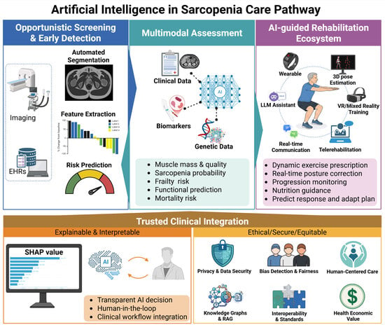
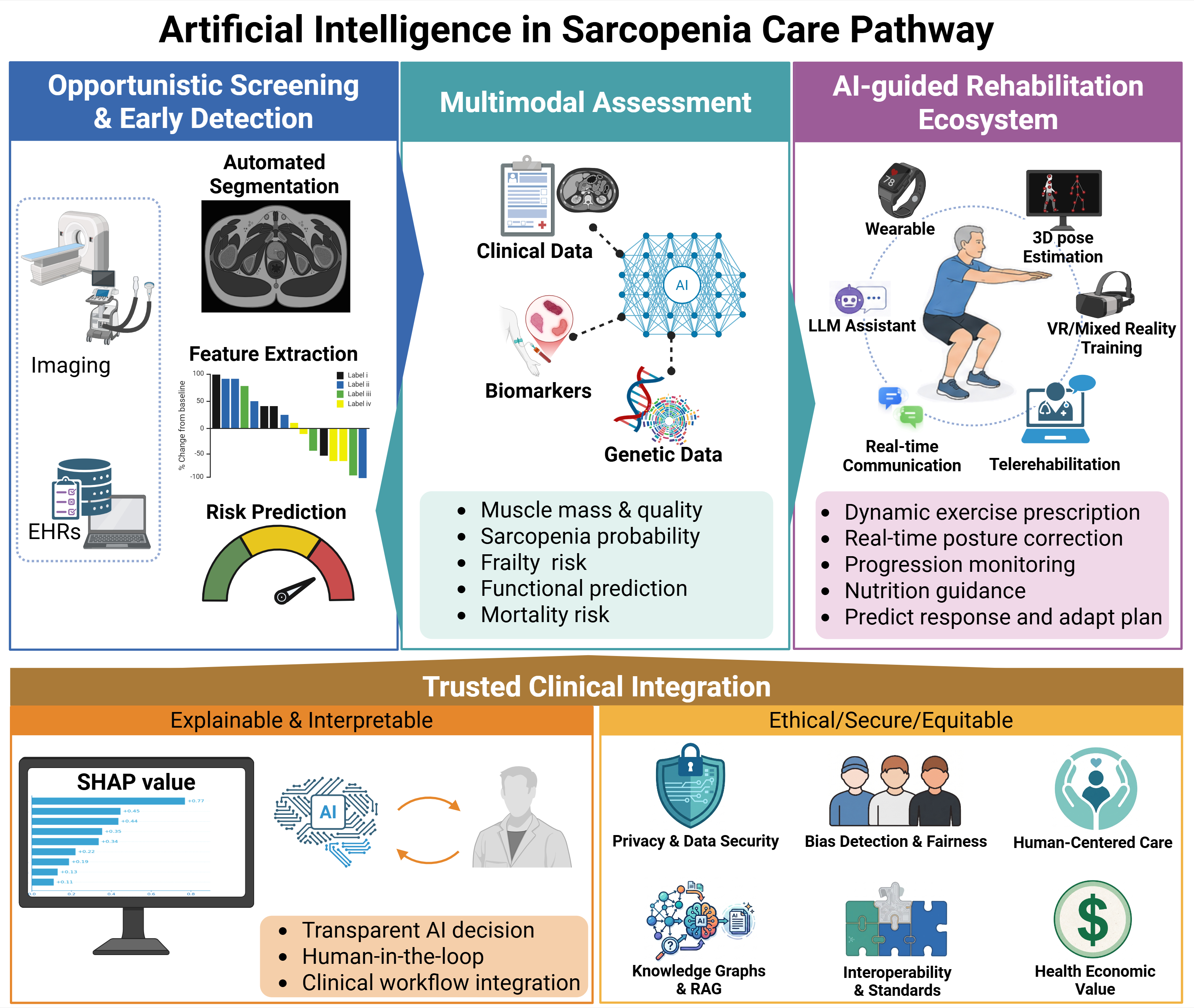
Graphical Abstract

1. Introduction

Sarcopenia is an age-related progressive skeletal muscle disorder characterized by the loss of muscle mass, strength, and function, and is increasingly recognized as a major health concern in aging societies [1]. It contributes to frailty, falls, disability, and mortality, affecting nearly one-third of community-dwelling older adults and over half of those in institutional care [2]. Although sarcopenia is clinically significant, it continues to be underdiagnosed and undertreated, primarily because of the inherent limitations of historical diagnostic approaches. Traditional diagnostic criteria, such as those previously endorsed by the European Working Group on Sarcopenia in Older People (EWGSOP2) in 2019 [2] and the Asian Working Group for Sarcopenia (AWGS) 2019 consensus [3], relied heavily on combined assessments of muscle mass, strength, and physical performance. However, these conventional methods are often constrained by cost, accessibility, operator dependency, and a lack of standardization, particularly in low-resource settings, making them poorly suited for large-scale screening or longitudinal monitoring. The newly released consensus of sarcopenia by AWGS in 2025 [4], introduces a critical paradigm shift. It simplifies the diagnostic algorithm by requiring only concurrent low muscle mass and strength, repositioning physical performance as a downstream outcome measure. However, executing this proactive strategy remains challenging. Because muscle deterioration often occurs insidiously, frequently masked by a normal or elevated body mass index (BMI), a vast population of at-risk individuals harbor “hidden” sarcopenia, evading early clinical detection [5,6].
The failure to identify these patients early in the disease trajectory is largely a consequence of existing diagnostic bottlenecks. Current clinical pathways typically rely on subjective, low-sensitivity screening questionnaires (such as the SARC-F) to justify formal imaging. While advanced modalities, including computed tomography (CT) and magnetic resonance imaging (MRI), serve as the definitive gold standards for quantifying skeletal muscle index (SMI) and microstructural tissue quality, their routine clinical use is severely restricted. Extracting meaningful quantitative data from these scans requires manual, slice-by-slice segmentation, which is prohibitively time-consuming, highly operator-dependent, and fundamentally unscalable for population-level screening [7,8].
Artificial intelligence (AI), encompassing both Deep Learning and classic Machine Learning, has emerged as a powerful technological solution to these systemic clinical barriers. By fully automating complex anatomical segmentation, AI has transformed body composition analysis from a specialized research endeavor into a highly accessible clinical utility. More importantly, AI algorithms can autonomously extract prognostic musculoskeletal data from existing medical records and standard diagnostic images originally ordered for unrelated reasons [5,9]. This capability unlocks the potential for “opportunistic screening” or prediction, allowing healthcare systems to passively identify high-risk patients operating invisibly within routine clinical workflows.
This review examines the integration of AI across the pathway of sarcopenia care. We map the rapid evolution of AI applications, beginning with its foundational role in opportunistic screening and multimodal predictive assessment. We then explore how AI is transitioning from a passive diagnostic tool into an active, longitudinal rehabilitation ecosystem powered by ubiquitous wearables and Large Language Models (LLMs). We also evaluate the indispensable role of Explainable AI (XAI) in establishing clinical trust, alongside the profound ethical and technical challenges that must be overcome to fully realize AI’s potential in preserving functional independence for aging populations.
While existing technical reviews frequently categorize advancements strictly by algorithm type to facilitate methodological comparisons [10], this review deliberately departs from that structure to adopt a clinical care pathway approach. We believe that integrating AI into routine practice requires bridging the translation gap. Clinicians and public health stakeholders evaluate technologies based on the patient journey, from opportunistic screening, diagnosis to rehabilitation, rather than underlying software architectures. Furthermore, organizing by clinical utility directly aligns with the paradigm shift introduced by the newly released AWGS consensus [4], which explicitly mandates a “life-course approach” to muscle health.

2. Methodology

Although this study is designed as a narrative review to provide a comprehensive, clinical pathway-oriented perspective, we adopted a structured search strategy across major databases, including PubMed, Web of Science, Scopus, and Google Scholar. The search targeted papers published up to February 2026. The search strategy utilized combinations of the following keywords: “sarcopenia”, “artificial intelligence”, “machine learning”, “deep learning”, “imaging-based assessment”, “risk prediction”, and “rehabilitation”.
We selected the key papers of (1) studies explicitly deploying AI, machine learning, or deep learning algorithms for the screening, diagnosis, risk stratification, or rehabilitation of sarcopenia; (2) studies involving human cohorts; and (3) articles published in English. The papers where AI was only tangentially mentioned without being the core methodological intervention were excluded.
During the screening phase, titles and abstracts were independently reviewed to eliminate irrelevant or duplicate records. To ensure that the underlying algorithmic methodologies can still be rigorously evaluated and compared side-by-side, we have synthesized the information of the key included studies within a comprehensive reference table (Table 1).

3. Artificial Intelligence in Sarcopenia Opportunistic Screening and Assessment

AI is now being integrated into the front end of the sarcopenia care pathway from opportunistic screening to quantitative assessment. We first summarize AI-enabled risk prediction using routinely available data (electronic health records and incidental imaging), then review imaging-based assessment across CT, MRI, and ultrasound, followed by non-imaging machine learning models that enable scalable community screening. Finally, we highlight emerging multimodal fusion approaches that combine complementary data streams to improve diagnostic accuracy and clinical utility.

3.1. Artificial Intelligence-Enabled Prediction and Opportunistic Screening

Routine clinical assessments frequently miss “hidden” sarcopenia. Because these patients often maintain a normal BMI or lack the overt physical frailty that triggers formal evaluation, they evade early detection. AI-enabled prediction and opportunistic screening bridge this diagnostic gap by extracting musculoskeletal data from existing electronic health records (EHRs) and medical images that were originally ordered for unrelated clinical reasons. For instance, Zambrano Chaves et al. analyzed abdominal CT scans and EHR data from 17,646 patients, demonstrating that automated predictive algorithms can identify at-risk individuals operating invisibly within standard radiologic workflows [5]. As demonstrated by Luo et al., AI algorithms can detect possible sarcopenia directly from standard EHRs, transforming routine medical documentation into an active screening tool [6]. Validating this proactive concept, automated deep learning tools applied to the L1 and L3 vertebral levels achieve high accuracy in predicting adverse clinical outcomes [9].
This opportunistic paradigm extends beyond hospital-centric EHRs or images into decentralized digital triage. Researchers have translated machine learning models into mobile applications and wearable ecosystems, enabling point-of-care risk prediction without requiring dedicated clinical infrastructure [45,54]. By shifting from reactive diagnosis to proactive identification, AI-driven screening operationalizes the recent consensus call for a “life-course approach” to muscle health. It facilitates the targeted identification of hidden sarcopenia in middle-aged adults (50–64 years) before irreversible frailty begins, potentially reducing the downstream socioeconomic burden of related complications [4].
While sarcopenia screening and prediction establish the foundation for early intervention, definitive clinical evaluation and comprehensive staging require more dedicated analytical frameworks. AI-augmented imaging platforms, including CT, MRI, and ultrasound, provide the necessary anatomical and microstructural quantification of muscle tissue. Meanwhile, non-imaging Machine Learning models employ diverse clinical, biological, and wearable datasets to offer highly accessible risk stratification. To capture the full physiological complexity of the syndrome, emerging multimodal architectures now synthesize these independent diagnostic streams into cohesive, high-dimensional predictive models.

3.2. Imaging-Based Assessment

While the sarcopenia consensus [4] establishes that clinical diagnosis fundamentally requires concurrent assessments of low muscle strength and mass, advanced medical imaging remains the definitive gold standard for deep anatomical phenotyping. These modalities, including CT, MRI, and ultrasound, enable both the quantitative measurement of skeletal muscle mass and the highly detailed evaluation of tissue composition. However, extracting this data via manual segmentation has presented a severe clinical bottleneck. The process is intensely time-consuming, highly operator-dependent, and vulnerable to significant inter-observer variability. These limitations make conventional analysis fundamentally unsuited for large-scale or population-level screening.
AI addresses this paradigm by automating complex segmentation and classification tasks. As comprehensively reviewed by Rozynek et al., AI-assisted imaging has transformed body composition analysis from a labor-intensive research task into a highly reliable clinical utility [7]. Deep Learning architectures, particularly those based on state-of-the-art U-Net frameworks, could achieve high anatomical precision, compressing analysis time from several minutes per image to seconds while enforcing absolute analytical reproducibility [8,21,22].

3.2.1. CT-Based Body Composition Analysis

CT imaging at the third lumbar vertebral (L3) level is internationally recognized as the definitive gold standard for estimating skeletal muscle index (SMI) and skeletal muscle area (SMA). The muscular compartments of the L3 cross-section correlate strongly with whole-body skeletal muscle volume and are easily identifiable on routine abdominal scans. While its diagnostic value is indisputable, the manual delineation of these compartments remains a prohibitive clinical limitation.
Deep Learning has effectively reduced this analytical barrier by fully automating the simultaneous quantification of skeletal muscle, visceral adipose tissue (VAT), and subcutaneous adipose tissue (SAT) [8]. Advanced convolutional neural networks (CNNs) have achieved anatomical fidelity that rivals expert radiologists. For instance, Weston et al. demonstrated that automated pipelines routinely yield Dice scores of 0.96 ± 0.02 for muscle and 0.97 ± 0.01 for VAT segmentation [11]. This technical precision is not an isolated phenomenon. It has been replicated across diverse multi-institutional datasets. Modern Machine Learning frameworks maintain cross-sectional area errors below 5% and Dice scores consistently exceeding 0.93, even when challenged with varying scanner protocols, different manufacturers, and large longitudinal screening cohorts [12,13,14,15,16].
Beyond geometric precision, these AI frameworks offer significant clinical and prognostic utility by enabling high-throughput analysis. In large-scale diagnostic evaluations, such as a cohort of 3096 cases analyzed by Onishi et al., AI systems delivered robust diagnostic sensitivities of 82.3%, specificities of 98.1%, and a positive predictive value of 89.5% for sarcopenia detection [20]. Comprehensive frameworks like the Artificial Intelligence Body Part Measure System (AIBMS) have reported area under the curve (AUC) values up to 0.874, uniquely supporting both rapid 2D L3 analysis and 3D volumetric assessment [21]. Modern AI applications in CT now extend beyond macroscopic anatomical boundaries into microstructural tissue characterization. By using CT radiomics, Machine Learning models can extract high-dimensional, subvisual texture features from skeletal muscle. These radiomic signatures capture intrinsic muscle quality changes, such as early myosteatosis and fibrous infiltration, supporting highly accurate sarcopenia identification that traditional cross-sectional area measurements alone might miss [23]. To facilitate rapid bedside integration, web-based platforms now translate these algorithms into standardized workflows, achieving classification accuracies of 97.5% and segmentation accuracies of 92% directly from routine imaging [22]. Additionally, automated chest CT assessments can now independently predict all-cause mortality in older adults [36].
Collectively, this body of work indicates that CT-based AI tools have definitively moved beyond proof-of-concept studies and into practical clinical implementation. Because CT scanners are widely deployed in modern healthcare, this modality provides an excellent platform for large-scale opportunistic screening. It allows clinicians to extract high-value musculoskeletal data without subjecting patients to additional radiation exposure, clinical visits, or financial burden beyond their original indication. Consequently, among all available imaging modalities, AI-augmented CT currently occupies the most mature stage of clinical validation, offering a readily deployable solution for identifying at-risk individuals.

3.2.2. MRI-Based Muscle and Fat Quantification

While CT drives high-throughput opportunistic screening, MRI provides a sophisticated, radiation-free counterpart equipped to decode the physiological changes in muscle tissue. Beyond its safety profile, which makes it the modality of choice for repeated longitudinal monitoring and interventional trials, MRI offers excellent soft-tissue contrast. Advanced conventional sequences, such as T1-weighted imaging, Dixon methods, and T2 mapping, enable the highly precise quantification of both muscle volume and intramuscular adipose tissue (IMAT). This serves as a definitive reference standard for evaluating myosteatosis and muscle atrophy, correlating strongly with bioelectrical impedance analysis [55,56,57,58]. However, the multi-parametric complexity and massive data volume of MRI have historically rendered its manual analysis clinically unfeasible.
The application of AI has successfully overcome these complexities. Fully automated Deep Learning pipelines utilizing chemical shift encoding-based water-fat MRI can now segment paraspinal and axial musculature with Dice coefficients exceeding 0.94, demonstrating strong test–retest reliability for tracking muscle degeneration over time [17,18]. What distinguishes AI-augmented MRI is its capacity to synthesize functional, subvisual biomarkers. By processing complex quantitative metrics like T2 relaxation times, which reflect underlying cellular degeneration, edema, and tissue inflammation, AI models generate a phenotype of muscle quality that attenuation-based CT simply cannot capture [59]. When fused with diffusion tensor imaging (DTI) for microstructural integrity and magnetic resonance spectroscopy (MRS) for intramyocellular lipid quantification, AI multi-parametric protocols offer a comprehensive view of muscle health [60]. The transformative potential of this technology lies in its newfound scalability. Defying the perception of MRI as a low-throughput research tool, Jung et al. deployed a Deep Learning framework across a massive cohort of 36,317 whole-body MRI scans. Their fully automated pipeline translated complex 3D volumetric data of skeletal muscle and IMAT into powerful prognostic biomarkers that independently predicted all-cause mortality. This landmark study demonstrated that AI-driven volumetric MRI profiling provides robust prognostic stability compared with conventional 2D single-slice measurements, even after adjusting for traditional risk factors [37].
However, the routine clinical adoption of MRI for sarcopenia assessment lagged behind CT, constrained by higher costs, prolonged acquisition times, and limited scanner accessibility. The convergence of accelerated imaging protocols, such as compressed sensing and parallel imaging, with AI-driven automated analysis and cloud-based institutional deployment has enhanced MRI’s clinical viability. Rather than competing with CT, MRI is now positioned as its essential complementary modality. While CT remains the practical choice for opportunistic screening in large populations, MRI fulfills a distinct, high-precision clinical niche. It provides a completely radiation-free alternative for patients requiring repeated longitudinal assessments, while offering a rich set of biomarkers for understanding the complex biology of muscle deterioration.

3.2.3. Ultrasound-Based Muscle Assessment

While CT provides large-scale screening and MRI offers detailed compositional phenotyping, ultrasound serves as the highly accessible counterpart, uniquely suited for point-of-care and community-based applications. It enables the real-time evaluation of fundamental muscular parameters, including muscle thickness, cross-sectional area, and echogenicity, making it ideal for primary care settings and bedside assessments of frail or immobile patients. However, the widespread clinical integration of conventional ultrasound has been restricted by some specific limitations: it is operator-dependent, suffers from inter-observer variability, and lacks standardized diagnostic cut-offs. The integration of AI alters this dynamic, shifting ultrasound toward an objective, reproducible science.
Deep Learning models can now automatically identify anatomical landmarks and extract standardized metrics in real time, neutralizing human operational bias. The clinical viability of this transformation was demonstrated in the DINOSAUR pilot study. By utilizing an automated ultrasound-derived IMAT index, the study validated that AI-assisted point-of-care evaluation could objectively assess sarcopenia with excellent intra-rater (ICC = 0.938) and robust inter-rater reproducibility (ICC = 0.776). This standardized metric achieved a reliable diagnostic performance (AUC = 0.727), proving its efficacy for community-based screening regardless of the examining clinician’s prior sonographic experience [24]. Recent prospective multi-center validations by Chen et al. have cemented the reliability of these automated tools. Their dedicated ultrasound AI system (the SARCO model) demonstrated robust generalizability, achieving a diagnostic AUC of 0.801 and an F1-score of 0.727 during independent external validation across diverse clinical environments [31].
Recent studies have successfully bridged the gap between point-of-care ultrasound and the CT gold standard. Notably, López-Gómez et al. demonstrated that AI-assisted ultrasonography of the rectus femoris can establish reliable diagnostic cut-offs for both muscle mass and quality [25,26,61]. In high-risk oncology cohorts, their AI-derived ultrasound metrics achieved statistically significant concordance with L3 CT scans (r = 0.47, p < 0.01), yielding acceptable diagnostic performance (AUC = 0.640) for identifying severe sarcopenia and myosteatosis [26]. This proves that AI-augmented ultrasound can serve as a clinically relevant diagnostic tool when CT is unavailable.
Beyond standardizing macro-anatomy, AI also extends the capability of using ultrasound imaging into the realm of microstructural tissue characterization. Traditionally, assessing muscle quality relied on the subjective visual grading of echogenicity to estimate fatty infiltration. Modern AI systems replace this flawed estimation with high-dimensional ultrasound radiomics, extracting subvisual texture features that objectively quantify myosteatosis and intramuscular fibrosis. Chen et al. [31] demonstrated that radiomics-based models significantly outperformed conventional echo-intensity assessments across multiple muscle groups. When these advanced radiomic features were integrated into cohesive predictive models, they achieved a strong diagnostic AUC of 0.85 for the rectus femoris, proving the prognostic value of subvisual texture analysis.
Meanwhile, the integration of shear-wave elastography (SWE) introduces tissue stiffness as a vital, quantifiable biomarker. In their foundational work, Yi et al. [27] demonstrated that applying deep convolutional neural networks (e.g., ResNet-50, DenseNet-121) directly to SWE images achieved sarcopenia classification accuracies of up to 80.0%, systematically outperforming models trained exclusively on conventional gray-scale sonograms (maximum accuracy 75.0%). Building upon this single-modality success, their subsequent research pioneered advanced Machine Learning frameworks that formally fuse standard morphological imaging with elastography [32]. By combining muscle architecture with these functional stiffness metrics using sophisticated techniques like feature-level fusion, these multi-parametric models captured complex pathophysiological changes, achieving high predictive accuracies (up to 83.55%) that isolated imaging modalities simply cannot achieve.
The integration of AI has redefined the clinical trajectory of ultrasound in sarcopenia management. While CT and MRI remain structurally anchored to specialized imaging centers, AI-augmented ultrasound democratizes high-precision diagnostics, bringing them directly to the patient’s bedside and into primary care communities. By automating morphological measurements, extracting subvisual radiomic textures, and quantifying functional tissue stiffness, AI neutralizes the historical limitations of operator dependency. As these multidimensional data streams are embedded into emerging clinical architectures, such as the DeepSarc-US framework [19] and the Sarcopenia Artificial Intelligence Diagnostic Decision Support System (SAID DSS) [35], they are instantly translated into actionable insights. Consequently, ultrasound functions as a highly scalable, precision-driven assessment engine for early intervention.

3.3. Non-Imaging Machine Learning Models

Although advanced imaging modalities provide precise anatomical data, their reliance on costly infrastructure limits their use as universal, first-line screening tools. In primary care and community settings, initial sarcopenia identification remains heavily dependent on traditional questionnaires, such as the SARC-F, which are notoriously limited by low sensitivity. To bridge this gap, researchers are developing non-imaging machine learning architectures that harness routinely available clinical data to stratify risk long before formal imaging is necessary.
Instead of processing diagnostic pixels, these models analyze rich tabular datasets, ranging from anthropometric measurements to structured EHRs, using classical, interpretable algorithms like Random Forest, eXtreme Gradient Boosting (XGBoost), and LightGBM. Luo et al. demonstrated that ML algorithms can successfully detect sarcopenia directly from standard EHRs [6]. Similarly, Yin et al. developed a “test-free” AI approach using baseline functional capacity indices to identify sarcopenia without any medical diagnostic equipment [28], while Kim et al. demonstrated that LightGBM models trained purely on physical and activity-related factors could achieve 84.8% predictive accuracy [40]. The integration of these models optimizes clinical workflows; for instance, the SARCO X Study revealed that ML-augmented algorithms could decrease the reliance on confirmatory ultrasound and handgrip assessments by up to 49.5% and 38.1%, respectively [29].
Beyond standard clinical metrics, non-imaging machine learning has proven highly effective at mining complex biological signatures. State-of-the-art frameworks systematically integrate multi-domain data, spanning biochemical, nutritional, and genetic profiles, to construct powerful predictive matrices. Urzi et al. leveraged multimodal variables, including micronutrient levels (folate, copper) and genetic features, to achieve an excellent diagnostic AUC of 0.951 [41]. Pushing this molecular frontier further, Chung et al. engineered a four-layer deep neural network (DSnet-v1) trained exclusively on multi-ethnic transcriptome datasets. By algorithmically distilling over 17,000 genes into a core signature of 27 prognostic RNA biomarkers, their AI model diagnosed sarcopenia with an unprecedented AUC of 0.99 [33]. These predictive pipelines are also actively used to predict whether sarcopenia can be reversed following specific therapeutic interventions, such as hybrid resistance training [47], and to predict long-term 4-year incident risks in large public health cohorts like the China Health and Retirement Longitudinal Study (CHARLS) [38] and to accurately predict mortality outcomes in older adults using profiles from the NHANES registry [39].
The clinical advantage of these predictive models lies in their widespread accessibility. They operate on low-dimensional data that are easily embedded into ubiquitous digital ecosystems, including smartphones, activity trackers, and smartwatches [54]. Recently, this digital triage has evolved to incorporate advanced wearable biomechanics. For example, Kim et al. developed an AI classification model utilizing smart insoles to capture subvisual spatiotemporal gait parameters and foot-pressure metrics. Tested specifically in vulnerable musculoskeletal patients, this Machine Learning framework successfully diagnosed sarcopenia entirely through unobtrusive, real-world walking analysis [30]. When integrated with wearable inertial sensors (IMUs), explainable AI algorithms can continuously analyze real-world gait sequences, achieving sarcopenia detection accuracy exceeding 93% during daily life activities [45]. As AI permeates the sarcopenia care pathway, non-imaging Machine Learning serves as a triage bridge. It ensures that high-resolution imaging resources are rationally allocated only to patients requiring anatomical confirmation.

3.4. Multimodal Approaches

While isolated imaging modalities have individually improved the quantitative and qualitative assessment of sarcopenia, relying exclusively on a single diagnostic lens limits the depth of clinical insight. Sarcopenia is a multifaceted geriatric syndrome where macroscopic muscle volume, microstructural tissue quality, and systemic metabolic health dynamically intersect. Mono-modal imaging captures only a fragmented snapshot of this complex pathophysiology. For instance, while CT excels at high-throughput macroscopic anatomical quantification, it cannot safely provide the high-frequency longitudinal microstructural tracking offered by MRI, nor the real-time bedside functional assessments of ultrasound. Consequently, single-modality AI models eventually encounter a predictive plateau, as they struggle to capture the frailty phenotype required for precision intervention.
Deep learning-driven multimodal fusion dismantles these isolated analytical silos. As highlighted by Huang et al. [62], while AI-assisted imaging has matured significantly for precise anatomical segmentation in high-risk cohorts, the next evolutionary step requires moving beyond single-modality boundaries. By mathematically integrating heterogeneous data streams, advanced neural networks can leverage the complementary strengths of different diagnostic tools. A demonstration of this shift is the recent development of multi-modality contrastive learning frameworks. For instance, Jin et al. [34] pioneered an architecture that formally fuses visual features extracted from hip X-rays with textual clinical parameters. Rather than relying on simple algorithmic concatenation, such advanced frameworks employ sophisticated visual-text feature fusion and non-local enhancement mechanisms. This allows the neural network to deeply mine the physiological correlations between structural imaging and systemic clinical data, achieving high predictive accuracy and systematically outperforming traditional models.
The clinical execution of these sophisticated architectures is already yielding substantial diagnostic capabilities. Modern synergistic frameworks seamlessly fuse macroscopic muscle indices derived from opportunistic CT scans with systemic physiological data and laboratory biomarkers extracted directly from EHRs [5]. Similarly, in the realm of point-of-care diagnostics, the formal integration of standard B-mode morphological imaging with SWE creates a multi-parametric model capturing both tissue architecture and functional stiffness, achieving predictive accuracies that isolated ultrasound simply cannot attain [32]. By converging structural, functional, and demographic data streams, these multimodal pipelines synthesize a comprehensive representation of muscle health.
The clinical implications of this multimodal convergence mark a transition from simple automated segmentation to systematic patient management. By processing these rich datasets, next-generation AI platforms are rapidly evolving into comprehensive predictive architectures, such as the SAID DSS [35] we mentioned before. These integrated clinical engines empower healthcare providers to synthesize complex anatomical, functional, Andrea systemic metabolic data into actionable risk stratifications at the bedside. Multimodal AI represents a major frontier in musculoskeletal assessment, equipping clinicians with the multidimensional insights necessary to combat sarcopenia.

4. Artificial Intelligence in Longitudinal Monitoring and Rehabilitation

The successful deployment of AI in prediction, screening, and multimodal assessments solves the challenge of early identification. However, sarcopenia is a dynamic, modifiable condition. The clinical continuity requires that high-precision diagnosis must be coupled with longitudinal monitoring and effective therapeutic interventions. The current gold standard for sarcopenia reversal is resistance training plus nutritional supplementation, which has suffered from exceptionally low long-term adherence outside supervised clinical settings. A comprehensive 2025 meta-analysis of randomized controlled trials by Chen et al. established the baseline efficacy of standard Digital Health Interventions (DHIs), confirming their ability to significantly improve skeletal muscle mass and grip strength. However, the study noted a critical limitation: conventional DHIs yield selective, often insufficient improvements in complex physical functions and Activities of Daily Living (ADL) [63]. Now, AI is gradually restructuring this process, evolving from a passive diagnostic tool into an active rehabilitation ecosystem.
The foundation of intelligent rehabilitation relies on continuous data acquisition and personalized prescription. Ubiquitous wearable technologies, such as smartwatches and inertial trackers, provide the necessary hardware to passively monitor longitudinal adherence and micro-functional improvements in real-world settings [54,64]. Translating this raw telemetry into actionable therapy requires advanced generative architectures. To address the complexity of multimorbidity, You et al. developed an intelligent exercise therapy system that integrates wearables with Large Language Models (LLMs), specifically ChatGPT-4. Deployed for patients suffering from concurrent sarcopenia and osteoarthritis, the LLM processes real-time physiological inputs to dynamically generate, adjust, and optimize highly personalized exercise prescriptions [49]. This shifts the AI’s role from a simple data aggregator to a cognitive prescribing assistant, ensuring interventions remain tailored to fluctuating patient capacities.
While LLMs handle the prescription of exercise, ensuring the correct execution of complex movements in unsupervised home environments demands sophisticated computer vision. Modern telerehabilitation frameworks extensively leverage deep learning and 3D human pose estimation to autonomously detect, classify, and correct rehabilitation postures in real-time [50]. This technology has proven particularly effective when applied to traditional Chinese mind–body exercises, which require meticulous postural precision. A series of rigorous clinical trials utilizing AI-guided visual feedback for Tai Chi, Baduanjin, and Yijinjing demonstrated that automated 3D pose correction is statistically non-inferior to traditional face-to-face clinical instruction, yielding significant improvements in muscle mass and gait speed [51,52,53]. Beyond 2D pose estimation, AI embedded within Mixed Reality (MR) environments offers deep kinematic analysis. Sung et al. utilized a Kinect-driven MR system to guide older adults through wide squats, employing machine learning to empirically quantify biomechanical adaptations, such as the significant reduction in abnormal knee rotation amplitudes following an 8-week intervention [46].
Intelligent rehabilitation is inherently multidimensional. As outlined in the stepwise algorithm proposed by Yim et al., optimal sarcopenia management must orchestrate AI-assisted telerehabilitation with advanced physical modalities, including Electrical Muscle Stimulation (EMS) and immersive virtual reality (VR) [65]. This physical exertion must be biochemically supported. Next-generation digital health frameworks utilize AI algorithms to precisely track dietary intake, optimizing the required protein synthesis pathways (e.g., ensuring ≥1.2 g/kg/day of high-quality protein, leucine-rich foods, and vitamin D supplementation) to complete the integrated exercise and nutritional loop [66]. As these comprehensive care models mature, AI facilitates the decentralization of rehabilitation. The integration of intelligent classification systems [67] and socially assistive robots is shifting the epicenter of care from acute hospital wards to sustainable, Community-Based Rehabilitation (CBR) networks, providing long-term cognitive and physical engagement for aging populations.
A highly impactful application of longitudinal AI is prognostic foresight. Rather than waiting for months to conduct post-intervention assessments, researchers are utilizing advanced interpretable machine learning (IML) frameworks, such as Stacking ensembles, at baseline. These models synthesize demographic, functional, and interventional data to accurately predict whether an individual older adult’s sarcopenia will be successfully “reversed” following a specific 24-week hybrid exercise regimen [47,48]. By predicting therapeutic responsiveness before the intervention even begins, AI empowers clinicians to adjust protocols for non-responders, finalizing the transition to predictive and precise sarcopenia rehabilitation.
In summary, the integration of AI into sarcopenia rehabilitation transcends traditional digital health tracking. By synthesizing generative prescriptions (LLMs), precise biomechanical monitoring (computer vision), and predictive foresight (IML), AI establishes a closed-loop therapeutic ecosystem. This paradigm shift empowers clinicians to deliver highly personalized rehabilitation directly into community and home settings. However, as these algorithms assume an increasingly authoritative role in patient management, their mathematical opacity introduces a significant clinical challenge. To safely transition these predictive models from experimental frameworks into trusted clinical protocols, the medical community must confront the algorithmic “black box”, necessitating a shift toward Explainable AI.

5. Explainable AI in Clinical Decision Making

While multimodal diagnostics and intelligent rehabilitation frameworks maximize predictive accuracy and therapeutic efficacy, their underlying algorithmic complexity often introduces a “black box” phenomenon, hindering real-world clinical adoption. In geriatric healthcare, where decisions directly impact patient frailty and survival, physicians cannot practically rely on an algorithm without understanding its physiological rationale. To bridge this trust gap, XAI has emerged as an essential safeguard across the entire sarcopenia care pathway. As recent literature emphasizes, for advanced multimodal models to safely transition into routine care, their outputs must be validated through explainable approaches that align with known biological etiopathogenesis [68]. Consequently, XAI serves as a vital tool for translating complex, opaque datasets, such as continuous gait sequences from wearable sensors, into readable clinical parameters, provided the data dimensionality is properly managed through feature extraction [45].
In diagnostic applications, XAI frameworks, most notably those utilizing SHapley Additive exPlanations (SHAP), transform binary risk scores into highly personalized intervention roadmaps. By applying SHAP to multimodal clinical matrices, recent studies have explicitly ranked the predictive weight of individual factors, revealing exactly how specific functional, nutritional, and genetic variables interact to drive an individual’s sarcopenic risk [41]. This transparency allows clinicians to tailor interventions for specific subgroups; for instance, recent XAI models have successfully identified unique cardiometabolic risk factors dictating sarcopenia onset in cardiovascular patients [42]. Furthermore, beyond cross-sectional snapshots, explainable models are increasingly applied longitudinally to track how the influence of these modifiable risk factors dynamically shifts over 2- to 4-year predictive horizons [43].
The utility of explainable models extends far beyond initial diagnosis, actively shaping the downstream prognosis and intelligent rehabilitation discussed previously. Advanced IML frameworks are currently deployed to map the clinical trajectory from baseline sarcopenia to severe disability, accurately predicting the eventual loss of ADL [44]. Most promisingly, XAI serves as a valuable predictive tool in therapeutic settings. Recent clinical trials have utilized interpretable models to forecast whether an older adult’s sarcopenia can be successfully reversed following targeted interventions, such as hybrid resistance training [47,48]. By providing transparent, evidence-based rationales across the entire continuum of care, XAI empowers healthcare providers to make informed decisions, finalizing AI’s role as a trusted clinical partner.
Despite these clinical advantages, the routine application of XAI remains profoundly challenging, heavily dependent on data dimensionality. In these scenarios of processing raw, ultra-high-dimensional biological data, interpretability tools may produce highly unstable feature rankings that lack clinical utility. However, this limitation could be mitigated by proper data management. As demonstrated by Urzi et al., when targeted genetic markers are integrated into multimodal clinical matrices and processed through progressive feature reduction, XAI frameworks like SHAP successfully maintain high interpretability [41]. Thus, to safely deploy XAI at the bedside, future computational frameworks must move beyond processing raw molecular arrays and actively constrain their interpretability mechanisms through rigorous dimensionality reduction, aligning predictive outputs with verified biological pathways.

6. Discussion, Challenges, and Future Perspectives

Integrating AI into the sarcopenia care pathway represents a fundamental shift in geriatric medicine. As detailed throughout this review, the major interconnected threads emerge: AI has systematically dismantled historical diagnostic bottlenecks [8,11], enabling automated opportunistic screening to identify “hidden” sarcopenia early. The diagnostic landscape is expanding toward high-dimensional multimodal architectures [41]. Furthermore, AI is actively transitioning management from reactive diagnosis to predictive, longitudinal rehabilitation [33,62]. However, translating these computational achievements from the laboratory into routine clinical practice reveals substantial barriers and critical open research questions that the medical community must resolve.
The majority of AI models currently deployed in sarcopenia research rely heavily on retrospective, single-center datasets [15]. This dependency creates a high risk of algorithmic overfitting and compromises external validity. For example, a deep learning model trained to segment L3 CT scans with high accuracy in a specific Asian cohort may experience severe performance degradation when applied to Caucasian or Hispanic populations. Although the updated sarcopenia consensus aligns closely with the Global Leadership Initiative in Sarcopenia (GLIS) [69] by simplifying the diagnostic algorithm to concurrent low muscle mass and strength, specific cut-off thresholds still vary globally based on population norms [4]. This introduces a pressing open research question: how can AI algorithms maintain diagnostic fidelity and equitable performance across diverse, multi-ethnic global populations with varying anthropometric norms? The immediate path forward requires designing massive, multi-center prospective clinical trials. By prioritizing diverse, multi-ethnic datasets and testing across varying scanner hardware, developers can mitigate intrinsic biases and ensure opportunistic screening performs equitably across global populations.
Meanwhile, even the most accurate XAI model will fail to achieve clinical adoption if it disrupts a physician’s established workflow. Currently, many diagnostic tools operate in isolated software silos [25,27]. Furthermore, extracting sarcopenia-related outcomes from unstructured EHRs presents a profound semantic hurdle. As demonstrated by Newbury et al., standard terminologies like the Unified Medical Language System (UMLS) and SNOMED-CT lack complete coverage for encoding complex clinical outcomes, which severely hinders interoperability across different healthcare networks [70]. This leads to the open question: How can we achieve seamless semantic interoperability across fragmented healthcare networks? Overcoming these terminology gaps is essential. Sarcopenia AI engines require native integration into existing EHR and Picture Archiving and Communication Systems (PACS). The ultimate objective is “zero-click” screening: an AI system that runs invisibly in the background, interpreting standardized data and generating actionable prognostic reports directly within the patient’s primary digital chart.
As intelligent rehabilitation frameworks increasingly rely on LLMs to generate dynamic exercise prescriptions discussed before, the risk of algorithmic hallucinations becomes a critical threat to patient safety. Generative models fundamentally lack inherent clinical reasoning. Grounding these models through clinical Knowledge Graphs (KGs) derived directly from EHRs provides a reliable, highly structured foundation for medical decision-making [71]. To further secure these systems, researchers are increasingly coupling KGs with Retrieval-Augmented Generation (RAG) architectures. RAG actively anchors generative outputs to external, real-time medical literature, drastically reducing the risk of fabricating clinical advice [72]. RAG actively anchors generative outputs to external, real-time medical literature, drastically reducing the risk of fabricating clinical advice. This framework addresses the static training limitations of LLMs by enforcing factual consistency; however, implementing RAG in clinical settings requires overcoming specific technical hurdles, such as generation latency and the “noise” inherent in retrieving data from highly heterogeneous medical records [73]. An urgent open research direction in this domain is optimizing specialty-focused RAG pipelines to eliminate latency and noise, thereby ensuring the absolute factual safety of generative AI in strict medical scenarios.
Because sarcopenia interventions inherently target older adults, deploying AI introduces distinct neuro-ethical and psychosocial challenges. The core ethical debates in eldercare technology center around balancing patient safety with the risk of dehumanizing the care experience [74]. With the expansion of smart home monitoring and continuous behavioral tracking, there is a pronounced risk of prioritizing surveillance efficiency over patient autonomy and privacy [75,76]. Older adults frequently express concern that digital rehabilitation tools might replace human caregivers or strip them of control over their personal health data [74,76]. A fundamental open question for the field is: how do we balance the clinical efficiency of digital surveillance with the preservation of patient dignity and autonomy? To prevent “ethical drift”, where supportive technologies silently morph into pervasive surveillance mechanisms, future frameworks must adopt a “Dignity-First” approach [75]. This paradigm shift requires moving beyond static consent forms to dynamic co-design methodologies, ensuring AI acts as an empowering tool that preserves human connection and agency rather than simply monitoring functional decline [75,76].
Ultimately, the widespread deployment of AI in sarcopenia management must be justified through rigorous health economic evaluations. While AI models successfully eliminate the time and labor costs associated with manual image segmentation, the initial IT integration, server maintenance, and regulatory licensing of these Software as Medical Device (SaMD) tools carry heavy financial burdens. This presents a final open research question: do the upstream costs of implementing AI screening and digital rehabilitation definitively offset the downstream clinical and socioeconomic burdens of sarcopenia? Future research must conduct comprehensive cost–benefit analyses. Investigators need to empirically demonstrate that the upstream costs of implementing AI screening and digital rehabilitation are offset by downstream savings. Proving that AI algorithms effectively prevent frailty cascades, reduce fall-related hospitalizations, and delay the loss of ADL [36,54] will be essential to securing reimbursement from healthcare payers and cementing AI’s role in value-based care.

7. Conclusions and Limitations

The integration of AI marks a transformative epoch in the clinical management of sarcopenia. As demonstrated throughout this review, AI has successfully evolved beyond the confines of basic anatomical segmentation, establishing a comprehensive, data-driven care pathway. By leveraging opportunistic screening through routine imaging and EHRs, predictive algorithms now enable the early identification of hidden muscle decline. Simultaneously, the convergence of advanced multimodal diagnostics with intelligent, LLM-guided telerehabilitation empowers healthcare providers to deliver highly personalized, dynamic interventions directly into community settings.
However, several methodological limitations within this review warrant consideration. First, although we employed a structured search strategy, the narrative format is inherently more susceptible to selection bias than a formal systematic review or meta-analysis. Second, the included studies exhibit profound heterogeneity. The wide variations in AI architectures, chosen imaging modalities, and regional diagnostic criteria preclude direct, quantitative comparisons of algorithmic performance across the broader literature. Finally, the field of AI evolves at an unprecedented pace. It is highly probable that newer predictive models or generative frameworks have emerged since the conclusion of our literature search.
Despite these constraints, this synthesis provides a necessary and cohesive roadmap for transitioning AI from experimental computing into the practical sarcopenia care pathway. By embracing robust ethical frameworks, privacy-preserving architectures, and seamless workflow integration, AI can transition from a collection of isolated diagnostic tools into a unified clinical ecosystem. This technological convergence provides an unprecedented opportunity to shift the sarcopenia care pathway from reactive treatment to proactive rehabilitation, safeguarding functional independence for aging populations worldwide.

Author Contributions

Conceptualization, N.Z. and W.H.C.; methodology, Q.W. and X.X.; validation, Q.W., X.X., X.L. and L.M.; data curation, Q.W.; writing—original draft preparation, Q.W., X.X. and N.Z.; writing—review and editing, X.L., L.M., C.C., L.Z., R.M.Y.W. and W.H.C.; funding acquisition, N.Z. and W.H.C. All authors have read and agreed to the published version of the manuscript.

Funding

This research was supported by the Hong Kong Health and Medical Research Fund-Research Fellowship Scheme (Ref. 10240097), CUHK Direct Grant for Research (Ref. 512109957 & 488036076), and the Strategic Seed Funding for Collaborative Research Scheme (Ref.: 498363366).

Institutional Review Board Statement

Not applicable.

Informed Consent Statement

Not applicable.

Data Availability Statement

No new data were created or analyzed in this study. Data sharing is not applicable to this article.

Acknowledgments

During the preparation of this work, the authors utilized Generative AI technology (Gemini 3.1 Pro) solely as an assistant for language polishing to enhance the readability of the manuscript and keyword recommendations. All literature, citations, and underlying ideas were personally sourced and rigorously verified by the human authors. The authors thoroughly reviewed and edited the final manuscript and take responsibility for the content and integrity of this publication.

Conflicts of Interest

The authors declare no conflicts of interest.

Abbreviations

The following abbreviations are used in this manuscript:
AHRAdjusted hazard ratio
AIBMSArtificial intelligence-based body part measure system
ANNArtificial neural network
CHARLSChina health and retirement longitudinal study
CoxCox proportional hazards regression
CSACross-sectional area
DenseNetDensely connected convolutional network
DiceDice similarity coefficient
DSnet-v1Deep learning network for molecular diagnosis (4-layer DNN)
DTWDynamic time warping
ESErector spinae muscle
F1F1 score
FCNFully convolutional network
FPAFeature pyramid attention
GBCGradient boosting classifier
GBDTGradient boosting decision tree
Grad-CAMGradient-weighted class activation mapping
GSUGray-scale ultrasonography
HRHazard ratio
IADLInstrumental activities of daily living
IMUInertial measurement unit
L1, L3, L4Lumbar vertebra 1, 3, 4
LASSOLeast absolute shrinkage and selection operator
LightGBMLight gradient boosting machine
LRLogistic regression
MAEMean absolute error (or median absolute error)
MFMultifidus muscle
MiTMuscle index (low echogenicity percentage)
MLPMultilayer perceptron
MMPOSEMulti-person pose estimation model
OROdds ratio
PDFFProton density fat fraction
POCUSPoint-of-care ultrasound
PSGPhotoplethysmography
ResNetResidual network
RFRandom forest
RFMARectus femoris muscle area
RFMTRectus femoris muscle thickness
RGRespiratory gas analysis
SARCOCNN & radiomics model for sarcopenia
SHAPShapley additive explanations
SVMSupport vector machine
SWEShear-wave elastography
T12Twelfth thoracic vertebra
U-NetU-shaped convolutional network
VGG19Visual geometry group 19-layer network
VIMATVolumetric intramuscular adipose tissue
VRVirtual reality
VSMVolumetric skeletal muscle
VSMFFVolumetric skeletal muscle fat fraction
XceptionXception deep learning architecture
XGBoostExtreme gradient boosting

References

  1. Larsson, L.; Degens, H.; Li, M.; Salviati, L.; Lee, Y.I.; Thompson, W.; Kirkland, J.L.; Sandri, M. Sarcopenia: Aging-Related Loss of Muscle Mass and Function. Physiol. Rev. 2019, 99, 427–511. [Google Scholar] [CrossRef]
  2. Cruz-Jentoft, A.J.; Bahat, G.; Bauer, J.; Boirie, Y.; Bruyère, O.; Cederholm, T.; Cooper, C.; Landi, F.; Rolland, Y.; Sayer, A.A.; et al. Sarcopenia: Revised European consensus on definition and diagnosis. Age Ageing 2019, 48, 16–31. [Google Scholar] [CrossRef] [PubMed]
  3. Chen, L.K.; Woo, J.; Assantachai, P.; Auyeung, T.W.; Chou, M.Y.; Iijima, K.; Jang, H.C.; Kang, L.; Kim, M.; Kim, S.; et al. Asian Working Group for Sarcopenia: 2019 Consensus Update on Sarcopenia Diagnosis and Treatment. J. Am. Med. Dir. Assoc. 2020, 21, 300–307.e302. [Google Scholar] [CrossRef]
  4. Chen, L.K.; Hsiao, F.Y.; Akishita, M.; Assantachai, P.; Lee, W.J.; Lim, W.S.; Muangpaisan, W.; Kim, M.; Merchant, R.A.; Peng, L.N.; et al. A focus shift from sarcopenia to muscle health in the Asian Working Group for Sarcopenia 2025 Consensus Update. Nat. Aging 2025, 5, 2164–2175. [Google Scholar] [CrossRef] [PubMed]
  5. Zambrano Chaves, J.M.; Hom, J.; Lenchik, L.; Chaudhari, A.S.; Boutin, R.D. Sarcopenia, Obesity, and Sarcopenic Obesity: Retrospective Audit of Electronic Health Record Documentation versus Automated CT Analysis in 17 646 Patients. Radiology 2025, 315, e243525. [Google Scholar] [CrossRef]
  6. Luo, X.; Ding, H.; Broyles, A.; Warden, S.J.; Moorthi, R.N.; Imel, E.A. Using machine learning to detect sarcopenia from electronic health records. Digit. Health 2023, 9, 20552076231197098. [Google Scholar] [CrossRef]
  7. Rozynek, M.; Kucybala, I.; Urbanik, A.; Wojciechowski, W. Use of artificial intelligence in the imaging of sarcopenia: A narrative review of current status and perspectives. Nutrition 2021, 89, 111227. [Google Scholar] [CrossRef]
  8. Bedrikovetski, S.; Seow, W.; Kroon, H.M.; Traeger, L.; Moore, J.W.; Sammour, T. Artificial intelligence for body composition and sarcopenia evaluation on computed tomography: A systematic review and meta-analysis. Eur. J. Radiol. 2022, 149, 110218. [Google Scholar] [CrossRef]
  9. Pickhardt, P.J.; Perez, A.A.; Garrett, J.W.; Graffy, P.M.; Zea, R.; Summers, R.M. Fully Automated Deep Learning Tool for Sarcopenia Assessment on CT: L1 Versus L3 Vertebral Level Muscle Measurements for Opportunistic Prediction of Adverse Clinical Outcomes. AJR Am. J. Roentgenol. 2022, 218, 124–131. [Google Scholar] [CrossRef]
  10. Yousaf, M.W.; Nadeem, A.H.; Nadeem, M.F.; Qaisar, R. The role of artificial intelligence in sarcopenia: Advances, applications, and future directions. Comput. Biol. Chem. 2026, 122, 108930. [Google Scholar] [CrossRef] [PubMed]
  11. Weston, A.D.; Korfiatis, P.; Kline, T.L.; Philbrick, K.A.; Kostandy, P.; Sakinis, T.; Sugimoto, M.; Takahashi, N.; Erickson, B.J. Automated Abdominal Segmentation of CT Scans for Body Composition Analysis Using Deep Learning. Radiology 2019, 290, 669–679. [Google Scholar] [CrossRef]
  12. Park, H.J.; Shin, Y.; Park, J.; Kim, H.; Lee, I.S.; Seo, D.W.; Huh, J.; Lee, T.Y.; Park, T.; Lee, J.; et al. Development and Validation of a Deep Learning System for Segmentation of Abdominal Muscle and Fat on Computed Tomography. Korean J. Radiol. 2020, 21, 88–100. [Google Scholar] [CrossRef]
  13. Burns, J.E.; Yao, J.; Chalhoub, D.; Chen, J.J.; Summers, R.M. A Machine Learning Algorithm to Estimate Sarcopenia on Abdominal CT. Acad. Radiol. 2020, 27, 311–320. [Google Scholar] [CrossRef]
  14. Graffy, P.M.; Liu, J.; Pickhardt, P.J.; Burns, J.E.; Yao, J.; Summers, R.M. Deep learning-based muscle segmentation and quantification at abdominal CT: Application to a longitudinal adult screening cohort for sarcopenia assessment. Br. J. Radiol. 2019, 92, 20190327. [Google Scholar] [CrossRef]
  15. Hemke, R.; Buckless, C.G.; Tsao, A.; Wang, B.; Torriani, M. Deep learning for automated segmentation of pelvic muscles, fat, and bone from CT studies for body composition assessment. Skelet. Radiol. 2020, 49, 387–395. [Google Scholar] [CrossRef] [PubMed]
  16. Paris, M.T.; Tandon, P.; Heyland, D.K.; Furberg, H.; Premji, T.; Low, G.; Mourtzakis, M. Automated body composition analysis of clinically acquired computed tomography scans using neural networks. Clin. Nutr. 2020, 39, 3049–3055. [Google Scholar] [CrossRef] [PubMed]
  17. Baum, T.; Lorenz, C.; Buerger, C.; Freitag, F.; Dieckmeyer, M.; Eggers, H.; Zimmer, C.; Karampinos, D.C.; Kirschke, J.S. Automated assessment of paraspinal muscle fat composition based on the segmentation of chemical shift encoding-based water/fat-separated images. Eur. Radiol. Exp. 2018, 2, 32. [Google Scholar] [CrossRef]
  18. Li, H.; Luo, H.; Liu, Y. Paraspinal Muscle Segmentation Based on Deep Neural Network. Sensors 2019, 19, 2650. [Google Scholar] [CrossRef]
  19. Behboodi, B.; Obrand, J.; Afilalo, J.; Rivaz, H. DeepSarc-US: A Deep Learning Framework for Assessing Sarcopenia Using Ultrasound Images. Appl. Sci. 2024, 14, 6726. [Google Scholar] [CrossRef]
  20. Onishi, S.; Kuwahara, T.; Tajika, M.; Tanaka, T.; Yamada, K.; Shimizu, M.; Niwa, Y.; Yamaguchi, R. Artificial intelligence for body composition assessment focusing on sarcopenia. Sci. Rep. 2025, 15, 1324. [Google Scholar] [CrossRef]
  21. Gu, S.; Wang, L.; Han, R.; Liu, X.; Wang, Y.; Chen, T.; Zheng, Z. Detection of sarcopenia using deep learning-based artificial intelligence body part measure system (AIBMS). Front. Physiol. 2023, 14, 1092352. [Google Scholar] [CrossRef] [PubMed]
  22. Jeong, C.W.; Lim, D.W.; Noh, S.H.; Lee, S.H.; Park, C. Development of an artificial intelligence-based application for the diagnosis of sarcopenia: A retrospective cohort study using the health examination dataset. BMC Med. Inform. Decis. Mak. 2025, 25, 61. [Google Scholar] [CrossRef]
  23. Kim, Y.J. Machine Learning Models for Sarcopenia Identification Based on Radiomic Features of Muscles in Computed Tomography. Int. J. Environ. Res. Public Health 2021, 18, 8710. [Google Scholar] [CrossRef]
  24. Yik, V.; Kok, S.S.X.; Chean, E.; Lam, Y.E.; Chua, W.T.; Tan, W.J.; Foo, F.J.; Ng, J.L.; Su, S.S.; Chong, C.X.; et al. Diagnosing Sarcopenia with AI-Aided Ultrasound (DINOSAUR)-A Pilot Study. Nutrients 2024, 16, 2768. [Google Scholar] [CrossRef]
  25. Lopez-Gomez, J.J.; Estevez Asensio, L.; Gonzalez Gutierrez, J.; Cebria, A.; Izaola Jauregui, O.; Perez Lopez, P.; Gomez-Hoyos, E.; Primo Martin, D.; Jimenez Sahagun, R.; Godoy, E.J.; et al. Evaluation of Muscle Mass and Quality With an AI-Based Muscle Ultrasound Imaging System in Patients at Risk of Malnutrition. J. Cachexia Sarcopenia Muscle 2025, 16, e70137. [Google Scholar] [CrossRef]
  26. Lopez-Gomez, J.J.; Sanchez-Lite, I.; Fernandez-Velasco, P.; Izaola-Jauregui, O.; Cebria, A.; Perez-Lopez, P.; Gonzalez-Gutierrez, J.; Estevez-Asensio, L.; Primo-Martin, D.; Gomez-Hoyos, E.; et al. Artificial intelligence-assisted rectus femoris ultrasound vs. L3 computed tomography for sarcopenia assessment in oncology patients: Establishing diagnostic cut-offs for muscle mass and quality. Front. Nutr. 2025, 12, 1678989. [Google Scholar] [CrossRef]
  27. Yi, J.; Shin, Y.; Hahn, S.; Lee, Y.H. Deep learning based sarcopenia prediction from shear-wave ultrasonographic elastography and gray scale ultrasonography of rectus femoris muscle. Sci. Rep. 2022, 12, 3596. [Google Scholar] [CrossRef]
  28. Yin, L.; Zhao, J. An Artificial Intelligence Approach for Test-Free Identification of Sarcopenia. J. Cachexia Sarcopenia Muscle 2024, 15, 2765–2780. [Google Scholar] [CrossRef]
  29. Kara, M.; Ceran, Y.; Analay, P.; Aksakal, M.F.; Durmus, M.E.; Tiftik, T.; Citir, B.; Sener, F.E.; Yilmaz, M.E.; Coskun, E.; et al. Screening/Diagnosing Sarcopenia with Machine Learning-Powered Risk Assessment: The SARCO X Study. J. Am. Med. Dir. Assoc. 2025, 26, 105683. [Google Scholar] [CrossRef] [PubMed]
  30. Kim, S.; Kim, H.S.; Yoo, J.I. Sarcopenia classification model for musculoskeletal patients using smart insole and artificial intelligence gait analysis. J. Cachexia Sarcopenia Muscle 2023, 14, 2793–2803. [Google Scholar] [CrossRef] [PubMed]
  31. Chen, Z.T.; Li, X.L.; Jin, F.S.; Shi, Y.L.; Zhang, L.; Yin, H.H.; Zhu, Y.L.; Tang, X.Y.; Lin, X.Y.; Lu, B.L.; et al. Diagnosis of Sarcopenia Using Convolutional Neural Network Models Based on Muscle Ultrasound Images: Prospective Multicenter Study. J. Med. Internet Res. 2025, 27, e70545. [Google Scholar] [CrossRef] [PubMed]
  32. Yi, J.; Hahn, S.; Oh, K.; Lee, Y.H. Sarcopenia prediction using shear-wave elastography, grayscale ultrasonography, and clinical information with machine learning fusion techniques: Feature-level fusion vs. score-level fusion. Sci. Rep. 2024, 14, 2769. [Google Scholar] [CrossRef]
  33. Chung, H.; Jo, Y.; Ryu, D.; Jeong, C.; Choe, S.K.; Lee, J. Artificial-intelligence-driven discovery of prognostic biomarker for sarcopenia. J. Cachexia Sarcopenia Muscle 2021, 12, 2220–2230. [Google Scholar] [CrossRef]
  34. Jin, Q.; Zou, C.; Cui, H.; Sun, C.; Huang, S.-W.; Kuo, Y.-J.; Xuan, P.; Cao, L.; Su, R.; Wei, L.; et al. Multi-modality Contrastive Learning for Sarcopenia Screening from Hip X-rays and Clinical Information. In Medical Image Computing and Computer Assisted Intervention—MICCAI 2023; Greenspan, H., Madabhushi, A., Mousavi, P., Salcudean, S., Duncan, J., Syeda-Mahmood, T., Taylor, R., Eds.; Springer Nature: Cham, Switzerland, 2023; pp. 85–94. [Google Scholar] [CrossRef]
  35. Brockhattingen, K.K.; Karlsson, E.H.; Bielefeldt, T.B.R.; Naemi, A.; Andersen-Ranberg, K.; Moradbeiki, P.; Ebrahimi, A.; Wiil, U.K. The sarcopenia artificial intelligence diagnostic decision support system (SAID DSS)—A multimodal deep learning model. BMC Geriatr. 2026, 26, 415. [Google Scholar] [CrossRef]
  36. Lenchik, L.; Barnard, R.; Boutin, R.D.; Kritchevsky, S.B.; Chen, H.; Tan, J.; Cawthon, P.M.; Weaver, A.A.; Hsu, F.C. Automated Muscle Measurement on Chest CT Predicts All-Cause Mortality in Older Adults from the National Lung Screening Trial. J. Gerontol. Ser. A 2021, 76, 277–285. [Google Scholar] [CrossRef] [PubMed]
  37. Jung, M.; Raghu, V.K.; Reisert, M.; Rieder, H.; Rospleszcz, S.; Pischon, T.; Niendorf, T.; Kauczor, H.-U.; Völzke, H.; Bülow, R.; et al. Deep learning-based body composition analysis from whole-body magnetic resonance imaging to predict all-cause mortality in a large western population. eBioMedicine 2024, 110, 105467. [Google Scholar] [CrossRef]
  38. Du, J.; Tao, X.; Zhu, L.; Wang, H.; Qi, W.; Min, X.; Wei, S.; Zhang, X.; Liu, Q. Development of a visualized risk prediction system for sarcopenia in older adults using machine learning: A cohort study based on CHARLS. Front. Public Health 2025, 13, 1544894. [Google Scholar] [CrossRef]
  39. Liu, M.; Guo, W.; Peng, J.; Wu, J. Multimodal data-driven prognostic model for predicting long-term outcomes in older adult patients with sarcopenia: A retrospective cohort study. Front. Public Health 2025, 13, 1614374. [Google Scholar] [CrossRef]
  40. Kim, J.H. Machine-learning classifier models for predicting sarcopenia in the elderly based on physical factors. Geriatr. Gerontol. Int. 2024, 24, 595–602. [Google Scholar] [CrossRef]
  41. Urzi, F.; Soberl, D.; Caputo, O.; Narici, M. Identifying risk factors for sarcopenia using machine learning: Insights from multimodal data. Eur. Geriatr. Med. 2025, 16, 1777–1788. [Google Scholar] [CrossRef] [PubMed]
  42. Yu, P.; Zhang, X.; Sun, G.; Zeng, P.; Zheng, C.; Wang, K. Sarcopenia prediction model based on machine learning and SHAP values for community-based older adults with cardiovascular disease in China. Front. Public Health 2025, 13, 1527304. [Google Scholar] [CrossRef] [PubMed]
  43. Chen, X.; Li, L. Prediction of sarcopenia at different time intervals: An interpretable machine learning analysis of modifiable factors. BMC Geriatr. 2025, 25, 133. [Google Scholar] [CrossRef] [PubMed]
  44. Song, Z.; Pan, T.; Yang, Y.; Zhang, Z. Application of interpretable machine learning to predict activities of daily living disability in sarcopenia: Insights from the CHARLS dataset. BMC Geriatr. 2025, 25, 827. [Google Scholar] [CrossRef]
  45. Kim, J.K.; Bae, M.N.; Lee, K.; Kim, J.C.; Hong, S.G. Explainable Artificial Intelligence and Wearable Sensor-Based Gait Analysis to Identify Patients with Osteopenia and Sarcopenia in Daily Life. Biosensors 2022, 12, 167. [Google Scholar] [CrossRef]
  46. Sung, Y.; Seo, J.W.; Lim, B.; Jiang, S.; Li, X.; Jamrasi, P.; Ahn, S.Y.; Ahn, S.; Kang, Y.; Shin, H.; et al. Machine Learning for Movement Pattern Changes during Kinect-Based Mixed Reality Exercise Programs in Women with Possible Sarcopenia: Pilot Study. Ann. Geriatr. Med. Res. 2024, 28, 427–436. [Google Scholar] [CrossRef] [PubMed]
  47. Wei, M.; Meng, D.; Guo, H.; He, S.; Tian, Z.; Wang, Z.; Yang, G.; Wang, Z. Hybrid Exercise Program for Sarcopenia in Older Adults: The Effectiveness of Explainable Artificial Intelligence-Based Clinical Assistance in Assessing Skeletal Muscle Area. Int. J. Environ. Res. Public Health 2022, 19, 9952. [Google Scholar] [CrossRef]
  48. He, S.; Wei, M.; Meng, D.; Wang, Z.; Yang, G.; Wang, Z. Self-determined sequence exercise program for elderly with sarcopenia: A Randomized controlled trial with clinical assistance from explainable artificial intelligence. Arch. Gerontol. Geriatr. 2024, 119, 105317. [Google Scholar] [CrossRef]
  49. You, M.; Chen, X.; Liu, D.; Lin, Y.; Chen, G.; Li, J. ChatGPT-4 and wearable device assisted Intelligent Exercise Therapy for co-existing Sarcopenia and Osteoarthritis (GAISO): A feasibility study and design for a randomized controlled PROBE non-inferiority trial. J. Orthop. Surg. Res. 2024, 19, 635. [Google Scholar] [CrossRef]
  50. Huang, P.-W.; Weng, Y.-C.; Chia, C.-H.; Chia, T.-L. Using Deep Learning to Detect Rehabilitation Exercise for Patients Diagnosed with Sarcopenia. In 2025 6th International Conference on Machine Learning and Human-Computer Interaction (MLHMI); IEEE: New York, NY, USA, 2025; pp. 17–24. [Google Scholar] [CrossRef]
  51. He, S.; Meng, D.; Wei, M.; Guo, H.; Yang, G.; Wang, Z. Proposal and validation of a new approach in tele-rehabilitation with 3D human posture estimation: A randomized controlled trial in older individuals with sarcopenia. BMC Geriatr. 2024, 24, 586. [Google Scholar] [CrossRef]
  52. Wei, M.; Meng, D.; He, S.; Lv, Z.; Guo, H.; Yang, G.; Wang, Z. Investigating the efficacy of AI-enhanced telerehabilitation in sarcopenic older individuals. Eur. Geriatr. Med. 2025, 16, 115–123. [Google Scholar] [CrossRef]
  53. Meng, D.; Wei, M.; He, S.; Lv, Z.; Guan, J.; Yang, G.; Wang, Z. Evaluating the efficacy of AI-enhanced 3D human pose estimation in telerehabilitation for older adults with sarcopenia. Sport Sci. Health 2025, 21, 979–990. [Google Scholar] [CrossRef]
  54. Turimov Mustapoevich, D.; Kim, W. Machine Learning Applications in Sarcopenia Detection and Management: A Comprehensive Survey. Healthcare 2023, 11, 2483. [Google Scholar] [CrossRef]
  55. Chianca, V.; Albano, D.; Messina, C.; Gitto, S.; Ruffo, G.; Guarino, S.; Del Grande, F.; Sconfienza, L.M. Sarcopenia: Imaging assessment and clinical application. Abdom. Radiol. 2022, 47, 3205–3216. [Google Scholar] [CrossRef]
  56. Lee, K.; Shin, Y.; Huh, J.; Sung, Y.S.; Lee, I.-S.; Yoon, K.-H.; Kim, K.W. Recent issues on body composition imaging for sarcopenia evaluation. Korean J. Radiol. 2019, 20, 205–217. [Google Scholar] [CrossRef] [PubMed]
  57. Chianca, V.; Vincenzo, B.; Cuocolo, R.; Zappia, M.; Guarino, S.; Di Pietto, F.; Del Grande, F. MRI quantitative evaluation of muscle fatty infiltration. Magnetochemistry 2023, 9, 111. [Google Scholar] [CrossRef]
  58. Kiefer, L.S.; Fabian, J.; Rospleszcz, S.; Lorbeer, R.; Machann, J.; Kraus, M.S.; Fischer, M.; Roemer, F.; Rathmann, W.; Meisinger, C.; et al. Population-based cohort imaging: Skeletal muscle mass by magnetic resonance imaging in correlation to bioelectrical-impedance analysis. J. Cachexia Sarcopenia Muscle 2022, 13, 976–986. [Google Scholar] [CrossRef]
  59. Engelke, K.; Chaudry, O.; Gast, L.; Eldib, M.A.; Wang, L.; Laredo, J.D.; Schett, G.; Nagel, A.M. Magnetic resonance imaging techniques for the quantitative analysis of skeletal muscle: State of the art. J. Orthop. Transl. 2023, 42, 57–72. [Google Scholar] [CrossRef]
  60. Hollingsworth, K.G.; de Sousa, P.L.; Straub, V.; Carlier, P.G. Towards harmonization of protocols for MRI outcome measures in skeletal muscle studies: Consensus recommendations from two TREAT-NMD NMR workshops, 2 May 2010, Stockholm, Sweden, 1–2 October 2009, Paris, France. Neuromuscul. Disord. 2012, 22, S54–S67. [Google Scholar] [CrossRef]
  61. Lopez-Gomez, J.J.; Estevez-Asensio, L.; Cebria, A.; Izaola-Jauregui, O.; Perez Lopez, P.; Gonzalez-Gutierrez, J.; Primo-Martin, D.; Jimenez-Sahagun, R.; Gomez-Hoyos, E.; Rico-Bargues, D.; et al. Artificial Intelligence-Assisted Muscular Ultrasonography for Assessing Inflammation and Muscle Mass in Patients at Risk of Malnutrition. Nutrients 2025, 17, 1620. [Google Scholar] [CrossRef]
  62. Huang, Y.T.; Tsai, Y.S.; Lin, P.C.; Yeh, Y.M.; Hsu, Y.T.; Wu, P.Y.; Shen, M.R. The Value of Artificial Intelligence-Assisted Imaging in Identifying Diagnostic Markers of Sarcopenia in Patients with Cancer. Dis. Markers 2022, 2022, 1819841. [Google Scholar] [CrossRef] [PubMed]
  63. Chen, W.; Yao, J.; Wang, H.; Jia, S.; Zhu, X.; Mao, L. Effects of digital health interventions on muscle mass, muscle strength, and physical function in older adults with sarcopenia: A systematic review and meta-analysis. Front. Public Health 2025, 13, 1711514. [Google Scholar] [CrossRef]
  64. Canzone, A.; Belmonte, G.; Patti, A.; Vicari, D.S.S.; Rapisarda, F.; Giustino, V.; Drid, P.; Bianco, A. The multiple uses of artificial intelligence in exercise programs: A narrative review. Front. Public Health 2025, 13, 1510801. [Google Scholar] [CrossRef] [PubMed]
  65. Jang, H.; Song, J.; Kim, J.; Lee, H.; Lee, H.; Park, H.-Y.; Shin, H.; Kwon, Y.-E.; Kim, Y.; Yim, J. The Present and Future of Sarcopenia Diagnosis and Exercise Interventions: A Narrative Review. Appl. Sci. 2025, 15, 12760. [Google Scholar] [CrossRef]
  66. Miao, C.; Dai, M.; Chen, X. A literature review on the effects of artificial intelligence-mediated nutrition counseling and exercise guidance on community-living sarcopenic obese elderly people. J. Food Nutr. Diet Sci. 2025, 3, 107–116. [Google Scholar] [CrossRef]
  67. Hung, Y.-T.; Liu, B.; Lin, Y.-C. AI classification system on sarcopenia for elderly. In 2023 IEEE 5th Eurasia Conference on Biomedical Engineering, Healthcare and Sustainability (ECBIOS); IEEE: New York, NY, USA, 2023; pp. 123–126. [Google Scholar] [CrossRef]
  68. Caputo, V.; Letteri, I.; Santini, S.J.; Sinatti, G.; Balsano, C. Towards Precision in Sarcopenia Assessment: The Challenges of Multimodal Data Analysis in the Era of AI. Int. J. Mol. Sci. 2025, 26, 4428. [Google Scholar] [CrossRef] [PubMed]
  69. Kirk, B.; Cawthon, P.M.; Arai, H.; Avila-Funes, J.A.; Barazzoni, R.; Bhasin, S.; Binder, E.F.; Bruyere, O.; Cederholm, T.; Chen, L.K.; et al. The Conceptual Definition of Sarcopenia: Delphi Consensus from the Global Leadership Initiative in Sarcopenia (GLIS). Age Ageing 2024, 53, afae052. [Google Scholar] [CrossRef]
  70. Newbury, A.; Liu, H.; Idnay, B.; Weng, C. The suitability of UMLS and SNOMED-CT for encoding outcome concepts. J. Am. Med. Inform. Assoc. 2023, 30, 1895–1903. [Google Scholar] [CrossRef] [PubMed]
  71. Civale, D.; De Maio, C.; Furno, D.; Senatore, S. Constructing a clinical knowledge graph from electronic health records for enhanced decision-making and disease diagnosis. Neurocomputing 2025, 668, 132358. [Google Scholar] [CrossRef]
  72. Yang, R.; Ning, Y.; Keppo, E.; Liu, M.; Hong, C.; Bitterman, D.S.; Ong, J.C.L.; Ting, D.S.W.; Liu, N. Retrieval-augmented generation for generative artificial intelligence in health care. npj Health Syst. 2025, 2, 2. [Google Scholar] [CrossRef]
  73. Neha, F.; Bhati, D.; Shukla, D.K. Retrieval-Augmented Generation (RAG) in Healthcare: A Comprehensive Review. AI 2025, 6, 226. [Google Scholar] [CrossRef]
  74. Klimova, B.; Kacetl, J. Ethical Considerations of AI Use by the Elderly. Int. J. Hum. Comput. Interact. 2026, 42, 3125–3136. [Google Scholar] [CrossRef]
  75. Joseph, J. Designing for dignity: Ethics of AI surveillance in older adult care. Front. Digit. Health 2025, 7, 1643238. [Google Scholar] [CrossRef] [PubMed]
  76. Choi, W.J.W.; Ahn, B.; Moorthy, G.; Do, K. AI-assisted care for older adults: A review of practical and ethical areas of concern. AI Ethics 2025, 5, 4681–4691. [Google Scholar] [CrossRef]

Table 1. A Comparative Synthesis of AI Methodologies Across the Sarcopenia Care Pathway.
ReferenceClinical ApplicationData Source/ModalityAlgorithm Type/ArchitectureKey Performance Metrics/Clinical Outcomes
Pickhardt et al. [9]Opportunistic ScreeningCT (Abdominal)Deep Learning (U-Net)Hip fracture AUC: 0.717 (L1), 0.709 (L3);
Death AUC: 0.737 (L1), 0.721 (L3)
Weston et al. [11]Opportunistic ScreeningCT (Abdominal)Deep Learning (CNNs)Dice: 0.96 ± 0.02 (Muscle); 0.97 ± 0.01 (VAT)
Park et al. [12]Opportunistic ScreeningCT (Abdominal)Deep Learning (FCN)Dice: 0.96 (Muscle);
CSA error: 2.1% (Muscle)
Burns et al. [13]Opportunistic ScreeningCT (Abdominal)Deep Learning (U-Net)Dice(test): 0.938 ± 0.028 (L3), 0.940 ± 0.026 (L4)
Graffy et al. [14]Opportunistic ScreeningCT (Abdominal)Deep Learning (U-Net)Muscle area: 190.6 vs. 133.3 cm2 (M/F);
Density: 34.3 vs. 27.3 HU
Hemke et al. [15]Opportunistic ScreeningCT (Pelvic)Deep Learning (U-Net)Dice: 0.95 (Muscle)
Paris et al. [16]Opportunistic ScreeningCT (L3)Deep Learning (U-Net)Dice: 0.983 ± 0.013 (Muscle)
Baum et al. [17]Opportunistic ScreeningMRI (L2-L5)Model-based (shape + feature) Dice: 0.83 (0.75–0.90)
Li et al. [18]Opportunistic ScreeningMRI (Lumbar)Deep Learning (U-Net + residual + FPA)Dice: 0.949 (MF), 0.913 (ES)
Behboodi et al. [19]Opportunistic ScreeningUltrasound (Quadriceps)Deep Learning (CNN, Transformer, U-Net)Dice: 0.90;
MAE: 0.13 cm
Onishi et al. [20]Opportunistic ScreeningCT (Abdominal)Machine LearningSensitivity: 82.3%;
Specificity: 98.1%
Gu et al. [21]Opportunistic ScreeningCT (L3 2D/3D)Deep Learning (U-Net, AIBMS)AUC: 0.874
Luo et al. [6]Diagnostic
Assessment
EHRMachine Learning (LR, SVM, MLP, RF, GBDT, XGBoost)AUC: 0.914 (LR, Sarcopenia-2);
AUC: 0.716 (LR, Sarcopenia-1)
Jeong et al. [22]Diagnostic
Assessment
CT (Abdominal)CNN (EfficientNetV2+ U-Net)L3 classification acc: 97.5%; Segmentation acc: 92%
Kim et al. [23]Diagnostic
Assessment
CT (L3)Machine Learning (XGBoost)AUC: 0.837;
Accuracy: 82.2%
Yik et al. [24]Diagnostic
Assessment
Ultrasound (Rectus femoris)AI-based automated analysis (MuscleSound)AUC: 0.727;
Sensitivity: 81.8%;
Specificity: 68.0%
López-Gómez et al. [25]Diagnostic
Assessment
Ultrasound (Rectus femoris)AI-based (U-Net, PIIXMED)OR (sarcopenia): 0.18 for RFMT;
OR (dynapenia): 0.07 for MiT
López-Gómez et al. [26]Diagnostic
Assessment
Ultrasound
(Rectus femoris) + CT (L3)
AI-based (U-Net)AUC: 0.636 (RFT for sarcopenia);
Sensitivity: 54.9%;
Specificity: 74.1%
Yi et al. [27]Diagnostic
Assessment
Ultrasound (Rectus femoris, GSU + SWE)Deep Learning (VGG19/ResNet-50/DenseNet-121)(SWE+VGG19): AUC 0.84;
Acc 80.0%;
Sensitivity: 88.9%;
Specificity: 72.7%
Yin et al. [28]Diagnostic
Assessment
demographics, ADL/IADLMachine Learning (GBC)AUC: 0.831;
Accuracy: 88.9%;
Sensitivity: 44.1%;
Specificity: 93.4%
Kara et al. [29]Diagnostic
Assessment
Clinical + functional + USMachine Learning (GBC, 3-stage cascaded)Accuracy: 98.0%;
Recall: 97.9%;
Precision: 92.6%
Kim et al. [30]Diagnostic
Assessment
Gait analysis Machine Learning (RF, SVM, ANN)RF: Accuracy 100%; F1-score 1.00
Chen et al. [31]Diagnostic
Assessment
Ultrasound (B-mode)CNN & Radiomics (SARCO)AUC: 0.801 (External Validation), 0.85 (Rectus Femoris) + 1
Yi et al. [32]Diagnostic
Assessment
Ultrasound (SWE + B-mode)Multimodal ML FusionAccuracy up to 83.55%
Chung et al. [33]Molecular DiagnosisTranscriptome (RNA)DSnet-v1 (4-layer DNN)AUC: 0.99 (using 27 RNA biomarkers)
Jin et al. [34]Multimodal
Diagnosis
Hip X-ray + Clinical TextMulti-modality contrastive learningAUC: 0.846
Brockhattingen et al. [35]Multimodal
Diagnosis
Ultrasound (POCUS) + Physical DataMultimodal Deep Learning (Xception + MLP) AUC: 0.84; Accuracy: 85%
Lenchik et al. [36]Risk
Stratification
CT (Chest, T12)Machine Learning (CNN)HR (male): 0.85 (SMA), 0.91 (SMD) for all-cause mortality
Jung et al. [37]Risk
Stratification
Whole-body MRIDeep LearningaHR: 0.88 (VSM), 1.06 (VSMFF), 1.19 (VIMAT) for all-cause mortality; Dice: 0.86-0.88
Du et al. [38]Risk
Stratification
Questionnaire data (CHARLS)Machine Learning (XGBoost)AUC: 0.70;
Sensitivity: 60.3%;
Specificity: 73.8%
Liu et al. [39]Risk
Stratification
Questionnaire data (CHARLS)Machine Learning (LASSO, XGBoost, RF + Cox)10-year AUC: 0.800
Kim et al. [40]Risk
Stratification
Physical/Activity DataLightGBMAccuracy: 84.8%
Urzi et al. [41]Risk
Stratification
Multimodal (Clinical, Diet, Genetic)Machine Learning (with SHAP)AUC: 0.951;
Accuracy: 93.62%
Yu et al. [42]Risk
Stratification
Physical/Activity DataMultimodal ML Fusion (with SHAP)Accuracy: 80.5% (XGBoost) optimal in prediction and risk stratification
Chen et al. [43]Risk
Stratification
Baseline Clinical MatricesMultimodal ML Fusion (with SHAP)AUC: 0.795 (XGBoost) in the 2-year prediction; 0.769 (LightGBM) in the 4-year prediction
Song et al. [44]Risk
Stratification
Physical/Activity DataInterpretable ML (with SHAP)AUC: 0.803 (training); 0.738 (test sets)
Kim et al. [45]Continuous MonitoringWearables (IMU/Smart Insoles)ML/Explainable AI (XAI)Accuracy: 88.69% for osteopenia;93.75% for sarcopenia during daily life activities
Sung et al. [46]Continuous MonitoringPhysical/Activity DataMachine Learning (Voting Classifier)AUC: 0.928 (PSG); 0.973 (RG)
Wei et al. [47]Reversibility PrognosisBaseline Clinical MatricesInterpretable ML (Stacking Ensembles)Accuracy: 85.7% (stacking model) in predicted responsiveness to hybrid resistance training
He et al. [48]Reversibility PrognosisBaseline Clinical MatricesExplainable ML (Stacking Ensemble + SHAP)ACU: 0.893 (stacking model)
You et al. [49]Dynamic
Prescription
Wearables + Physiological DataLLM (ChatGPT-4)Generated optimized and personalized exercise prescriptions
Huang et al. [50]RehabilitationWearables (IMU)Deep Learning (MMPOSE model and DTW)High-accuracy detection of human keypoints and dynamic posture analysis
He et al. [51]TelerehabilitationPhysical/Activity DataDeep LearningSignificant improvements in ASMI, 6 m walking pace, and QoL
Wei et al. [52]TelerehabilitationPhysical/Activity DataDeep LearningSignificant improvements and comparable therapeutic efficacy
Meng et al. [53]TelerehabilitationPhysical/Activity DataDeep LearningViable and alternative to traditional face-to-face and remote interventions
Disclaimer/Publisher’s Note: The statements, opinions and data contained in all publications are solely those of the individual author(s) and contributor(s) and not of MDPI and/or the editor(s). MDPI and/or the editor(s) disclaim responsibility for any injury to people or property resulting from any ideas, methods, instructions or products referred to in the content.