Article

Predicting Clinical Outcomes and Symptom Relief in Uterine Fibroid Embolization Using Machine Learning on MRI Features

1
Dalio Institute of Cardiovascular Imaging, Weill Cornell Medicine, New York, NY 10065, USA
2
Department of Radiology, Weill Cornell Medicine, New York, NY 10065, USA
3
School of Electrical and Computer Engineering, Cornell Tech, Cornell University, New York, NY 10044, USA
*
Author to whom correspondence should be addressed.
AI 2025, 6(9), 200; https://doi.org/10.3390/ai6090200
Submission received: 23 July 2025 / Revised: 11 August 2025 / Accepted: 18 August 2025 / Published: 25 August 2025
(This article belongs to the Section Medical & Healthcare AI)

Abstract

Uterine fibroids are one of the leading health concerns for women worldwide, affecting up to 80% of women by the age of 50. While recent advancements have improved the diagnosis and treatment of fibroids, the current standard of care still faces important limitations due to the need for a personalized approach to treatment. Uterine fibroid embolization (UFE) has emerged as a promising minimally invasive alternative to traditional surgery, offering advantages such as shorter recovery times, fewer complications, and the preservation of the uterus. However, despite their highly reported effectiveness, only about 1% of eligible patients are offered UFE. This drastic underutilization is partially due to limited physician confidence in predicting patient-specific outcomes. To address this challenge, in this study, we aim to present an objective analysis of the factors influencing UFE success and introduce a scalable and interpretable machine learning (ML) system designed to support clinical decision-making. We have curated a dataset that includes 74 patients, with a total of 311 fibroids for our analysis. We have also developed two sets of ML models for predicting UFE procedure success based on a pre-operative MRI scan as the input. The first model predicts overall procedure success and the likelihood of relieving specific symptoms, achieving an accuracy of 75% (AUC = 0.74) for procedure outcome and 81–88% (AUC = 0.81–0.87) for different symptoms, respectively. The second set of models predicts the success of each individual fibroid responding to the treatment, achieving a 76% accuracy and 75% F-1 score. The AI models in this study can potentially provide patient-specific prediction of procedure effectiveness on both patient-level and fibroid-level, enhancing procedure referral accuracy.

1. Introduction

Uterine fibroids are one of the leading health concerns for women, with estimates suggesting that up to 80% of women will develop fibroids by the age of 50 [1]. Notably, women of African descent are at significantly higher risk, with studies indicating up to a threefold increased risk [2]. Beyond their individual health implications (such as pain, fertility complications, and pregnancy-related risks), fibroids also pose a substantial economic burden. The direct and indirect costs associated with fibroid management, including treatments and fertility services, as well as the loss of productivity, are estimated to exceed USD 42 billion annually [3]. Fibroids can commonly exhibit significant variability in their characteristics, such as size, type, location, and degeneration [4]. This variability means that a one-size-fits-all approach is not always effective; instead, treatment must be highly individualized based on factors such as the number of fibroids and their characteristics, as well as symptoms, age, and fertility goals [5,6]. For instance, while aggressive treatments such as open myomectomy or hysterectomy may be recommended for larger and more symptomatic fibroids [7,8], minimally invasive treatments such as laparoscopic myomectomy or uterine fibroid embolization (UFE) are often more suitable for small-midsize fibroids [9]. Fibroid type can also play a crucial role in determining the most appropriate treatment strategy. For instance, hysteroscopic myomectomy is an effective option for treating submucosal fibroids, while less invasive approaches like laparoscopic myomectomy or UFE are often preferred for cases involving multiple intramural fibroids [10,11]. Despite the importance of selecting the right intervention, the current standard of care still has major limitations. Treatment decisions can be complex, and physicians often lack confidence in predicting accurate individual treatment success for procedures. As an example, while UFE is considered a highly effective minimally invasive procedure (with a success rate of up to 92%) [12], it is only recommended to 1% of patients [13]. This is particularly surprising considering that, compared to open myomectomy, UFE has a shorter recovery time (2–3 weeks vs. 4–6 weeks for myomectomy) [9], 40–50% less post-operative pain [14], three times lower risk of complications and infections, and significantly less blood loss [15,16,17]. This referral gap is mainly rooted in a lack of confidence by physicians regarding its effect on fertility and the exact success rate for each patient. A better understanding of factors contributing to UFE success can help educate both physicians and patients about its availability as an option [18,19]. As such, there is a critical need for interpretable intelligent models that not only predict procedural outcomes but also identify the key factors contributing to their effectiveness, to support more informed decision-making and promote wider adoption [20,21,22,23].
There has been a growing body of work applying machine learning to uterine fibroids [24]. Some studies, such as [25,26], focus on automating fibroid identification from MRI rather than predicting procedure outcomes. These approaches are complementary to ours and could be integrated with our methods to enable end-to-end automation—from fibroid detection to outcome prediction. Another line of research, such as [27], uses radiomics features extracted from MRI combined with linear discriminant analysis to predict intrinsic fibroid properties, for example, the growth risk of tumors. A further set of studies focuses directly on predicting procedure outcomes. For instance, works such as [28,29,30,31] train machine learning models, including random forests and XGBoost, as well as deep learning-based models, among others, to predict the likelihood of successful high-intensity focused ultrasound (HIFU) treatment, defined as achieving a non-perfused volume ratio (NPVR) ≥ 80%. There have also been efforts towards predicting the difficulty of HIFU ablation, such as the study provided in [32]. Most of the existing machine learning research in predicting fibroid treatment procedure outcome has focused on HIFU so far. In contrast, we aim to study UFE, which has been shown to be a more effective procedure, associated with significantly lower fibroid-related symptoms and lower re-intervention rates [33]. The closest study to ours is [34], where the authors use a deep ResNet model on fibroid MR images to predict UFE symptom improvement. Unlike their work, which considers only single fibroids, our method can model multiple fibroids per patient. Furthermore, while they predict overall symptom improvement, our approach predicts resolution for five individual symptoms as well as clinical shrinkage independently. Methodologically, they use raw images as model inputs, whereas we use extracted quantitative features—such as vascularity, viable tissue volume, and total fibroid volume. Furthermore, unlike their method, our method leverages full 3D fibroid volumes, which yield more accurate features and are also more intuitive for visual understanding compared to the 2D slices used in their study.
In this study, we have curated a dataset of 74 patients who underwent UFE. For each patient, all uterine fibroids were identified along with their characteristics, including FIGO classification (International Federation of Gynecology and Obstetrics classification), volume, and vascularity, totaling 311 fibroids. We then used this dataset to train two sets of machine learning–based models. One set of models is designed to predict overall clinical success and the likelihood of post-operative symptoms on a patient-level. The second set of models is trained specifically to predict the per-fibroid likelihood of success. In addition to outcome prediction, the system identifies the most influential factors contributing to success or failure, offering an interpretable framework to support clinical decision-making. Unlike previous approaches [34] our model is capable of processing patients with multiple fibroids by accounting for all of them collectively. Although these models are easily adaptable to other gynecology procedures as well, this study focuses on UFE due to its proven effectiveness as a minimally invasive treatment and the limited attention it currently receives in clinical practice.

2. Materials and Methods

2.1. Dataset Curation

To conduct our analysis, from a cohort of 573 individuals who underwent UFE at New York Presbyterian Weill Cornell Medical Center between 2021 and 2023, we curated a dataset of 74 patients with 311 fibroids. A representative subset of all the patients was included in this study—patients for whom both pre- and post-treatment contrast-enhanced imaging is available, and there are no confounding conditions such as adenomyosis or endometriosis. Each patient’s MRI was manually annotated to identify anatomical structures of interest, including the uterus, bladder, endometrium, cervix, and all visible fibroids. For each fibroid, we extracted key features, including FIGO classification, spatial location within the uterus (e.g., anterior, posterior, fundal), 3D volume, vascularity, and tissue viability, primarily from sagittal T2-weighted slices. Fibroid segmentation is performed manually using 3D Slicer; however, this process can be automated using the automatic segmentation methods [35]. Annotations were validated by two board-certified radiologists, each with over ten years of experience. Post-contrast MRI was used to categorize fibroids based on tissue viability into three groups: viable (more than 80% viable tissue), mixed, and non-viable (less than 20% viable tissue). Mixed composition fibroid vascularity was also quantified and categorized into low, medium, or high vascularity. Vascularity was found to correlate with both tissue viability and the fibroid’s brightness intensity on T2-weighted MRI relative to the surrounding muscle and myometrium [36]. Figure 1 presents a representative case from the dataset. The top row shows slices from the pre-operative MRI, and the bottom row shows slices from the post-operative MRI. Figure 1C illustrates examples of the vascularity categories. These features are critical for characterizing fibroids and predicting their response to UFE treatment.
Table 1 includes the statistics of our patient cohort, which contains 74 patients predominantly perimenopausal (mean age 49.5 ± 7.0 years), with an average BMI in the upper–normal range (25.9 ± 6.0 kg/m2). Gravidity averaged fewer than two pregnancies, suggesting most patients were multiparous but not extensively, with 26% having had no prior pregnancies. Participants self-identified 16% as African American, 28% as Caucasian, and 37% as Other/Declined.
By analyzing the morphologic and volumetric response to UFE, we observe several key findings: the mean uterine volume fell by roughly 35% (629 ± 374 cm3 to 407 ± 237 cm3) following UFE, and the total fibroid burden declined by 44% (286 ± 243 cm3 to 160 ± 158 cm3). Although the average number of fibroids per patient remained stable (4.2 ± 3.4 vs. 3.8 ± 3.1), the dramatic reduction in volume indicates a larger shrinkage of individual lesions rather than the complete disappearance of smaller ones. This volumetric response is consistent with prior reports that UFE preferentially induces ischemic necrosis in hypervascular fibroids rather than completely shrinking smaller ones. By analyzing symptom relief, we notice that in our cohort, UFE has improved symptoms across all five symptom domains. Heavy menstrual bleeding—which affected over 80% of patients at baseline—dropped to under 31.7% post-embolization. Pelvic pain prevalence declined by more than one-third (91.8% to 58.1%) and back pain by nearly 40 percentage points. Bloating and urinary frequency were almost halved after treatment, highlighting UFE’s ability to relieve both compressive and ischemic consequences of fibroids.
For each patient, we have further identified all the fibroids and extracted relevant clinical features (Table 2). Among the 311 fibroids analyzed, 24% were intramural, 7% submucosal, and 23% subserosal. Pedunculated lesions were rarer (2%), while “hybrid” (mixed submucosal–subserosal) fibroids represented 44% of all the fibroids. Fibroid location was evenly distributed across the anterior, posterior, and fundal walls, with only 6% of the fibroids being large enough to cover most of the uterus. Vascularity categorizations showed most fibroids (56%) were highly perfused at baseline, with fewer demonstrating medium (32%) or low (12%) blood flow. Post-embolization, 61% of fibroids became predominantly non-viable, 30% mixed, and 9% still retained mostly viable tissue.

2.2. Machine Learning Model Pipeline

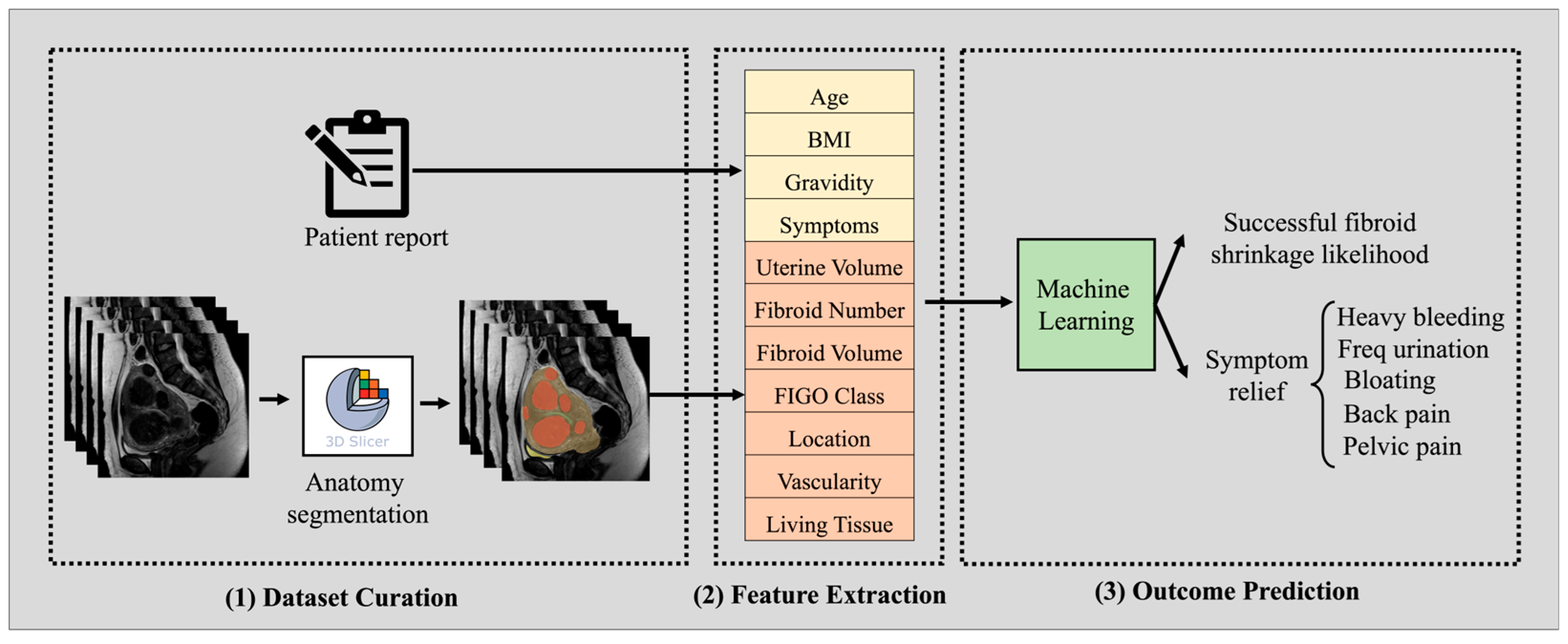
Figure 2 illustrates the pipeline of our ML prediction model. Steps 1 and 2 involve dataset curation and relevant feature extraction, as detailed in the previous section. For each case, individual fibroid features are extracted and augmented with corresponding features from the patient’s medical report. Our ML model is specifically designed to predict not only overall clinical outcomes (likelihood of fibroid shrinkage), but also the patient’s response to symptoms. The likelihood of fibroid shrinkage is defined as >50% shrinkage in patient fibroid volume, which is typically considered procedure success [37,38]. To enable patient-level predictions, it is essential for the machine learning model to aggregate fibroid-level features (characteristics of individual fibroids) in a meaningful and order-invariant manner and combine them with patient-level clinical features. An ideal architecture for this task is Deep Set Networks, a deep learning-based model specifically designed to process feature sets in an order-invariant manner [39,40].
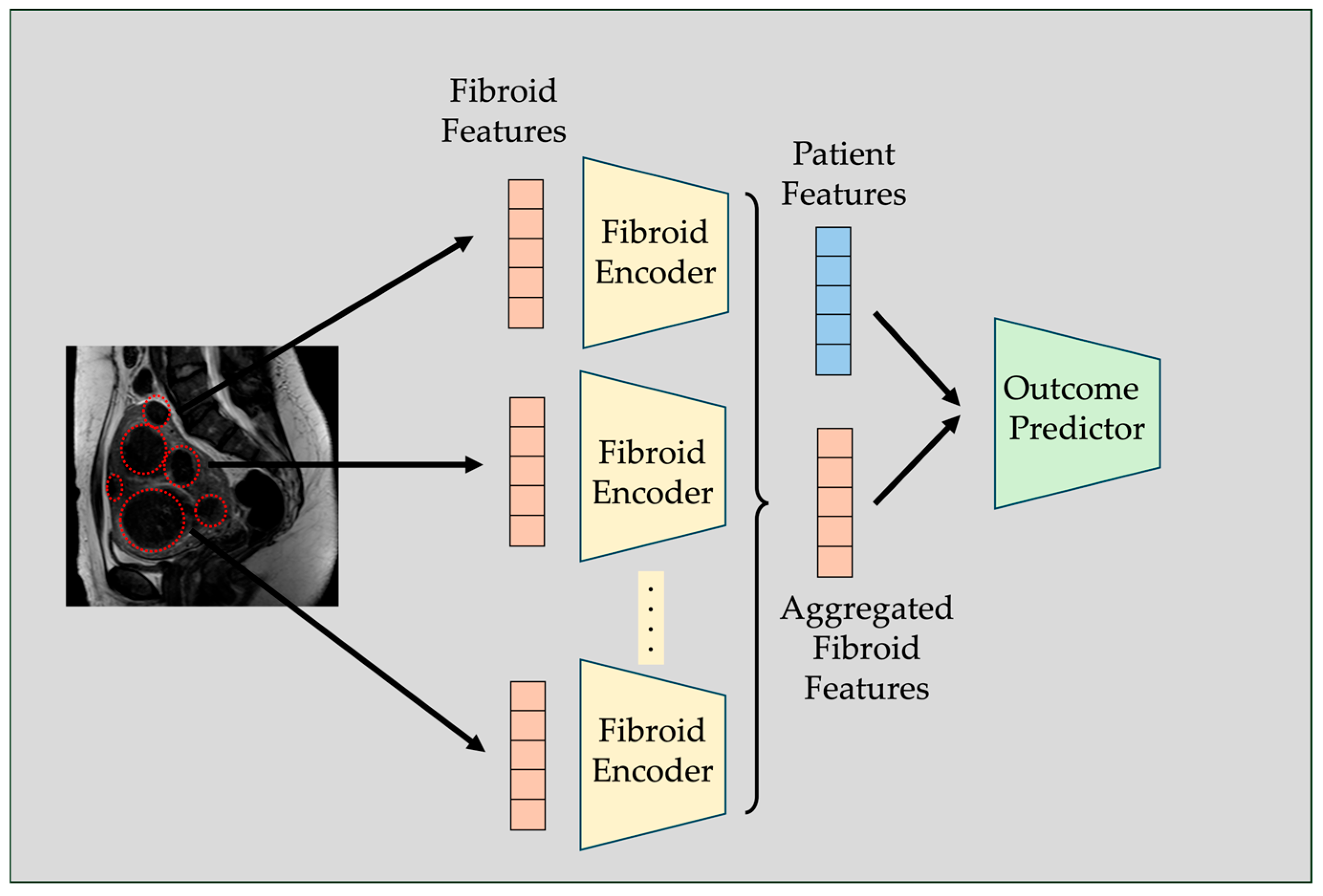
Deep Set Networks [40] represent a specialized class of deep learning architectures specifically designed to operate on unordered sets, where the permutation of elements should not influence the model’s output. This architecture is ideal for problems where each data sample has a variable number of features, and these features do not naturally follow any order. Traditional neural networks process a fixed set of features where the order of the features matters—i.e., the network’s output can change if the order of inputs changes. However, in many clinical applications such as ours, we are dealing with patients each having a different number of tumors/fibroids. Furthermore, in the set of fibroids for each patient, individual fibroids have no inherent or meaningful order. Therefore, concatenating these features imposes an unnatural order on the features, which can lead to inconsistent or biased representations. Another issue with traditional neural networks is that they are inherently unable to process samples with a varying number of features. Deep Set models address this challenge by applying an element-wise transformation to each member, aggregating the transformed elements through a permutation-invariant operation (e.g., summation or mean pooling), and subsequently applying a global transformation to predict the final outcome [40]. This ensures that the model’s output is unaffected by the ordering of inputs, as well as enables it to process a variable type of fibroiod for each sample. Figure 3 further illustrates the internal structure of the Deep Set model used in this study. In our study, each patient is characterized by a variable number of fibroids, with each fibroid having features such as size, location, vascularity, etc. To enable patient-level prediction of clinical outcomes, the Deep Set model first encodes each fibroid’s feature vector via a shared network. The encoded representations are then aggregated across all fibroids for a given patient and concatenated with patient-specific clinical features (e.g., age, BMI, baseline symptom severity). The combined representation is then processed by a subsequent network to predict the outcomes of interest. This approach allows the model to accommodate patients with differing numbers of fibroids, and it is particularly important, as the sequence in which fibroids are processed should not affect the model’s predictive outcomes. Instead, the model focuses on the relative importance of individual features, such as fibroid characteristics, regardless of their order. The implementation of this work was performed using Pytorch 2.4.1 and Python 3.9. In the Deep Set Network, both the fibroid encoder and the outcome predictor modules consisted of three linear layers, each followed by batch normalization and leaky ReLU activation. We used mean aggregation across fibroid embeddings, with masking applied to handle nonexistent fibroids. A dropout rate of 0.3 was applied during training. For optimization, we used the Adam optimizer with a learning rate of 0.01 and weight decay of 0.0001. Binary cross-entropy was used as the loss function. A cutoff threshold of 0.5 was applied to the predicted probabilities to obtain binary outcomes. Hyperparameters were selected using random search over a predefined search space. 20% percent of the dataset was reserved as a hold-out test set, and the best model parameters were determined using 5-fold cross-validation.

2.3. Fibroid-Level Prediction

In addition to predicting overall clinical outcomes at the patient level, it is also beneficial to assess the success of the procedure for each fibroid in order to identify which fibroids responded well and which did not. This can further help identify the features that make fibroids more likely to respond to treatment. According to the standard of care, a fibroid is considered successfully treated if it shows more than a 50% reduction in volume on post-operative imaging [37,38].
To enable individual fibroid analysis, we use the same dataset of our patient cohort to train a set of machine learning models to predict outcomes at the fibroid level. Our models include both simple linear baselines, such as logistic regression, and more advanced non-linear models, such as random forests [41] and XGBoost [42]. Random forests are ensembles of decision trees that excel at capturing non-linear relationships and feature interactions—patterns commonly found in medical data. Additionally, they naturally provide measures of feature importance, helping us identify which fibroid characteristics (e.g., vascularity, location, baseline volume) most strongly influence shrinkage outcomes. These per-fibroid predictions not only improve interpretability but may also support clinical decision-making, especially in patients with multiple fibroids of varying sizes and types.

3. Results

3.1. Procedure Outcome and Post-Op Symptom Prediction

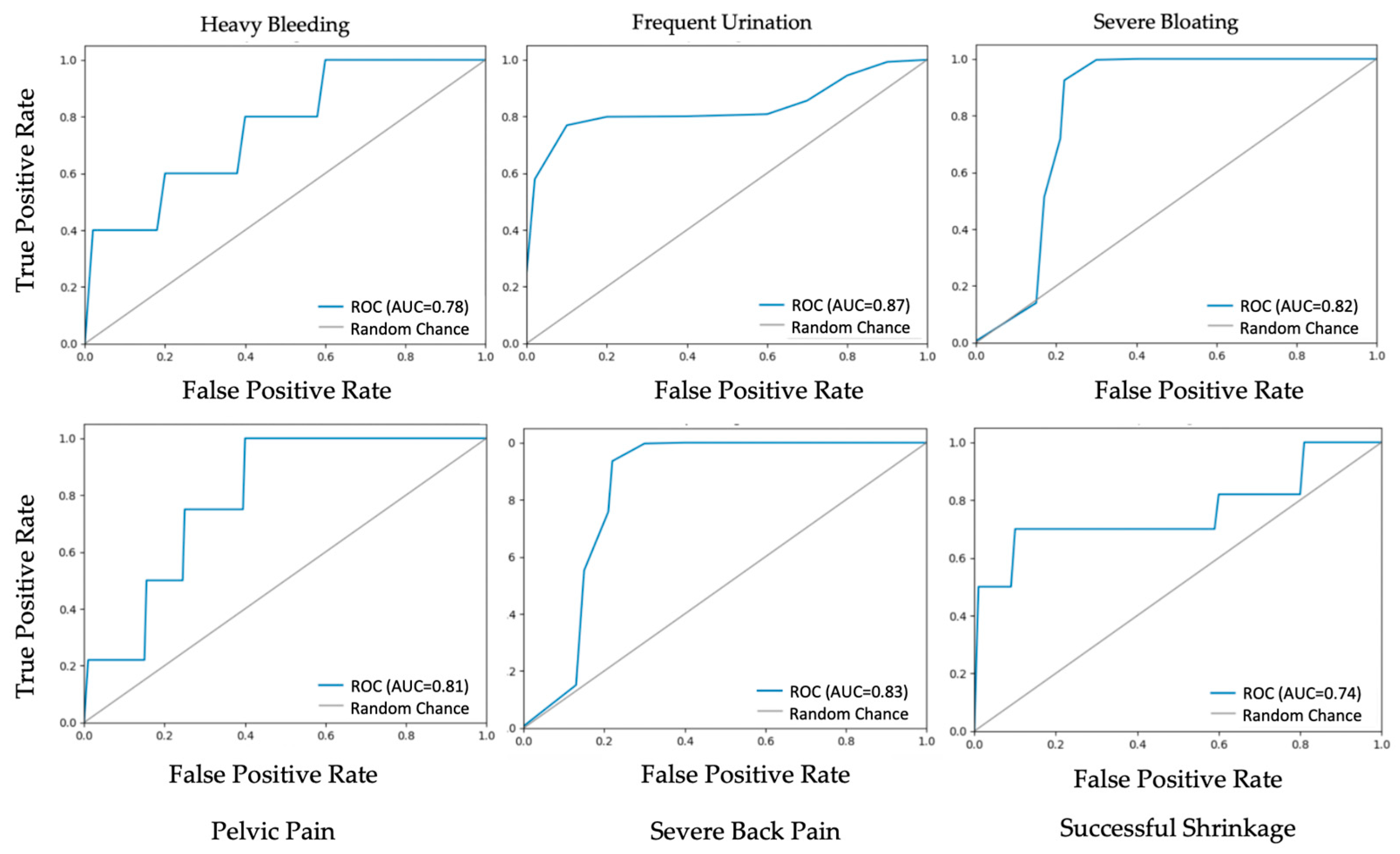
Table 3 presents the performance of the Deep Set Network model in predicting post-operative symptoms and fibroid shrinkage outcomes. The model achieved an accuracy ranging from 75% (for clinical outcome prediction) to 88% (for the frequent urination symptom). The highest F1 score was observed in predicting frequent urination (F1 = 88%). Similarly, the model performed reasonably well in predicting post-op pelvic pain and severe back pain, each achieving an F1 score of 81% with balanced precision and recall. Prediction of heavy bleeding achieved a slightly lower F1 score of 78%.
Figure 4 illustrates the corresponding ROC curve for the classification task. We can see that the AUC is aligned with our previous results. As before, frequent urination has the highest AUC among the symptoms (AUC ≈ 0.87), rising steeply at low false positive rates.

3.2. Ablation Study

As an ablation study to assess the value of the order-invariant aggregation mechanism in the Deep Set architecture, and to examine the necessity of using Deep Set Networks over traditional machine learning and deep learning methods, we conducted additional experiments with conventional models. As previously noted, the variable number of fibroids per patient makes it challenging to process this data using traditional machine learning approaches, which typically assume a fixed number of features. To address this limitation, we have concatenated the features of all fibroids in a random order and used this as the input for the traditional models. For non-existent fibroids, we masked the corresponding features so they would not influence the model’s predictions. The results of these models are presented in Table 4 and Table 5. We can see that the Deep Set Network consistently outperforms both traditional machine learning as well as deep learning models, considering accuracy and F-1 score. Deep Set Networks can outperform traditional models for our problem, since it is inherently designed to handle variable-size feature sets through permutation-invariant aggregation. However, traditional models introduce bias by imposing an arbitrary order on the fibroids through the feature and also lead to overfitting due to the larger feature dimensionality. This experiment further confirms our hypothesis that Deep Set Networks are a suitable choice for this problem.

3.3. Individual Fibroid Outcome Prediction

Table 6 summarizes the results of the fibroid success prediction models. Our results indicated that ensemble tree methods outperformed both linear and instance-based classifiers. Random forests and XGBoost achieved the highest accuracy and F1 score. Support vector machines and K-nearest neighbors delivered moderate performance, while logistic regression lagged behind. The superior performance of random forests and XGBoost likely stems from their ability to model non-linear feature interactions and robustly handle heterogeneous, high-dimensional inputs without extensive feature engineering.

4. Discussion

In this study, we demonstrated the potential of machine learning models to predict both clinical success and post-operative symptom persistence following UFE. We curated a dataset of 74 patients, comprising 311 fibroids, with each case annotated at both the patient and fibroid levels. The dataset includes patient-specific clinical features as well as fibroid-level characteristics such as FIGO classification, volume, location, and vascularity. By integrating patient clinical information with fibroid-specific features, our approach aims to capture complex patterns that may influence treatment response and symptom resolution. We observed that our models had varying prediction abilities for different symptoms. Our models achieved the highest performance in predicting the persistence of frequent urination, suggesting that symptoms strongly linked to uterine size and mass effect are more easily captured using baseline imaging and clinical features. This is likely due to the direct relationship between fibroid shrinkage and relief of pressure-related symptoms, which are well represented in imaging-derived variables. Prediction of pelvic pain and back pain also showed strong performance (F1 = 81% for both), indicating that the models effectively learned fibroid-related contributors to post-operative pain, such as size, location, and vascularity. In contrast, the prediction of heavy bleeding was more challenging. This is not surprising, given the complex nature of this symptom. Unlike symptoms such as frequent urination—which are largely driven by mechanical compression and can be directly inferred from fibroid size and location—heavy menstrual bleeding is influenced by a combination of hormonal factors, vascularization, endometrial involvement, and fibroid subtype. While our dataset included vascularity and FIGO classification, it lacked detailed hormonal profiles, menstrual history, and endometrial imaging features that could more directly account for bleeding patterns. Moreover, bleeding is often a subjective symptom, varying in how patients perceive and report it, adding noise to the labels used for training. These factors likely contributed to the lower accuracy observed in our model and highlight the need for more comprehensive clinical and imaging data to improve the prediction of this outcome.
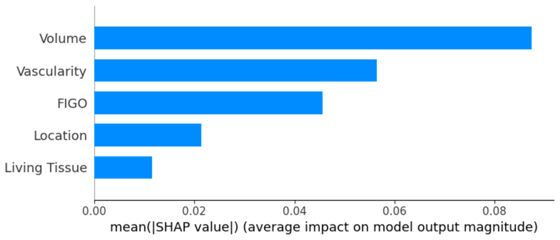
In order to gain better insights into the variables influencing fibroid response to UFE, we also conducted feature-importance analysis using Shapley additive explanations (SHAP) [43] to investigate which fibroid characteristics drive predictive performance (Figure 5). Figure 5 illustrates the feature importance obtained from random forests. Notably, fibroid volume and vascularity emerged as the strongest predictors of treatment response. This finding aligns with clinical expectations, as larger fibroids with rich vascularity tend to respond more effectively to embolization. High vascularity facilitates better delivery of embolic agents, resulting in more substantial infarction and subsequent volume reduction. Conversely, poorly vascularized fibroids may receive insufficient embolic material, leading to suboptimal treatment outcomes. FIGO subtype and anatomic location exert moderate effects, reflecting that hybrid/submucosal classifications tend to favor better outcomes. Similarly, due to the correlation between vascularity and living tissue, most of the effect size is captured through vascularity, making living tissue less significant.
Predicting successful clinical outcome and fibroid shrinkage after UFE is generally a more challenging problem than forecasting post-operative symptom change. Shrinkage can depend on multiple lesion-level biological factors—such as fibroid vascularity, collateral flow, embolic endpoint, particle size, degeneration type, and hormonal milieu—that can be difficult to observe or quantify. In some cases, different lesions of the same fibroid can have different vascularity. In contrast, symptom improvement is shaped largely by patient-level factors, including baseline symptom burden, expectations, and the degree of bulk or bleeding reduction, even if modest. Moreover, different fibroid lesions within the same uterus may respond unevenly, making it inherently difficult to translate these mixed responses into a single shrinkage measure. Nevertheless, a patient may still experience substantial symptom improvement if the most symptomatic fibroid becomes ischemic, even without a significant reduction in total fibroid volume. Finally, shrinkage often unfolds over several months and is strongly affected by variation in follow-up timing, further complicating prediction compared with earlier, easier-to-capture symptom changes.
In this study, we aimed to highlight the clinical relevance of incorporating fibroid-level imaging features into predictive models and demonstrate their potential role in pre-procedural treatment planning. These models could be integrated into radiology or EMR systems to generate a success probability score during preoperative assessments or assist gynecologists in making appropriate referrals. Additionally, the approach could be extended to other procedures to support optimal treatment selection. Such tools would help physicians counsel patients more effectively, set realistic expectations, and guide the selection of the most appropriate intervention. Ultimately, this could enhance personalized care and improve treatment outcomes.
Building on these findings, several areas deserve further investigation. First, validating the model in larger, multi-center cohorts can illustrate its generalization across different patient groups and imaging settings, especially given the retrospective nature and limited sample size of our analysis. These larger-scale multicenter cohorts may provide a less biased estimate of the model’s actual accuracy in unobserved clinical settings. Furthermore, it can be beneficial to further incorporate additional features such as hormonal profiles, menstrual history, endometrial characteristics, other chronic diseases, as well as comorbidities. In addition, we plan to investigate the prediction of longer-term outcomes, including long-term recurrence data, in the future. Automating the feature extraction process, particularly from imaging data, would help scale the approach to larger datasets and reduce manual effort. Adding advanced imaging features—like dynamic contrast-enhanced MRI or diffusion metrics—could also help improve predictions, especially for fibroids with moderate vascularity. Furthermore, exploring more complex models such as convolutional neural nets and self-attention-based models could be another avenue to explore on larger cohort sizes. Using real-time angiographic data during the procedure (such as particle flow patterns) may also make shrinkage forecasts more personalized. Exploring deep learning models that combine 3D fibroid shape with clinical outcomes over time could also capture more complex patterns than traditional features. Finally, linking model predictions to long-term symptom relief and quality-of-life outcomes will help make the forecasts more useful for guiding patient-centered treatment decisions.

Author Contributions

Conceptualization, B.M. and S.J.; methodology, S.J., B.M. and B.C.L.; software, S.J. and A.C.; validation, L.S., M.S. and N.A.L.; formal analysis, S.J.; investigation, S.J.; resources, B.M. and M.S.; data curation, A.C., S.J., M.S., N.A.L. and L.S.; writing—original draft preparation, S.J., A.C. and B.M.; writing—review and editing, B.M., B.C.L., M.R.S.; visualization, S.J.; supervision, B.M. and M.R.S.; funding acquisition, B.M. and M.R.S. All authors have read and agreed to the published version of the manuscript.

Funding

This research was supported by the National Institute of Child Health and Human Development grant number HD112975-01.

Institutional Review Board Statement

This study was reviewed by the Weill Cornell Medicine Institutional Review Board (IRB), protocol number 22-08025173.

Informed Consent Statement

This study used retrospective data, and informed consent was waived due to the use of de-identified records and minimal risk to participants.

Data Availability Statement

The raw data supporting the conclusions of this article could be made available by the authors upon reasonable request.

Acknowledgments

We gratefully acknowledge Mousumi Dhara for her contribution to the data curation process. During the preparation of this manuscript, the authors used ChatGPT 4 to improve the language, grammar, and overall clarity of the text. The authors have reviewed and edited the output and take full responsibility for the content of this publication.

Conflicts of Interest

Corresponding author Mosadegh has a financial interest in SmartHER Inc., a pre-seed startup company aiming to commercialize this tool, which was disclosed and managed by WCM’s conflicts office. SmartHER Inc. had no role in the design, collection, analyses, or interpretation of data of the study, nor in the writing of the manuscript, or in the decision to publish the results.

References

  1. De La Cruz, M.S.; Buchanan, E.M. Uterine Fibroids: Diagnosis and Treatment. Am. Fam. Physician 2017, 95, 100–107. [Google Scholar]
  2. Stewart, E.A.; Nicholson, W.K.; Bradley, L.; Borah, B.J. The burden of uterine fibroids for African-American women: Results of a national survey. J. Women’s Health 2013, 22, 807–816. [Google Scholar] [CrossRef] [PubMed]
  3. Hazimeh, D.; Coco, A.; Casubhoy, I.; Segars, J.; Singh, B. The Annual Economic Burden of Uterine Fibroids in the United States (2010 Versus 2022): A Comparative Cost-Analysis. Reprod. Sci. 2024, 31, 3743–3756. [Google Scholar] [CrossRef] [PubMed]
  4. Wilde, S.; Scott-Barrett, S. Radiological appearances of uterine fibroids. Indian J. Radiol. Imaging 2009, 19, 222–231. [Google Scholar] [CrossRef]
  5. El Sabeh, M.; Borahay, M.A. The Future of Uterine Fibroid Management: A More Preventive and Personalized Paradigm. Reprod. Sci. 2021, 28, 3285–3288. [Google Scholar] [CrossRef]
  6. Mension, E.; Calaf, J.; Chapron, C.; Dolmans, M.M.; Donnez, J.; Marcellin, L.; Petraglia, F.; Vannuccini, S.; Carmona, F. An update on the management of uterine fibroids: Personalized medicine or guidelines? J. Endometr. Uterine Disord. 2024, 7, 100080. [Google Scholar] [CrossRef]
  7. Mettler, L.; Schollmeyer, T.; Tinelli, A.; Malvasi, A.; Alkatout, I. Complications of Uterine Fibroids and Their Management, Surgical Management of Fibroids, Laparoscopy and Hysteroscopy versus Hysterectomy, Haemorrhage, Adhesions, and Complications. Obstet. Gynecol. Int. 2012, 2012, 791248. [Google Scholar] [CrossRef]
  8. Donnez, J.; Dolmans, M.M. Uterine fibroid management: From the present to the future. Hum. Reprod. Update 2016, 22, 665–686. [Google Scholar] [CrossRef]
  9. Young, M.; Mikes, B.A. Uterine Fibroid Embolization. In StatPearls; StatPearls Publishing LLC: Petersburg, FL, USA, 2025. [Google Scholar]
  10. Mas, A.; Tarazona, M.; Dasí Carrasco, J.; Estaca, G.; Cristóbal, I.; Monleón, J. Updated approaches for management of uterine fibroids. Int. J. Womens Health 2017, 9, 607–617. [Google Scholar] [CrossRef]
  11. Balamurugan, S.; Shah, R.; Panganiban, K.; Lehrack, M.; Agrawal, D.K. Uterine Artery Embolization: A Growing Pillar of Gynecological Intervention. J. Radiol. Clin. Imaging 2025, 8, 1–17. [Google Scholar] [CrossRef]
  12. Raikhlin, A.; Baerlocher, M.O.; Asch, M.R. Uterine fibroid embolization: CME update for family physicians. Can. Fam. Physician 2007, 53, 250–256. [Google Scholar]
  13. Kubiszewski, K.; Maag, B.; Hunsaker, P.; Iwai, T.; Lim, J.; Duffey, E.; Varlamov, A.; Yoo, R. Investigating the Underutilization of Uterine Fibroid Embolization by Surveying Practice Preferences of Obstetricians/Gynecologists. J. Vasc. Interv. Radiol. 2023, 34, 1430–1434. [Google Scholar] [CrossRef] [PubMed]
  14. Razavi, M.K.; Hwang, G.; Jahed, A.; Modanlou, S.; Chen, B. Abdominal myomectomy versus uterine fibroid embolization in the treatment of symptomatic uterine leiomyomas. Am. J. Roentgenol. 2003, 180, 1571–1575. [Google Scholar] [CrossRef]
  15. Lukies, M.; Clements, W. Current Strategies for Prevention of Infection After Uterine Artery Embolisation. Cardiovasc. Interv. Radiol. 2022, 45, 911–917. [Google Scholar] [CrossRef] [PubMed]
  16. Dumousset, E.; Chabrot, P.; Rabischong, B.; Mazet, N.; Nasser, S.; Darcha, C.; Garcier, J.M.; Mage, G.; Boyer, L. Preoperative uterine artery embolization (PUAE) before uterine fibroid myomectomy. Cardiovasc. Interv. Radiol. 2008, 31, 514–520. [Google Scholar] [CrossRef]
  17. LaMorte, A.I.; Lalwani, S.; Diamond, M.P. Morbidity associated with abdominal myomectomy. Obstet. Gynecol. 1993, 82, 897–900. [Google Scholar] [PubMed]
  18. Mailli, L.; Patel, S.; Das, R.; Chun, J.Y.; Renani, S.; Das, S.; Ratnam, L. Uterine artery embolisation: Fertility, adenomyosis and size—What is the evidence? CVIR Endovasc. 2023, 6, 8. [Google Scholar] [CrossRef]
  19. Kaufman, C.S. Improving Access to UFE: What Are the Barriers? Endovasc. Today 2022, 21, 74–76. [Google Scholar]
  20. Stiglic, G.; Kocbek, P.; Fijacko, N.; Zitnik, M.; Verbert, K.; Cilar, L. Interpretability of machine learning-based prediction models in healthcare. Wiley Interdiscip. Rev. Data Min. Knowl. Discov. 2020, 10, e1379. [Google Scholar] [CrossRef]
  21. Tonekaboni, S.; Joshi, S.; McCradden, M.D.; Goldenberg, A. What clinicians want: Contextualizing explainable machine learning for clinical end use. In Proceedings of the Machine Learning for Healthcare Conference, Ann Arbor, MI, USA, 8–10 August 2019; pp. 359–380. [Google Scholar]
  22. Raji, H.; Tayyab, M.; Sui, J.; Mahmoodi, S.R.; Javanmard, M. Biosensors and machine learning for enhanced detection, stratification, and classification of cells: A review. Biomed. Microdevices 2022, 24, 26. [Google Scholar] [CrossRef]
  23. Raji, H.; Xie, P.; Mahmoodi, S.R.; Javanmard, M. Wireless power-up and readout from a label-free biosensor. Biomedical Microdevices. 2025, 27, 2. [Google Scholar] [CrossRef]
  24. Tinelli, A.; Morciano, A.; Sparic, R.; Hatirnaz, S.; Malgieri, L.E.; Malvasi, A.; D’Amato, A.; Baldini, G.M.; Pecorella, G. Artificial Intelligence and Uterine Fibroids: A Useful Combination for Diagnosis and Treatment. J. Clin. Med. 2025, 14, 3454. [Google Scholar] [CrossRef]
  25. Zhang, C.; Shu, H.; Yang, G.; Li, F.; Wen, Y.; Zhang, Q.; Dillenseger, J.-L.; Coatrieux, J.-L. HIFUNet: Multi-class segmentation of uterine regions from MR images using global convolutional networks for HIFU surgery planning. IEEE Trans. Med. Imaging 2020, 39, 3309–3320. [Google Scholar] [CrossRef]
  26. Zhang, J.; Liu, Y.; Chen, L.; Ma, S.; Zhong, Y.; He, Z.; Li, C.; Xiao, Z.; Zheng, Y.; Lv, F. DARU-Net: A dual attention residual U-Net for uterine fibroids segmentation on MRI. J. Appl. Clin. Med. Phys. 2023, 24, e13937. [Google Scholar] [CrossRef]
  27. Drukker, K.; Medved, M.; Harmath, C.B.; Giger, M.L.; Madueke-Laveaux, O.S. Radiomics and quantitative multi-parametric MRI for predicting uterine fibroid growth. J. Med. Imaging 2024, 11, 054501. [Google Scholar] [CrossRef]
  28. Yang, M.; Chen, Y.; Zhou, X.; Yu, R.; Huang, N.; Chen, J. Machine learning models for prediction of NPVR ≥ 80% with HIFU ablation for uterine fibroids. Int. J. Hyperth. 2025, 42, 2473754. [Google Scholar] [CrossRef] [PubMed]
  29. Akpinar, E.; Bayrak, O.-C.; Nadarajan, C.; Müslümanoğlu, M.-H.; Nguyen, M.-D.; Keserci, B. Role of machine learning algorithms in predicting the treatment outcome of uterine fibroids using high-intensity focused ultrasound ablation with an immediate nonperfused volume ratio of at least 90%. Eur. Rev. Med. Pharmacol. Sci. 2022, 26, 8376–8394. [Google Scholar] [PubMed]
  30. Li, C.; He, Z.; Lv, F.; Liu, Y.; Hu, Y.; Zhang, J.; Liu, H.; Ma, S.; Xiao, Z. An interpretable MRI-based radiomics model predicting the prognosis of high-intensity focused ultrasound ablation of uterine fibroids. Insights Into Imaging 2023, 14, 129. [Google Scholar] [CrossRef] [PubMed]
  31. Li, C.; He, Z.; Lv, F.; Liao, H.; Xiao, Z. Predicting the Prognosis of HIFU Ablation of Uterine Fibroids Using a Deep Learning-Based 3D Super-Resolution DWI Radiomics Model: A Multicenter Study. Acad. Radiol. 2024, 31, 4996–5007. [Google Scholar] [CrossRef]
  32. Shen, L.; Huang, X.; Liu, Y.; Bai, S.; Wang, F.; Yang, Q. Multi-sequence magnetic resonance imaging radiomics combined with imaging features predicts the difficulty of HIFU treatment of uterine fibroids. Sci. Rep. 2025, 15, 3259. [Google Scholar] [CrossRef]
  33. Liu, L.; Wang, T.; Lei, B. Uterine artery embolization compared with high-intensity focused ultrasound ablation for the treatment of symptomatic uterine myomas: A systematic review and meta-analysis. J. Minim. Invasive Gynecol. 2021, 28, 218–227. [Google Scholar] [CrossRef]
  34. Luo, Y.H.; Xi, I.L.; Wang, R.; Abdallah, H.O.; Wu, J.; Vance, A.Z.; Chang, K.; Kohi, M.; Jones, L.; Reddy, S.; et al. Deep Learning Based on MR Imaging for Predicting Outcome of Uterine Fibroid Embolization. J. Vasc. Interv. Radiol. 2020, 31, 1010–1017 e1013. [Google Scholar] [CrossRef]
  35. Isensee, F.; Jaeger, P.F.; Kohl, S.A.A.; Petersen, J.; Maier-Hein, K.H. nnU-Net: A self-configuring method for deep learning-based biomedical image segmentation. Nat. Methods 2021, 18, 203–211. [Google Scholar] [CrossRef]
  36. Duc, N.M.; Keserci, B. Review of influential clinical factors in reducing the risk of unsuccessful MRI-guided HIFU treatment outcome of uterine fibroids. Diagn. Interv. Radiol. 2018, 24, 283–291. [Google Scholar] [CrossRef]
  37. Faye, N.; Pellerin, O.; Thiam, R.; Chammings, F.; Brisa, M.; Marques, E.; Cuénod, C.A.; Sapoval, M.; Fournier, L.S. Diffusion-weighted imaging for evaluation of uterine arterial embolization of fibroids. Magn. Reson. Med. 2013, 70, 1739–1747. [Google Scholar] [CrossRef]
  38. Lee, M.S.; Kim, M.D.; Jung, D.C.; Lee, M.; Won, J.Y.; Park, S.I.; Lee, D.Y.; Lee, K.H. Apparent diffusion coefficient of uterine leiomyoma as a predictor of the potential response to uterine artery embolization. J. Vasc. Interv. Radiol. 2013, 24, 1361–1365. [Google Scholar] [CrossRef]
  39. Zhang, Y.; Hare, J.; Prugel-Bennett, A. Deep Set Prediction Networks. Adv. Neural Inf. Process. Syst. 2019, 32, 3207–3217. [Google Scholar]
  40. Zaheer, M.; Kottur, S.; Ravanbakhsh, S.; Poczos, B.; Salakhutdinov, R.R.; Smola, A.J. Deep Sets. Adv. Neural Inf. Process. Syst. 2017, 30, 3391–3401. [Google Scholar]
  41. Breiman, L. Random forests. Mach. Learn. 2001, 45, 5–32. [Google Scholar] [CrossRef]
  42. Chen, T.; Guestrin, C. Xgboost: A scalable tree boosting system. In Proceedings of the 22nd Acm Sigkdd International Conference on Knowledge Discovery and Data Mining, San Francisco, CA, USA, 13–17 August 2016; pp. 785–794. [Google Scholar]
  43. Lundberg, S.M.; Lee, S.-I. A unified approach to interpreting model predictions. Adv. Neural Inf. Process. Syst. 2017, 30, 4768–4777. [Google Scholar]
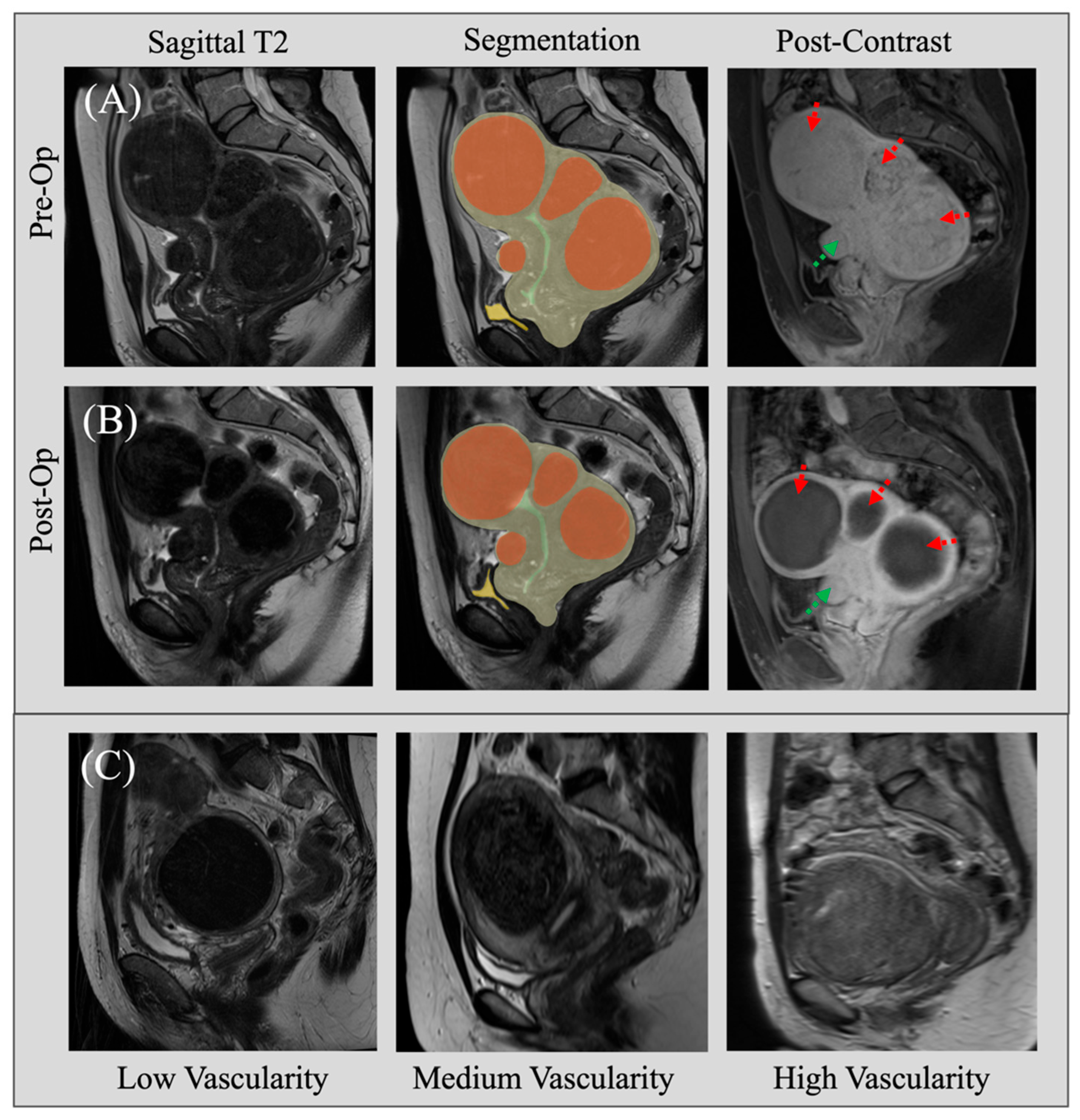
Figure 1. Sample images from the dataset. Row (A) displays a sample pre-operative MRI with sagittal T2 images, annotations, and contrast. Row (B) shows the same slice for the same patient’s post-UFE MRI scan. As observed, three of the fibroids have reduced viability (dashed red label), while one remains viable (dashed green label). Row (C) provides 3 samples of patients with variable vascularity.
Ai 06 00200 g001
Figure 2. Workflow of our ML-powered system. Image features are first extracted from MRI scans and then combined with patient-specific features. A Machine Learning model then predicts overall clinical success as well as post-op symptoms.
Ai 06 00200 g002
Figure 3. Architecture of the Deep Set–based Network used for procedure outcome and symptom prediction. Fibroid features are first encoded using shared encoders, then aggregated and combined with patient-level features, and passed to the second-stage predictor.
Ai 06 00200 g003
Figure 4. ROC curves corresponding to the outcome/symptom prediction tasks.
Ai 06 00200 g004
Figure 5. Feature importance scores for the different variables determined by the model.
Ai 06 00200 g005
Table 1. Summary of clinical statistics of the patient cohort.
Patient Cohort (n = 74)Pre-OpPost-Op
Uterus volume (cm3)629 ± 374407 ± 237
Fibroid number4.2 ± 3.43.8 ± 3.1
Fibroid total volume (cm3)286 ± 243160 ± 158
Symptom Prevalence
 ●
Heavy Bleeding
83.8%31.7%
 ●
Frequent Urination
54.0%31.1%
 ●
Bloating
58.1%24.3%
 ●
Pelvic Pain
91.8%58.1%
 ●
Back Pain
78.4%48.6%
Age (year)49.5 ± 7.0--
Weight (kg)70.4 ± 15.9--
BMI (kg/m2)25.9 ± 6.0--
Gravidity1.7 ± 2.0--
Racial Demographics --
 ●
Caucasian
28%--
 ●
African American
16%--
 ●
Other
13%--
 ●
Declined
24%--
Table 2. Per-fibroid variable statistics.
Fibroid Characteristics (n = 311)Frequency
FIGO Classification
 ●
Submucosal
7%
 ●
Intramural
24%
 ●
Subserosal
23%
 ●
Pedunculated
2%
 ●
Hybrid
44%
Vascularity
 ●
Low
12%
 ●
Medium
32%
 ●
High
56%
Location
 ●
Posterior
30%
 ●
Anterior
27%
 ●
Fundus
24%
 ●
Middle
13%
 ●
Whole
6%
Living Tissue
 ●
Non-Viable
9%
 ●
Mixed
30%
 ●
Viable
61%
Table 3. Summary of model accuracy in predicting overall outcome prediction and post-op symptoms.
ModelAccuracyPrecisionRecallF-1 Score
Heavy Bleeding81%85%81%78%
Frequent Urination88%90%88%88%
Severe Bloating81%91%81%83%
Pelvic Pain82%81%82%81%
Severe Back Pain82%80%81%81%
Successful Shrinkage75%75%75%75%
Table 4. Accuracy of Deep Set Network vs. traditional methods.
ModelHeavy
Bleeding
Freq
Urination
BloatingPelvic PainBack PainSuccessful
Shrinkage
Deep Set Networks81%88%81%82%82%75%
Traditional Neural Net75%75%75%75%75%69%
Light GBM56%75%69%75%62%50%
SVM62%87%75%69%75%56%
Table 5. F-1 Score of Deep Set Network vs. traditional methods.
ModelHeavy
Bleeding
Freq
Urination
BloatingPelvic PainBack
Pain
Successful
Shrinkage
Deep Set Networks78%88%83%81%81%75%
Traditional Neural Net73%71%77%75%75%68%
Light GBM57%74%70%73%59%50%
SVM53%85%77%65%75%55%
Table 6. Performance of different machine learning models in predicting fibroid shrinkage outcome.
ModelAccuracyPrecisionRecallF1 Score
Logistic Regression57%61%57%58%
Support Vector Machine61%64%61%62%
Random Forests76%75%76%75%
XGBoost71%69%71%67%
K-Nearest Neighbors63%65%63%64%
Light GBM66%67%65%66%
Neural Nets70%75%70%71%
Disclaimer/Publisher’s Note: The statements, opinions and data contained in all publications are solely those of the individual author(s) and contributor(s) and not of MDPI and/or the editor(s). MDPI and/or the editor(s) disclaim responsibility for any injury to people or property resulting from any ideas, methods, instructions or products referred to in the content.