
How Did This Happen? Xenograft Conversion to Dermal Scaffolding after Scalding Grease Burn

 ,
, 
Abstract
:1. Introduction
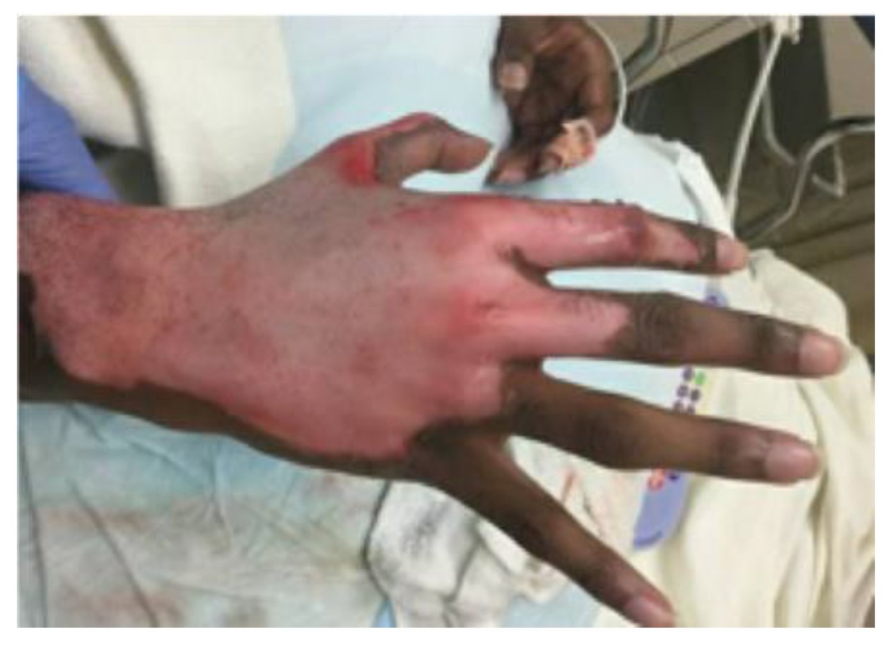
2. Case Presentation
3. Discussion
4. Conclusions
Author Contributions
Funding
Institutional Review Board Statement
Informed Consent Statement
Conflicts of Interest
References
- Hermans, M.H.E. Porcine xenografts vs. (cryopreserved) allografts in the management of partial thickness burns: Is there a clinical difference? Burns 2014, 40, 408–415. [Google Scholar] [CrossRef] [PubMed]
- Baronio, G. Degli Innesti Animali; Dalla Stamperia e Fonderia del Genio: Milan, Italy, 1804. [Google Scholar]
- Girdner, J.H. Skin-grafting with grafts taken from the dead subject. Med. Rec. 1881, 20, 119. [Google Scholar]
- Chick, L.R. Brief History and Biology of Skin Grafting. Ann. Plast. Surg. 1988, 21, 358–365. [Google Scholar] [CrossRef] [PubMed]
- Sheridan, R.L.; Tompkins, R.G. Skin Substitutes in Burns. Burns 1999, 25, 97–103. [Google Scholar] [CrossRef]
- Saffle, J.R. Closure of the Excised Burn Wound: Temporary Skin Substitutes. Clin. Plast. Surg. 2009, 36, 627–641. [Google Scholar] [CrossRef] [PubMed]
- Yamamoto, T.; Iwase, H.; King, T.W.; Hara, H.; Cooper, D.K.C. Skin xenotransplantation: Historical review and clinical potential. Burns 2018, 44, 1738–1749. [Google Scholar] [CrossRef] [PubMed]
- Troy, J.; Karlnoski, R.; Downes, K.; Brown, K.S.; Cruse, C.W.; Smith, D.J.; Payne, W.G. The Use of EZ Derm® in Partial-Thickness Burns: An Institutional Review of157 Patients. Eplasty 2013, 13, e14. [Google Scholar] [PubMed]
- Latarjet, J. The management of pain associated with dressing changes in patients with burns. EWMA J. 2002, 2, 5–9. [Google Scholar]
- Diegidio, P.; Hermiz, S.J.; Ortiz-Pujols, S.; Jones, S.W.; van Duin, D.; Weber, D.J.; Cairns, B.A.; Hultman, C.S. Even Better Than the Real Thing? Xenografting in Pediatric Patients with Scald Injury. Clin. Plast. Surg. 2017, 44, 651–656. [Google Scholar] [CrossRef] [PubMed]
- Luterman, A.; Kraft, E.; Bookless, S. Biologic dressings: An appraisal of current practices. J. Burn Care Rehabil. 1980, 1, 18–23. [Google Scholar] [CrossRef]
- Saricilar, E.C.; Huang, S. Comparison of porcine and human acellular dermal matrix outcomes in wound healing: A deep dive into the evidence. Arch. Plast. Surg. 2021, 48, 433–439. [Google Scholar] [CrossRef] [PubMed]
- Burkey, B.; Davis, W., 3rd; Glat, P.M. Porcine xenograft treatment of superficial partial-thickness burns in paediatric patients. J. Wound Care 2016, 25, S10–S15. [Google Scholar] [CrossRef] [PubMed]
- Castagnoli, C.; Alotto, D.; Cambieri, I.; Casimiri, R.; Aluffi, M.; Stella, M.; Alasia, S.T.; Magliacani, G. Evaluation of donor skin viability: Fresh and cryopreserved skin using tetrazolioum salt assay. Burns 2003, 29, 759–767. [Google Scholar] [CrossRef] [PubMed]
- Rezaei, E.; Beiraghi-Toosi, A.; Ahmadabadi, A.; Tavousi, S.H.; Alipour Tabrizi, A.; Fotuhi, K.; Nooghabi, M.J.; Manafi, A.; Ahmadi Moghadam, S. Can Skin Allograft Occasionally Act as a Permanent Coverage in Deep Burns? A Pilot Study. World J. Plast. Surg. 2017, 6, 94–99. [Google Scholar] [PubMed]
- Atiyeh, B.S.; Hayek, S.N.; Gunn, S.W. New technologies for burn wound closure and healing—Review of the literature. Burns 2005, 31, 944–956. [Google Scholar] [CrossRef] [PubMed]
- Dakour Aridi, H.; Arhuidese, I.; Scudder, M.; Reifsnyder, T.; Malas, M.B. A prospective randomized study of bovine carotid artery biologic graft and expanded polytetrafluoroethylene for permanent hemodialysis access. J. Vasc. Surg. 2018, 67, 1606–1612.e4. [Google Scholar] [CrossRef] [PubMed]
- Chen, X.; Feng, X.; Xie, J.; Ruan, S.; Lin, Y.; Lin, Z.; Shen, R.; Zhang, F. Application of acellular dermal xenografts in full-thickness skin burns. Exp. Ther. Med. 2013, 6, 194–198. [Google Scholar] [CrossRef] [PubMed] [Green Version]
- Christian, R.A.; Stabile, K.J.; Gupta, A.K.; Leckey, B.D., Jr.; Cardona, D.M.; Nowinski, R.J.; Kelly, J.D., 2nd; Toth, A.P. Histologic Analysis of Porcine Dermal Graft Augmentation in Treatment of Rotator Cuff Tears. Am. J. Sports Med. 2021, 49, 3680–3686. [Google Scholar] [CrossRef] [PubMed]


Publisher’s Note: MDPI stays neutral with regard to jurisdictional claims in published maps and institutional affiliations. |
© 2022 by the authors. Licensee MDPI, Basel, Switzerland. This article is an open access article distributed under the terms and conditions of the Creative Commons Attribution (CC BY) license (https://creativecommons.org/licenses/by/4.0/).
Share and Cite
Tran, A.; Windell, E.; Pumiglia, L.; Bettencourt, A.; Vercruysse, G. How Did This Happen? Xenograft Conversion to Dermal Scaffolding after Scalding Grease Burn. Eur. Burn J. 2022, 3, 401-406. https://doi.org/10.3390/ebj3030035
Tran A, Windell E, Pumiglia L, Bettencourt A, Vercruysse G. How Did This Happen? Xenograft Conversion to Dermal Scaffolding after Scalding Grease Burn. European Burn Journal. 2022; 3(3):401-406. https://doi.org/10.3390/ebj3030035
Chicago/Turabian StyleTran, Aurelie, Elizabeth Windell, Luke Pumiglia, Amanda Bettencourt, and Gary Vercruysse. 2022. "How Did This Happen? Xenograft Conversion to Dermal Scaffolding after Scalding Grease Burn" European Burn Journal 3, no. 3: 401-406. https://doi.org/10.3390/ebj3030035
APA StyleTran, A., Windell, E., Pumiglia, L., Bettencourt, A., & Vercruysse, G. (2022). How Did This Happen? Xenograft Conversion to Dermal Scaffolding after Scalding Grease Burn. European Burn Journal, 3(3), 401-406. https://doi.org/10.3390/ebj3030035