Article

A Pilot Trial Assessing the Feasibility and Efficacy of a Novel Powder for Rapid Wound Healing

1
Department of Medicine, University of British Columbia, Vancouver, BC V6T 1Z4, Canada
2
International Collaboration on Repair Discoveries (ICORD), Vancouver, BC V5Z 1M9, Canada
3
Department of Biomedical and Molecular Sciences, Queen’s University, Kingston, ON K7L 3N6, Canada
4
Division of Plastic Surgery, Department of Surgery, University of British Columbia, Vancouver, BC V6T 1Z4, Canada
5
Department of Biochemistry and Microbiology, University of Victoria, Victoria, BC V8P 5C2, Canada
*
Author to whom correspondence should be addressed.
Eur. Burn J. 2021, 2(4), 238-248; https://doi.org/10.3390/ebj2040018
Submission received: 31 August 2021 / Revised: 19 October 2021 / Accepted: 29 October 2021 / Published: 1 November 2021

Abstract

:
It is well-understood that wound care poses a significant burden on the healthcare system and patient well-being. As such, it is imperative to develop efficient methods that facilitate tissue repair. Our group previously developed a nutritional gel scaffold, proven to accelerate wound repair. Due to its gel-like properties, this scaffold requires a time-consuming reconstitution, and is optimized for cavernous wounds. This pilot study examined the feasibility of a powdered form of this scaffold to accelerate healing of full-thickness wounds, thus broadening the range of applications, while providing a practical product. Splinted full-thickness wounds were generated on the backs of 6 mice, and treated with either powder, the original gel scaffold, or no treatment. Feasibility and efficacy of the powder was assessed through comparison of clinical wound measurements and histological assessments. There was a significant effect of treatment on rate of epithelialization [H(3) = 8.346, p = 0.0024] and on days to epithelial closure [H(3) = 8.482, p = 0.0061]. Post hoc analysis revealed that while requiring no reconstitution and simple to apply, the powder was sufficient to accelerate epithelialization compared to untreated wounds (p < 0.05). Furthermore, our results suggest that application of this powder did not alter certain processes associated with healing progress, such as epidermal thickness and collagen deposition. As such, this powder may provide a novel alternative to our previously developed gel scaffold by accelerating epithelialization, while providing a practical product. Future studies necessitate further evaluation of healing measures with a larger sample size.

Graphical Abstract

1. Introduction

It is estimated that every year, greater than 8 million patients seek medical assistance for wound management [1]. Wound healing involves a series of processes, including inflammation, proliferation, and remodeling [2]; however, various patient groups, such as the elderly, obese patients, those with diabetes mellitus, coronary artery disease, or cancer, have impeded healing [3]. It is estimated that USD 10 billion is spent annually in North America alone on complex wound care [4]. This issue is also seen across Europe, where an estimated 2–3% of healthcare expenditure is spent on wound management [5]. Given the impact of wounds on patient wellbeing and the healthcare system, it is imperative to develop efficient methods that facilitate tissue repair.
Although tissue-engineered skin substitutes have been used to facilitate wound healing, these can elicit acute rejection [6,7]. While scaffolds, including autografts, circumvent antigenicity, these are often composed of solid materials restricting the ability of the product to conform to wound topography [8,9]. This inability to integrate with the wound surface impedes vascular perfusion and innervation, increasing the risk of graft loss [8,9]. To circumvent these issues, several powder scaffolds have been developed [10,11,12,13,14,15,16]. These have been reported to be effective in conforming to wound irregularities and help maintain a protective environment. Additionally, powder scaffolds have been reported to enable a cellular response and promote angiogenesis, while offering hemostatic properties [13].
The use of collagen-based biomaterials predates the 1900s, and has since accounted for a myriad of innovations aimed at improving soft-tissue repair [12,17]. The enthusiasm surrounding collagen-based biomaterials originates from the dominant role of collagen in the extracellular matrix (ECM) [12]. Collagen is commonly used in biomaterials for its biodegradable and non-toxic properties, whose fibers confer high tensile strength and stability [12,18]. To the authors’ knowledge, however, there have been no reports of a collagen-based powder scaffold which accelerates wound epithelialization and closure.
Our group previously developed a nutritional and flowable cross-linked collagen-glycosaminoglycan-based scaffold with polyvinyl alcohol (PVA)-borate able to rapidly solidify once at 35–37 °C [8,9]. Once reconstituted, this gel scaffold was shown to have higher in vitro tensile strength, faster fibril formation, and reduced collagenase digestion compared to other collagen-based materials [8]. Furthermore, this scaffold contains the necessary amino acids, vitamins, and minerals required for cell growth [8,9,19]. Likewise, this product has been shown to accelerate healing, while enabling linear cellular organization and formation of a skin-like keratinized epidermis [8,9,19].
Unfortunately, this scaffold has practical limitations; with respect to application, the gel needs to be reconstituted with a sterile liquid and requires a polymerization time of 10 to 20 min [8,9]. This time-consuming reconstitution and application process is undesirable and limiting in many clinical settings. Furthermore, given the flowable nature of the gel scaffold, it is optimized for deep and cavernous wounds [19]. As such, the gel scaffold may be substandard for full-thickness injuries, wherein movement of the affected area risks displacing the scaffold. This pilot study aimed to address the limitations associated with currently available treatments for wound healing by broadening the range of applications of our previously developed gel scaffold. To do so, we investigated whether a powder version of the original gel scaffold could function as a novel alternative to improve healing in full-thickness wounds while providing a practical product.

2. Materials and Methods

2.1. Animal Model and Wound Generation

Following a previously described model of excisional wound healing, six female CD1 mice were purchased from Charles River Laboratories [20]. The mice were housed in an animal care facility under standardized conditions for a one week acclimation period. All procedures were completed in accordance with the ethics approval outlined by the University of British Columbia and the Canadian Guidelines on Animal Care. The mice were individually anesthetized using 2% isoflurane in oxygen (flow rate of 1.0 L/min) and kept under anesthesia throughout the procedure. Prior to wounding, the dorsum of each mouse was shaved and disinfected using 10% Povidone-Iodine (Betadine Purdue Pharma, Pickering, ON, Canada). Following aseptic surgical techniques, two full thickness, 7 mm diameter circular wounds were created bilaterally on either side of the vertebral column of each mouse. To prevent premature wound closure due to contraction of the underlying panniculus carnosus, each wound was splinted using annular silicone splints (Grace Bio-Labs, Bend, OR, USA) with an inner and outer diameter of 7 and 14 mm, respectively. The splints were secured using six interrupted sutures (6-0 Ethilon Nylon Suture, Ethicon LLC, Cornelia, GA, USA).

2.2. Preparation and Application of Treatment Conditions

Treatment conditions were defined as follows: (a) acellular in situ forming gel scaffold (G), (b) acellular freeze-dried powdered (P), or (c) no treatment (NT). For the (G) condition, the acellular in situ forming gel scaffold was prepared and freeze dried into a powder, then reconstituted at the time of application [8,9]. The powder was reconstituted to 80 mg/mL with sterile water, vortexed for one minute, and then centrifuged for 5 min at 1000 revolutions per minute (RPM) with a relative centrifugal force (RCF) of 92 G. Since this mouse model was limited to two wounds per animal, it was not possible to assess all three treatment conditions simultaneously on each mouse. To circumvent this limitation and control for confounding factors, the six mice were randomly assigned to one of six treatment pairs (G-P, P-G, G-NT, NT-G, P-NT, NT-P). For wounds assigned to the (G) condition, 0.1 mL of gel was applied and allowed to polymerize over approximately 10 min. For the (P) condition, 8 mg of un-reconstituted freeze-dried powder was distributed over the entire surface of the wound using a small scoopula. The time taken for the powder to become visibly humidified was measured. Prior to reconstitution and application, the freeze-dried powder was stored at 4 °C. During the wound generation procedure, both the gel (G) and powder (P) were kept on ice, and then removed to warm to room temperature immediately prior to application. No-treatment (NT) conditions received no gel or powder.
Following the treatment applications, all wounds were covered with a semi occlusive dressing (Tegaderm Film, 3M Health Care, St. Paul, MN, USA). A secondary layer of dressing (3MTM CobanTM, Maplewood, MN, USA) was applied over the Tegaderm to prevent the mice from removing the splints and interfering with the wound healing process. Treatments were applied twice, with the first application at the time of injury. The second application occurred at the onset of granulation tissue development within the wound bed. The study endpoint was determined as the time at which each mouse had reached complete closure of both wounds. To control for differences in healing speeds between mice, negative control (NT) wounds were monitored until closure. At the study endpoint, mice were first anesthetized using isoflurane, and euthanized by CO2 asphyxia.

2.3. Wound Epithelialization and Closure

Wounds were photographed twice per week; mice were first anesthetized, and the CobanTM and TegardermTM dressings were then removed, photographed, and re-bandaged as described above. Using the photos acquired throughout the study, areas of open wound were measured in pixels with ImageJ (National Institute of Health, Bethesda, MD, USA) and normalized as a percentage of the inner splint area. The rate of wound closure for each mouse was determined via linear regression of open wound (%) as a function time (days). All wound measurements were conducted blindly. The same principles were used to calculate rate of epithelialization, where un-epithelialized regions of wound were normalized as a percentage of inner splint area, and the rate determined as a function of time. In addition to rate, days until complete epithelialization, and subsequently complete closure were noted.

2.4. Histological Analysis

All wound sites were harvested using a 12-mm circular biopsy punch. Tissue samples were fixed in 10% neutral buffered formalin solution (Sigma-Aldrich, St. Louis, MO, USA) for 24 h before being processed and embedded in paraffin blocks. Tissue samples were sectioned at 5 μm thickness and stained using either a standard hematoxylin and eosin stain (H&E) protocol, or Masson’s Trichrome stain. All histology slides were imaged using a Nikon Eclipse 80i microscope (Nikon Corporation, Tokyo, Japan). Histological measurements were performed using AxioVision (Carl Zeiss MicroImaging Inc., Thornwood, NY, USA) and ImageJ. All image acquisitions and measurements were performed blindly.

2.5. Epidermal Thickness

There have been previous reports suggesting that epidermal thickness (ET) is associated with healing progress, whereby a thinner epidermis correlates with maturation of the neo-epidermis [21,22]. In addition to measuring rate of epidermal regeneration as described above, newly formed ET was measured throughout the wound bed, as a secondary measure of wound maturity. Using Axiovision to assess the H&E-stained samples, the ET for regions of fully formed epidermis were measured at 200 µm increments spanning the wound bed. All images and measurements were acquired blindly.

2.6. Collagen Deposition

Samples stained with Masson’s trichrome were evaluated for collagen deposition using ImageJ, whereby color deconvolution allowed for the separation of blue-stained collagen fibers [23]. Collagen deposition was calculated as the percentage of collagen deposition in wound bed and normalized to percent of collagen deposition in peripheral healthy dermis. All samples were assessed blindly. In addition, wounds were assessed for the presence of collagen deposition in a classical “basket-weave” pattern as a secondary measure of organized wound healing. Samples stained using Masson’s Trichrome were visualized and evaluated by a blinded individual.

2.7. Statistical Analysis

A graphical abstract was created using BioRender (Toronto, ON, Canada). Statistical analysis and figures were generated using GraphPad Prism (Sandiego, CA, USA). Normality of the data’s distribution was assessed using the d’Agostino–Pearson omnibus test for normality. Because of sample size, data normality could not be ascertained. Rates of epithelialization and wound healing were determined via linear regression as a function of wound area (%) to time (days). Comparisons between groups were determined using the Kruskal–Wallis test using the Dunn’s multiple comparisons correction test (α = 0.05).

3. Results

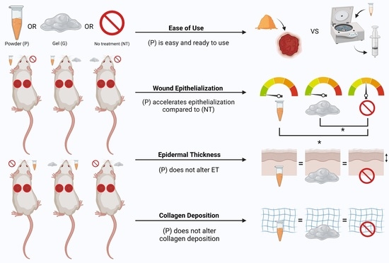
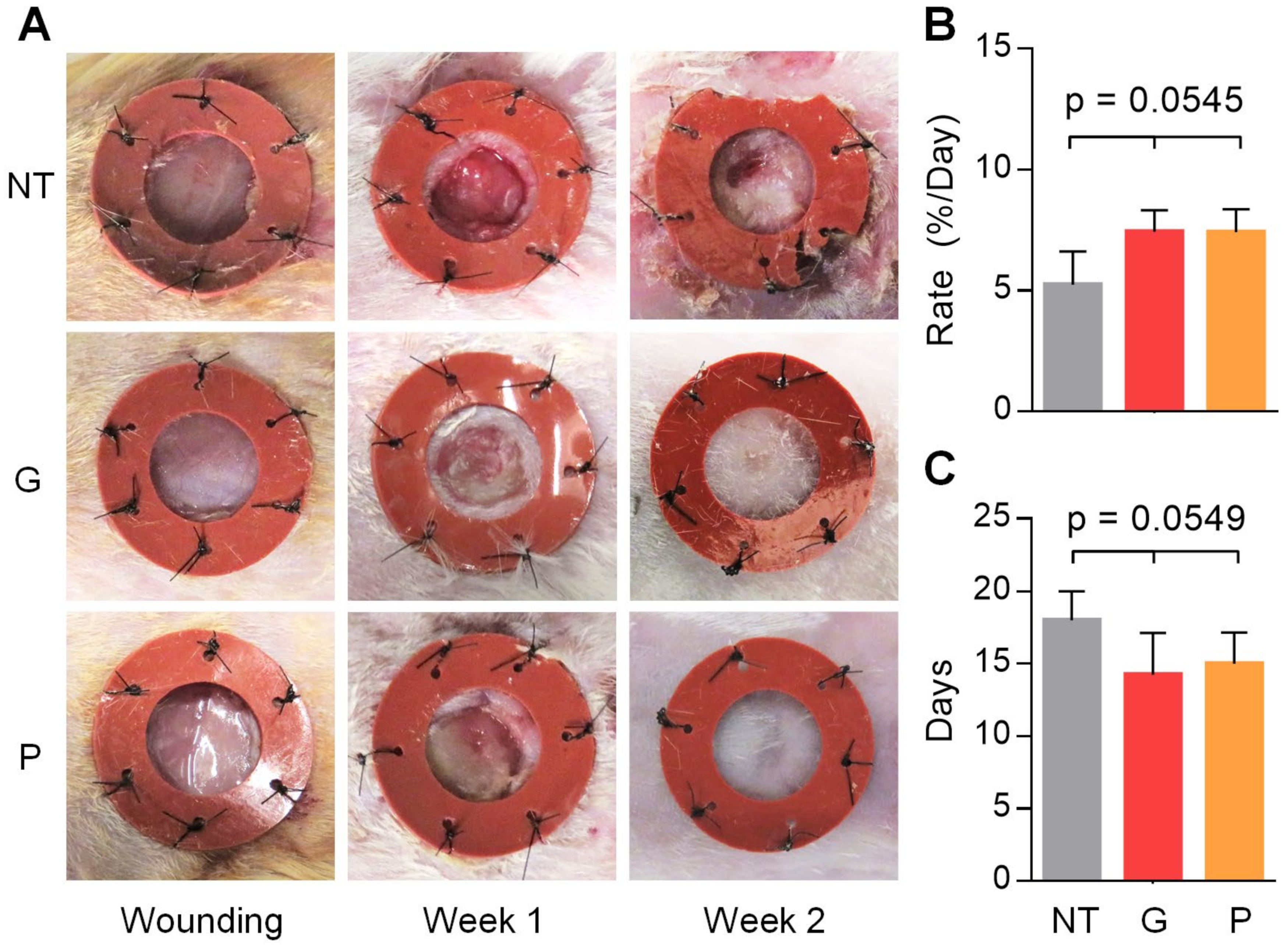
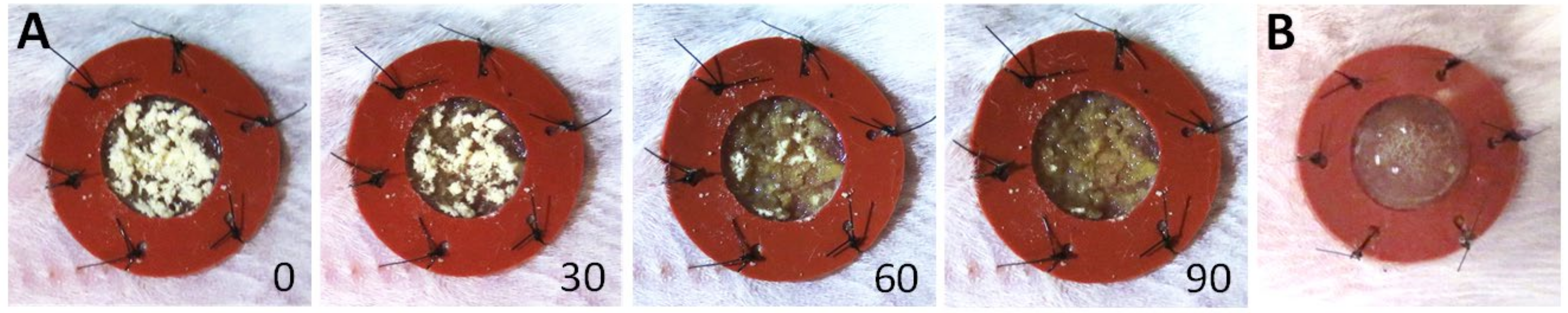
3.1. Use of This Powder Is Feasible for Full-Thickness Wounds in a Murine Model

Comparison of the powder with its gel counterpart is provided (Table 1). This powder is a freeze-dried, PVA borate, cross-linked collagen-glycosaminoglycan-based scaffold, which was stored as a powder at 4 °C throughout the length of the study. The application and distribution of the powder throughout the wound bed was subjectively noted to be feasible using a small scoopula. Once applied, moisture from the wound environment was sufficient to enable the powder rehydration process to begin spontaneously at the wound site (Figure 1A). Representative photographs of the powder in comparison to the gel scaffold are provided (Figure 1A,B). The time in which it took for the powder to humidify in situ was 90 s (Figure 1A).

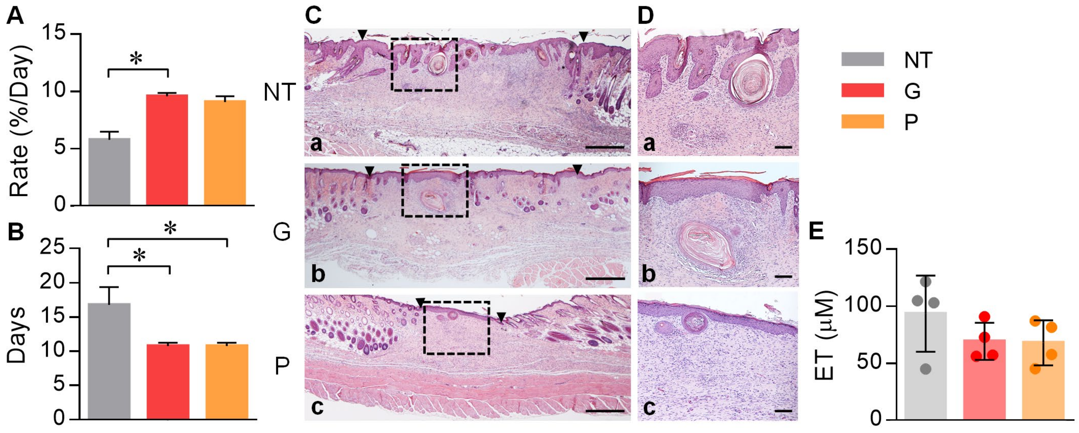
3.2. Powder Application May Accelerate Wound Closure

Progression of wound healing was observed through photographs acquired twice per week (Figure 2A). The effect of treatment condition on the rate of wound closure [(H(3) = 5.654, p = 0.0545] and days required to complete closure [H(3) = 5.570, p = 0.0549] are depicted in Figure 2B,C, respectively. While not statistically significant, both powder and gel conditions had nearly identical rates of closure, both of which were increased in comparison to NT x ¯ = 5.2 ± 1.4, x ¯ = 7.4 ± 0.9, x ¯ = 7.4 ± 1.0, for NT, G, and P conditions, respectively, Figure 2B). Additionally, both powder and gel conditions required a similar duration to close, both of which were decreased in comparison to NT ( x ¯ = 18.0 ± 2.0, x ¯ = 14.3 ± 2.9, x ¯ = 15.0 ± 2.2), for NT, G, and P conditions, respectively, Figure 2C).

3.3. Powder Application Accelerates Epithelialization, without Altering Epidermal Thickness

A significant effect of treatment condition on rate of epithelialization [(H(3) = 8.346, p = 0.0024, Figure 3A] was observed. Post hoc analysis using Dunn’s multiple comparisons test indicated that gel-treated wounds had significantly greater rates of epithelialization compared to NT (p < 0.05, Figure 3A). Additionally, the powder-treated wounds were observed to have comparable epithelialization rates to gel-treated wounds, which was increased in comparison to NT ( x ¯ = 5.8 ± 0.7, x ¯ = 9.6 ± 0.3, x ¯ = 9.1 ± 0.5, for NT, G, and P conditions, respectively, Figure 3A). Furthermore, a significant effect of treatment condition on days required to achieve complete epithelial closure was found [H(3) = 8.482, p = 0.0061, Figure 3B). Post hoc analysis using Dunn’s multiple comparisons test indicated that both powder-treated and gel-treated wounds required significantly fewer days to achieve epithelial closure compared to NT (p < 0.05, Figure 3B). Representative samples illustrating ET using H&E staining are provided at 2× and 10× magnification (Figure 3C,D). No statistically significant difference in days required to achieve complete epithelialization was found between the gel and powder treatments. Despite an increased epithelialization rate and fewer days required to achieve complete epithelialization of the wound bed, no difference in ET was found between groups (Figure 3E). Mean ET was noted to be 93.5 ± 33.4, 69.2 ± 16.3, and 67.9 ± 19.8 for NT, G, and P, respectively.

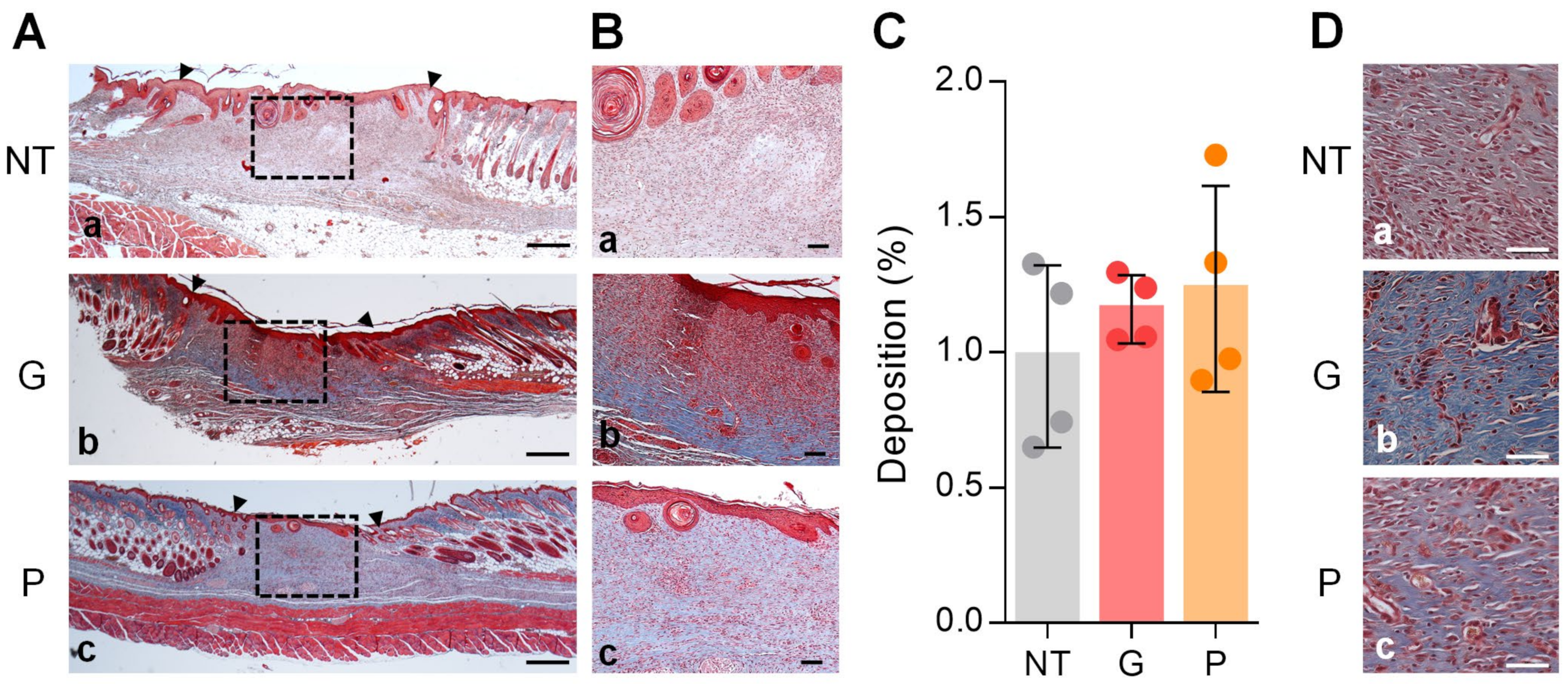
3.4. Powder Application Does Not Alter Collagen Deposition

Granulation tissue and matrix formation was determined by calculating total percent collagen deposition. Representative samples illustrating collagen deposition using Masson’s Trichrome are provided at 2× and 10× magnification (Figure 4A,B). No significant difference was found in quantitative measurement of collagen deposition among treatment groups (Figure 4C). Wounds were examined at 40× magnification (Figure 4D) to assess for patterns of collagen deposition. The majority of NT and P-treated wounds had predominantly mixed presentations of classic “basket-weave” collagen depositions, with regions of collagen deposition in thinner, parallel bundles. Collagen deposition resembling that of a basket weave was primarily found in the upper papillary dermis, whereas the lower reticular dermis was primarily composed of parallel collagen bundles. Of all treatment conditions, G-treated conditions were observed to have a basket-weave collagen deposition, which most closely resembled that of unwounded skin. Nonetheless, no clear difference in collagen patter deposition was identified between groups.

4. Discussion

It is understood that healthcare benefits from improvements in wound healing technologies at both an individual and institutional level [24]. To address this need, our group previously developed an in situ-forming nutritional scaffold proven to accelerate wound repair [8,9]. However, our gel scaffold is limited by its need for reconstitution and restricted range of applications. In this pilot study, we found that application of a novel powdered version of the original gel scaffold is a feasible approach for accelerating epithelialization in a murine model. Furthermore, our results suggest that application of this powder did not disrupt certain processes associated with healing progress and maturation, including that of epidermal thickness and collagen deposition.
Although it was not quantified, the authors noticed that the powder’s ease of use was improved, compared to the original gel scaffold. Unlike the gel, the powder did not require reconstitution prior to application [8]. Beyond being an inherently simpler product to use, providing it as a ready-to-use powder would circumvent additional costs associated with reconstitution (materials, equipment, and time lost). The application and distribution of the powder throughout the wound was also found to be feasible using a small scoopula. However, the presence of a broader standard deviation in collagen deposition was noted amongst powder-treated wounds in comparison to those treated with gel. The authors have considered that this may be due to the powder rehydrating in heterogeneous clumps when compared to the gel’s homogeneous distribution. Accordingly, further optimization of the application technique may be beneficial. Interestingly, the wound environment was sufficient to enable the powder rehydration process to begin spontaneously and rapidly at the wound site. The authors speculate that, compared to the original gel scaffold, this property may provide an inherent advantage in absorbing wound exudate. Future studies may benefit from assessing whether this powder offers any benefit in wounds with increased exudate, such as in burn wounds.
In addition to providing an easy-to-use and feasible product, the powder and original gel scaffold significantly accelerated epithelialization compared to NT, with similar efficacy. These results are corroborated by previous studies, demonstrating that our gel scaffold accelerated healing in murine wound models [9,19]. Interestingly, a significant difference in rate of epithelialization was only found between the gel and NT groups, despite powder-treated wounds having nearly identical epithelialization rates as those treated with gel. This is thought to be a consequence of the Kruskall–Wallis test comparing value ranks as opposed to group means. It is therefore likely that, had the sample size been larger and a one-way ANOVA been appropriate to assess differences, significance would have been observed. This observation is corroborated by the finding that powder-treated wounds required significantly fewer days than NT to completely epithelialize, for which an increased epithelialization rate is thus anticipated. Acceleration of wound healing and prompt re-epithelialization is associated with a decrease in infection rates, scarring, morbidity, and relieves financial burdens on the health care system [24,25]. Open wounds provide a region in which invasion and colonization of microbes can occur, rendering these sites less receptive to treatment and prolonging wound healing [25]. As such, prompt re-epithelialization has proven advantageous in limiting complications of the repair process [25].
In conjunction with measuring speed of wound healing, features suggestive of healing progress and maturity were also assessed. Two measures were used: ET and collagen deposition. Wound closure is achieved via keratinocyte proliferation and full reepithelization of the wound [21]. Secondary to rapid keratinocyte proliferation, newly regenerated epidermis is initially thick, and progressively thins as the neo-epidermis matures [21]. As such, ET can be considered as an index of healing progress [21,22]. It is also widely accepted that collagen formation and deposition constitutes wound healing by facilitating tissue growth and repair [26,27,28,29]. Specifically, the deposition of collagen fibers has been associated with mature granulation tissue, and thus is characteristic of a more advanced healing stage [30,31,32,33,34]. Furthermore, the classic “basket-weave” deposition pattern of collagen has been associated with a decreased incidence of scar tissue, whereas the formation of collagen bundles parallel to the basement membrane have been associated with increased scar tissue formation in rodents [35].
Despite an observed increase in wound healing rate, no significant difference in either measure of ET, percent collagen deposition, nor pattern of collagen deposition were observed, suggesting that application of the powder did not alter healing progress and wound maturation as per the measures used in this study. Interestingly, however, ET in the powder group was 27.4% thinner than the NT group, corroborating with the hypothesis that powder application accelerates wound healing. It may be that statistical significance was not observed due to the limited sample size of our pilot study. Furthermore, our study was conducted using a healthy mouse model, and thus NT wounds are presumed to have undergone normal healing physiology. It may be possible that application of this powder would improve healing outcomes if assessed in a model of aberrant wound healing, and thus would warrant further investigation.
While it aimed to investigate the efficacy and feasibility of a novel powder as an adjunct to wound healing, this pilot study is limited in certain respects. Assessment of the feasibility was limited by the subjectivity of the analysis. Additionally, due to the pilot nature of this study, the authors recognize that findings were largely limited by sample size. Future studies using a greater study sample to increase statistical power whilst improving the generalization of our findings are warranted. An increase in sample size would likely elucidate the importance of results which were found to be of near significance in this study, notably wound closure and epidermal thickness. Additionally, the powder contains a nearly identical composition to its gel counterpart, and thus it is presumed that the powder likely has similar scaffolding properties once reconstituted in vivo. However, this was not specifically assessed in this study. Thus, further evaluation of the powder’s scaffolding properties such as its role in supporting cell attachment and migration would be of benefit.
As alluded to above, elucidating the potential of this powder in accelerating and ameliorating healing quality warrants additional studies using models of aberrant healing. Such research would enable a better understanding of the role for this powder in patients who suffer from pathological wounds. Another important consideration is the potential for use of this powder in burn wounds. It is estimated that every year, 6 million people seek assistance for burn wounds, and burn care treatment is a well-known economic burden [1,36,37,38,39]. As such, investigating this powder using a burn wound model would be of great value. Nonetheless, our findings suggest that this powder may provide a promising alternative to our previously developed gel scaffold by accelerating healing while providing a practical product. As such, the culmination of additional studies would be to assess this powder in patients with full-thickness wounds, including that of burn injuries.

Author Contributions

Author contributions are as follows, with order corresponding to degree of contribution: Conceptualization, A.G., R.J., M.V., E.M.; methodology, A.G., R.J.; software, E.M.; validation, A.G., R.J.; formal analysis, M.V., E.M.; investigation, M.V., E.M., R.J., B.R.; resources, R.T.K.; data curation, E.M., M.V.; writing—original draft preparation, M.V., E.M.; writing—review and editing, S.S.-O., R.J., A.G.; visualization, M.V.; supervision, R.J., A.G.; project administration, M.V., R.J.; funding acquisition, R.T.K., A.G. All authors have read and agreed to the published version of the manuscript.

Funding

This research was funded by grants from the Michael Smith Foundation for Health Research Innovation to Commercialization (I2C) Program (Sponsor Identifier: 17257) and an Innovation Challenge Award, funded by VGH &UBC Hospital Foundation (F18-02825).

Institutional Review Board Statement

The study was conducted in accordance with the ethics approval outlined by the University of British Columbia (protocol code A18-0366, valid through 2018–2022) and the Canadian Guidelines on Animal Care.

Informed Consent Statement

Not applicable.

Data Availability Statement

The data that support the findings of this study are available from the corresponding author upon reasonable request.

Acknowledgments

The authors would like to recognize that this study was conducted on the traditional, ancestral, and unceded lands of Musqueam, Squamish, and Tsleil-Waututh First Nations.

Conflicts of Interest

A.G. is the leading inventor of gel scaffold technology. The funders had no role in the design of the study; in the collection, analyses, or interpretation of data; in the writing of the manuscript, or in the decision to publish the results.

References

  1. Sen, C.K. Human Wounds and Its Burden: An Updated Compendium of Estimates. Adv. Wound Care 2019, 8, 39–48. [Google Scholar] [CrossRef] [Green Version]
  2. Gonzalez, A.C.D.O.; Andrade, Z.D.A.; Costa, T.F.; Medrado, A.R.A.P. Wound healing-A literature review. An. Bras. Dermatol. 2016, 91, 614–620. [Google Scholar] [CrossRef] [PubMed] [Green Version]
  3. Thomas Hess, C. Checklist for factors affecting wound healing. Adv. Skin Wound Care 2011, 24, 192. [Google Scholar] [CrossRef] [PubMed]
  4. Swanson, L. Solving stubborn-wound problem could save millions, team says. CMAJ 1999, 160, 556. [Google Scholar]
  5. Posnett, J.; Gottrup, F.; Lundgren, H.; Saal, G. The resource impact of wounds on health-care providers in Europe. J. Wound Care 2009, 18, 154. [Google Scholar] [CrossRef] [Green Version]
  6. Zaulyanov, L.; Kirsner, R.S. A review of a bi-layered living cell treatment (Apligraf) in the treatment of venous leg ulcers and diabetic foot ulcers. Clin. Interv. Aging 2007, 2, 93–98. [Google Scholar] [CrossRef]
  7. FDA. Technology Assessment: Skin Substitutes for Treating Chronic Wounds; FDA publication Agency for Healthcare: Silver Spring, MD, USA, 2011. [Google Scholar]
  8. Hartwell, R.; Leung, V.; Chavez-Munoz, C.; Nabai, L.; Yang, H.; Ko, F.; Ghahary, A. A novel hydrogel-collagen composite improves functionality of an injectable extracellular matrix. Acta Biomater. 2011, 7, 3060–3069. [Google Scholar] [CrossRef]
  9. Hartwell, R.; Poormasjedi-Meibod, M.S.; Chavez-Munoz, C.; Jalili, R.B.; Hossenini-Tabatabaei, A.; Ghahary, A. An in-situ forming skin substitute improves healing outcome in a hypertrophic scar model. Tissue Eng.-Part A 2015, 21, 1085–1094. [Google Scholar] [CrossRef] [PubMed] [Green Version]
  10. Kallis, P.J.; Friedman, A.J. Collagen powder in wound healing. J. Drugs Dermatol. 2018, 17, 403–408. [Google Scholar]
  11. Ahmed, T.A.E.; Suso, H.P.; Maqbool, A.; Hincke, M.T. Processed eggshell membrane powder: Bioinspiration for an innovative wound healing product. Mater. Sci. Eng. C 2019, 95, 192–203. [Google Scholar] [CrossRef] [PubMed]
  12. Chattopadhyay, S.; Raines, R.T. Review collagen-based biomaterials for wound healing. Biopolymers 2014, 101, 821–833. [Google Scholar] [CrossRef] [Green Version]
  13. Peng, X.; Xu, X.; Deng, Y.; Xie, X.; Xu, L.; Xu, X.; Yuan, W.; Yang, B.; Yang, X.; Xia, X.; et al. Ultrafast Self-Gelling and Wet Adhesive Powder for Acute Hemostasis and Wound Healing. Adv. Funct. Mater. 2021, 31, 2102583. [Google Scholar] [CrossRef]
  14. Murphy, S.V.; Skardal, A.; Nelson, R.A.; Sunnon, K.; Reid, T.; Clouse, C.; Kock, N.D.; Jackson, J.; Soker, S.; Atala, A. Amnion membrane hydrogel and amnion membrane powder accelerate wound healing in a full thickness porcine skin wound model. Stem Cells Transl. Med. 2020, 9, 80–92. [Google Scholar] [CrossRef] [Green Version]
  15. Chen, X.; Peng, L.H.; Chee, S.S.; Shan, Y.H.; Liang, W.Q.; Gao, J.Q. Nanoscaled pearl powder accelerates wound repair and regeneration in vitro and in vivo. Drug Dev. Ind. Pharm. 2019, 45, 1009–1016. [Google Scholar] [CrossRef]
  16. Tottoli, E.M.; Dorati, R.; Genta, I.; Chiesa, E.; Pisani, S.; Conti, B. Skin wound healing process and new emerging technologies for skin wound care and regeneration. Pharmaceutics 2020, 12, 735. [Google Scholar] [CrossRef] [PubMed]
  17. Gibson, T. Evolution of catgut ligatures: The endeavours and success of Joseph Lister and William Mace wen. Br. J. Surg. 2005, 77, 824–825. [Google Scholar] [CrossRef] [PubMed]
  18. Maeda, M.; Tani, S.; Sano, A.; Fujioka, K. Microstructure and release characteristics of the minipellet, a collagen-based drug delivery system for controlled release of protein drugs. J. Control. Release 1999, 62, 313–324. [Google Scholar] [CrossRef]
  19. Forbes, D.; Russ, B.; Kilani, R.; Ghahary, A.; Jalili, R. Liquid Dermal Scaffold with Adipose-Derived Stem Cells Improve Tissue Quality in a Murine Model of Impaired Wound Healing. J. Burn Care Res. 2019, 40, 550–557. [Google Scholar] [CrossRef]
  20. Galiano, R.D.; Michaels, V.J.; Dobryansky, M.; Levine, J.P.; Gurtner, G.C. Quantitative and reproducible murine model of excisional wound healing. Wound Repair Regen. 2004, 12, 485–492. [Google Scholar] [CrossRef]
  21. Teller, P.; White, T.K. The physiology of wound healing: Injury through maturation. Perioper. Nurs. Clin. 2011, 6, 159–170. [Google Scholar] [CrossRef]
  22. Smink, D.S. Schwartz’s Principles of Surgery, 10th Edition. Ann. Surg. 2015, 261, 1026. [Google Scholar] [CrossRef]
  23. Chen, Y.; Yu, Q.; Xu, C.B. A convenient method for quantifying collagen fibers in atherosclerotic lesions by imagej software. Int. J. Clin. Exp. Med. 2017, 10, 14904–14910. [Google Scholar]
  24. Nussbaum, S.R.; Carter, M.J.; Fife, C.E.; DaVanzo, J.; Haught, R.; Nusgart, M.; Cartwright, D. An Economic Evaluation of the Impact, Cost, and Medicare Policy Implications of Chronic Nonhealing Wounds. Value Health 2018, 21, 27–32. [Google Scholar] [CrossRef] [PubMed] [Green Version]
  25. Guo, S.; DiPietro, L.A. Critical review in oral biology & medicine: Factors affecting wound healing. J. Dent. Res. 2010, 89, 219–229. [Google Scholar] [CrossRef]
  26. Mathew-Steiner, S.S.; Roy, S.; Sen, C.K. Collagen in Wound Healing. Bioengineering 2021, 8, 63. [Google Scholar] [CrossRef]
  27. Frantz, C.; Stewart, K.M.; Weaver, V.M. The extracellular matrix at a glance. J. Cell Sci. 2010, 123 Pt 24, 4195–4200. [Google Scholar] [CrossRef] [PubMed] [Green Version]
  28. Hynes, R.O. Stretching the boundaries of extracellular matrix research. Nat. Rev. Mol. Cell Biol. 2014, 15, 761–763. [Google Scholar] [CrossRef] [PubMed]
  29. Mouw, J.K.; Ou, G.; Weaver, V.M. Extracellular matrix assembly: A multiscale deconstruction. Nat. Rev. Mol. Cell Biol. 2014, 15, 771–785. [Google Scholar] [CrossRef] [PubMed]
  30. Richardson, M. Acute wounds: An overview of the physiological healing process. Nurs. Times 2004, 100, 50–53. [Google Scholar]
  31. Singer, A.J.; Clark, R.A.F. Cutaneous Wound Healing. N. Engl. J. Med. 1999, 341, 738–746. [Google Scholar] [CrossRef]
  32. Braiman-Wiksman, L.; Solomonik, I.; Spira, R.; Tennenbaum, T. Novel Insights into Wound Healing Sequence of Events. Toxicol. Pathol. 2007, 35, 767–779. [Google Scholar] [CrossRef]
  33. Kirsner, R.S.; Eaglstein, W.H. The wound healing process. Dermatol. Clin. 1993, 11, 629–640. [Google Scholar] [CrossRef]
  34. Ruszczak, Z. Effect of collagen matrices on dermal wound healing. Adv. Drug Deliv. Rev. 2003, 55, 1595–1611. [Google Scholar] [CrossRef] [PubMed]
  35. McDougall, S.; Dallon, J.; Sherratt, J.; Maini, P. Fibroblast migration and collagen deposition during dermal wound healing: Mathematical modelling and clinical implications. Phil. Trans. R. Soc. A. 2006, 364, 1385–1405. [Google Scholar] [CrossRef]
  36. Hop, M.J.; Polinder, S.; Van Der Vlies, C.H.; Middelkoop, E.; Van Baar, M.E. Costs of burn care: A systematic review. Wound Repair Regen. 2014, 22, 436–450. [Google Scholar] [CrossRef] [PubMed] [Green Version]
  37. Sanchez, J.L.A.; Bastida, J.L.; Martínez, M.M.; Moreno, J.M.M.; Chamorro, J.J. Socio-economic cost and health-related quality of life of burn victims in Spain. Burns 2008, 34, 975–981. [Google Scholar] [CrossRef] [PubMed]
  38. Hemington-Gorse, S.J.; Potokar, T.S.; Drew, P.J.; Dickson, W.A. Burn care costing: The Welsh experience. Burns 2009, 35, 378–382. [Google Scholar] [CrossRef]
  39. Koljonen, V.; Laitila, M.; Rissanen, A.M.; Sintonen, H.; Roine, R.P. Treatment of patients with severe burns-costs and health-related quality of life outcome. J. Burn Care Res. 2013, 34, e318–e325. [Google Scholar] [CrossRef]
Figure 1. Powder vs gel scaffold comparison and rehydration time within the wound bed. (A) Upon application of the powder scaffold to the open wound, representative images of powder rehydration as a function of time are depicted. Photographs were acquired at time of application—0 s, 30 s, 60 s, and 90 s. (B) Depiction of the original gel scaffold is provided for comparison.
Ebj 02 00018 g001
Figure 2. Assessment of wound closure following treatment. Splinted full thickness wounds were treated with either gel scaffold (G), powder (P), or left untreated (NT). (A) Qualitative overview of wound healing across treatment conditions for the time of wounding, week one, and week two, is shown. Representative examples of each treatment condition were chosen for the selected time points. (B) Differences between rates of wound closure are depicted. The results are expressed as mean ± standard deviation, n = 4 for all treatments. (C) Days to wound closure for each treatment condition is depicted. The results are expressed as mean ± standard deviation, n = 4 for all treatments.
Ebj 02 00018 g002
Figure 3. Histological evaluation of wound tissue. Color legend is provided in the top right. (A) Differences between rates of wound closure across treatment conditions are depicted. The results are expressed as mean ± standard deviation, n = 4 for all treatments. * p < 0.05. (B) Days to full epithelialization for each treatment is depicted. The results are expressed as mean ± standard deviation, n = 4 for all treatments, * p < 0.05. (C,D) Representative histological analysis by H&E used to quantify epidermal thickness is shown. The wound bed at 2× magnification (C) and within the wound at 10× magnification (D) is shown for NT (a), G (b), and P (c) conditions. Black dashed rectangles demarcate the wound areas shown at higher magnification on the right. Black triangles indicate the wound edges, whereby the wound is located between these markings. Scale bars for (C,D) represent 500 µM and 100 µM, respectively. (E) The mean ET for each treatment is displayed. Using H&E-stained slides, the ET of each wound was measured every 200 µm increments for regions of fully formed epidermis. Results are expressed as mean ± standard deviation, n = 4 for all treatments groups.
Ebj 02 00018 g003
Figure 4. Analysis of collagen deposition in wound tissue. Histological analysis by Masson’s Trichrome is shown. (A,B) The whole wound at 2× magnification (A) and within the wound at 10× magnification (B) is shown for NT (a), gel (b), and powder (c) conditions. Black dashed rectangles demarcate the wound areas shown at higher magnification on the right. Black triangles indicate the wound edges, whereby the wound is located between these markings. Scale bars for (A,B) represent 500 µM and 100 µM, respectively. (C) The ratio of collagen deposition across the wound bed for all treatment groups is displayed. The results are expressed as mean ± standard deviation, n = 4 across all treatment groups. (D) The wounds at 40× magnification are shown for NT (a), gel (b), and powder (c) conditions. The scale bars represent 50 µM.
Ebj 02 00018 g004
Table 1. Comparison of scaffold characteristics and properties. The characteristics and properties of the powder and the original gel scaffold are compared in this table. Summarized are the powder’s properties determined as per this study and the properties of the original gel scaffold determined through previously validated studies. Composition, reconstitution, feasibility of application, and properties are discussed.
PowderGel
CompositionCross-linked collagen-glycosaminoglycan-based scaffold with polyvinyl alcohol (PVA)-borate (freeze-dried/powdered)Cross-linked collagen-glycosaminoglycan-based scaffold with polyvinyl alcohol (PVA)-borate reconstituted with sterile water [8]
ReconstitutionNot requiredRequires reconstitution via vortexing for one minute in sterile water and centrifugation for 5 min at 1000 RPM and 92 G RCF prior to application [8]
Feasibility of
Application
Feasible to apply and observed to distribute throughout the wound bed in this murine model
Rehydration of the powder into a gel scaffold begins once applied into the wound and visibly humidifies in approximately 90 s
Feasible to apply. Noted to distribute evenly in cavernous and tunneling wounds [9]
PropertiesAccelerates epithelialization
Does not alter certain features suggestive of wound maturity, including ET and collagen deposition
Superior mechanical and physical properties in vitro compared to other collagen-based materials [8]
Contains the necessary amino acids, vitamins, and minerals required for cell growth [8,9]
Accelerates healing while enabling linear cellular organization and formation of a skin-like keratinized epidermis [8]
Non-toxic to human fibroblasts [8]
Optimized for deep and cavernous wounds due to its flowable nature [8,9]
Demonstrated to ameliorate healing in a hypertrophic scarring model [9]
Publisher’s Note: MDPI stays neutral with regard to jurisdictional claims in published maps and institutional affiliations.

