Article

Epidermal Protein C Levels Correspond to Local Injury Severity and Increased Clinical Support in Burn Patients

1
Sutton Arthritis Research Laboratory, Kolling Institute of Medical Research, University of Sydney at Royal North Shore Hospital, St Leonards, NSW 2065, Australia
2
Royal North Shore Hospital, St Leonards, NSW 2065, Australia
3
St Vincent’s Hospital, Darlinghurst, NSW 2010, Australia
*
Author to whom correspondence should be addressed.
Eur. Burn J. 2021, 2(4), 226-237; https://doi.org/10.3390/ebj2040017
Submission received: 1 August 2021 / Revised: 20 October 2021 / Accepted: 27 October 2021 / Published: 1 November 2021
(This article belongs to the Special Issue The Global Burden of Burns, Burn Care Management and Outcome)

Abstract

:
The protein C (PC) system has proven to be a crucial cascade in systemic inflammatory and coagulopathic disorders such as severe sepsis and, more recently, in severe burns. We aimed to conflate our recent systemic findings with further investigations in the local tissue effects of a severe burn injury on the expression of PC and its main receptor endothelial protein C receptor (EPCR). Of the 86 patients enrolled in our recent study, 34 consented to biopsies of both normal and burn edge tissue. These were examined histologically and immunostained for PC, EPCR, and CD68. The burn samples expressed lower PC (p = 0.0027) and higher EPCR (p = 0.0253) than the normal samples in a histological severity-dependent manner. There was also a negative association between PC expression and CD68 positive macrophage infiltration (τb = −0.214, p = 0.020), which was expectedly higher in burn edge samples (p < 0.0005). Interestingly, while there were no correlations between tissue and plasma PC or EPCR, local PC expression was also prognostic of our previously established outcome of a patient requiring increased medical support (OR 0.217 (95%CI 0.052 to 0.901), p = 0.035). The results suggest that local PC cascade changes from a burn injury may be a separate process to the systemic effects and that the local levels may provide useful information in addition to the diagnostic and prognostic abilities we previously found in the circulating PC system.

1. Introduction

Protein C (PC) is a vitamin K-dependent serine protease that plays key roles in homeostasis [1]. The zymogen is converted into activated protein C (APC) by thrombin complexed with thrombomodulin, a process strongly augmented by first binding to endothelial protein C receptor (EPCR) [2,3]. APC performs its more well-known anti-coagulation actions through the cleavage of factors Va and VIIIa [4], with its absence leading to fatal purpura fulminans [5]. Independent of this pathway, APC also exhibits several cytoprotective properties, including anti-inflammation, anti-apoptosis, and endothelial and epithelial barrier stabilisation [1]. Many such properties require EPCR to present APC to its cleavage site on protease-activated receptor (PAR)-1 [6]. Straddling both the coagulation and inflammatory cascades uniquely places PC/APC as a diagnostic and prognostic biomarker; their systemic changes has been well studied in sepsis [7,8,9] and reported in various other pathologies including severe acute pancreatitis [10] and trauma-induced coagulopathy [11], where they have been shown to be associated with complications such as infection, organ failure, and death.
We have recently shown that burn injuries cause early decreases in plasma PC and APC [12,13]. This was followed by recovery to a steady state after around one week, consistent with several other longitudinal coagulation studies in burn patients [14,15,16,17]. Additionally, we found that early changes were associated with injury severity and that their overall levels were negatively correlated with circulating inflammatory cytokines including C reactive protein (CRP), tumour necrosis factor (TNF)-α, interleukin (IL)-1β, IL-6, IL-8, and IL-17. We further demonstrated that a low initial PC was the most important cofactor in a multivariate regression model that predicted whether patients required increased clinical support (see below in the Study Design section). The results portrayed PC as a promising biomarker that is both diagnostic and prognostic, with high sensitivity and specificity, while being easy to measure and reproducible with a currently available standardised pathology assay.
PC was once thought to be primarily synthesised in hepatocytes and endothelial cells as a circulating molecule; however, the epidermis has since been found to possess its own independent PC system that can synthesise and activate PC and that can regulate its function via receptors and inhibitors [18]. Indeed, keratinocytes have been shown to express all components of the PC axis, including EPCR [19], PAR-1 [19], and other related receptors [18]. Through such pathways, PC stimulates keratinocyte survival, proliferation, and migration and enhances their barrier integrity [18,19]. These are critical actions for restoring form and function following cutaneous injuries including burns.
It is unknown whether the reduction in post-burn plasma PC we previously reported is reflective of local cutaneous changes. This study aimed to compare PC and EPCR expression in normal versus burn damaged skin, along with macrophage infiltration as a surrogate marker of local inflammation, and to determine whether they correlate with plasma levels and patient outcomes.

2. Materials and Methods

2.1. Study Design

This report comprises new data from a single-centre prospective observational study, as previously described [12]. Briefly, we recruited 86 patients with severe burn injuries over a two-year period: 2015–2017. They were required to be over 18 years of age and to have 10–80% of total body surface area (TBSA) burns, where at least some areas were partial or full thickness. The participants could not be pregnant or lactating, have clinically significant clotting or bleeding disorders, or have active local or systemic infections. The blood collected on the third daily included PC, APC, soluble EPCR, CRP, TNF-α, IL-1β, IL-6, IL-8, and IL-17. Of these, 34 patients consented to punch biopsies being taken during excision and grafting surgery. Biopsies were taken as early as possible, all within the first week of admission, with a median of within 2 days. We used our previously described binary composite outcome termed “increased support” [12]. This was a binary composite outcome that identified patients who (i) received large amounts of intravenous fluid (≥5 L/day over the first 72 h of admission), which could lead to complications such as pulmonary oedema or abdominal compartment syndrome, with the highest risks in the first 72 h [20]; (ii) had an extended LOS in the ICU (≥5 days), which correlates strongly with mortality [21]; and/or (iii) had more than an average number of surgical interventions (≥5), with related perioperative issues of infection, bleeding, scarring, and general anaesthesia [22,23,24]. Together, they painted a picture of a patient requiring greater medical intervention who was at a greater risk of adverse effects from these interventions.

2.2. Histology

Two 6 mm punch biopsies were taken from each consenting patient. One biopsy was taken from undamaged skin remote from the burn sites, and one was taken at the burn edge. The tissue samples were formalin fixed and paraffin embedded. Histological sections (4 µm) were cut and stained with hematoxylin and eosin (H&E) or immunostained.

2.3. Immunohistochemistry

Primary antibodies used were PC (P4680, Merck), EPCR (BAF2245, R&D Systems, Minneapolis, MN, USA), and CD68 (MA5-13324, ThermoFisher Scientific, Waltham, MA, USA). Standard immunohistochemistry techniques were used. Briefly, tissue sections of wound samples were dewaxed in xylene and rehydrated through graduated ethanol baths. After heat retrieval, endogenous peroxidase activity was blocked using 0.3% hydrogen peroxide and nonspecific staining was blocked by Serum-Free Protein Block (X0909, DAKO). The slides were then incubated in primary antibodies at 4 °C overnight in a Shandon Sequenza (ThermoFisher Scientific, Waltham, MA, USA). On the following day, the slides were incubated in the appropriate ready-to-use secondary antibody from DAKO for 1 h at room temperature. This was followed by 10 min detection using the Liquid DAB+ Substrate Chromogen System (K3468, DAKO). Finally, the slides were counterstained and mounted.
Two independent assessors counted the number of CD68 positively staining macrophages in three random 40× objective fields of view within the papillary dermis of each sample, taking care to avoid any capillaries with a high density of cells. With an eyepiece magnification of 10×/22 mm, this field of view was 0.238 mm2.

2.4. Image Analysis

An independent pathologist qualitatively assessed the H&E sections. Wound images were taken using a camera (Nikon DS-Ri1) mounted to a light microscope (Nikon Eclipse Ci). The quantitative scores of these images were objectively determined by a cell count for CD68 stained slides or by the IHC Profiler plugin for ImageJ software for the immunostained PC and EPCR slides, which gave scores of negative (0), low positive (1), positive (2), and high positive (3) [25].

2.5. Statistal Analyses

The statistical tests used in analyses of the data are as described in each section. The Marginal Homogeneity Program was used for the Bhapkar and McNemar tests [26]. The IBM® SPSS® Statistics v20 package was used for all other tests. Statistical significance was taken at p < 0.05.

3. Results

3.1. Clinical Details

The cohort, as previously described, consisted of 86 patients with a mean age of 44 years, one quarter of whom were female. The vast majority presented with a thermal burn. Approximately two thirds primarily suffered burns of partial thickness, with one third having primarily full thickness. The mean burn size was 21%TBSA. The 34 patients who consented to a biopsy shared similar baseline characteristics with the total population (Table 1).

3.2. Expression of PC Is Decreased and EPCR Increased in Burn-Damaged Epidermis

Figure 1 reveals the typical PC and EPCR staining from a paired (normal and burn edge) biopsy of a patient in this study. A Bhapkar test for marginal homogeneity of the paired ordinal data in the PC contingency table (Table 2) revealed that a direct burn injury had a significant effect on the overall protein C staining scores, p = 0.0022. The proportion of high positively staining burn samples (12%) was significantly lower than the proportion of highly positive normal samples (41%) (McNemar post hoc test with Bonferroni adjustment, p = 0.0075). The burn samples had lower PC than the normal samples (McNemar test of overall bias, p = 0.0027).
A direct burn injury also had a significant effect on the overall EPCR staining scores (Bhapker, p = 0.0238) (Table 3), with the proportion of low positive burn samples (27%) being significantly lower than the proportion of low positive normal samples (56%) (p = 0.0124). The EPCR levels from burnt skin generally scored higher than on normal skin samples (p = 0.0253). These results highlight a dichotomy for the PC axis in burn tissue, with the PC level being low but with its receptor with similar anti-inflammatory properties, EPCR, being high.
There were negative correlations between total burn size (%TBSA) and the expression of both PC (τb = −0.313, p = 0.028) and EPCR (τb = −0.304, p = 0.033) in burn edge skin but not in normal skin biopsies. The Mann–Whitney U tests further showed that the median expression of burn edge EPCR (1 vs. 2, U = 63.000, p = 0.022), but not PC (1.5 vs. 2, U = 72.500, p = 0.051), was lower in primarily full thickness compared with partial thickness burns. The expression of PC and EPCR in normal skin did not correlate with %TBSA or whether the burn injury was predominantly partial or full thickness.

3.3. Progressive Burn Damage Is Correlated with Decreased PC and Increased EPCR Expression

Biopsies from burn edges showed progressive damage from normal to necrotic tissue, with distinctly different pathological zones in the epidermis. To further analyse the PC and EPCR expressions with burn injury severity, we subdivided the burns into five distinct zones, as identified by a pathologist and shown in Figure 2. Zone A shows a normal epidermis with basket-weave keratin overlaying the stratified squamous epithelium, consisting of four discernible layers: the stratum basale, stratum spinosum, stratum granulosum, and stratum corneum (Figure 2A). Connective tissue, neurovascular structures, and occasional inflammatory cells are seen in the dermis. Zone B shows intercellular oedema (spongiosis), vacuolation, and large keratinocytes in the epithelium (Figure 2B). Clusters of mixed inflammatory cells are present in the dermis, though a normal architecture is maintained. Zone C demonstrates epidermal thinning, parakeratosis, and nuclear elongation (Figure 2C). Epidermal thickness is significantly reduced, and there is loss of keratinocyte maturation, as indicated by a lack of well-defined layers. Focally, remnants of the normal epidermal architecture are present. This architecture is completely lost in Zone D (Figure 2D). There are no discernible layers in the epidermis, and the keratinocytes do not demonstrate any maturation. The dermis shows scattered inflammatory cells. Zone E (Figure 2E) reveals an epidermis that is necrotic and/or separating from the underlying dermis. The epidermis completely ulcerated in some parts, while in other parts, it was reduced in thickness, with small, pyknotic nuclei. There are erythrocytes and chronic inflammatory cells in the dermis, consistent with an inflammatory process. The overall qualitative assessment of our burn edge biopsies was consistent with the well-described morphological characterisations of superficial and partial thickness burns [27].
PC staining tended to be higher in normal zones compared with more damaged zones (Figure 2F), whereas EPCR staining tended to be lower in normal compared with more damaged zones (Figure 2G). Somers’ d was run to determine the association between PC or EPCR staining scores (dependent variables) with progressive histological zones of burn damage (independent variable) in 124 zones from all samples combined, as classified above. There was a negative correlation between PC expression and degree of burn damage (d = −0.361, p < 0.0005). In contrast, there was a positive correlation between EPCR expression and degree of burn damage (d = 0.264, p < 0.0005).

3.4. Macrophage Infiltration Is Greater in Burn Edge Dermis and Is Associated with Epidermal PC Expression

The mean density of macrophages from the dermis of each sample was calculated for analysis (Figure 3A,B). As these data sets were positively skewed (as assessed by visual inspection of their histograms and Normal Q–Q Plots), a Wilcoxon signed-rank test showed that there was a highly significant increase in median macrophage infiltration in the burn edge dermis (55.4/mm2) compared with biopsies from the normal dermis (26.7/mm2), p < 0.0005. Additionally, there was a strong positive correlation between macrophage infiltration in the normal sample and macrophage infiltration in the burn edge sample for each patient (ρs = 0.629, p < 0.0005). An increase in time from injury to biopsy was also associated with an increase in macrophage infiltration in both normal (ρs = 0.374, p = 0.027) and burn samples (ρs = 0.393, p = 0.018). There were no correlations between time from injury to biopsy, and PC and EPCR expression in either burn edge or normal skin.
The total burn percentage was not correlated with macrophage infiltration in either normal or burn edge samples. There was significantly greater macrophage infiltration in burn edge biopsies from primarily full thickness burns (134.3/mm2) compared with primarily partial thickness burns (50.9/mm2) (Mann–Whitney U test, p = 0.035). There were no differences in macrophage infiltration of normal dermis from patients with primarily full thickness burns (39.3/mm2) compared with those with primarily partial thickness burns (26.7/mm2), p = 0.368. A Kendall’s tau-b correlation was run to determine the relationship between epidermal PC or EPCR scores with dermal macrophage infiltration amongst 72 matched pairs (both burn and normal tissue combined). There was a negative association between PC scores and macrophage infiltration (τb = −0.214, p = 0.020), but no association of macrophages with EPCR scores (τb = 0.170, p = 0.129). While it was noted that PC and EPCR staining was present on dermal appendages including follicles and sweat glands, these were too inconsistently present on the samples (and at varying depths) for meaningful quantification and correlation with dermal macrophage infiltration.

3.5. PC and EPCR Levels Are Not Associated with Their Levels in Plasma

Kendall’s tau-b correlation was run to determine the relationship between burn tissue PC and EPCR scores versus plasma PC, APC, sEPCR, and the selected cytokine levels. For each patient, the closest timepoint to the time of biopsy was selected to collect data from the corresponding plasma level. There were no significant associations between plasma or skin levels for either PC (τb = 0.164, p = 0.294) or EPCR (τb = 0.074, p = 0.616). There was also no association between plasma APC and burn tissue PC (τb = 0.010, p = 0.946) or EPCR (τb = 0.047, p = 0.753) expression. In the normal epidermis, there was a negative correlation between PC scores and both plasma sEPCR (τb = −0.382, p = 0.013) and IL-6 (τb = −0.345, p = 0.028). There were no other significant associations between normal and burn epidermal PC and EPCR expression with plasma PC, EPCR, APC, CRP, TNF-α, IL-1β, IL-6, IL-8, and IL-17. There were also no correlations between these plasma markers, and normal or burn dermal macrophage infiltration. These data suggest that skin biopsy samples from burn patients may provide additional PC-related information over that from plasma samples.

3.6. PC Levels in Burn Tissue Are Directly Associated with Patients Who Require Increased Support

Table 4 compares the main clinical features of those requiring increased support with those who did not. A univariate binomial logistic regression was performed to ascertain the effects of PC or EPCR expression (treated as continuous variables) in burn-injured skin on the likelihood that participants require increased support. The model for PC was statistically significant, p = 0.019. It explained 21.7% (Nagelkerke R2) of the variance and correctly classified 72.7% of cases with 50.0% sensitivity and 82.6% specificity. Higher PC expression in burn epidermis was associated with a decreased likelihood of requiring increased support, odds ratio 0.217 (95%CI 0.052 to 0.901), p = 0.035. Regression for EPCR yielded an equivocal result. The model was statistically significant, p = 0.035. It explained 17.9% (Nagelkerke R2) of the variance and correctly classified 69.7% of cases, with 50.0% sensitivity and 78.3% specificity. However, within this model, a higher EPCR was not significantly associated with an increased likelihood of requiring increased support (OR 0.267 (95%CI 0.070–1.015), p = 0.053). Neither PC nor EPCR expression in normal skin correlated with increased support. Macrophage infiltration in normal and burn tissue also did not predict for this outcome. The main findings here are that patients with higher PC but not EPCR levels in burnt skin require less overall hospital support.

4. Discussion

The present results demonstrate that burn-damaged epidermis expresses less PC and more EPCR than normal skin in a local burn damage severity-dependent manner. PC as an anti-inflammatory agent was highlighted by its negative correlations with macrophage infiltration as a surrogate marker of inflammation, which was noted to be substantially higher in burns compared with normal tissue. We found that skin tissue PC acted in a similar manner to plasma PC and was directly associated with the requirement for increased clinical support [12]. However, the tissue PC (and EPCR) expressions were not correlated with their plasma levels, suggesting a separate local response that may facilitate more information about outcome than plasma markers alone.
Both PC and EPCR were abundantly expressed in the epidermis and dermal vascular endothelial cells, similar to that previously described [18,19]. Quantitative scoring provided objective evidence for the first time that epidermal PC expression was reduced in burn edge skin compared with normal skin, whereas EPCR expression was increased. Indeed, there was a gradual change in both, based on histological categorisation of the epidermal burn damage severity, with PC expression being negatively associated with progressive epidermal damage and vice versa for EPCR. The reduction in PC in burnt skin may be explained by increased activation/consumption, which is consistent with our previous report showing that plasma PC levels are acutely low following a severe burns injury associated with higher plasma APC/PC activation. It is thought that more extensive burns trigger excessive systemic activation of PC to meet the physiological demand for preventing microvascular thrombosis, and endothelial and epithelial destruction. This has also been reported in reperfusion injury following coronary artery bypass surgery [28] and liver transplantation [29]. The activation of PC to APC is increased by 20 fold by EPCR [2]. Greater epidermal PC activation would also explain the increased epidermal EPCR expression seen, which is upregulated by APC [19]. EPCR itself has vital anti-inflammatory functions; baboons treated with an antibody that blocks PC and APC binding to EPCR died following sublethal concentrations of Escherichia coli due to loss of its anticoagulation and anti-inflammatory responses [30]. The protective nature of EPCR against acute inflammation was further supported by the findings that mice with a severe deficiency have reduced survival following endotoxin infusion [31], whereas mice overexpressing EPCR exhibit reduced mortality [32]. Thus, the low PC and high EPCR expressions could be attributed to PC consumption and EPCR upregulation in response to local tissue damage and inflammation. Interestingly, we found no correlations between skin versus systemic PC and EPCR levels, of which neither had any positive correlations. This suggested that the local burn wound may undergo a separate inflammatory and coagulation process than the systemic changes that have been so well described in burns [33,34] and hints that, while APC or PC can be used systemically in a critically burned patient for treatment of complications such as sepsis [35], there may be another role for topical therapy to help heal the burn wound. There is extensive evidence of the APC’s wound healing effects when applied topically in both preclinical [36,37] and clinical [38,39,40] studies.
Topical APC therapy could further mitigate the inevitable hypertrophic scarring resultant from excessive inflammation in burns [41]. A significant thermal injury triggers a dysfunctional immune response, leading to progressive capillary permeability with protein leakage, hypovolaemic shock, multi-organ failure, sepsis, and death [42,43]. A vital culprit of this is hyperactive macrophages, primed by the burn injury with enhanced production of proinflammatory cytokines [44]. The unsurprisingly increased macrophage infiltration in burn dermis was reflective of greater local inflammation. There was a negative correlation between epidermal PC expression and dermal macrophage density. We found that PC was also expressed in dermal skin adnexa including the epithelial cells of hair follicles, glands, and vessels, as previously reported [18]. More PC in the dermis suggests greater local anti-inflammatory actions as APC can diminish dermal macrophage activity by reducing the release of cytokines including macrophage inflammatory protein-1α, monocyte chemoattractant protein-1, and TNF-α [45,46] and can colocalise with EPCR on endothelium to maintain its barrier function and to downregulate vascular adhesion molecules such as intercellular adhesion molecule-1 [47,48]. Depletion of macrophages during the inflammatory phase of healing lessens scarring [49]. Wound models in Acomys and PU.1 knockout mice have little or no macrophages, respectively, and heal with minimal scarring [50]. Thus, topical APC or PC treatment could both hasten wound healing and reduce the scar burden in burn patients through its anti-inflammatory actions that act at least in part by reducing macrophage infiltration.
Lastly, we found the level of burn damaged epidermal PC was negatively associated with the previously defined composite clinical outcome of increased support. That is, lower PC expression at the injury site predicted for greater likelihood of requiring increased support in the form of greater fluid resuscitation, longer ICU stay, and/or more surgeries. This was likely related to the observation that local skin PC expression was lower in larger burns and primarily full thickness burns. Both burn size and area were strong predictors of this outcome [12]. Taken together, it can be seen that more severe burns reduced not only circulating PC but also PC expression in the injury area, with an associated EPCR increase. In this respect, tissue PC acts as a surrogate marker of local injury severity and the subsequent inflammatory response. Hence, low epidermal PC are expected to correlate with poorer clinical outcomes, i.e., the need for increased support, which was demonstrated in the results, and augmenting its levels may assist recovery. The incongruence between tissue and systemic PC, and the prognostic value of tissue PC expression hints that local PC levels may provide more information about burns outcomes than plasma PC alone.
In conclusion, we found reduced PC expression and increased EPCR expression in burn tissue, likely due to the increased activation of PC to APC as an anti-inflammatory agent. Local PC levels, although also prognostic for outcome, did not correlate with systemic levels, suggesting a separate local process. PC expression was further negatively associated with macrophage infiltration. Taken together, local PC levels may provide more information about burn injuries, and topical treatment with APC/PC may help improve local inflammatory damage. Future studies could involve more participants with tissue samples longitudinally to track whether tissue PC levels parallel local recovery, inflammation, and scarring.

Author Contributions

Conceptualisation, J.V., G.F. and C.J.J.; data curation, R.Z., T.C.L. and A.K.; formal analysis, R.Z., D.W., R.M. and H.L.; funding acquisition, R.Z. and T.C.L.; investigation, R.Z. and H.L.; methodology, T.C.L., A.W., J.V. and C.J.J.; project administration, T.C.L. and A.K.; software, R.Z.; supervision, J.V., M.X. and C.J.J.; visualisation, R.Z.; writing—original draft, R.Z. and D.W.; writing—review and editing, M.X. and C.J.J. All authors have read and agreed to the published version of the manuscript.

Funding

Funding for this study was supported by the Ramsay Research and Teaching Fund and by a Sydney Medical School Foundation Scholarship Grant.

Institutional Review Board Statement

The study was conducted according to the guidelines of the Declaration of Helsinki and approved by the Northern Sydney Local Health District Human Research Ethics Committee of Royal North Shore Hospital (protocol number 1312-409M, approved in 2014).

Informed Consent Statement

Informed consent was obtained from all subjects involved in the study.

Data Availability Statement

The data presented in this study are available on request from the corresponding author.

Acknowledgments

We thank Nancy Huang and Andrew Zimmerman for their assistance with data collection and the staff of the Emergency Department, Intensive Care Unit, and the Severe Burns Unit at Royal North Shore Hospital for their assistance in this project. We also thank Sue Smith for the technical support with histology and immunohistochemistry and Dukyeon Kim for his advice on statistics.

Conflicts of Interest

Christopher Jackson, John Vandervord, Gregory Fulcher, and Aruna Wijewardena are shareholders in a company undertaking a trial of 3K3A-APC in diabetic ulcers. No competing financial interests exist for the other authors. The funders had no role in the design of the study; in the collection, analyses, or interpretation of data; in the writing of the manuscript; or in the decision to publish the results.

References

  1. Griffin, J.H.; Fernandez, J.A.; Gale, A.J.; Mosnier, L.O. Activated protein C. J. Thromb. Haemost. 2007, 5 (Suppl. 1), 73–80. [Google Scholar] [CrossRef] [PubMed]
  2. Stearns-Kurosawa, D.J.; Kurosawa, S.; Mollica, J.S.; Ferrell, G.L.; Esmon, C.T. The endothelial cell protein C receptor augments protein C activation by the thrombin-thrombomodulin complex. Proc. Natl. Acad. Sci. USA 1996, 93, 10212–10216. [Google Scholar] [CrossRef] [PubMed] [Green Version]
  3. Fukudome, K.; Esmon, C.T. Identification, cloning, and regulation of a novel endothelial cell protein C/activated protein C receptor. J. Biol. Chem. 1994, 269, 26486–26491. [Google Scholar] [CrossRef]
  4. Esmon, C.T. Molecular Events that Control the Protein C Anticoagulant Pathway. Thromb. Haemost. 1993, 70, 29–35. [Google Scholar] [CrossRef] [PubMed]
  5. Chalmers, E.; Cooper, P.; Forman, K.; Grimley, C.; Khair, K.; Minford, A.; Morgan, M.; Mumford, A.D. Purpura fulminans: Recognition, diagnosis and management. Arch. Dis. Child. 2011, 96, 1066–1071. [Google Scholar] [CrossRef] [Green Version]
  6. RRiewald, M.; Petrovan, R.J.; Donner, A.; Mueller, B.M.; Ruf, W. Activation of Endothelial Cell Protease Activated Receptor 1 by the Protein C Pathway. Science 2002, 296, 1880–1882. [Google Scholar] [CrossRef]
  7. Fisher, C.J., Jr.; Yan, S.B. Protein C levels as a prognostic indicator of outcome in sepsis and related diseases. Crit. Care Med. 2000, 28 (Suppl. 9), S49–S56. [Google Scholar] [CrossRef]
  8. Lang, T.C.; Zhao, R.; Kim, A.; Wijewardena, A.; Vandervord, J.; McGrath, R.; Fitzpatrick, S.; Fulcher, G.; Jackson, C.J. Plasma protein C levels are directly associated with better outcomes in patients with severe burns. Burn. J. Int. Soc. Burn Inj. 2019, 45, 1659–1672. [Google Scholar] [CrossRef] [PubMed]
  9. Liaw, P.C.; Esmon, C.T.; Kahnamoui, K.; Schmidt, S.; Kahnamoui, S.; Ferrell, G.; Beaudin, S.; Julian, J.A.; Weitz, J.I.; Crowther, M.; et al. Patients with severe sepsis vary markedly in their ability to generate activated protein C. Blood 2004, 104, 3958–3964. [Google Scholar] [CrossRef] [PubMed]
  10. Shorr, A.F.; Bernard, G.R.; Dhainaut, J.-F.; Russell, J.R.; Macias, W.L.; Nelson, D.R.; Sundin, D.P. Protein C concentrations in severe sepsis: An early directional change in plasma levels predicts outcome. Crit. Care 2006, 10, R92. [Google Scholar] [CrossRef] [Green Version]
  11. Lindstrom, O.; Kylanpaa, L.; Mentula, P.; Puolakkainen, P.; Kemppainen, E.; Haapiainen, R.; Fernandez, J.A.; Griffin, J.H.; Repo, H.; Petaja, J. Upregulated but insufficient generation of activated protein C is associated with development of multiorgan failure in severe acute pancreatitis. Crit. Care 2006, 10, R16. [Google Scholar] [CrossRef] [Green Version]
  12. Cohen, M.J.; Call, M.; Nelson, M.; Calfee, C.S.; Esmon, C.T.; Brohi, K.; Pittet, J.F. Critical Role of Activated Protein C in Early Coagulopathy and Later Organ Failure, Infection and Death in Trauma Patients. Ann. Surg. 2012, 255, 379–385. [Google Scholar] [CrossRef] [Green Version]
  13. Zhao, R.; Lang, T.C.; Kim, A.; Wijewardena, A.; Vandervord, J.; McGrath, R.; Fitzpatrick, G.; Xue, M.; Jackson, C. Early protein C activation is reflective of burn injury severity and plays a critical role in inflammatory burden and patient outcomes. Burn. J. Int. Soc. Burn. Inj. 2021. [Google Scholar] [CrossRef] [PubMed]
  14. Kowal-Vern, A.; Gamelli, R.L.; Walenga, J.M.; Hoppensteadt, D.; Sharp-Pucci, M.; Schumacher, H.R. The effect of burn wound size on hemostasis: A correlation of the hemostatic changes to the clinical state. J. Trauma 1992, 33, 50–56; discussion 56–57. [Google Scholar] [CrossRef] [PubMed]
  15. Garcia-Avello, A.; Lorente, J.A.; Cesar-Perez, J.; Garcia-Frade, L.J.; Alvarado, R.; Arevalo, J.M.; Navarro, J.L.; Esteban, A. Degree of hypercoagulability and hyperfibrinolysis is related to organ failure and prognosis after burn trauma. Thromb. Res. 1998, 89, 59–64. [Google Scholar] [CrossRef]
  16. Lavrentieva, A.; Kontakiotis, T.; Bitzani, M.; Papaioannou-Gaki, G.; Parlapani, A.; Thomareis, O.; Tsotsolis, N.; Giala, M.-A. Early coagulation disorders after severe burn injury: Impact on mortality. Intensive Care Med. 2008, 34, 700–706. [Google Scholar] [CrossRef]
  17. Van Haren, R.M.; Thorson, C.M.; Valle, E.J.; Busko, A.M.; Guarch, G.A.; Andrews, D.M.; Pizano, L.R.; Schulman, C.I.; Namias, N.; Proctor, K.G. Hypercoagulability after burn injury. J. Trauma Acute Care Surg. 2013, 75, 37–43; discussion 43. [Google Scholar] [CrossRef] [PubMed]
  18. Xue, M.; Campbell, D.; Jackson, C. Protein C Is an Autocrine Growth Factor for Human Skin Keratinocytes. J. Biol. Chem. 2007, 282, 13610–13616. [Google Scholar] [CrossRef] [Green Version]
  19. Xue, M.; Campbell, D.; Sambrook, P.N.; Fukudome, K.; Jackson, C. Endothelial Protein C Receptor and Protease-Activated Receptor-1 Mediate Induction of a Wound-Healing Phenotype in Human Keratinocytes by Activated Protein C. J. Investig. Dermatol. 2005, 125, 1279–1285. [Google Scholar] [CrossRef] [Green Version]
  20. Brandstrup, B.; Tønnesen, H.; Beier-Holgersen, R.; Hjortsø, E.; Ørding, H.; Lindorff-Larsen, K.; Rasmussen, M.S.; Lanng, C.; Wallin, L.; Iversen, L.H. Effects of intravenous fluid restriction on postoperative complications: Comparison of two perioperative fluid regimens: A randomized assessor-blinded multicenter trial. Ann. Surg. 2003, 238, 641. [Google Scholar] [CrossRef] [PubMed]
  21. Minne, L.; Abu-Hanna, A.; De Jonge, E. Evaluation of SOFA-based models for predicting mortality in the ICU: A systematic review. Crit. Care 2009, 12, R161. [Google Scholar] [CrossRef] [Green Version]
  22. Abbott, T.; Ahmad, T.; Phull, M.; Fowler, A.; Hewson, R.; Biccard, B.; Chew, M.; Gillies, M.; Pearse, R.; Beattie, S.; et al. The surgical safety checklist and patient outcomes after surgery: A prospective observational cohort study, systematic review and meta-analysis. Br. J. Anaesth. 2018, 120, 146–155. [Google Scholar] [CrossRef] [PubMed] [Green Version]
  23. Gangemi, E.N.; Gregori, D.; Berchialla, P.; Zingarelli, E.; Cairo, M.; Bollero, D.; Ganem, J.; Capocelli, R.; Cuccuru, F.; Cassano, P.; et al. Epidemiology and Risk Factors for Pathologic Scarring After Burn Wounds. Arch. Facial Plast. Surg. 2008, 10, 93–102. [Google Scholar] [CrossRef] [PubMed] [Green Version]
  24. Mason, S.E.; Noel-Storr, A.; Ritchie, C.W. The Impact of General and Regional Anesthesia on the Incidence of Post-Operative Cognitive Dysfunction and Post-Operative Delirium: A Systematic Review with Meta-Analysis. J. Alzheimer’s Dis. 2010, 22, S67–S79. [Google Scholar] [CrossRef]
  25. Varghese, F.; Bukhari, A.; Malhotra, R.; De, A. IHC Profiler: An Open Source Plugin for the Quantitative Evaluation and Automated Scoring of Immunohistochemistry Images of Human Tissue Samples. PLoS ONE 2014, 9, e96801. [Google Scholar] [CrossRef] [PubMed] [Green Version]
  26. Uebersax, J.S. User Guide for the MH Program (Version 1.1). Available online: http://ourworld.compuserve.com/homepages/jsuebersax/mh.htm (accessed on 12 January 2019).
  27. Moritz, A.R. Studies of Thermal Injury: III. The Pathology and Pathogenesis of Cutaneous Burns. An Experimental Study. Am. J. Pathol. 1947, 23, 915–941. [Google Scholar] [PubMed]
  28. Raivio, P.; Fernandez, J.; Kuitunen, A.; Griffin, J.H.; Lassila, R.; Petäjä, J. Activation of protein C and hemodynamic recovery after coronary artery bypass surgery. J. Thorac. Cardiovasc. Surg. 2007, 133, 44–51. [Google Scholar] [CrossRef] [PubMed] [Green Version]
  29. Ilmakunnas, M.; Petäjä, J.; Höckerstedt, K.; Mäkisalo, H.; Fernandez, J.A.; Griffin, J.H.; Jansson, S.-E.; Repo, H.; Pesonen, E.J. Activation of protein C during reperfusion in clinical liver transplantation. Transplantation 2003, 75, 467–472. [Google Scholar] [CrossRef]
  30. Taylor, F.B., Jr.; Stearns-Kurosawa, D.J.; Kurosawa, S.; Ferrell, G.; Chang, A.C.K.; Laszik, Z.; Kosanke, S.; Peer, G.; Esmon, C.T. The endothelial cell protein C receptor aids in host defense against Escherichia coli sepsis. Blood 2000, 95, 1680–1686. [Google Scholar] [CrossRef]
  31. Iwaki, T.; Cruz, D.T.; Martin, J.A.; Castellino, F.J. A cardioprotective role for the endothelial protein C receptor in lipopolysaccharide-induced endotoxemia in the mouse. Blood 2005, 105, 2364–2371. [Google Scholar] [CrossRef] [Green Version]
  32. Li, W.; Zheng, X.; Gu, J.; Hunter, J.; Ferrell, G.L.; Lupu, F.; Esmon, N.L.; Esmon, C.T. Overexpressing endothelial cell protein C receptor alters the hemostatic balance and protects mice from endotoxin. J. Thromb. Haemost. 2005, 3, 1351–1359. [Google Scholar] [CrossRef] [PubMed]
  33. Farina, J.A.; Rosique, M.J.; Rosique, R.G. Curbing Inflammation in Burn Patients. Int. J. Inflamm. 2013, 2013, 715645. [Google Scholar] [CrossRef] [PubMed] [Green Version]
  34. Dobson, G.P.; Letson, H.L.; Sharma, R.; Sheppard, F.R.; Cap, A.P. Mechanisms of early trauma-induced coagulopathy: The clot thickens or not? J. Trauma Acute Care Surg. 2015, 79, 301–309. [Google Scholar] [CrossRef] [PubMed] [Green Version]
  35. Weintraub, J.; Satterwhite, T.; Allo, M. The use of drotrecogin alfa recombinant activated protein C for severe sepsis in the critically burned patient: A new treatment approach. Burns 2006, 32, 783–787. [Google Scholar] [CrossRef] [PubMed]
  36. Uchiba, M.; Okajima, K.; Oike, Y.; Ito, Y.; Fukudome, K.; Isobe, H.; Suda, T. Activated Protein C Induces Endothelial Cell Proliferation by Mitogen-Activated Protein Kinase Activation In Vitro and Angiogenesis In Vivo. Circ. Res. 2004, 95, 34–41. [Google Scholar] [CrossRef] [Green Version]
  37. Jackson, C.J.; Xue, M.; Thompson, P.; Davey, R.A.; Whitmont, K.; Smith, S.; Buisson-Legendre, N.; Sztynda, T.; Furphy, L.J.; Cooper, A.; et al. Activated protein C prevents inflammation yet stimulates angiogenesis to promote cutaneous wound healing. Wound Repair Regen. Off. Publ. Wound Health Soc. Eur. Tissue Repair Soc. 2005, 13, 284–294. [Google Scholar] [CrossRef] [PubMed]
  38. Whitmont, K.; Reid, I.; Tritton, S.; March, L.; Xue, M.; Lee, M.; Fulcher, G.; Sambrook, P.; Slobedman, E.; Cooper, A.; et al. Treatment of Chronic Leg Ulcers With Topical Activated Protein C. Arch. Dermatol. 2008, 144, 1479–1483. [Google Scholar] [CrossRef] [Green Version]
  39. Wijewardena, A.; Vandervord, E.; Lajevardi, S.S.; Vandervord, J.; Jackson, C.J. Combination of Activated Protein C and Topical Negative Pressure Rapidly Regenerates Granulation Tissue Over Exposed Bone to Heal Recalcitrant Orthopedic Wounds. Int. J. Low. Extrem. Wounds 2011, 10, 146–151. [Google Scholar] [CrossRef]
  40. Kapila, S.; Reid, I.; Dixit, S.; Fulcher, G.; March, L.; Jackson, C.; Cooper, A. Use of dermal injection of activated protein C for treatment of large chronic wounds secondary to pyoderma gangrenosum. Clin. Exp. Dermatol. 2014, 39, 785–790. [Google Scholar] [CrossRef]
  41. Zhao, R.; Jackson, C.J.; Xue, M. Extracellular Matrix and Other Factors that Impact on Cutaneous Scarring. In Chronic Wounds, Wound Dressings and Wound Healing; Shiffman, M.A., Low, M., Eds.; Springer International Publishing: Cham, Switzerland, 2018; pp. 135–178. [Google Scholar]
  42. Rae, L.; Fidler, P.; Gibran, N. The Physiologic Basis of Burn Shock and the Need for Aggressive Fluid Resuscitation. Crit. Care Clin. 2016, 32, 491–505. [Google Scholar] [CrossRef]
  43. Lopez, O.N.; Cambiaso-Daniel, J.; Branski, L.K.; Norbury, W.B.; Herndon, D.N. Predicting and managing sepsis in burn patients: Current perspectives. Ther. Clin. Risk Manag. 2017, 13, 1107–1117. [Google Scholar] [CrossRef] [PubMed] [Green Version]
  44. Schwacha, M.G. Macrophages and post-burn immune dysfunction. Burns 2003, 29, 1–14. [Google Scholar] [CrossRef]
  45. Brueckmann, M.; Hoffmann, U.; de Rossi, L.; Weiler, H.M.; Liebe, V.; Lang, S.; Kaden, J.J.; Borggrefe, M.; Haase, K.K.; Huhle, G. Activated protein C inhibits the release of macrophage inflammatory protein-1-alpha from THP-1 cells and from human monocytes. Cytokine 2004, 26, 106–113. [Google Scholar] [CrossRef] [PubMed]
  46. Yuksel, M.; Okajima, K.; Uchiba, M.; Horiuchi, S.; Okabe, H. Activated protein C inhibits lipopolysaccharide-induced tumor necrosis factor-alpha production by inhibiting activation of both nuclear factor-kappa B and activator protein-1 in human monocytes. Thromb. Haemost. 2002, 88, 267–273. [Google Scholar] [CrossRef]
  47. Franscini, N.; Bachli, E.B.; Blau, N.; Leikauf, M.-S.; Schaffner, A.; Schoedon, G. Gene Expression Profiling of Inflamed Human Endothelial Cells and Influence of Activated Protein C. Circulation 2004, 110, 2903–2909. [Google Scholar] [CrossRef] [PubMed] [Green Version]
  48. Jackson, C.; Xue, M. Anti-inflammatory actions of the anticoagulant, activated protein C. In Inflammatory Diseases–A Modern Perspective; Nagal, A., Ed.; InTech: Rijeka, Croatia, 2011; pp. 42–74. [Google Scholar]
  49. Lucas, T.; Waisman, A.; Ranjan, R.; Roes, J.; Krieg, T.; Müller, W.; Roers, A.; Eming, S.A. Differential Roles of Macrophages in Diverse Phases of Skin Repair. J. Immunol. 2010, 184, 3964–3977. [Google Scholar] [CrossRef] [Green Version]
  50. Brant, J.; Yoon, J.H.; Polvadore, T.; Barbazuk, W.; Maden, M. Cellular events during scar-free skin regeneration in the spiny mouse, Acomys. Wound Repair Regen. 2016, 24, 75–88. [Google Scholar] [CrossRef] [PubMed]
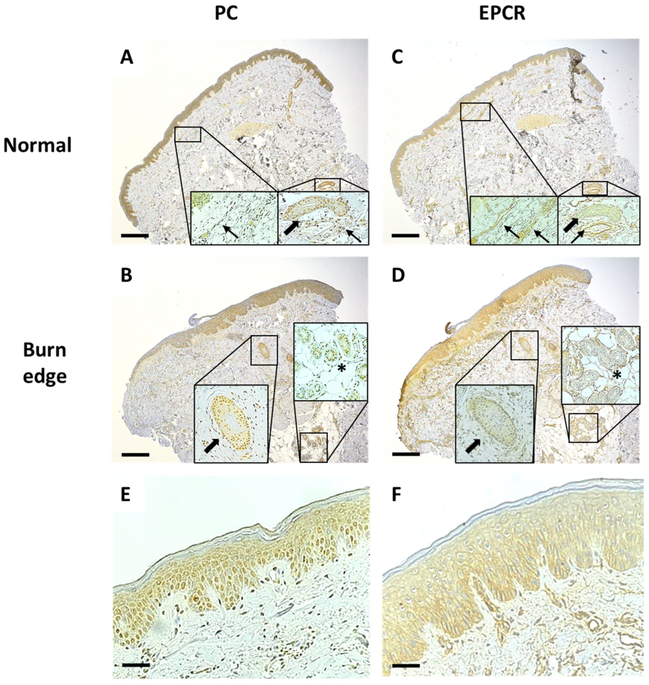
Figure 1. PC and EPCR staining of normal and burn edge biopsies from a typical patient of this study. PC staining in normal (A) and burn edge (B) biopsies alongside EPCR epithelial staining in normal (C) and burn edge (D) biopsies. Both PC and EPCR stained structures in the dermis including hair follicles (thick arrow), vessels (thin arrow), and sweat glands (asterisk). Scale bar = 1 mm. Insets are 2.5× greater magnification. PC demonstrated mostly nuclear staining (E), whereas EPCR was mostly cytoplasmic (F). Scale bar = 200 µm.
Ebj 02 00017 g001
Figure 2. Progressive burn damage in a typical burn edge biopsy. Zone A shows a normal keratinised epidermis with four discernible layers: the stratum basale (SB), stratum spinosum (SP), stratum granulosum (SG), and stratum corneum (SC) (A). Inset is 1.5× greater magnification. Zone B shows spongiosis (thin arrows), vacuolation (asterisk), and large keratinocytes (B). Inset is 2× greater magnification. Zone C demonstrates epidermal thinning, parakeratosis (hash), and nuclear elongation (thick arrow), with a loss of well-defined layers (C). Inset is 2.5× greater magnification. Zone D shows a complete loss of discernible layers or keratinocyte maturation (D). Inset is 2× greater magnification. Zone E shows a necrotic epidermis that is separating from the dermis (E). Inset is 1.5× greater magnification. Scattered inflammatory cells can be seen in the dermis of zones B–E. PC staining tended to be higher in less damaged compared with more severely damaged zones (F), whereas EPCR staining tended to be lower in the less damaged zones and higher in the more severely damaged zones (G). N = 124 zones from 68 biopsies. Scale bar = 200 μm.
Ebj 02 00017 g002
Figure 3. Dermal macrophage infiltration. CD-68-positive macrophage concentration in normal (A) and burn edge (B) dermis. This was quantified by the mean number of macrophages/mm2, as determined by two independent assessors by counting the number of CD68 positively stained macrophages in three random 40× objective fields of view within the papillary dermis of each sample, taking care to avoid any capillaries with a high density of cells. There was greater median macrophage infiltration in the burn edge (55.4/mm2) compared with normal biopsies (26.7/mm2), **** p < 0.0005 (C). N = 34. Scale bar = 100 μm. Insets are 2.5× greater magnification.
Ebj 02 00017 g003
Table 1. Clinical overview of patient and burn characteristics on admission.
Biopsied PatientsAll Patients
Total patients3486
 Female (%)8 (24)22 (26)
 Male (%)26 (76)64 (74)
Age, mean ± SD43 ± 1644 ± 19
Burn size, mean ± SD22 ± 1621 ± 13
Burn depth
 Partial (%)24 (71)59 (69)
 Full (%)10 (29)27 (31)
Table 2. Contingency table for PC staining in normal versus burn edge biopsies.
Burn Biopsy PCTotal
NegativeLow PositivePositiveHigh Positive
Normal biopsy PCNegative00000 (0%)
Low positive01304 (12%)
Positive266216 (47%)
High positive0111214 (41%)
Total2 (6%)8 (24%)20 (59%)4 (12%)34
Table 3. Contingency table for EPCR staining in normal versus burn edge biopsies.
Burn Biopsy EPCRTotal
NegativeLow PositivePositiveHigh Positive
Normal biopsy EPCRNegative00011 (3%)
Low positive1611019 (56%)
Positive038213 (38%)
High positive00101 (3%)
Total1 (3%)9 (27%)20 (59%)4 (12%)34
Table 4. Clinical characteristics for patients requiring increased support versus those who had a standard admission.
Standard AdmissionIncreased Support
Total patients2410
 Female (%)6 (67)3 (33)
 Male (%)18 (72)7 (28)
Age, mean ± SD43 ± 1741 ± 14
Burn size, mean ± SD19 ± 1230 ± 21
Burn depth
 Partial (%)20 (83)4 (17)
 Full (%)4 (4)6 (60)
Length of stay, mean ± SD19 ± 3160 ± 56
ICU length of stay, mean ± SD0 ± 121 ± 30
Number of surgeries, mean ± SD2 ± 18 ± 5
Mean IV fluids per day over first 72 h, mean ± SD2 ± 15 ± 3
Publisher’s Note: MDPI stays neutral with regard to jurisdictional claims in published maps and institutional affiliations.