Review

Diagnosis and Management of Invasive Fungal Wound Infections in Burn Patients

by
Kaitlin A. Pruskowski
1,2,*,
Thomas A. Mitchell
1,
John L. Kiley
3,
Trevor Wellington
3,
Garrett W. Britton
1 and
Leopoldo C. Cancio
1,4
1
US Army Institute of Surgical Research, JBSA Fort Sam Houston, TX 78234, USA
2
Department of Medicine, F. Edward Hebert School of Medicine, Uniformed Services University of the Health Sciences, Bethesda, MD 20814, USA
3
Brooke Army Medical Center, JBSA Fort Sam Houston, TX 78234, USA
4
Department of Surgery, Long School of Medicine, University of Texas Health San Antonio, San Antonio, TX 78229, USA
*
Author to whom correspondence should be addressed.
Eur. Burn J. 2021, 2(4), 168-183; https://doi.org/10.3390/ebj2040013
Submission received: 27 July 2021 / Revised: 10 September 2021 / Accepted: 23 September 2021 / Published: 1 October 2021
(This article belongs to the Special Issue Critical Care in Burns)

Abstract

:
Invasive fungal wound infection (FWI) after burn injury, while uncommon, is associated with significant morbidity and mortality. There are numerous risk factors for FWI, including large burn size and incomplete excision of burn wounds. FWI can be challenging to diagnose. Close attention to changes in the physical examination and, in particular, to the appearance of burn wounds leads the burn team to be suspicious of FWI. Once FWI is suspected, histopathological evaluation of an incisional biopsy provides definitive diagnosis, while tissue culture enables identification of the causative organism to the species level and facilitates targeted antifungal therapy. Management of FWI focuses largely on aggressive surgical intervention, in addition to adjunctive systemic and topical antifungals and nonpharmacologic therapies. Treatment of FWI involves a multifaceted approach, which requires expertise from the entire multidisciplinary burn team.

1. Introduction

Invasive fungal wound infection (FWI) after burn injury, while uncommon, is associated with significant morbidity and mortality. Early recognition, diagnosis, and treatment (both operative and medical) are essential to optimizing the patient’s survival. Clinicians must remain vigilant for the occurrence of opportunistic fungal infection in burn wounds that may originate from any etiology: blast, thermal, chemical, electrical, or friction mechanisms. There are numerous predisposing risk factors for FWI in burn patients, which are listed in Table 1. Postburn immunosuppression, including impaired neutrophil and T-cell function, further predisposes to fungal infections [1]. The goals of this review are to summarize the current state of knowledge on the diagnosis and medical and surgical treatment (including topical and systemic pharmacotherapeutics) of FWI, while exploring the limitations of each of these potential treatments.
In multivariate analysis, FWI was an independent predictor of mortality in burns of intermediate size (30–60% TBSA) and often carries a mortality that exceeds 90% [5,6]. Importantly, the risk of FWI increases if efforts to excise, graft, and heal the burn wound have been delayed or are unsuccessful. Important contextual points are the geographical location of injury, and whether the wounds were contaminated upon injury. For example, Fusarium spp. and Apophysomyces variabilis originate from soil and plants, and contamination in the field may introduce these organisms into the burn wound. Thus, contamination by fungi at the point of injury likely led to an increased incidence of FWI in blast-injured casualties from recent combat operations in Iraq and Afghanistan [7].
The loss of the cutaneous barrier provides a common portal of entry [8]. Furthermore, the longer patients remain hospitalized with open wounds, other events such as ventilator-associated pneumonia, central-line-associated bloodstream infection, acute respiratory distress syndrome, and renal failure comprise a short list of complications that may impede timely wound closure. The widespread utilization of topical and intravenous broad-spectrum antibiotics selects for the growth of yeast. The identification of fungal infections has been growing steadily since the late 1960s in direct correspondence with the widespread utilization of antimicrobials [6].
It is helpful to understand the broader epidemiologic context. FWIs in burn patients are a continued threat, in large part because of increased survival secondary to burn-shock resuscitation, topical antibacterial therapy, and improvements in general critical care. Patients with larger burn sizes are surviving past the resuscitation period and are hospitalized for longer periods of time. Thus, Sarabahi et al. found that FWIs in their unit often emerged late in a burn patient’s clinical course and that non-albicans species were common [9].

2. Causative Organisms

Although the taxonomy of fungi is complex, a useful clinical classification is provided in Table 2. This clinical classification divides the fungi into four main groups: (1) yeasts, i.e., Candida; (2) the hyaline, septate, 45-angle branching molds such as Aspergillus and Fusarium; (3) the broad, ribbon-like, pauciseptate, right-angle branching Mucorales; (4) the dematiaceous (pigmented) fungi [10].
Yeasts largely refer to the genus Candida. Candida is a unicellular organism which may form multicellular linear aggregates called pseudohyphae; these may be mistaken for filamentous fungi on histopathology. Although candidemia and other non-wound infections are relatively common in burn and other critically ill patients, they are infrequent causes of invasive FWI, and the occurrence thereof is thought to represent severe immunosuppression. Table 3 displays organisms that have been isolated from burn wounds with FWI and their associated mortality.
The hyaline molds, including Aspergillus and Fusarium, form true hyphae that are separated from each other by septa and which branch at 45 degrees or dichotomously (forming equal branches). In burn patients, these fungi are considered intermediate between Candida and Mucorales in terms of pathogenicity. Aspergillus is named after its conidiophores, which resemble holy-water sprinklers (aspergilla). Conidiophores produce and release spores, or conidia, via asexual reproduction. In the case of Aspergillus, these dust-like conidia are readily inhaled and explain why this organism is an important cause of pulmonary fungal infection in immunosuppressed persons. In burn patients, however, the wound by far is the primary site of Aspergillus infection. Murray et al. showed that Aspergillus and Candida were frequently recovered in 97 patients who underwent autopsy after succumbing to their burn injuries, and that Aspergillus was the species to which the patient’s deaths were most commonly attributable. Fusarium is another hyaline filamentous fungus [12]. It is named after its likeness to a spindle or fusus. In fact, Fusarium may swell between its septa and even become globose; this may make it resemble the Mucorales on histopathology. In addition to causing human infections, Fusarium is a troublesome agricultural pathogen and produces mycotoxins that can enter the food supply.
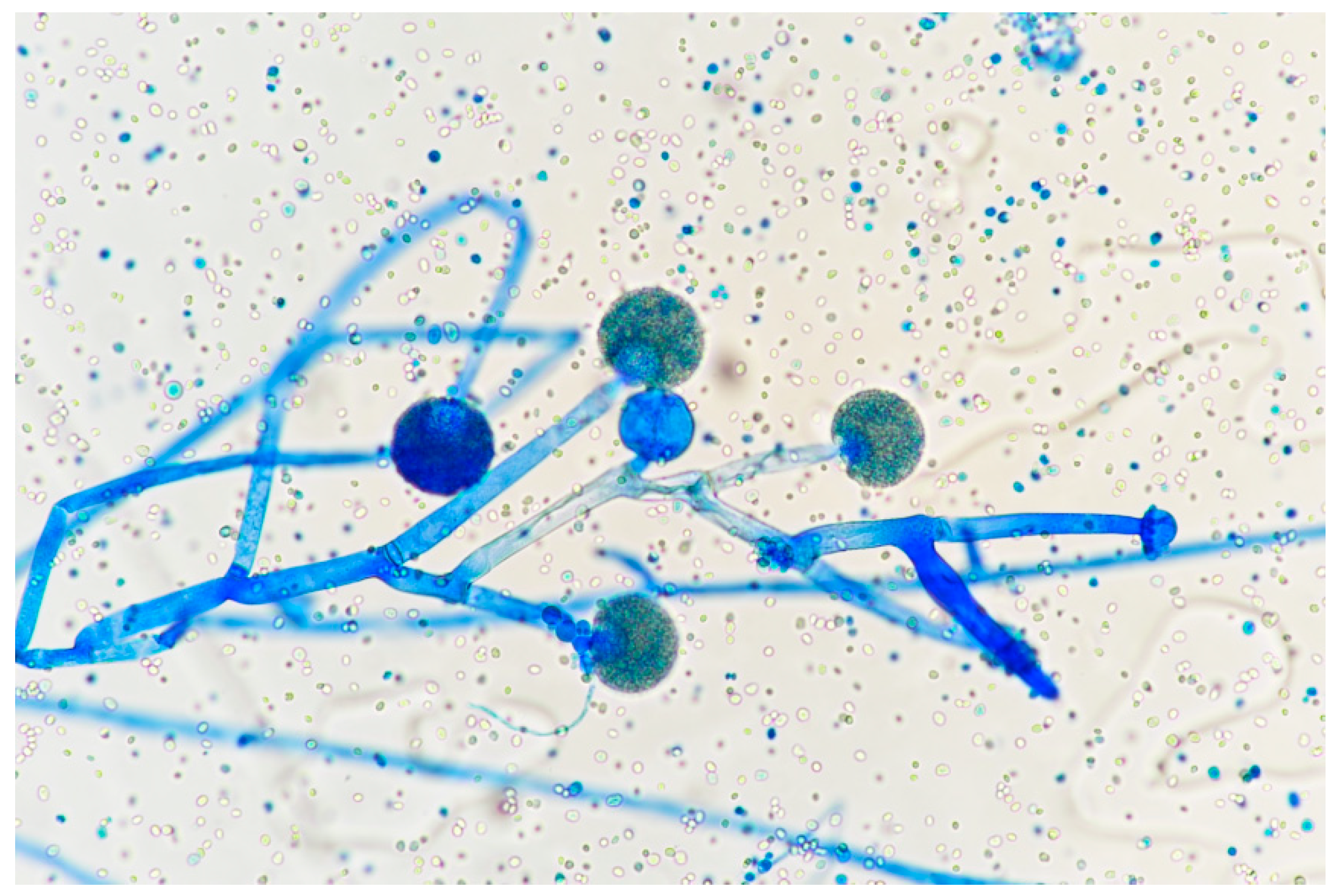
The Mucorales order contains a large group of filamentous fungi (e.g., Mucor, Rhizopus, and others) with distinct morphology. They are broader, branch at right angles, and are pauciseptate (i.e., with few septa). They cause the most aggressive FWIs, often crossing fascial planes and necessitating a more radical approach to wound care, including amputations, in order to eradicate the process. An infection caused by one of these organisms is called a mucormycosis; older literature used the terms zygomycosis (caused by “zygomycetes”) or phytomycosis (caused by “phycomycetes”).
Lastly, the dematiaceous fungi are less common causes of FWI. These brown-pigmented organisms derive their color from dense melanin content, which is thought to serve both protective and virulence-enhancing purposes.
Despite the above classification scheme, two important caveats must be considered. First, it is not possible to distinguish among these groups on the basis of histopathology alone; that is, morphological diagnosis is error-prone. Rather, identification of genus and species requires concomitant tissue culture (see below). Secondly, FWIs are often polymicrobial; cultures may reveal more than one fungal pathogen or coincidence of bacterial infection. An experienced clinical mycologist can be invaluable in making a preliminary determination as to what potential fungal organisms are infecting the burn patient. This makes fungal diagnosis and treatment ideally the work of a multidisciplinary team including the surgeon, pathologist, mycologist, and infectious disease specialist.

3. Diagnosis

Timely diagnosis of FWI in burn patients is challenging for multiple reasons. The diagnosis of an FWI occurs at a median of 10 days after injury, but can occur anytime during hospitalization [30]. Currently, there is no widely accepted fungal screening assay that correlates with FWI. Therefore, diagnosis must rely heavily upon a high level of clinical suspicion, physical examination of the patient’s burn wounds, and assessment of the patient’s clinical status. Complicating this even more are the varying distinctions between fungal wound colonization (FWC) and fungal wound infection (FWI). Lastly, the European Organization for Research and Treatment of Cancer/Mycoses Study Group (EORTC/MSG) guidelines, which are often referenced by clinicians to help with the diagnosis of invasive fungal infections, are only helpful within the appropriate clinical context [31]. These were developed with hematologic malignancy patients as their focus; hence, they cannot be broadly applied to burn patients [32].
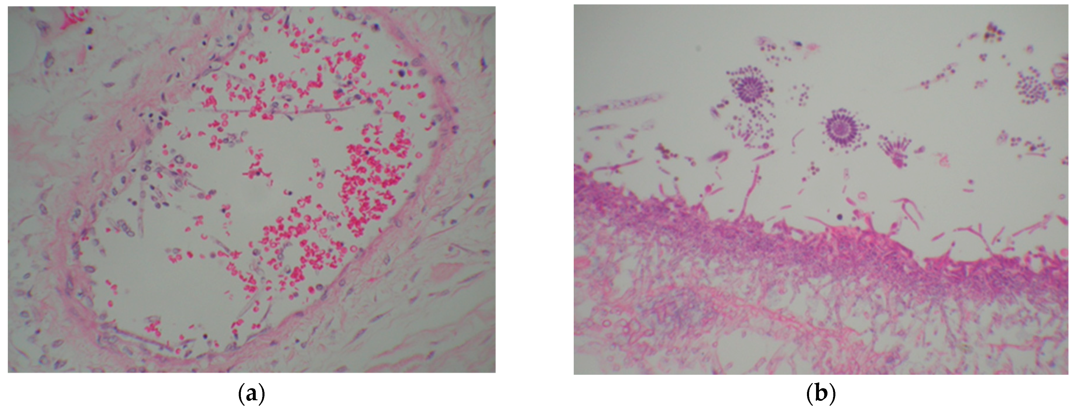
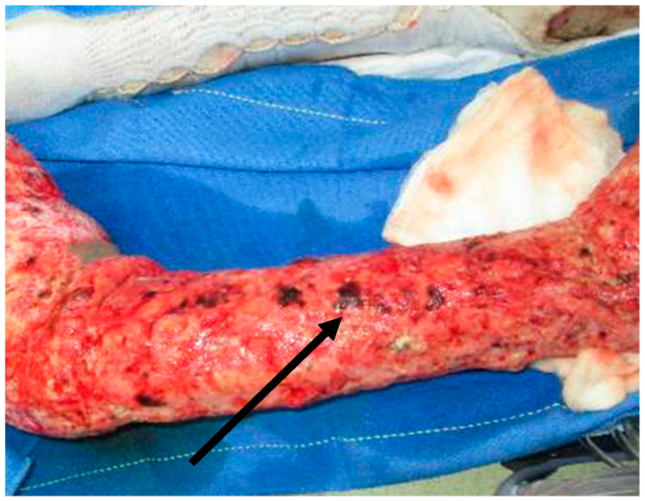
On physical examination, wound findings consistent with FWI include “tinctorial changes”, i.e., a change in the color of the burn wound [33]. These findings may include the appearance of white-appearing clusters of Aspergillus (Figure 1 and Figure 2), black-appearing Mucor (Figure 3 and Figure 4), or frankly nonviable tissue. Attention should be paid to the specific anatomical distribution of these findings in order to plan subsequent surgical operations. Such findings and clinical deterioration should prompt the team to implement multiple tactics: full-thickness incisional biopsy for histopathology and tissue culture, often followed by surgical debridement to viable-appearing tissue, appropriate anti-fungal dressings, and intravenous antifungal agents that cover broadly for both yeasts and molds.
Diagnosis should include a full-thickness biopsy that includes subjacent viable-appearing tissue for histopathological diagnosis and culture. The biopsy may be performed at bedside or in the operating theater. Coordination with pathology enables a “rush” specimen to be performed, as well as a diagnosis within 24 h [34]. A particular caveat in histopathological biopsy is ensuring that viable tissue is present within the specimen. Specifically, a scalpel should be utilized to obtain a 500 mg lenticular biopsy including eschar and deeper unburned hypodermis in the anatomical location of highest concern. If the biopsy is not performed in the operating room, local anesthetic should be injected to perform a field block without distorting the surgical specimen. Furthermore, half of the specimen should be sent for cultures (aerobic, anaerobic, and fungal), and the other half should be sent to pathology for histological examination. Although a frozen-section technique can yield results in as little as 45 min, it is associated with a significant false-negative rate and should not be used without confirmatory permanent section [35]. Importantly, fungal cultures may take several days to weeks to enable growth of the pathogen, which are not relevant to diagnosis of infection per se. The eventual growth of a specific fungus may connote further prognostic information and facilitate tailoring of antifungals. Blood cultures should also be obtained.
Pruitt and colleagues developed a definition of microbial colonization and invasion, as shown in Table 4 [34]. This definition was based on studies of Pseudomonas aeruginosa burn-wound infection in the Walker–Mason rat model, in which bacteria enter the burn eschar from the surface, progressively invade through nonviable into viable tissue, and then spread hematogenously to distant organs. Wound colonization is defined as histopathologic evidence of microorganisms in nonviable tissue, whereas infection occurs when microorganisms invade viable tissue. This schema is clinically meaningful; Horvath et al. showed that FWI, but not FWC, is associated with increased mortality [36]. However, the histopathological differentiation of FWI and FWC should be performed with the following caveats in mind: (1) the propensity of fungal infections to cause necrosis of viable tissue as they progress means that some biopsies with fungal elements in nonviable tissue, particularly if they demonstrate angioinvasion, may actually represent FWI; (2) it is unknown whether histopathological diagnosis is subject to inter-rater reliability problems; (3) failure to diagnose FWI may represent sampling error at the time of biopsy; (4) some patients with FWC will progress to FWI; thus, a change in therapy and increased vigilance, rather than reassurance, is appropriate in the presence of FWC [36]; (5) the original classification scheme was based on microbial invasion of intact burn eschar. Today, given the practice of early excision of the burn wound, it is often applied to tissue which has already been excised and which is now newly necrotic; this outer layer of nonviable tissue has been called “neo-eschar”.
The recovery of fungal organisms on tissue culture is useful (see below) but is not the essential step in diagnosis. Conversely, accurate identification of fungal genus on histopathology is not always possible. Thus, the two procedures, histopathology and tissue culture, should be performed concurrently. Schofield et al. found that histopathology did not often correlate with culture in FWC and FWI [19]. That is to say, histopathologic morphology was not adequate to predict what would grow from fungal cultures. This work builds an important argument supporting not only the role of histopathology, but also culture as an important tool for selecting the appropriate antifungal therapy.
Several experimental techniques exist to improve the diagnostic accuracy of histopathology. Ganesan and colleagues assessed a pan-fungal DNA sequencing assay based on the polymerase chain reaction for rapid identification of filamentous fungi in formalin-fixed paraffin-embedded (FFPE) specimens from 64 combat casualties [37]. They found this assay to be specific (99%) but not very sensitive (63%) in comparison to histopathology. Sensitivity was higher for Mucorales and for angioinvasion. Other researchers have developed assays for individual genera [38].
There are two techniques which employ morphologic techniques to augment the capabilities of histopathology: immunohistochemistry and in situ hybridization. One example of immunohistochemistry-based staining techniques was described by Son et al., who aimed to distinguish between Mucorales or Aspergillus infections. The antibodies used in this study were anti-Rhizopus arrhizus and anti-Aspergillus mouse monoclonal antibodies [39]. The importance of validating such assays locally should be emphasized [38].
Lastly, in situ hybridization uses probes to detect organism-specific ribosomal RNA (rRNA) targets. A proposed approach is to first identify the presence of a fungal infection by routine fungal stains, and then to use in situ hybridization to identify the specific genus. This two-stage approach was used by Hayden and colleagues to differentiate Aspergillus, Fusarium, and Pseudallescheria in tissue sections with high accuracy [40]. Again, local validation of these techniques is required.
The utility of a serologic test that could predict or support an earlier diagnosis of fungal infection has the potential to be high in burn patients. Important examples of such assays include (1,3)-β-d-glucan (BG) (Associates of Cape Cod Inc., East Falmouth, MA, USA) for the detection largely of Candida and Aspergillus spp., as well as galactomannan (GM) (Platelia Aspergillus EIA, Bio-Rad, Hercules, CA, USA) for the detection of Aspergillus. In large part, however, this work has been unsuccessful in demonstrating clinical utility, in burns, outside the diagnosis of candidemia. Shupp et al. evaluated the BG levels of 18 burn patients without candidemia early in their admission and found that half of the group had positive levels at baseline [41]. Kaita et al. studied 51 patients retrospectively who had BG levels measured due to suspicion of candidemia and found that higher cutoff levels than the recommended by the manufacturer did support the diagnosis of candidemia [42]. Lastly, in a retrospective review of a single burn center, Blyth et al. studied 54 patients that had fungal screening assays sent due to high suspicion of invasive fungal infection. They found a high rate of BG positivity (81%) but no significant association with fungal infection, FWC, or fungal species identified [43]. Ultimately, outside of candidemia, BG is unlikely to be a helpful clinical tool in burn patients based on the current published data.

4. Treatment

4.1. Surgical Management

Obtaining effective surgical source control is the key to successful management; the remaining interventions are ancillary. An FWI portends a poor prognosis and will require multiple operative debridements to obtain viable margins. This is in contrast to a colonized wound, which may or may not require immediate operation.
Operative planning is based upon the physical examination and physiological status of the patient. Patients who have an FWI are often in distributive shock and require pre-operative optimization to include fluid resuscitation and attention to multiorgan system failure; they may require continuous renal replacement therapy and/or correction of coagulopathy. After hemodynamic and biochemical optimization, the surgical plan is dictated by the physical examination. Specifically, all wounds need to be examined closely to ascertain the viability of tissue and the presence of lesions that are suspicious for FWI. A fascial excision, rather than excision to fat, is often warranted. If the infection extends into muscle, a proximal amputation may be needed.
Surgical extirpation of invasive fungal disease is pivotal, as the intravenous antifungals are unable to penetrate nonviable tissue. FWI of the face, sinuses, and/or orbits often requires specialist consultation to obtain definitive source control [8]. FWI of an extremity may require aggressive amputation; mucormycosis is notorious for invading across fascial planes. We ensure that each anatomical region has a clean set of instruments and we change gloves to prevent cross-contamination between the different operative fields. Between surgical takebacks, wound care with a topical antifungal should be performed. Options for topical antifungal therapy are discussed below.
Biopsies should be obtained at subsequent operations to ensure that the margins remain free from recurrent or residual FWI. We recommend performing daily operations until the wound is free of infection. After verifying successful source control, temporary wound coverage can be achieved with cadaver allograft, followed by split-thickness skin grafting or more advanced reconstructive procedures if indicated. Importantly, a frank conversation should occur with the patient, patient’s family, or surrogate decision maker regarding an individual’s wishes after the diagnosis of an FWI, as the prognosis is poor, and the likelihood of a debilitating amputation is high. Understanding a patient’s goals for care and quality of life are vital when combating an FWI.

4.2. Systemic Antifungals

Systemic antifungals are often started for FWI as adjuncts to surgical management. The systemic antifungal regimen needs to provide coverage of most common pathogens (Mucor, Fusarium, Aspergillus, Candida). At this time, there are few systemic antifungal options available, and most patients are initiated on a triazole antifungal plus amphotericin B. Due to the paucity of available literature, empiric dose adjustments to account for the pharmacokinetic alterations that occur in burn patients cannot be recommended.

4.2.1. Amphotericin B

Amphotericin B is broadly active against many fungal organisms, including Candida, Aspergillus, and Mucor. Amphotericin B is often added to a burn patient’s antifungal regimen (in addition to a broad-spectrum triazole) for double coverage against Aspergillus species. Amphotericin B causes fungal cell death by binding to ergosterol and changing the permeability of the fungal cell membrane. It is known for its adverse effect of nephrotoxicity, which is mitigated by liposomal formulation. However, due to resistance of Aspergillus terreus to amphotericin B, this agent should not empirically be used as monotherapy for the treatment of fungal infection [44,45]. Amphotericin B also has activity against Mucorales and Fusarium, which, as previously mentioned, are common organisms seen in FWI after burn injury. Amphotericin B should be considered a first-line systemic antifungal agent for FWI, in additional to a broad-spectrum triazole [46,47].

4.2.2. Triazole Antifungals

Fluconazole, posaconazole, voriconazole, and isavuconazole make up the class of triazole antifungals. These agents work by inhibiting fungal cytochrome p450 activity, thereby decreasing ergosterol synthesis and preventing fungal cell membrane formation. In the treatment of FWI, fluconazole is of limited utility, as it lacks coverage of molds. The other azoles are options for systemic therapy in the setting of FWI, as they cover Aspergillus, Fusarium, and Mucor. However, each azole has gaps in the coverage of Aspergillus species, and double coverage with amphotericin B is usually warranted [48,49]. The pharmacokinetic literature is limited to a single case series on voriconazole, in which high inter-patient variability was observed [50]. Therapeutic drug monitoring is recommended when available to ensure that pharmacokinetic goals are being achieved.

4.2.3. Echinocandins

The echinocandins (micafungin, caspofungin, and anidulfungin) inhibit 1,3-β-glucan synthase, decreasing the glucan content of fungal cell walls. This in turn leads to instability of the fungal cell wall and cellular lysis. In their 2016 update on the management of invasive aspergillosis, the Infectious Diseases Society of America (IDSA) recommends the addition of echinocandins as salvage therapy. Empiric use of an echinocandin or the use of echinocandins as monotherapy is not recommended [32].

4.3. Topical Antifungals

Postoperatively, the patient’s wounds should be dressed with agents that have antifungal properties. Amphotericin B (possibly in combination with mafenide acetate), nystatin, voriconazole, Manuka honey, silver products, and Dakin’s solution all show antifungal properties and may be considered for such treatment, although none of these is ideal.

4.3.1. Sulfamylon + Amphotericin B (SMAT)

Whereas mafenide acetate (Sulfamylon) has no antifungal activity, a combination of Sulfamylon aqueous solution and amphotericin B deoxycholate (‘SMAT’) has been used with the intent of covering both bacteria and fungi in one topical solution. However, a recent study tested this combination in a variety of strengths and conditions and found that the amphotericin B component was undetectable by day 2 after compounding [51]. Additionally, Aspergillus growth was seen on day 0. However, SMAT showed activity against Candida for up to 45 days after compounding, depending on storage conditions.

4.3.2. Nystatin

Nystatin topical powder has been studied as an adjunct for the treatment of FWI in burn patients. Nystatin alters fungal cell-wall permeability by binding to sterols in the fungal cell membrane, allowing cellular contents to leak out. In a case series that included four pediatric burn patients, nystatin 6,000,000 units/g was used in conjunction with wet-to-dry dressings, which were changed every 6 h [1]. All patients included had biopsy-proven FWI secondary to Fusarium or Aspergillus. All patients had continuous improvement of their wounds over the 2 week treatment period and were eventually discharged home. Despite these promising results, there are several limitations to nystatin. Nystatin 6,000,000 unit/g is no longer available in the United States; nystatin powder is now only commercially available in a strength of 100,000 units/g. It is not known if this lower strength will yield the same results. Additionally, when used in combination with mafenide, the antimicrobial effects of both agents are lost [52]. However, when used in combination with silver sulfadiazine, the antimicrobial effects of both agents are preserved [52]. More data are needed before nystatin powder can be routinely recommended as a topical adjunct for the treatment of FWI.

4.3.3. Voriconazole

Voriconazole, as discussed earlier, is a triazole antifungal agent. While typically used systemically, a case report described the use of voriconazole as a topical solution [53]. Voriconazole powder for injection was mixed with normal saline in a 1% w/v topical solution. A bone-marrow-transplant patient with a 5 × 5 cm nonhealing wound, infected with Aspergillus flavus, was treated with topical voriconazole plus systemic amphotericin B. Topical voriconazole (applied twice daily) was continued for 5 weeks, at which time she was transitioned to topical nystatin. The wound measured 2.5 × 3 cm when voriconazole was discontinued. No adverse effects due to topical voriconazole were noted.

4.3.4. Manuka Honey

Manuka honey has recently been gaining popularity as a topical antimicrobial. Methylglyoxal is the major antimicrobial compound in Manuka honey; however, its mechanism of action remains unknown. Data from the cosmetic industry show that the minimum inhibitory concentration (MIC) of Manuka honey is low for Candida sp. (MIC = 1.25 mg/mL), but is higher for Aspergillus spp. (MIC = 10 mg/mL) [54]. It is unknown if these concentrations can be achieved with topical application to a burn wound. A recent in vitro study included multiple concentrations of Manuka honey and various exposure times [55]. Manuka honey demonstrated time-dependent activity against Fusarium, Aspergillus, and Mucor. However, while Manuka honey was toxic to fungal cells, it also demonstrated toxicity to human keratinocytes and other cell lines, which may limit its use in the burn population.

4.3.5. Silver

Silver products exert antifungal effects by disrupting the structure of the fungal cell membrane, thereby inhibiting the budding process. Silver sulfadiazine, silver nitrate (0.5% aqueous solution), and nanocrystalline silver have been shown to have efficacy against Aspergillus fumigatus spores. Candida spp. have also shown susceptibility to silver products, although C. albicans and C. glabrata have reduced susceptibility to silver nitrate, as compared to silver sulfadiazine [6]. Additionally, C. tropicalis has been shown to be resistant to silver nitrate. Mucor is not susceptible to most silver products, although it has some susceptibility to silver sulfadiazine. In addition to its antifungal properties, silver has other positive effects on wound beds, which may be helpful in the burn population. A limitation of silver products is limited penetration into the wound [56].

4.3.6. Dakin’s Solution

Dakin’s solution (buffered sodium hypochlorite) was developed during WW I for the treatment of war wounds as one component of what we would now call a bundle of care, which included aggressive surgical management, implantation of catheters for solution delivery, and infusion of the solution as frequently as every 2 h [57]. It is available as a “full-strength” solution (0.5%) or as dilutions thereof (0.25%, 0.125%). The primary limitations of Dakin’s solution are rapid inactivation, lack of penetration into the wound, and local toxicity. A recent in vitro study tested the efficacy and toxicity of each strength of Dakin’s solution at various exposure times [58]. All strengths of Dakin’s were effective in killing all molds tested, but it also had dose-dependent toxicity to keratinocytes, fibroblasts, and osteoblasts. These risks must be weighed against the antifungal benefits of Dakin’s solution.

4.3.7. Cerium

Cerium is a rare earth metal with the potential to be used as a topical antifungal. The antifungal mechanism of action is uncertain [59]. Cerium has been shown to have in vitro activity against Candida species. Its activity against molds, such as Aspergillus and Mucor, remains unknown [60]. At this time, cerium is not commercially available in the United States, but is available (in combination with silver sulfadiazine as Flammacerium) in other parts of the world.

4.4. Nonpharmacologic Treatments

In addition to systemic and topical pharmacotherapy, there are several nonpharmacologic adjuncts for the management of IFI.

4.4.1. Hyperbaric Oxygen Therapy

Hyperbaric oxygen (HBO) therapy may enhance leukocyte activity, increase tissue repair, and exhibit synergy with systemic antimicrobial agents [43]. HBO has been studied as an adjunct to systemic antifungals with or without surgical infection. Additionally, HBO therapy has been shown to reduce the fungal biofilm of wounds [61]. It is recommended that 40–80 HBO sessions (90–120 min each) be used for this purpose [43]. In a case series of 14 oncology patients with diagnosed mucormycosis or invasive aspergillosis, all patients received an average of 21–90 min HBO sessions [62]. In this series, 50% of patients survived their invasive fungal infections. Furthermore, in a case report, a tsunami survivor with cutaneous mucormycosis underwent HBO therapy as an adjunct to aggressive surgical management and systemic antifungals [63]. After 3 weeks of treatment, the patient demonstrated healing wounds and was without evidence of systemic fungal infection. However, there are no data on HBO in burn patients.

4.4.2. Ultraviolet-C Light

While not yet studied in humans, ultraviolet (UV-C) light has been shown to have antifungal activity both in vitro and in animal models. UV-C damages fungal DNA, preventing replication. In a murine model, UV-C was shown to be superior to nystatin cream in reducing the bioburden of Candida albicans without causing damage to healthy skin [64]. In addition to Candida, UV-C light has also shown in vitro efficacy against Aspergillus and Fusarium spp. [65,66].

4.5. Immune-Enhancing Treatments

The observation that invasive fungal infection occurs most frequently in immunosuppressed patients has led to efforts to identify and correct such immunosuppression during treatment of an infection. For example, Unsinger and colleagues evaluated the use of IL-7 in a mouse model of cecal ligation and puncture followed by intravenous infusion of Candida albicans. IL-7 treatment improved T-cell activation, proliferation, adhesion-molecule expression, and IFN-γ production, resulting in increased survival [67]. Studies in burn patients have not been performed. However, the same group recently described successful IL-7 treatment of a patient with a massive necrotizing FWI caused by Trichosporon and Saksenaea spp. following a high-speed motorcycle accident, which was resistant to antifungal and surgical therapy [68]. Other immune-based therapies under development for FWI include nivolumab, an anti-PD-1 monoclonal antibody which enhances T-cell proliferation and cytokine production [69].

5. Prophylaxis

Given the high morbidity and mortality of FWI in burn patients, prevention is key, but there is no specific systemic or topical regimen that is recommended for this purpose. However, the clinician should be mindful of the spectrum of coverage of the topical wound care agents. The prolonged use of broad-spectrum topical antibacterial agents may create an optimal environment in which yeast and mold thrive. It may be prudent to rotate topical antimicrobial agents over a patient’s clinical course to include a product with antifungal activity.

6. Limitations

Several limitations are present in the existing data and review. The available literature is limited to retrospective reviews, case series, and case studies. Each of these publications had their own inherent limitations. Additionally, other studies focused on invasive fungal infections in other patient populations, mostly patients who are immunocompromised secondary to cancer or other disease processes, and these findings may not be applicable to the burn population. Lastly, this review focused on FWI, but patients may experience bacterial wound infections or other types of fungal infections (i.e., candidemia) after burn injury that contribute to morbidity and mortality unrelated to FWI. The literature and the treatment options presented may not be applicable or ideal for other types of infections.

7. Future Directions

Future research should focus on early recognition and diagnosis of FWI, as well as the application of novel adjunct systemic therapies. As limited treatment options exist for the management of FWI, prevention and early recognition are key. Identifying the patients who are most likely to develop to FWI can help the clinician take steps to mitigate this. New diagnostic criteria that are both sensitive and specific for FWI after burn injury can potentially help the clinician more quickly identify FWI and initiate earlier operative, systemic, and topical treatments. For systemic therapy, there are number of novel antifungal agents in various stages of the drug development process, several of which have spectra of activity which cover organisms commonly seen in FWI after burn injury [70]. Lastly, immune-enhancing treatments show promising results when used for the management of FWI in non-burn patients.

8. Conclusions

In summary, the timely diagnosis of FWI in a burn patient is difficult. Recurrent necrosis or gross changes in the appearance of burn wounds should be aggressively addressed with histopathologic analysis, early surgical debridement, and culture. Upon suspicion of FWI, systemic antifungals, topical agents, and nonpharmacologic therapies may be useful adjuncts, but aggressive surgical management is the main effort. The concerted activities of the entire multidisciplinary team are needed to achieve successful outcomes in patients with FWI.

Author Contributions

K.A.P. was responsible for design of the review; K.A.P., T.A.M., J.L.K., G.W.B., T.W. and L.C.C. contributed to data acquisition and to drafting the manuscript; L.C.C. revised it critically for important intellectual content. All authors have read and agreed to the published version of the manuscript.

Funding

This review received no external funding.

Institutional Review Board Statement

Not applicable, as this is a review.

Informed Consent Statement

Not applicable for this review paper. All photographs used are stock images.

Data Availability Statement

Not applicable.

Acknowledgments

The authors would like to thank Jesse Salinas, Department of Microbiology, Brooke Army Medical Center, for his assistance with obtaining the photograph seen in Figure 4.

Conflicts of Interest

All authors are employees of the US Government (as civilian or active-duty service members). No authors have any other conflict of interest.

Disclaimer

The opinions or assertions contained herein are the private views of the authors and are not be construed as official or as reflecting the views of the Department of the Army or the Department of Defense. No authors have any other conflict of interest.

References

  1. Barret, J.P.; Ramzy, P.I.; Heggers, J.P.; Villareal, C.; Herndon, D.N.; Desai, M.H. Topical nystatin powder in severe burns: A new treatment for angioinvasive fungal infections refractory to other topical and systemic agents. Burns 1999, 25, 505–508. [Google Scholar] [CrossRef]
  2. Dean, D.A.; Burchard, K.W. Surgical perspective on invasive Candida infections. World J. Surg. 1998, 22, 127–134. [Google Scholar] [CrossRef]
  3. Codish, S.D.; Sheridan, I.D.; Monaco, A.P. Mycotic wound infections. A new challenge of the surgeon. Arch. Surg. 1979, 114, 831–835. [Google Scholar] [CrossRef] [PubMed]
  4. Blyth, D.M.; Chung, K.K.; Cancio, L.C.; King, B.T.; Murray, C.K. Clinical utility of fungal screening assays in adults with severe burns. Burns 2013, 39, 413–419. [Google Scholar] [CrossRef] [PubMed]
  5. D’Avignon, L.C.; Chung, K.K.; Saffle, J.R.; Renz, E.M.; Cancio, L.C. Prevention of infections associated with combat-related burn injuries. J. Trauma 2011, 71 (Suppl. S2), S282–S289. [Google Scholar] [CrossRef] [PubMed]
  6. Wright, J.B.; Lam, K.; Hansen, D.; Burrell, R.E. Efficacy of topical silver against fungal burn wound pathogens. Am. J. Infect. Control 1999, 27, 344–350. [Google Scholar] [CrossRef]
  7. Tribble, D.R.; Rodriguez, C.J. Combat-Related Invasive Fungal Wound Infections. Curr. Fungal Infect. Rep. 2014, 8, 277–286. [Google Scholar] [CrossRef] [Green Version]
  8. Burchard, K.W. Fungal sepsis. Infect. Dis. Clin. N. Am. 1992, 6, 677–692. [Google Scholar] [CrossRef]
  9. Sarabahi, S.; Tiwari, V.K.; Arora, S.; Capoor, M.R.; Pandey, A. Changing pattern of fungal infection in burn patients. Burns 2012, 38, 520–528. [Google Scholar] [CrossRef]
  10. Wiederhold, N.P.; Gibas, C.F.C. From the Clinical Mycology Laboratory: New Species and Changes in Fungal Taxonomy and Nomenclature. J. Fungi 2018, 4, 138. [Google Scholar] [CrossRef] [Green Version]
  11. Moore, E.C.; Padiglione, A.A.; Wasiak, J.; Paul, E.; Cleland, H. Candida in burns: Risk factors and outcomes. J. Burn Care Res. 2010, 31, 257–263. [Google Scholar] [CrossRef] [PubMed]
  12. Murray, C.K.; Loo, F.L.; Hospenthal, D.R.; Cancio, L.C.; Jones, J.A.; Kim, S.H.; Holcomb, J.B.; Wade, C.E.; Wolf, S.E. Incidence of systemic funcal infection and related mortality following severe burns. Burns 2008, 34, 1108–1112. [Google Scholar] [CrossRef] [PubMed]
  13. Muhammed, M.; Anagnostou, T.; Desalermos, A.; Kourkoumpetis, T.K.; Carneiro, H.A.; Glavis-Bloom, J.; Coleman, J.J.; Mylonakis, E. Fusarium infection: Report of 26 cases and review of 97 cases from the literature. Medicine 2013, 92, 305–316. [Google Scholar] [CrossRef] [PubMed] [Green Version]
  14. Cawley, M.J.; Braxton, G.R.; Haith, L.R.; Reilly, K.J.; Guilday, R.E.; Patton, M.L. Trichosporon beigelii infection: Experience in a regional burn center. Burns 2000, 26, 483–486. [Google Scholar] [CrossRef]
  15. Mitchell, T.A.; Hardin, M.O.; Murray, C.K.; Ritchie, J.D.; Cancio, L.C.; Renz, E.M.; White, C.E. Mucormycosis attributed mortality: A seven-year review of surgical and medical management. Burns 2014, 40, 1689–1695. [Google Scholar] [CrossRef]
  16. dela Cruz, W.P.; Calvano, T.P.; Griffith, M.E.; White, C.E.; Kim, S.H.; Sutton, D.A.; Thompson, E.H.; Fu, J.; Wickes, B.L.; Guarro, J.; et al. Invasive Apophysomyces variabilis infection in a burn patient. J. Clin. Microbiol. 2012, 50, 2814–2817. [Google Scholar] [CrossRef] [Green Version]
  17. Hospenthal, D.R.; Chung, K.K.; Lairet, K.; Thompson, E.H.; Guarro, J.; Renz, E.M.; Sutton, D.A. Saksenaea erythrospora infection following combat trauma. J. Clin. Microbiol. 2011, 49, 3707–3709. [Google Scholar] [CrossRef] [Green Version]
  18. Kucan, J.O.; Hall, S. Alternaria burn wound sepsis. J. Burn Care Rehabil. 1985, 6, 501–502. [Google Scholar] [CrossRef]
  19. Schofield, C.M.; Murray, C.K.; Horvath, E.E.; Cancio, L.C.; Kim, S.H.; Wolf, S.E.; Hospenthal, D.R. Correlation of culture with histopathology in fungal burn wound colonization and infection. Burns 2007, 33, 341–346. [Google Scholar] [CrossRef]
  20. Beckett, A.R.; Kahn, S.A.; Seay, R.; Lintner, A.C. Invasive Curvularia infections in burn patients: A case series. Surg. Infect. Case Rep. 2017, 2, 76–79. [Google Scholar] [CrossRef] [Green Version]
  21. Marcus, J.E.; Piper, L.C.; Ainsworth, C.R.; Sams, V.G.; Batchinsky, A.; Okulicz, J.F.; Barsoumian, A.E. Infections in patients with burn injuries receiving extracorporeal membrane oxygenation. Burns 2019, 45, 1880–1887. [Google Scholar] [CrossRef] [PubMed]
  22. Maurel, V.; Denis, B.; Camby, M.; Jeanne, M.; Cornesse, A.; Glavnik, B.; Alanio, A.; Rousseau, A.F.; Lefloch, R.; Lagrange-Xelot, M.; et al. Outcome and characteristics of invasive fungal infections in critically ill burn patients: A multicenter retrospective study. Mycoses 2020, 63, 535–542. [Google Scholar] [CrossRef]
  23. Van Bang, B.N.; Thanh Xuan, N.; Xuan Quang, D.; Ba Loi, C.; Thai Ngoc Minh, N.; Nhu Lam, N.; Ngoc Anh, D.; Thi Thu Hien, T.; Xuan Su, H.; Tran-Anh, L. Prevalence, species distribution, and risk factors of fungal colonization and infection in patients at a burn intensive care unit in Vietnam. Curr. Med. Mycol. 2020, 6, 42–49. [Google Scholar]
  24. Kaur, R.; Bala, K.; Ahuja, R.B.; Srivastav, P.; Bansal, U. Primary cutaneous mucormycosis in a patient with burn wounds due to Lichtheimia ramosa. Mycopathologia 2014, 178, 291–295. [Google Scholar] [CrossRef]
  25. Moon, P.; Jithendran, N. Invasive Fungal Infection with Absidia Corymbifera in Immunocompetent Patient with Electrical Scalp Burn. World J. Plast. Surg. 2018, 7, 249–252. [Google Scholar]
  26. Farmer, A.R.; Murray, C.K.; Driscoll, I.R.; Wickes, B.L.; Wiederhold, N.; Sutton, D.A.; Sanders, C.; Mende, K.; Enniss, B.; Feig, J.; et al. Combat-Related Pythium aphanidermatum Invasive Wound Infection: Case Report and Discussion of Utility of Molecular Diagnostics. J. Clin. Microbiol. 2015, 53, 1968–1975. [Google Scholar] [CrossRef] [Green Version]
  27. Tamayo Lomas, L.; Domínguez-Gil González, M.; Martín Luengo, A.I.; Eiros Bouza, J.M.; Piqueras Pérez, J.M. Nosocomial infection due to Trichosporon asahii in a critical burned patient. Rev. Iberoam. Micol. 2015, 32, 257–260. [Google Scholar] [CrossRef] [PubMed]
  28. Tram, Q.A.; Minh, N.T.N.; Anh, D.N.; Lam, N.N.; Dung, T.N.; Thi Minh Chau, N.; Tran-Anh, L. A Rare Case of Fungal Burn Wound Infection Caused by Fusarium solani in Vietnam. J. Investig. Med. High Impact Case Rep. 2020, 8, 2324709620912122. [Google Scholar] [CrossRef] [PubMed]
  29. Que, A.T.; Nguyen, N.T.; Do, N.A.; Nguyen, N.L.; Tran, N.D.; Le, T.A. Infection of burn wound by Aspergillus fumigatus with gross appearance of fungal colonies. Med. Mycol. Case Rep. 2019, 24, 30–32. [Google Scholar] [CrossRef] [PubMed]
  30. Akers, K.S.; Rowan, M.P.; Niece, K.L.; Graybill, J.C.; Mende, K.; Chung, K.K.; Murray, C.K. Antifungal wound penetration of amphotericin and voriconazole in combat-related injuries: Case report. BMC Infect. Dis. 2015, 15, 184. [Google Scholar] [CrossRef] [PubMed] [Green Version]
  31. Hoenigl, M.; Strenger, V.; Buzina, W.; Valentin, T.; Koidl, C.; Wölfler, A.; Seeber, K.; Valentin, A.; Strohmeier, A.T.; Zollner-Schwetz, I.; et al. European Organization for the Research and Treatment of Cancer/Mycoses Study Group (EORTC/MSG) host factors and invasive fungal infections in patients with haematological malignancies. J. Antimicrob. Chemother. 2012, 67, 2029–2033. [Google Scholar] [CrossRef] [PubMed] [Green Version]
  32. Patterson, T.F.; Thompson, G.R., 3rd; Denning, D.W.; Fishman, J.A.; Hadley, S.; Herbrecht, R.; Kontoyiannis, D.P.; Marr, K.A.; Morrison, V.A.; Nguyen, M.H.; et al. Practice Guidelines for the Diagnosis and Management of Aspergillosis: 2016 Update by the Infectious Diseases Society of America. Clin. Infect. Dis. 2016, 63, e1–e60. [Google Scholar] [CrossRef] [PubMed]
  33. Ladhani, H.A.; Yowler, C.J.; Claridge, J.A. Burn Wound Colonization, Infection, and Sepsis. Surg. Infect. 2021, 22, 44–48. [Google Scholar] [CrossRef]
  34. Pruitt, B.A., Jr.; McManus, A.T.; Kim, S.H.; Goodwin, C.W. Burn wound infections: Current status. World J. Surg. 1998, 22, 135–145. [Google Scholar]
  35. Heaton, S.M.; Weintrob, A.C.; Downing, K.; Keenan, B.; Aggarwal, D.; Shaikh, F.; Tribble, D.R.; Wells, J. Histopathological techniques for the diagnosis of combat-related invasive fungal wound infections. BMC Clin. Pathol. 2016, 16, 11. [Google Scholar] [CrossRef] [Green Version]
  36. Horvath, E.E.; Murray, C.K.; Vaughan, G.M.; Chung, K.K.; Hospenthal, D.R.; Wade, C.E.; Holcomb, J.B.; Wolf, S.E.; Mason, A.D., Jr.; Cancio, L.C. Fungal wound infection (not colonization) is independently associated with mortality in burn patients. Ann. Surg. 2007, 245, 978–985. [Google Scholar] [CrossRef]
  37. Ganesan, A.; Wells, J.; Shaikh, F.; Peterson, P.; Bradley, W.; Carson, M.L.; Petfield, J.L.; Klassen-Fischer, M.; Akers, K.S.; Downing, K.; et al. Molecular Detection of Filamentous Fungi in Formalin-Fixed Paraffin-Embedded Specimens in Invasive Fungal Wound Infections Is Feasible with High Specificity. J. Clin. Microbiol. 2019, 58, 58. [Google Scholar] [CrossRef]
  38. Guarner, J.; Brandt, M.E. Histopathologic diagnosis of fungal infections in the 21st century. Clin. Microbiol. Rev. 2011, 24, 247–280. [Google Scholar] [CrossRef] [Green Version]
  39. Son, H.J.; Song, J.S.; Choi, S.; Jung, J.; Kim, M.J.; Chong, Y.P.; Lee, S.O.; Choi, S.H.; Kim, Y.S.; Woo, J.H.; et al. A comparison of histomorphologic diagnosis with culture- and immunohistochemistry-based diagnosis of invasive aspergillosis and mucormycosis. Infect. Dis. 2020, 52, 279–283. [Google Scholar] [CrossRef] [PubMed]
  40. Hayden, R.T.; Isotalo, P.A.; Parrett, T.; Wolk, D.M.; Qian, X.; Roberts, G.D.; Lloyd, R.V. In situ hybridization for the differentiation of Aspergillus, Fusarium, and Pseudallescheria species in tissue section. Diagn. Mol. Pathol. Am. J. Surg. Pathol. Part B 2003, 12, 21–26. [Google Scholar] [CrossRef] [PubMed]
  41. Shupp, J.W.; Petraitiene, R.; Jaskille, A.D.; Pavlovich, A.R.; Matt, S.E.; Nguyen do, T.; Kath, M.A.; Jeng, J.C.; Jordan, M.H.; Finkelman, M.; et al. Early serum (1→3)-β-D-glucan levels in patients with burn injury. Mycoses 2012, 55, 224–227. [Google Scholar] [CrossRef]
  42. Kaita, Y.; Tarui, T.; Otsu, A.; Tanaka, Y.; Suzuki, J.; Yoshikawa, K.; Yamaguchi, Y. The Clinical Significance of Serum 1,3-β-D-Glucan For the Diagnosis of Candidemia in Severe Burn Patients. J. Burn Care Res. 2019, 40, 104–106. [Google Scholar] [CrossRef] [PubMed]
  43. Kaide, C.G.; Khandelwal, S. Hyperbaric oxygen: Applications in infectious disease. Emerg. Med. Clin. N. Am. 2008, 26, 571–595. [Google Scholar] [CrossRef]
  44. Blum, G.; Hörtnagl, C.; Jukic, E.; Erbeznik, T.; Pümpel, T.; Dietrich, H.; Nagl, M.; Speth, C.; Rambach, G.; Lass-Flörl, C. New insight into amphotericin B resistance in Aspergillus terreus. Antimicrob. Agents Chemother. 2013, 57, 1583–1588. [Google Scholar] [CrossRef] [PubMed] [Green Version]
  45. Steinbach, W.J.; Benjamin, D.K., Jr.; Kontoyiannis, D.P.; Perfect, J.R.; Lutsar, I.; Marr, K.A.; Lionakis, M.S.; Torres, H.A.; Jafri, H.; Walsh, T.J. Infections due to Aspergillus terreus: A multicenter retrospective analysis of 83 cases. Clin. Infect. Dis. 2004, 39, 192–198. [Google Scholar] [CrossRef] [PubMed] [Green Version]
  46. Warkentien, T.; Rodriguez, C.; Lloyd, B.; Wells, J.; Weintrob, A.; Dunne, J.R.; Ganesan, A.; Li, P.; Bradley, W.; Gaskins, L.J. Invasive mold infections following combat-related injuries. Clin. Infect. Dis. 2012, 55, 1441–1449. [Google Scholar] [CrossRef] [PubMed] [Green Version]
  47. Rodriguez, C.J.; Tribble, D.R.; Malone, D.L.; Murray, C.K.; Jessie, E.M.; Khan, M.; Fleming, M.E.; Potter, B.K.; Gordon, W.T.; Shackelford, S.A. Treatment of Suspected Invasive Fungal Infection in War Wounds. Mil. Med. 2018, 183 (Suppl. S2), 142–146. [Google Scholar] [CrossRef] [PubMed] [Green Version]
  48. Mayr, A.; Lass-Flörl, C. Epidemiology and antifungal resistance in invasive Aspergillosis according to primary disease: Review of the literature. Eur. J. Med. Res. 2011, 16, 153–157. [Google Scholar] [CrossRef] [PubMed] [Green Version]
  49. Snelders, E.; van der Lee, H.A.; Kuijpers, J.; Rijs, A.J.; Varga, J.; Samson, R.A.; Mellado, E.; Donders, A.R.; Melchers, W.J.; Verweij, P.E. Emergence of azole resistance in Aspergillus fumigatus and spread of a single resistance mechanism. PLoS Med. 2008, 5, e219. [Google Scholar] [CrossRef]
  50. Schlotman, T.; Akers, K. 381 Pharmacokinetics and pharmacodynamics of voriconazole in burn patients: A case series. J. Burn Care Res. 2018, 39 (Suppl. S1), S161. [Google Scholar] [CrossRef]
  51. Rizzo, J.A.; Martini, A.K.; Pruskowski, K.A.; Rowan, M.P.; Niece, K.L.; Akers, K.S. Thermal stability of mafenide and amphotericin B topical solution. Burns 2018, 44, 475–480. [Google Scholar] [CrossRef]
  52. Heggers, J.P.; Robson, M.C.; Herndon, D.N.; Desai, M.H. The efficacy of nystatin combined with topical microbial agents in the treatment of burn wound sepsis. J. Burn Care Rehabil. 1989, 10, 508–511. [Google Scholar] [CrossRef]
  53. Klein, K.C.; Blackwood, R.A. Topical voriconazole solution for cutaneous aspergillosis in a pediatric patient after bone marrow transplant. Pediatrics 2006, 118, e506–e508. [Google Scholar] [CrossRef]
  54. Juliano, C.; Magrini, G.A. Methylglyoxal, the major antibacterial factor in manuka honey: An alternative to preserve natural cosmetics? Cosmetics 2019, 6, 1. [Google Scholar] [CrossRef] [Green Version]
  55. Yabes, J.M.; White, B.K.; Murray, C.K.; Sanchez, C.J.; Mende, K.; Beckius, M.L.; Zera, W.C.; Wenke, J.C.; Akers, K.S. In Vitro activity of Manuka Honey and polyhexamethylene biguanide on filamentous fungi and toxicity to human cell lines. Med. Mycol. 2017, 55, 334–343. [Google Scholar]
  56. Atiyeh, B.S.; Costagliola, M.; Hayek, S.N.; Dibo, S.A. Effect of silver on burn wound infection control and healing: Review of the literature. Burns 2007, 33, 139–148. [Google Scholar] [CrossRef]
  57. Cancio, L.C. Topical Antimicrobial Agents for Burn Wound Care: History and Current Status. Surg. Infect. 2021, 22, 3–11. [Google Scholar] [CrossRef] [PubMed]
  58. Barsoumian, A.; Sanchez, C.J.; Mende, K.; Tully, C.C.; Beckius, M.L.; Akers, K.S.; Wenke, J.C.; Murray, C.K. In vitro toxicity and activity of Dakin’s solution, mafenide acetate, and amphotericin B on filamentous fungi and human cells. J. Orthop. Trauma 2013, 27, 428–436. [Google Scholar] [CrossRef] [PubMed]
  59. Farias, I.A.P.; Dos Santos, C.C.L.; Sampaio, F.C. Antimicrobial Activity of Cerium Oxide Nanoparticles on Opportunistic Microorganisms: A Systematic Review. BioMed Res. Int. 2018, 2018, 1923606. [Google Scholar] [CrossRef] [PubMed] [Green Version]
  60. Silva-Dias, A.; Miranda, I.M.; Branco, J.; Cobrado, L.; Monteiro-Soares, M.; Pina-Vaz, C.; Rodrigues, A.G. In vitro antifungal activity and in vivo antibiofilm activity of cerium nitrate against Candida species. J. Antimicrob. Chemother. 2015, 70, 1083–1093. [Google Scholar] [CrossRef] [PubMed] [Green Version]
  61. Memar, M.Y.; Yekani, M.; Alizadeh, N.; Baghi, H.B. Hyperbaric oxygen therapy: Antimicrobial mechanisms and clinical application for infections. Biomed. Pharmacother. Biomed. Pharmacother. 2019, 109, 440–447. [Google Scholar] [CrossRef] [PubMed]
  62. Segal, E.; Menhusen, M.J.; Shawn, S. Hyperbaric oxygen in the treatment of invasive fungal infections: A single-center experience. Isr. Med Assoc. J. IMAJ 2007, 9, 355–357. [Google Scholar] [PubMed]
  63. Andresen, D.; Donaldson, A.; Choo, L.; Knox, A.; Klaassen, M.; Ursic, C.; Vonthethoff, L.; Krilis, S.; Konecny, P. Multifocal cutaneous mucormycosis complicating polymicrobial wound infections in a tsunami survivor from Sri Lanka. Lancet 2005, 365, 876–878. [Google Scholar] [CrossRef]
  64. Dai, T.; Kharkwal, G.B.; Zhao, J.; St Denis, T.G.; Wu, Q.; Xia, Y.; Huang, L.; Sharma, S.K.; d’Enfert, C.; Hamblin, M.R. Ultraviolet-C light for treatment of Candida albicans burn infection in mice. Photochem. Photobiol. 2011, 87, 342–349. [Google Scholar] [CrossRef] [Green Version]
  65. Jun, S.; Irudayaraj, J.; Demirci, A.; Geiser, D. Pulsed UV-light treatment of corn meal for inactivation of Aspergillus niger spores. Int. J. Food Sci. Technol. 2003, 38, 883–888. [Google Scholar] [CrossRef]
  66. Córdova-Alcántara, I.M.; Venegas-Cortés, D.L.; Martínez-Rivera, M.; Pérez, N.O.; Rodriguez-Tovar, A.V. Biofilm characterization of Fusarium solani keratitis isolate: Increased resistance to antifungals and UV light. J. Microbiol. 2019, 57, 485–497. [Google Scholar] [CrossRef]
  67. Unsinger, J.; Burnham, C.A.; McDonough, J.; Morre, M.; Prakash, P.S.; Caldwell, C.C.; Dunne, W.M., Jr.; Hotchkiss, R.S. Interleukin-7 ameliorates immune dysfunction and improves survival in a 2-hit model of fungal sepsis. J. Infect. Dis. 2012, 206, 606–616. [Google Scholar] [CrossRef] [Green Version]
  68. Turnbull, I.R.; Mazer, M.B.; Hoofnagle, M.H.; Kirby, J.P.; Leonard, J.M.; Mejia-Chew, C.; Spec, A.; Blood, J.; Miles, S.M.; Ransom, E.M.; et al. IL-7 Immunotherapy in a Nonimmunocompromised Patient With Intractable Fungal Wound Sepsis. Open Forum Infect. Dis. 2021, 8, ofab256. [Google Scholar] [CrossRef]
  69. Grimaldi, D.; Pradier, O.; Hotchkiss, R.S.; Vincent, J.L. Nivolumab plus interferon-γ in the treatment of intractable mucormycosis. Lancet Infect. Dis. 2017, 17, 18. [Google Scholar] [CrossRef] [Green Version]
  70. Rauseo, A.M.; Coler-Reilly, A.; Larson, L.; Spec, A. Hope on the Horizon: Novel Fungal Treatments in Development. Open Forum Infect. Dis. 2020, 7, ofaa016. [Google Scholar] [CrossRef] [Green Version]
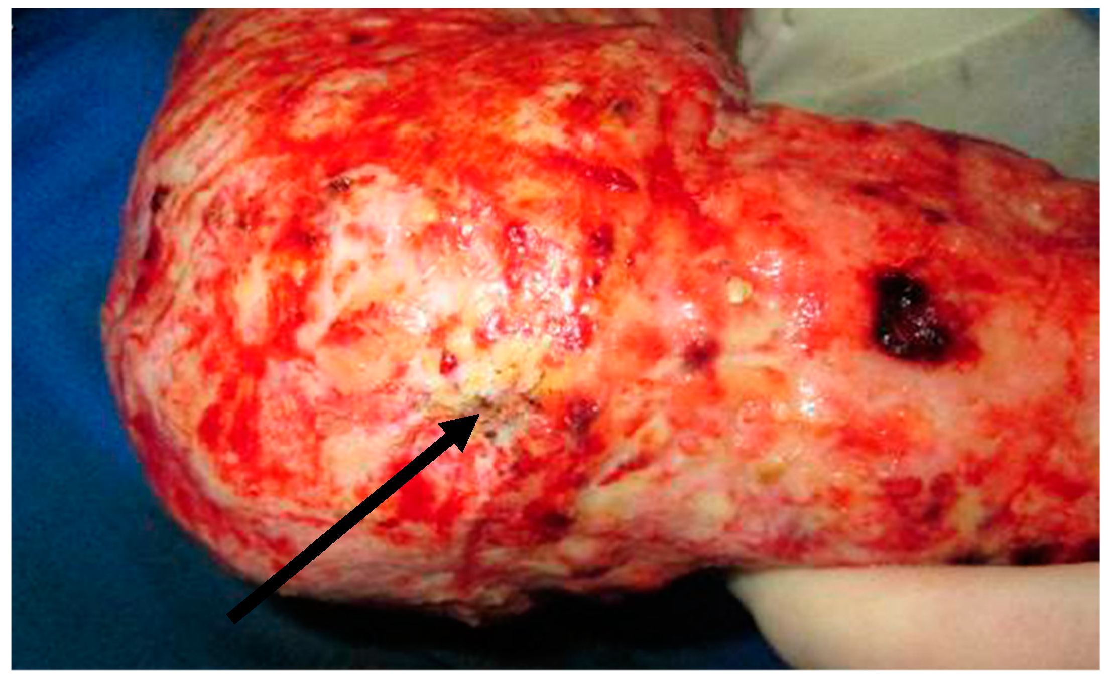
Figure 1. Aspergillus on a previously excised burn wound.
Ebj 02 00013 g001
Figure 2. (a) Aspergillus vascular invasion on histopathology: septate hyphae with approximately 10 μm thick branch at 45 degrees. (b) Aspergillus fruiting bodies (“aspergilla”) seen on the surface of the wound.
Ebj 02 00013 g002
Figure 3. Mucor on previously excised burn wound.
Ebj 02 00013 g003
Figure 4. Mucor spp. recovered on culture stained with lactophenol blue demonstrating classic ribbon-like aseptate hyphae with a mature sporangium and spores throughout the field.
Ebj 02 00013 g004
Table 1. Risk factors for FWI [2,3,4].
Risk Factor
Burn-related>60% TBSA burns
Full-thickness burns
Total parenteral nutrition (TPN)
Multisystem organ failure (MSOF)
Serum glucose > 200 mg/dL
>7 days of systemic antibiotic therapy
Table 2. Clinical classification of fungi.
GroupSyndromePathophysiologyMorphology References
Candida spp.CandidiasisFrequent colonizer but infrequent invader of burn wounds; increasingly non-albicans speciesBudding yeasts or rounded, yeast-like structures, with or without septa or pseudohyphae[11]
Hyaline, septate molds (Aspergillus, Fusarium spp.)Aspergillosis; hyalohyphomycosis (non-Aspergillus infections)Aspergillus is the most common cause of lethal FWI in the modern era; Fusarium has a propensity to enter bloodstreamThin (3–12 μm), septate, acute-angle (45°) or dichotomous branching hyphae; nonpigmented[12,13,14]
Mucorales order (Apophysomyces, Mucor, Rhizomucor, Rhizopus, Saksenaea spp.)Mucormycosis (previously zygomycosis, Phyto mycosis)Most aggressive invader; causes edema then angioinvasion, thrombosis, and necrosisBroad (5–20 μm), pauciseptate, thin-walled, right-angle-branching, ribbon-like, folded or crinkled hyphae[15,16,17]
Dematiaceous fungi (Alternaria, Bipolaris, Curvularia spp.)PhaeohyphomycosisRare in burn patientsPigmented hyphae[18,19,20]
Table 3. Organisms isolated from burn wounds with FWI.
AuthorsStudy TypeOrganisms IsolatedType of BurnSurgical InterventionOutcomes
Marcus et al. [21]Retrospective reviewCandida albicans, Candida rugosa, Mucor sp., and Fusarium sp.Thermal burns, electrical injury, inhalation injury only, toxic epidermal necrolysisNot reported In-hospital mortality: 40%
Maurel et al. [22]Retrospective reviewCandida albicans, C. parapsilosis, C. glabrata, C. krusei, C. tropicalis, Trichosporon, Aspergillus fumigatus, A. flavus, A. terreus Mucor sp., M. circinelloides, Rhizopus, Rhizomucor, Lichteimia (Absidia), Fusarium sp., ScedosporiumThermal burn, electrical injuryNot reported90-day mortality: 37.2%
Sarabahi et al. [9]Prospective observationalNon-albicans Candida sp., C. albicans, Aspergillus sp.Not reportedNot reported43% mortality
Van Bang et al. [23]Prospective observationalCandida albicans, C. tropicalis, C. parapsilosis, C. duobushaemulonis, Kodameae ohmeri, Aspergillus fumigatus, A. flavus, A. oryzae, A. chevalieri, A. nominus, Fusarium soliumNot reportedNot reported19.5% mortality
Kaur et al. [24]Case reportLichtheimia ramosaFlame burnDaily surgical debridementSurvived
Mitchell et al. [15]Retrospective studyMucor circinelloides, Saksenaea vasoformis, S. erythroospora, Pythium aphanidermatumBlast injury, motor vehicle collision)Average of 2.5 operative procedures 83% had an amputation92% mortality
Dela Cruz et al. [16]Case reportApophysomyces variabilisFlame burnAggressive debridement of infected tissue after FWI suspicionDied
Moon et al. [25]Case reportAbsidia corymbiferaHigh-voltage electrical injuryWide debridement after clinical concern for FWI, then anterior lateral thigh flapSurvived
Farmer et al. [26]Case reportAlternaria sp., Aspergillus sp., C. elegans, Geotrichum sp., and Pythium aphanidermatumBlast injurySerial irrigation and debridementDied
Tamayo Lomas et al. [27]Case reportTrichosporon asahiiNot reportedSix surgical procedures including escaharotomies, debridements, and autograftsSurvived
Tram et al. [28]Case reportFusarium solaniFlame burnSurgical measures for debridement and skin transplantationDied
Que et al. [29] Case reportAspergillus fumigatusNot reportedNot reportedDied
Table 4. Definition and staging of wound colonization and infection. Adopted from Pruitt et al (table 4) [34].
Colonization
Stage 1AMicroorganisms present on wound surface
Stage 1BMicrobial penetration of eschar
Stage 1CProliferation of microorganisms at interface between viable and nonviable tissue (subeschar space)
Infection
Stage IIAFoci of microinvasion in uppermost viable tissue
Stage IIBPenetration of microbes to variable depth within viable tissue
Stage IICAngioinvasion (microorganisms within small blood vessels and/or lymphatics)
Publisher’s Note: MDPI stays neutral with regard to jurisdictional claims in published maps and institutional affiliations.