Article

Systematic Quantification of Hypertrophic Scar in Adult Burn Survivors

by
Zoë Edger-Lacoursière
1,
Bernadette Nedelec
1,2,3,*,
Elisabeth Marois-Pagé
2,
Ana de Oliveira
2,3,
Marie-Andrée Couture
2,
Valérie Calva
2 and
José A. Correa
4
1
School of Physical and Occupational Therapy, McGill University, Montreal, QC H3A 0G4, Canada
2
Hôpital de Réadaptation Villa Medica, Montreal, QC H2X 1C9, Canada
3
Centre de Recherche du Centre Hospitalier de l’Université de Montréal (CHUM), Montreal, QC H2X 0A9, Canada
4
Department of Mathematics and Statistics, McGill University, Montreal, QC H3A 0G4, Canada
*
Author to whom correspondence should be addressed.
Eur. Burn J. 2021, 2(3), 88-105; https://doi.org/10.3390/ebj2030008
Submission received: 16 June 2021 / Revised: 20 July 2021 / Accepted: 21 July 2021 / Published: 29 July 2021

Abstract

:
Very few objective scar evaluations have been conducted with burn survivors, which limits our knowledge of the clinical recovery profile of hypertrophic scars (HSc) and donor site scars (D). The purpose of this study was to prospectively quantify the skin characteristics of post-burn HSc in different anatomical locations (D) and normal skin (N) using objective instrumentation. The skin characteristics of HSc, D, and N in 44 burn survivors were measured at 2, 3, 4, 5, 6, and 7 months post-burn using validated instrumentation: a high-frequency ultrasound (for thickness), Cutometer® (for pliability), and Mexameter® (for erythema and pigmentation). Up to five sites were assessed on the same participant, if their scar was located on the upper extremity (UE), lower extremity (LE), and trunk. A mixed model two-way analysis of variance was used to investigate the differences in means between sites at each time point and between time points at each site. The results revealed that the HSc sites were thicker than the D and N at all time points; the UE and trunk HSc were thicker than the LE HSc at 7 months post-burn; the pliability of the trunk HSc did not improve over time; and the UE HSc was more erythematous at 7 months, compared to other anatomical sites, whereas the D erythema decreased from 2 to 7 months. As clinicians have prioritized UE treatments due to their functional importance, this study provides objective measurements to further support this practice and encourages clinicians to also prioritize trunk HSc treatments after burn injuries.

1. Introduction

The prevention and treatment of hypertrophic scars (HSc) is still one of the most important issues in burn rehabilitation [1] and has been described as “the greatest unmet challenge following burn injury” [2]. This is the case, because HSc formation is associated with contracture development, severe pruritus, persisting functional limitations and psychological distress, decreased quality of life [3], and delayed reintegration into society [2]. In the literature, between 33 and 91% of burn survivors are expected to develop HSc, depending on various patient and injury characteristics [4,5,6,7,8].
Very few objective scar evaluations have been conducted with the burn survivor population, which limits our knowledge of the clinical recovery profile of HSc, thereby causing an impact on rehabilitation intervention and treatment prioritization. However, scar quantification in burn survivors using objective instrumentation has been investigated at 3, 6, and 12 months post-burn [9]. Nonetheless, the data before 3 months and between 3 and 6 and 6 and 12 months have not been produced, which could allow for a better understanding of the early recovery profile of HSc and donor site scars of the burn survivor population. Thus, objective scar evaluations would be valuable, since it would provide clinicians with data to establish HSc and donor site scar treatment priorities and objectives.
Furthermore, little is known about the similarities or differences in the recovery profile of post-burn scars at specific anatomical locations. However, the normative data for pliability, erythema, melanin, and thickness specific to 16 different anatomical locations were reported using the DermaScan C (Cortex Technology, Handsund, Denmark), Cutometer®(MPA 580, Courage & Khazaka Electronic GmbH, Koln, Germany) and Mexameter® (MX18, Courage & Khazaka Electronic GmbH, Koln, Germany) [10]. This study provided objective measurements that showed how skin characteristics differed by their specific anatomical locations. Therefore, it is now possible to compare HSc and donor site skin characteristics to normative data for specific anatomical locations and better understand the effect of anatomic location on the development and resolution of HSc and normal scars.
Therefore, the objective of this study was to prospectively quantify the thickness, pliability, erythema, and pigmentation of post-burn HSc, donor sites (normal scar), and normal skin in different anatomical locations between 2 and 7 months post-burn using objective instrumentation.

2. Materials and Methods

A prospective, observational time-series study was conducted at the Montreal Burn Center. This study was approved by the McGill Faculty of Medicine Institutional Review Board, the ethics and scientific committees of the Centre de Recherche Centre Hospitalier de l’Université de Montréal (CRCHUM), and the Villa Medica Rehabilitation Hospital Research Approval Committee.

2.1. Study Population

The participants were recruited from the Villa Medica Rehabilitation Hospital, where all evaluations and follow-ups were performed. Subjects who had developed HSc were recruited for this study according to the following inclusion criteria: (1) ≥18 years old, (2) admitted to Villa Medica Rehabilitation Hospital of the treatment of a burn injury, and (3) informed consent was obtained. The exclusion criteria included: (1) Was unable to understand French or English, (2) had a suspected or known allergy to ultrasound gel, (3) had dermatological conditions, which may interfere with the study results, such as psoriasis, eczema, etc., (4) had refused to give informed consent, or (5) had a psychiatric illness that impaired their ability to participate in the study or provide informed consent. Burn survivors who met the eligibility criteria were identified and were approached by one of their treating clinicians. These clinicians briefly described the study to the patients, and if they were interested in participating, their names were provided to the research assistant. The research assistant approached potential participants regarding the study, provided them with additional information, and gave them a copy of the informed consent form. Those who provided written consent were screened and examined to ascertain study eligibility. Eligible participants were identified by their initials and a preset 3-digit code to ensure confidentiality.

2.2. Measurement Procedures

Up to five sites were identified and selected for evaluation of the same participant if they were located in one of these three general anatomical locations: (1) upper extremity; (2) lower extremity; or (3) trunk. If no burn injury was sustained at each of these locations, the scar measurement was only performed at the locations that had sustained a burn injury. Within these site locations, the most severe scar was selected for evaluation if it had been rated as >2.034 mm thick, as measured by high-frequency ultrasound, and was hyperemic, defined as a Mexameter erythema index of >300.
If a grafting procedure was performed, a representative donor site was selected. A standard normal skin site was evaluated on the medial surface of the upper arm, if available. When unavailable, the closest unburned site to the medial surface of the upper arm was selected, and sites with substantial sun exposure were avoided.
Measurements were conducted in a temperature-controlled room (22 ± 1 °C), with the participant in the same position for each consecutive measurement (generally, sitting comfortably in a chair, with their arms resting at their sides, unless otherwise specified). If the participants were wearing pressure garments, they were asked to remove them 15 min prior to the scar assessment. A flexible, transparent film (Cling Vinyl Grafix, Creative-Coldsnow Artist Materials, Overland Park, KS, USA) was applied to the measurement site, the scar was traced, and a hole was cut over the exact site to be measured. Photographs of the hypertrophic scar sites were obtained to facilitate relocation for serial measurements, but not for evaluation purposes.
All patients were evaluated at baseline (admission to Villa Medica Rehabilitation Hospital) and re-evaluated monthly from 2 up to 7 months post-burn by the same evaluator. The thickness, pliability, erythema, and melanin were evaluated using several instruments: the DermaScan C (Cortex Technology, Handsund, Denmark) to measure thickness, the Cutometer® (MPA 580, Courage & Khazaka Electronic GmbH, Koln, Germany) to measure skin pliability, and the Mexameter® (MX18, Courage & Khazaka Electronic GmbH, Koln, Germany) to measure skin erythema and melanin. The participant’s current level of pain and itch were evaluated at each follow-up visit. Pain and itch have been previously assessed with the burn population and are clinically important when investigating HSc post-burn [11,12].

2.3. Measurement Tools

Scar thickness was measured using the DermaScan C (Cortex Technology, Handsund, Denmark), a high-frequency (20 MHz) ultrasound scanner with image-processing software (Dermavision 2D, Dermascan C v. 3, Cortex Technology, Handsund, Denmark), which captures and reproduces high-resolution images that allow skin thickness measurements to be generated. A medium-focus transducer with a 12 mm wide viewing field that was able to penetrate to 15 mm below the skin surface was used for this study. Prior to each measurement, a thin layer of conducting ultrasound gel (EcoGel 100 Imaging Ultrasound Gel, Eco-med Pharmaceutical Inc., Mississauga, ON, Canada) was applied to the transducer to provide contact between the clear plastic diaphragm and the skin surface. The transducer was held perpendicular to the site, while the echographic image was recorded. The research assistant, using the dedicated computer software, later generated the thickness measurements. The mean of three evenly spaced measures of the distance between the outer surface of the echogenic stratum corneum and the inner surface of the dermis, which is the boundary of the hypoechogenic subcutaneous fat, was recorded as the total skin thickness in millimeters. All measurements were performed with the ultrasound velocity set at 1580 m/s. High-frequency ultrasound has been shown to be a reliable and valid assessment of post-burn scar thickness in both adults [13,14] and children [15].
Skin elasticity was measured with the Cutometer® (MPA 580, Courage & Khazaka Electronic GmbH, Koln, Germany), which is based on the suction and elongation measuring principle. The device generates negative pressure, which draws the skin into a hollow aperture in the center of the probe and estimates the skin penetration depth with an optical measuring system. Four different measurement modes feature pre-programmed sequences of “on/off” pressure cycles. For this study, mode 1 was chosen, as it delivers three cycles of negative air pressure (450 mbar) for 2 s, followed by 2 s of no pressure. The results are expressed as the mean of three measurement cycles. The probe with a 6 mm hollow aperture was used for this study. Prior to the measurement cycle, the probe is applied lightly to the skin, without the outer ring contacting the skin surface, at a perpendicular axis to the skin. During the measurement cycle, it is important that neither the subject nor the probe move. Skin pliability parameters are traditionally expressed as either absolute parameters (Ua, Ue, Uf, Ur, Uv) or relative parameters (R-parameters). However, since the R0 = Uf parameter (which represents the maximum deformation or total distention of the skin) has previously been shown to provide the most reliable measurement of post-burn scar tissue when using the Cutometer [14,16,17], only this measurement was used for the analysis in this study. The Cutometer was cleaned after every use and calibrated biweekly, as per the manufacturer’s specifications. The Cutometer has been shown to be a reliable and valid assessment of post-burn scar pliability [14,16,17].
Erythema and melanin were evaluated with the Mexameter® (MX18, Courage & Khazaka Electronic GmbH, Koln, Germany), which is based on the tissue’s narrow wavelength light absorption. The probe has 16 light-emitting diodes that send three defined wavelengths of light (568, 660, and 880 nm). A receiver then measures the light reflected by the skin. Since the quantity of emitted light is known, the absorption rate of defined wavelengths can be ascertained, which are selectively absorbed by melanin (660 nm) pigments or hemoglobin (568 nm). For each measurement, the probe was held perpendicular to the skin. It was lightly applied to the skin surface, without the outer ring making contact, thus activating the light emitter. The reflected light was measured by the receiver, and the erythema and melanin index (range 1–1000) was immediately displayed on the console; thus, the probe only remains in contact with the skin for a few seconds. The Mexameter probe was cleaned and the accuracy checked monthly, as per the manufacturer’s specifications. The Mexameter has been shown to be a reliable and valid assessment of post-burn erythema and melanin [13,14,18].
The level of pain and itch were measured with a visual analogue scale (VAS). The participants were presented with a 10 cm line, anchored with no pain (itch) to the worse pain (itch) imaginable. The participants were asked to mark their current level of pain and itch on the line, and once selected, the measure in centimeters was recorded. The duration of the itch was also recorded using the following scale: 0 =no itch, 1 ≤ 5 min, 2 ≥ 5 min < 30 min, 3 ≥ 30 min < 60 min, and 4 ≥ 60 min. The weekly frequency of itch was recorded with a 0–7 day scale, and the daily frequency was recorded with an approximate number of times per day.

2.4. Statistical Analyses

Descriptive statistics for categorical variables are reported as frequency counts and percentages. For continuous variables, we report the means and standard deviations (SD) if there was evidence that the distribution of values followed a normal distribution, and medians and inter-quartile ranges (IQR: 25th percentile–75th percentile) otherwise.
Differences in the means between scar sites at each time point, as well as differences in the means between times at each scar site, were investigated for each skin characteristic separately with a mixed model two-way analysis of variance (ANOVA) model, taking into account two sources of within-subject correlation: one due to measurements of intra-individual scars and the other due to repeated measurements over time. The p-values were not adjusted for multiple testing in order to avoid being overly conservative and subsequently missing important treatment effects [19,20].
All hypothesis tests were two-sided and were performed at the 0.05 level of significance. All data analyses were generated using the SAS software, version 9.4 (SAS Institute Inc., Cary, NC, USA).

3. Results

3.1. Study Population

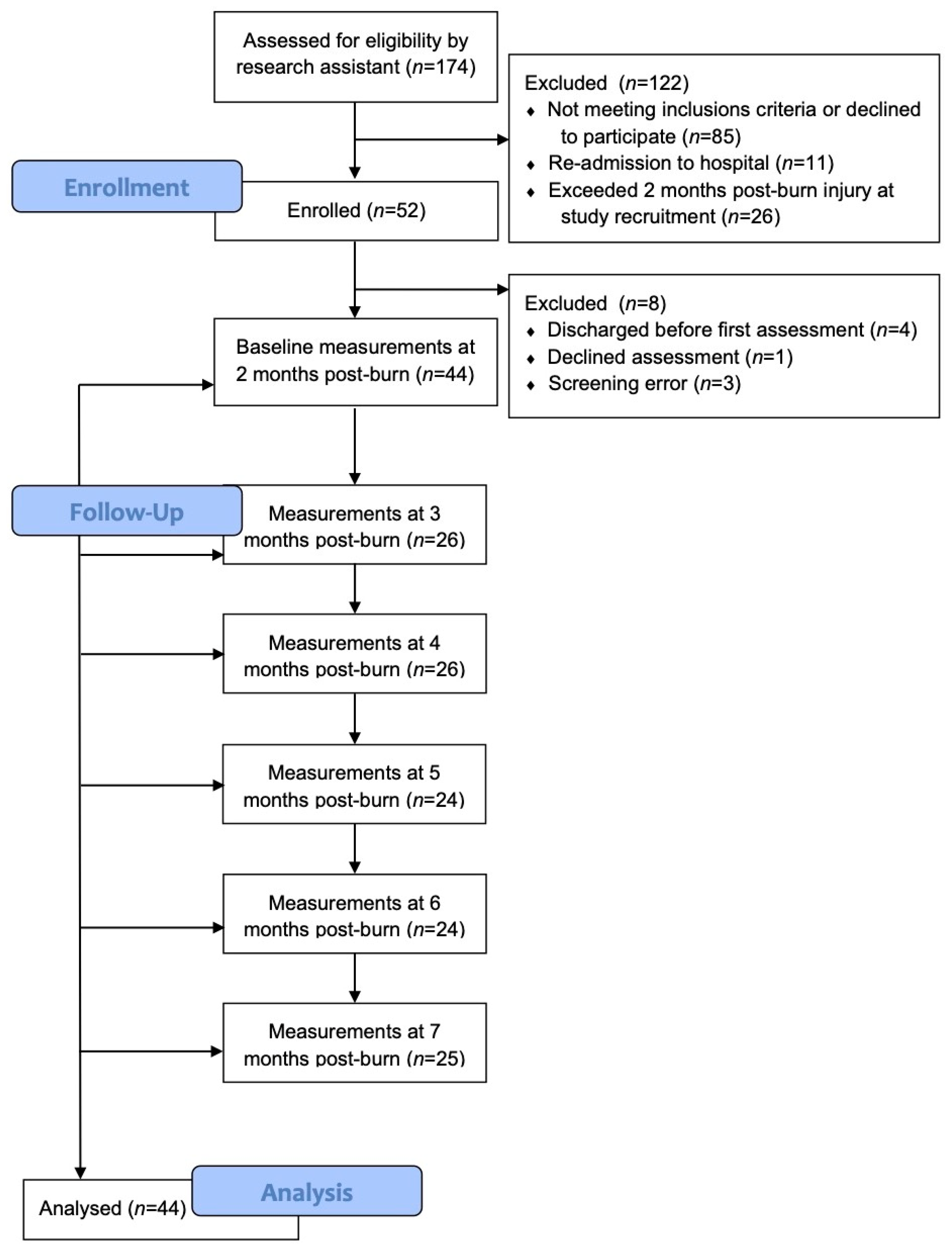
The participants (n = 174) were assessed for eligibility, and 52 patients were enrolled in the study. However, 3 screening errors occurred, one participant declined to participate before the baseline measures were obtained, and 4 patients were discharged before taking the baseline measures. Thus, baseline measures were performed with 44 participants, and measurements were taken every month up to 7 months with as many participants as possible. Analyses were completed, with all measurements from the 44 participants. As per the CONSORT guidelines [21], Figure 1 details the number of participants recruited, the number of participants enrolled and evaluated at each follow-up time point, and the reasons for exclusions throughout this study. The demographics of the participants and the clinical characteristics of the sites chosen for evaluation are reported in Table 1. A total of 44 participants were included in this study, with the majority being male (79.5%). They had a median age of 38 (IQR 28–56) years and a median percentage total body surface area (TBSA) burned of 16% (IQR 9.5–28.5). The median time post-burn to enrollment in the study was 56 (IQR 49.5–63) days. The most common cause for their burn injury was fire/flame (61.4%), followed by scalding (22.7%). The participants were classified using the Fitzpatrick’s skin type I-VI scale, with the majority being categorized as types II-IV (97%). They had a median inpatient length of stay (LOS) in the acute care hospital of 22 (IQR 12.5–27.5) days and in the rehabilitation hospital of 17.5 (IQR 9.5–32) days. The median number of surgeries was 2 (IQR 1–3), with the first surgery occurring a median of 5 (IQR 4–6) days post-burn injury. The scar sites were more commonly located on the upper extremity, but the donor sites were mostly on the lower extremity. All participants had been prescribed pressure garments and moisturizers or lubricants for the treatment of their scar site and moisturizers or lubricants for their donor sites, but pressure therapy is generally not prescribed for donor sites, unless HSc develops; however, pressure therapy adherence was not monitored as part of this study.

3.2. Skin Characteristics

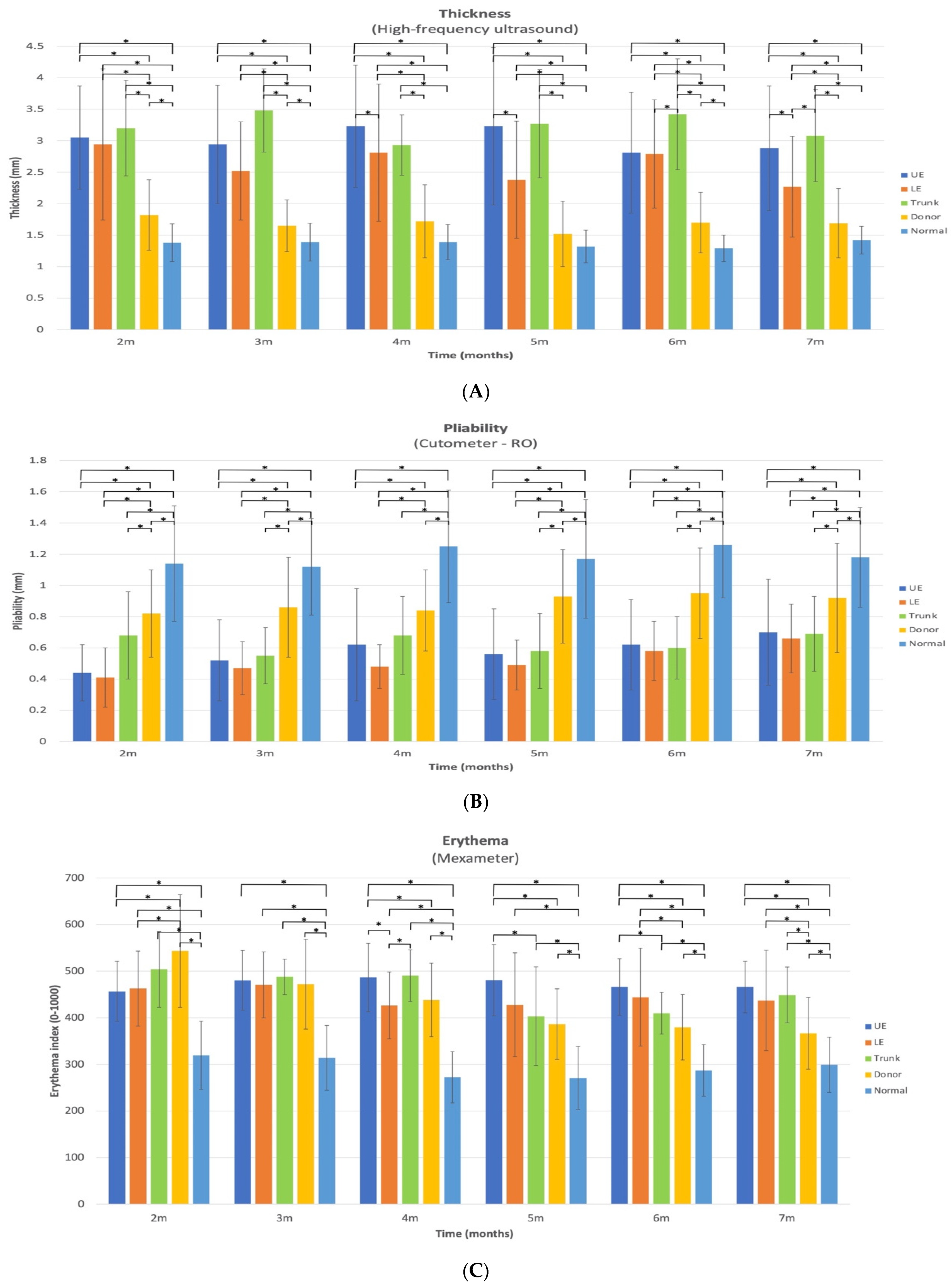
The differences in mean between the five different scar sites (upper extremity, lower extremity, trunk, donor, and normal) at 2, 3, 4, 5, 6, and 7 months are presented in Figure 2A–D for thickness, pliability, erythema, and melanin, respectively. The significant differences have been represented in these figures. The differences in mean between the combined HSc sites, donor sites, and normal skin sites are graphically represented in Figure 3A–D for thickness, pliability, erythema, and melanin, respectively. The differences in mean over time (2 to 7 months) for the upper extremity, lower extremity, trunk, donor, and normal sites are presented in Table 2, Table 3, Table 4 and Table 5 for thickness, pliability, erythema, and melanin, respectively.

3.3. Thickness

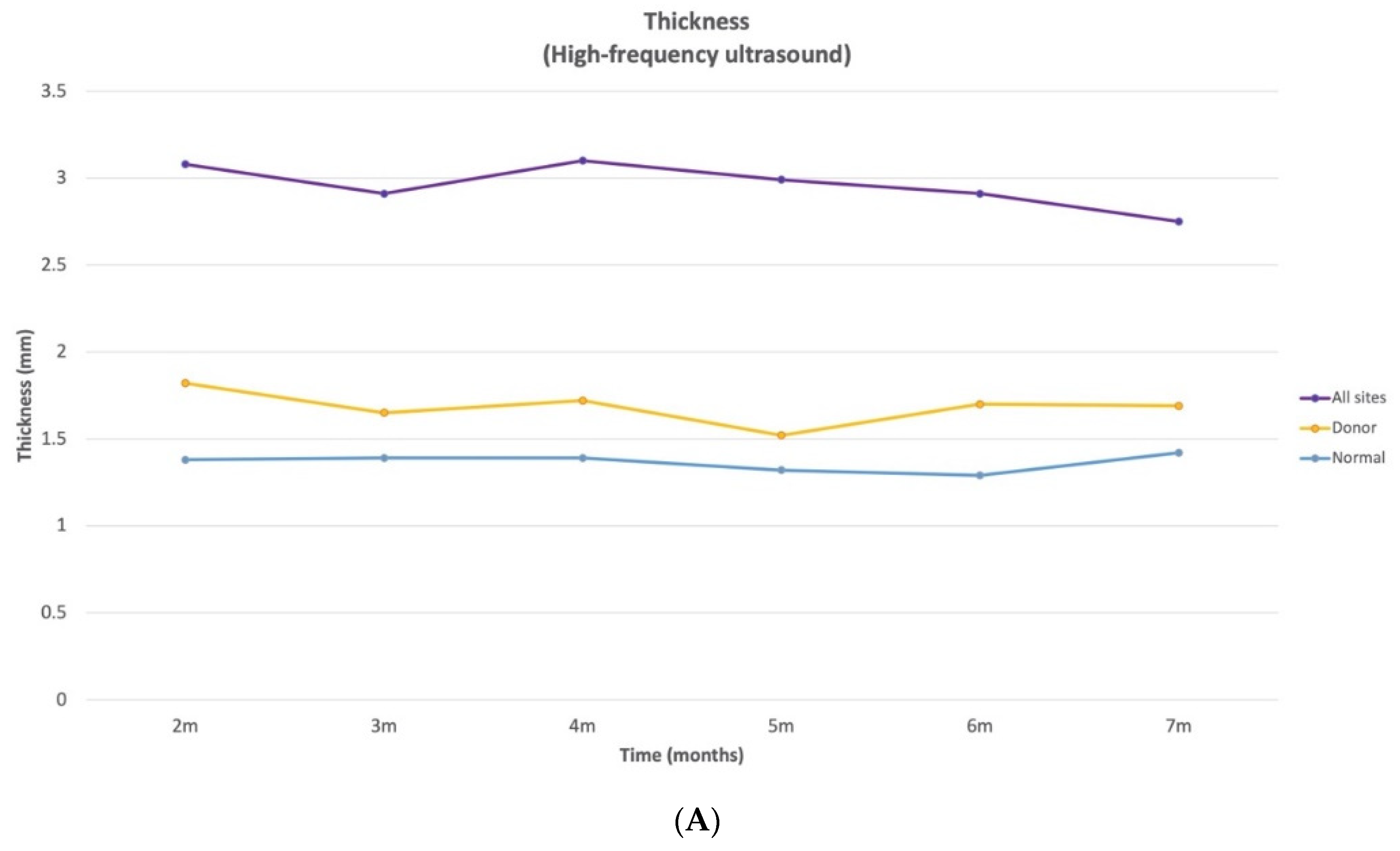
There were no significant differences in thickness between the donor site and normal site at 4, 5, and 7 months. The LE and trunk sites had significant differences in thickness at 6 and 7 months. The LE and UE sites had significant differences in thickness at 4, 5, and 7 months. At all time points, the following sites had significant differences in thickness: UE and donor, UE and normal, trunk and donor, trunk and normal, LE and donor, and LE and normal. Across all time points, there were no significant differences in thickness between the trunk and UE sites (Figure 2A). The average total reduction in HSc thickness of the combined locations between 2 and 7 months was 0.33 mm. The HSc in the combined anatomical sites are thicker than the donor and normal thickness at all time points (Figure 3A).
The mean thickness of the HSc in the UE sites significantly increased over time between 2 (3.05 (SD 0.82) mm) and 4 months (3.23 (SD 0.97) mm) and 3 (2.94 (SD 0.94) mm) and 4 months. It significantly decreased over time between 3 and 6 (2.81 (SD 0.96) mm), 3 and 7 months (2.88 (SD 0.99) mm), 4 and 6 months, 4 and 7 months, 5 (3.23 (SD 1.25) mm) and 6 months, and 5 and 7 months. The thickness of the HSc in the trunk sites significantly decreased over time between 3 (3.48 (SD 0.66) mm) and 4 months (2.93 (SD 0.48) mm) and 3 and 7 months (3.08 (SD 0.73) mm). Finally, the thickness of the HSc in the LE sites significantly decreased over time between 2 (2.94 (SD 1.2) mm) and 7 months (2.27 (SD 0.8) mm) and 3 (2.52 (SD 0.78) mm) and 7 months (Table 2 and Figure 3A). In spite of this decrease in HSc thickness in the upper and lower extremity sites and trunk sites, the thickness did not return to normal or the donor site thickness by 7 months after injury and, in fact, continued to be approximately twice as thick as normal and donor skin 7 months after injury. At all time points, the thickness of normal skin and scars in the donor sites did not significantly change over time.

3.4. Pliability

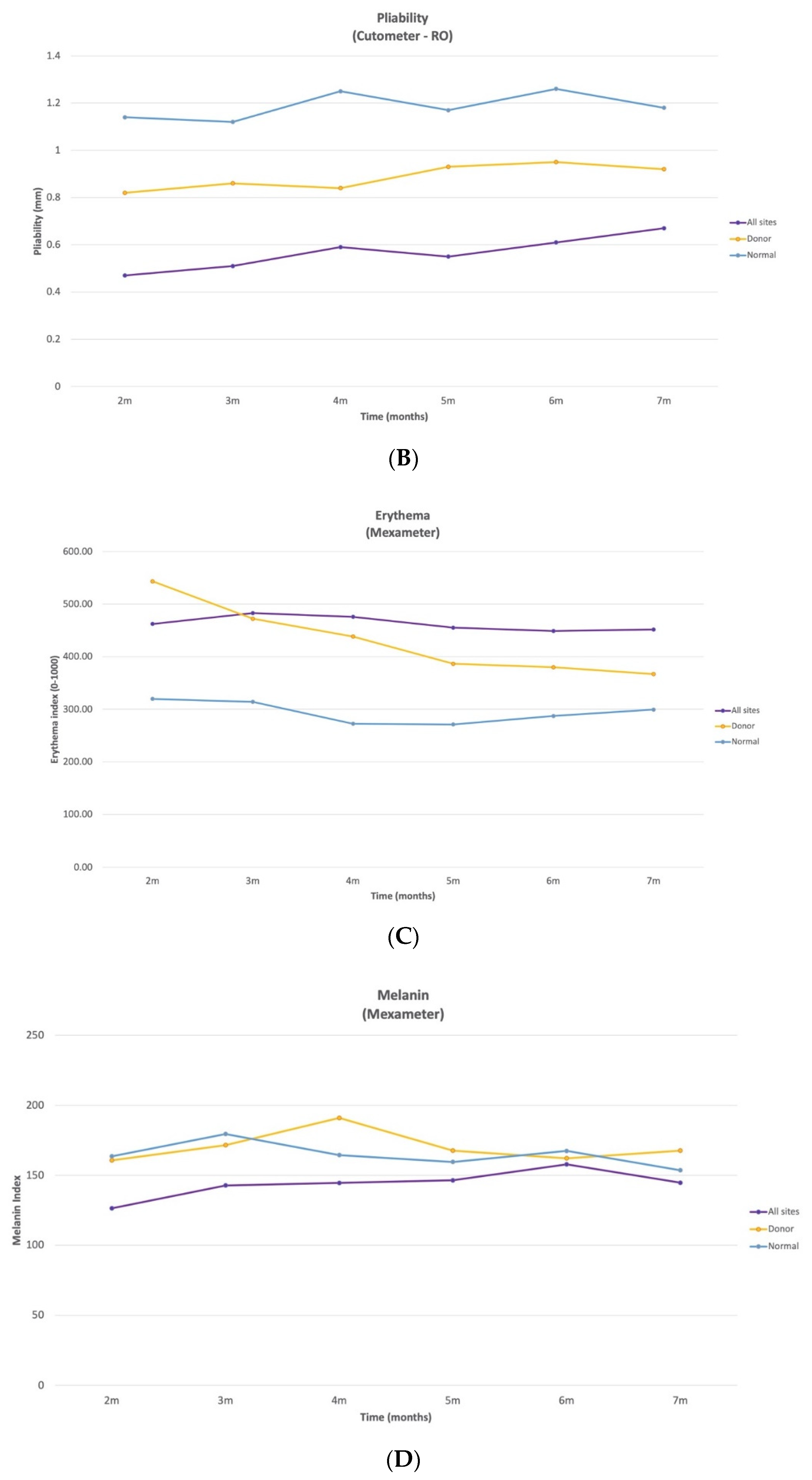
The pliability of the normal skin was significantly different from HSc in the UE, LE, and trunk sites and from the scars in the donor sites at all time points. The donor site was also significantly different from UE and LE at all time points. The pliability of the trunk site was significantly different from the donor site at all time points, except at 4 months (Figure 2B). The HSc in the combined anatomical sites were less pliable than the donor and normal thickness at all time points (Figure 3B).
There was no significant change in the mean pliability of normal skin and HSc in the trunk site over time. However, the pliability of the HSc in the UE significantly increased between 2 (0.44 (SD 0.18) mm) and 4 months (0.62 (SD 0.36) mm), 2 and 5 months (0.56 (SD 0.29) mm), 2 and 6 months (0.62 (SD 0.29) mm), 2 and 7 months (0.7 (SD 0.34) mm), 3 (0.52 (SD 0.26) mm) and 7 months, 4 and 7 months, 5 and 7 months, and 6 and 7 months (Table 3).
The mean pliability of the HSc in the LE also significantly increased between 2 (0.41 (SD 0.19) mm) and 6 months (0.58 (SD 0.19) mm), 2 and 7 months (0.66 (SD 0.22) mm), 3 (0.47 (SD 0.17) mm) and 7 months, 4 (0.48 (SD 0.14 mm) and 6 months, 4 and 7 months, and 5 (0.49 (SD 0.16) mm) and 7 months. Finally, the pliability of the scars in the donor sites significantly increased between 2 (0.82 (SD 0.28) mm) and 5 months (0.93 (SD 0.3) mm), 2 and 6 months (0.95 (SD 0.29) mm), 2 and 7 months (0.92 (SD 0.35) mm), 4 (0.84 (SD 0.26) mm) and 6 months, and 4 and 7 months. By 7 months, neither the donor sites nor the HSc of all sites reached the mean normal skin pliability (1.9 (SD 0.35) mm). By 7 months, neither the UE, LE, nor trunk HSc reached the donor site pliability (0.92 (SD 0.35) mm) (Table 3 and Figure 3B).

3.5. Erythema

Across all time points, the normal site had significantly different erythema indexes from all other sites. The donor and LE sites were not significantly different at 3, 4, and 5 months. The donor and UE sites were not significantly different at 3 months only. The trunk and donor sites were not significantly different at any time point, except at 7 months. The UE and LE sites and LE and trunk sites were significantly different at 4 months only (Figure 2C). The HSc in the combined anatomical sites were less erythematous than the donor site at 2 months post-burn. The donor site erythema was slightly less significant than the HSc, starting at 3 months and continuing to reduce across time, while remaining more erythematous than normal skin at 7 months post-burn (Figure 3C).
The UE site’s mean erythema index significantly increased over time between 2 (456.86 (SD 64.23)) and 3 months (480.31 (SD 63.88)) and decreased between 3 and 7 months (466.02 (SD 55.1)). The trunk site’s erythema index significantly decreased over time between 2 (503.88 (SD 81.15)) and 5 months (403.29 (SD 105.73)), 2 and 6 months (410.08 (SD 44.7)), 2 and 7 months (449 (SD 59.77)), 3 (487.79 (SD 37.94)) and 5 months, 3 and 6 months, 3 and 7 months, 4 (490.38 (SD 55.21)) and 5 months, and 4 and 6 months. The LE site’s mean erythema index significantly decreased over time between 3 (470.62 (SD 70.51)) and 4 months (426.6 (SD 71.29)). The donor site’s erythema index also significantly decreased over time between 2 (543.44 (SD 120.8)) and 3 months (472.26 (SD 92.3)), 2 and 4 months (438.39 (SD 78.65)), 2 and 5 months (386.5 (SD 75.53)), 2 and 6 months (379.91 (SD 70.2)), 2 and 7 months (366.91 (SD 77)), 3 and 4 months, 3 and 5 months, 3 and 6 months, 3 and 7 months, 4 and 5 months, 4 and 6 months, 4 and 7 months, and 5 and 7 months. The normal site’s mean erythema index significantly decreased over time between 2 (319.64 (SD 73.28)) and 4 months (272.5 (SD 55.01)), 2 and 5 months (271.04 (SD 67.73)), 3 (314.12 (SD 69.52)) and 4 months, 3 and 5 months, and 3 and 6 months (287.25 (SD 55.43)). Even though the donor site’s mean erythema index decreased over time, it did not return to the normal site’s (299.36 (SD 59.26)) redness after 7 months (Table 4 and Figure 3C).

3.6. Pigmentation

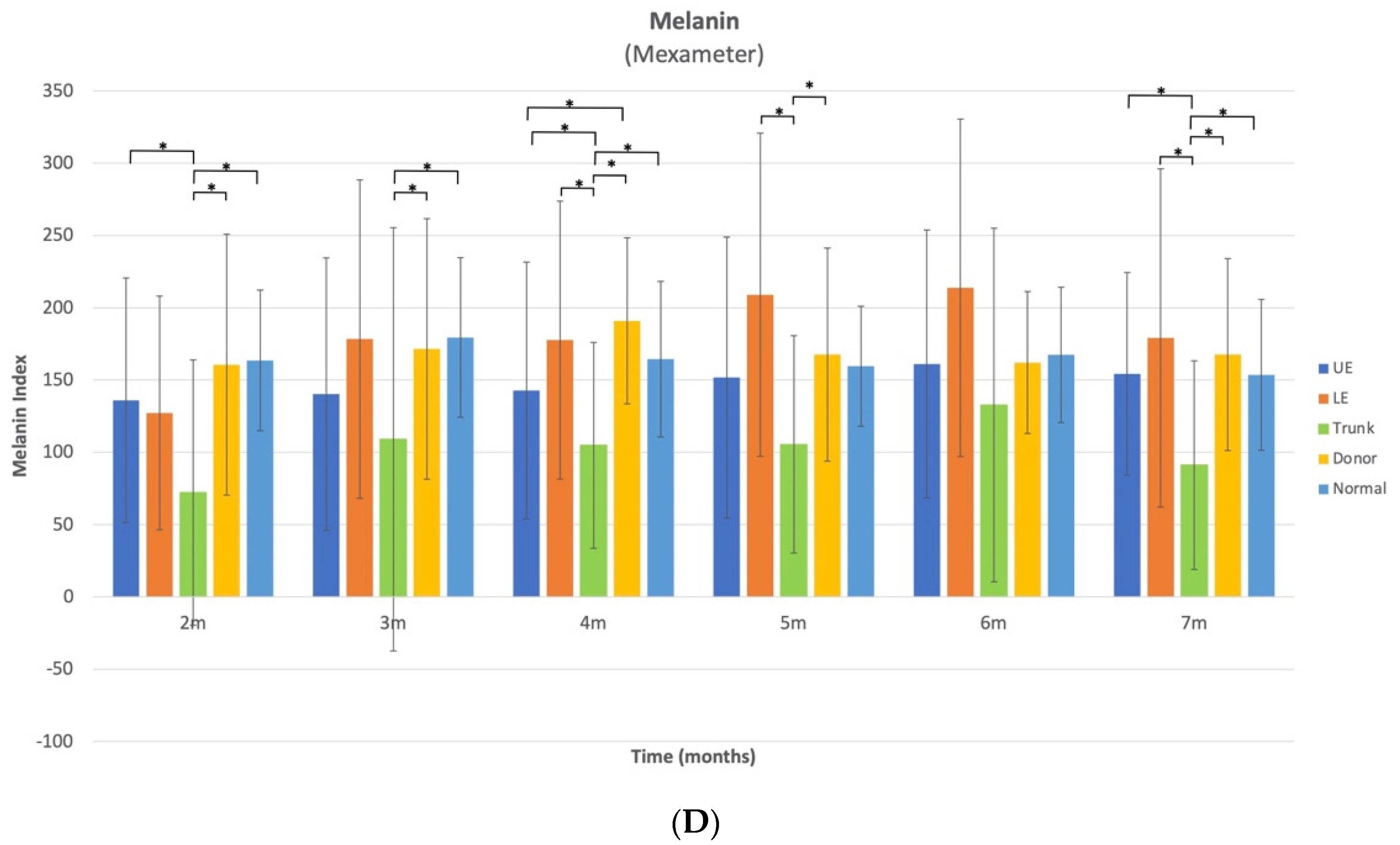
The melanin index means for the HSc in the trunk sites were 72.17 (SD 91.8), 109 (SD 146.39), 104.81 (SD 71.2), 105.5 (SD 75.17), 132.75 (SD 122.26), and 91.14 units (SD 72.13), and for the scars in the donor sites, they were 160.67 (SD 90.24), 171.52 (SD 90.16), 190.96 (SD 57.43), 167.59 (SD 73.7), 162.14 (SD 49.12), and 167.59 units (SD 66.42) at 2, 3, 4, 5, 6, and 7 months, respectively. They were significantly different at all time points, except at 6 months. The mean melanin index for skin in the normal sites were 163.57 (SD 48.66), 179.46 (SD 55.23), 164.42 (SD 53.78), 159.5 (SD 41.5), 167.42 (SD 46.8), and 7153.56 units (SD 52.22) at 2, 3, 4, 5, 6, and 7 months, respectively. They were significantly different than the mean melanin index for the HSc in the trunk sites at 2, 3, 4, and 7 months. The melanin index means for the HSc in the UE sites were 136 (SD 84.59), 140.24 (SD 94.24), 142.8 (SD 88.79), 151.75 (SD 97.14), 161.11 (SD 92.64), and 154.33 units (SD 70.04) at 2, 3, 4, 5, 6, and 7 months, respectively. They were significantly different than the means for HSc in the trunk sites at 2, 4, and 7 months. The mean melanin index for the HSc in the LE sites were 127.24 (SD 80.83), 178.31 (SD 110.15), 177.6 (SD 96.14), 209 (SD 111.78), 213.78 (SD 116.72), and 179.08 units (SD 117.02) at 2, 3, 4, 5, 6, and 7 months, respectively. They were significantly different than the means for the HSc in the trunk sites at 4 and 5 months. The melanin index significantly varied between the HSc in UE sites and scars in the donor sites at 4 months exclusively (Figure 2D). The melanin index of the HSc in the combined anatomical sites was less than the donor and normal site across time. However, the melanin index of the combined HSc, donor scars, and normal skin was similar at 6 months post-burn (Figure 3D).
The UE sites’ melanin index significantly increased over time between 3 (110.5 units) and 6 months (150.25 units). The trunk site’s melanin index significantly increased over time between 2 (43 units) and 6 months (86.75 units). The LE site’s melanin index significantly increased over time between 2 (97 units) and 3 months (141 units), 2 and 4 months (150 units), 2 and 5 months (197.5 units), 2 and 6 months (211 units), and 2 and 7 months (164.5 units) (Table 5 and Figure 3D).

3.7. Pain and Itch

Only one participant reported pain (VAS 1/10) at an upper extremity scar site once at 3 months. No other participants reported pain at any of the sites evaluated at any of the time points. Out of the 44 participants included in the final analysis, 20 reported itch on at least one occasion. Three of the participants only reported itch at one time point, and for all of these participants, their itch was located at the donor site or the lower extremity. Twelve of the participants reported itch at the donor site, with the majority reporting itch at 2 months, but several reported donor site itch at later time points as well. However, most participants reported zero itch at all sites at most, if not all, time points.

4. Discussion

Thickness, pliability, erythema, and pigmentation of HSc, donor sites, and normal skin have previously been measured over time in burn survivors using objective instrumentation [9], but the time spread between measures was at least three months. To the best of our knowledge, this is the first time that HSc characteristics have been compared over time on a monthly basis between 2 and 7 months post-burn. These data provide a better understanding of the recovery profile of HSc sites, relative to normal skin and donor sites in the same individual, and expands our understanding of specific anatomical site differences.
Scar thickness demonstrated notable site-specific changes with time that have important clinical implications. Between 2 and 7 months post-burn, the thickness of HSc in the combined anatomical sites (UE, LE, and trunk) was approximately 2 times thicker than normal skin and 1.6 times thicker than donor site scars. When examining specific anatomical locations, the three sites (UE, LE, and trunk) demonstrated both increases and decreases in thickness over time. However, in all 3 anatomical locations, there was a significant decrease in thickness from 3 to 7 months. The thickness of UE HSc sites were at their peak at 4 months post-burn and decreased by 10.8% between 4 and 7 months post-burn. For the HSc in the LE site, they were at their peak of thickness at 2 months and decreased by 22.8% between 2 and 7 months post-burn. The thickness of the HSc in the trunk sites were at their peak at 3 months and demonstrated an improvement of 11.5% between 3 and 7 months. Thus, scars appear to increase in thickness for a maximum of 4 months, then plateau or reduce across time.
While HSc thickness appears to vary by anatomical location, for example, the trunk sites were slightly but not significantly thicker than the UE and were significantly thicker than the LE at 7 months, this may be related to pre-burn thickness. The normative data for the trunk thickness of normal skin is approximately 0.73 mm and 0.65 mm thicker than normal UE and LE skin, respectively, for 20–59 year-old males and females [10]. Therefore, the pre-burn difference may account for some of the site-specific differences in HSc thickness.
The comparison of different anatomical HSc sites to donor scars at 7 months post-burn (LE HSc 1.3 times thicker, UE 1.7 times thicker, and trunk 1.8 times thicker) suggests that the LE HSc improved at a faster rate. Nonetheless, at 7 months LE, the HSc thickness remained statistically different from the donor site scars, meaning its thickness did not return to a normal scar thickness at 7 months post-burn.
The donor site thickness was significantly thicker than the normal skin at all time points, except at 4, 5, and 7 months. This is consistent with a previous study, where the donor sites were not significantly thicker than the normal sites, starting at 3 months [10]. Of all the skin characteristics that were evaluated in this study, thickness most clearly distinguished normal scar (donor site) and normal skin from HSc at all time points. Interestingly, in a clinical study where the Vancouver scar scale (VSS) (a subjective scar assessment) was used to evaluate HSc, the height (thickness) subscale was the most specific (85.9%) and sensitive (99.5%) skin characteristic for the diagnosis of HSc [22].
Since usual care was continued during the data collection, the impact of pressure therapy on the scar thickness measures must be considered. Pressure garments have been shown to improve the thickness of HSc in burn survivors and compression garment wear is now considered a gold standard treatment for pathological scarring, like HSc [23,24]. However, studies have shown that pressure garments should be monitored regularly, since optimal pressure may not be achieved [25,26], and pressure may reduce with time [25] or at different anatomical sites, partially due to the fact that pressure loss correlates to the radius of the curvature of that anatomical region [27,28,29,30].
Another factor that could have an impact on the variability of pressure benefits, depending on the anatomical location, is patient adherence to this treatment. A systematic review and meta-ethnography of the non-adherence with compression garment wear in adult burn patients revealed six themes impacting adherence: sensory factors (such as itch, pain, sensitivity, or body temperature), psychological state, impact of the garment on the patient’s function, availability of social support, degree of choice, and education provided to patients by their therapists [31]. Since the UE is the anatomical site most solicited during functional activities, and the ribcage expands when breathing, perceived tightness may specifically limit patients adherence to UE and trunk garment wear. In fact, verbal reports from clinicians confirmed that they rarely aim to ensure adequate pressure for chest and trunk garments, because patients simply do not tolerate them. Thus, trunk HSc may take longer to improve because of this lack of adequate pressure dosage applied to this area. However, to the best of our knowledge, no study has systematically investigated the different factors associated with the anatomical variation of pressure tolerance, treatment adherence, or therapeutic benefit.
The pliability of HSc in the UE and LE and normal scar sites significantly increased between 2 and 7 months by 59%, 61%, and 12%, respectively. To the best of our knowledge, this study is the first to demonstrate an improvement in pliability for HSc and donor site scars as early as 4 months post-burn. On the other hand, the pliability for the trunk HSc did not demonstrate a significant change between at any time point and only increased by 1.5% between 2 and 7 months, which could demonstrate a lack of spontaneous resolution of pliability for this anatomical site and/or a lack of response to pressure therapy.
In this study, even though the donor site scar pliability significantly improved between 2 and 7 months, it remained significantly less pliable than normal skin. Thus, although a donor site scar is considered a normal scar, it does not return to a normal pliability by 7 months post-burn.
There were significant changes over time in the erythema index for all measured sites, including the normal skin. Between 2 and 7 months, the HSc redness in the UE was the only anatomical location where it significantly increased and peaked at 4 months post-burn. There was a significant decrease in UE HSc erythema between 3 and 7 months, but it remained the most erythematous at 7 months. Trunk HSc redness peaked at 2 months post-burn and significantly decreased from 2 to 7 months. LE HSc peaked at 3 months post-burn and significantly decreased from 3 to 4 months only. Conversely, donor site erythema decreased significantly (32.5%) between 2 and 7 months and almost returned to normal skin levels (ration 1.2:1), which has previously been reported [9]. Surprisingly, at 2 months, donor site scars were redder than all HSc sites, making it difficult to distinguish donor from HSc based on erythema, until 5 months post burn. Similarly, clinicians found that vascularity (erythema) was very difficult to judge in subjective scar assessments, like the VSS [22]. Thus, erythema alone should not be used to distinguish HSc from normal scars in the first months post-burn; thickness is a more reliable indicator.
To the best of our knowledge, this is the first time that normal skin erythema has been reported to vary significantly with time, where it peaked at 2 months and significantly decreased from 2 to 5 and 3 to 6 months. Since 61% of the normal skin measurements were taken on the UE, 34% on the LE, and 5% on the trunk, we compared the erythema index of normal skin at 7 months to the normative data [10] for each of these anatomical locations for 20–59-year-old male and females. Normal skin at 7 months was 10%, 55%, and 12% more erythematous than the average normative data in the UE, LE, and trunk, respectively. Therefore, normal skin in this cohort did not return to normative data redness at 7 months post-burn. Further research is needed to determine why burn survivors demonstrate elevated levels of normal skin erythema and when it returns to normal. To the best of our knowledge, there are no other longitudinal studies that examined erythema in burn survivors using the Mexameter; therefore, it is not possible to make further comparisons.
The melanin index of trunk HSc was lower than that of all other sites at all time points with sporadic significant variation across time. However, it is important to consider normative anatomical data, since melanin varies greatly based upon sun exposure, and the UE and LE are more likely to be exposed to the sun, compared to the trunk. When comparing to normative data, trunk melanin in normal skin for 20–59-year-old Caucasian male and females, it is approximately 5.4% and 7% less pigmented than normal skin in the UE and LE, respectively [10]. However, this difference cannot be entirely responsible for the fact that the melanin index of HSc in the trunk site at 7 months was approximately 61% lower than UE HSc and 65% lower than LE HSc sites. Therefore, for this cohort, the HSc on the trunk were significantly less pigmented than the HSc on the UE at 2, 4, and 7 months post-burn and significantly less pigmented than the HSc on the LE sites at 4, 5, and 7 months, and normal anatomic variation did not entirely account for this difference. When examining the combined anatomical HSc results, the melanin index is quite similar for HSc, donor scar, and normal skin at 6 months post-burn. To the best of our knowledge, no other reports have used the Mexameter to examine melanin in burn survivors; thus, further comparisons are not possible.
Since only 11 of the 44 participants reported itch at baseline, it was generally episodic, and there was substantial variability in the location and itch intensity across time, there did not appear to be any discernable patterns, nor a persistence problem at specific locations. This may be related to the fact that our research group has been very involved in a number of research projects examining itch evaluation and treatment; therefore, clinicians may be attentive to addressing any issues that do arise.

Study Limitations

As with any study, the limitations associated with this study have to be considered when interpreting the results. All participants were recruited from a single burn center in North America, so these results are not generalizable to other burn centers. Since the participants predominantly fell into category II or III of the Fitzpatrick scale, these data may not be generalizable to populations in higher categories. Unfortunately, the sample size in the different categories does not allow us to make comparisons across the Fitzpatrick scale. As with any longitudinal study with burn survivors, it is challenging to obtain consistent data at set time points. Since the participants’ physical presence is required for the data reported in this study, and many participants live far away from the burn center, the extent of missing data is higher than what we would have hoped for, in spite of the fact that the data collection was coordinated with patients during follow-up clinic appointments. Future studies should monitor different treatment benefits and objectively measure their effects on HSc and normal scar characteristics for the burn population at specific anatomical locations. Larger samples from multiple burn centers and for longer periods of time should also be investigated.
5. Conclusions

These results provide a better understanding of the recovery profile of HSc in different anatomical locations and donor site scars secondary to burn injuries. In addition, the data support the suggestion that scar management treatments should prioritize the UE and trunk sites that develop HSc during the first two to four months post-burn and continue to vary significantly from normal scar and normal skin at 7 months. This recommendation is based on the findings that UE and trunk HSc tend to be thicker than LE HSc at 7 months post-burn, the pliability of trunk HSc did not improve over time, and UE HSc continued to be more erythematous at 7 months, compared to other anatomical sites. While many clinicians have historically prioritized UE treatments due to their functional importance, these data provide objective measurements to further support this practice and encourage clinicians to also prioritize trunk HSc treatments. These results also provide evidence that the recovery profile of HSc varies at different anatomical sites and that thickness is the characteristic that distinguishes HSc from normal scar and normal skin. Further research is required to continue to explore the recovery profile of HSc and therapeutic approaches to optimize outcomes.

Author Contributions

Conceptualization, B.N., V.C., M.-A.C.; methodology, B.N.; formal analysis, J.A.C.; investigation, A.d.O., V.C., M.-A.C., E.M.-P.; resources, B.N.; data curation, B.N., A.d.O.; writing—original draft preparation, Z.E.-L.; writing—review and editing, Z.E.-L., B.N.; supervision, B.N.; project administration, A.d.O.; funding acquisition, B.N. All authors have read and agreed to the published version of the manuscript.

Funding

This work was supported by the Fondation des pompiers du Québec pour les Grands Brûlés.

Institutional Review Board Statement

IRB Study Number: A10-E69-10B.

Informed Consent Statement

Informed consent was obtained from all subjects involved in the study.

Acknowledgments

The authors thank the burn survivors, clinicians, and research team members from the Villa Medica Rehabilitation Hospital who participated in this study for their valuable contributions. This work was supported by the Fondation des pompiers du Québec pour les Grands Brûlés.

Conflicts of Interest

The authors declare no conflict of interest.

References

  1. Esselman, P.C.; Thombs, B.; Magyar-Russell, G.; Fauerbach, J.A. Burn Rehabilitation. Am. J. Phys. Med. Rehabil. 2006, 85, 383–413. [Google Scholar] [CrossRef]
  2. Finnerty, C.C.; Jeschke, M.G.; Branski, L.K.; Barret, J.P.; Dziewulski, P.; Herndon, D.N. Hypertrophic scarring: The greatest unmet challenge after burn injury. Lancet 2016, 388, 1427–1436. [Google Scholar] [CrossRef] [Green Version]
  3. Bock, O.; Schmid-Ott, G.; Malewski, P.; Mrowietz, U. Quality of life of patients with keloid and hypertrophic scarring. Arch. Dermatol. Res. 2006, 297, 433–438. [Google Scholar] [CrossRef]
  4. Deitch, E.A.; Wheelahan, T.M.; Rose, M.P.; Clothier, J.; Cotter, J. Hypertrophic burn scars: Analysis of variables. J. Trauma Inj. Infect. Crit. Care 1983, 23, 895–898. [Google Scholar] [CrossRef]
  5. Gangemi, E.N.; Gregori, D.; Berchialla, P.; Zingarelli, E.; Cairo, M.; Bollero, D.; Ganem, J.; Capocelli, R.; Cuccuru, F.; Cassano, P.; et al. Epidemiology and Risk Factors for Pathologic Scarring After Burn Wounds. Arch. Facial Plast. Surg. 2008, 10, 93–102. [Google Scholar] [CrossRef] [PubMed] [Green Version]
  6. Lawrence, J.W.; Mason, S.T.; Schomer, K.; Klein, M.B. Epidemiology and Impact of Scarring After Burn Injury. J. Burn Care Res. 2012, 33, 136–146. [Google Scholar] [CrossRef] [PubMed]
  7. Wallace, H.J.; Fear, M.W.; Crowe, M.M.; Martin, L.J.; Wood, F.M. Identification of factors predicting scar outcome after burn injury in children: A prospective case-control study. Burn Trauma 2017, 5, 19. [Google Scholar] [CrossRef]
  8. Wallace, H.J.; Fear, M.W.; Crowe, M.M.; Martin, L.J.; Wood, F.M. Identification of factors predicting scar outcome after burn in adults: A prospective case–control study. Burns 2017, 43, 1271–1283. [Google Scholar] [CrossRef]
  9. Nedelec, B.; Correa, J.A.; De Oliveira, A.; LaSalle, L.; Perrault, I. Longitudinal burn scar quantification. Burns 2014, 40, 1504–1512. [Google Scholar] [CrossRef]
  10. Nedelec, B.; Forget, N.J.; Hurtubise, T.; Cimino, S.; De Muszka, F.; Legault, A.; Liu, W.L.; Oliveira, A.C.M.T.G.; Calva, V.; A Correa, J. Skin characteristics: Normative data for elasticity, erythema, melanin, and thickness at 16 different anatomical locations. Ski. Res. Technol. 2015, 22, 263–275. [Google Scholar] [CrossRef] [PubMed]
  11. Nedelec, B.; Rachelska, G.; Parnell, L.K.; LaSalle, L. Double-Blind, Randomized, Pilot Study Assessing the Resolution of Postburn Pruritus. J. Burn Care Res. 2012, 33, 398–406. [Google Scholar] [CrossRef] [PubMed]
  12. Parnell, L.K.; Nedelec, B.; Rachelska, G.; LaSalle, L. Assessment of Pruritus Characteristics and Impact on Burn Survivors. J. Burn Care Res. 2012, 33, 407–418. [Google Scholar] [CrossRef]
  13. Nedelec, B.; Correa, J.A.; Rachelska, G.; Armour, A.; LaSalle, L. Quantitative Measurement of Hypertrophic Scar: Interrater Reliability and Concurrent Validity. J. Burn Care Res. 2008, 29, 501–511. [Google Scholar] [CrossRef] [PubMed]
  14. Nedelec, B.; Correa, J.A.; Rachelska, G.; Armour, A.; LaSalle, L. Quantitative Measurement of Hypertrophic Scar: Intrarater Reliability, Sensitivity, and Specificity. J. Burn Care Res. 2008, 29, 489–500. [Google Scholar] [CrossRef]
  15. Simons, M.; Kee, E.G.; Kimble, R.; Tyack, Z. Ultrasound is a reproducible and valid tool for measuring scar height in children with burn scars: A cross-sectional study of the psychometric properties and utility of the ultrasound and 3D camera. Burns 2017, 43, 993–1001. [Google Scholar] [CrossRef] [PubMed]
  16. Rennekampff, H.-O.; Rabbels, J.; Reinhard, V.; Becker, S.T.; Schaller, H.-E. Comparing the Vancouver Scar Scale With the Cutometer in the Assessment of Donor Site Wounds Treated With Various Dressings in a Randomized Trial. J. Burn Care Res. 2006, 27, 345–351. [Google Scholar] [CrossRef]
  17. Lee, K.; Bamford, A.; Gardiner, F.; Agovino, A.; ter Horst, B.; Bishop, J.; Sitch, A.; Grover, L.; Logan, A.; Moiemen, N. Investigating the intra- and inter-rater reliability of a panel of subjective and objective burn scar measurement tools. Burns 2019, 45, 1311–1324. [Google Scholar] [CrossRef]
  18. Van Der Wal, M.; Bloemen, M.; Verhaegen, P.; De Vet, H.; van Zuijlen, P.; Tuinebreijer, W.; Middelkoop, E. Objective Color Measurements. J. Burn Care Res. 2013, 34, e187–e194. [Google Scholar] [CrossRef]
  19. Rothman, K.J. No Adjustments Are Needed for Multiple Comparisons. Epidemiology 1990, 1, 43–46. [Google Scholar] [CrossRef] [Green Version]
  20. Althouse, A.D. Adjust for Multiple Comparisons? It’s Not That Simple. Ann. Thorac. Surg. 2016, 101, 1644–1645. [Google Scholar] [CrossRef] [Green Version]
  21. Schulz, K.F.; Altman, D.G.; Moher, D. WITHDRAWN: CONSORT 2010 statement: Updated guidelines for reporting parallel group randomised trials. Int. J. Surg. 2010. [Google Scholar] [CrossRef]
  22. Thompson, C.M.; Sood, R.F.; Honari, S.; Carrougher, G.J.; Gibran, N. What score on the Vancouver Scar Scale constitutes a hypertrophic scar? Results from a survey of North American burn-care providers. Burns 2015, 41, 1442–1448. [Google Scholar] [CrossRef] [PubMed] [Green Version]
  23. Atiyeh, B.; El Khatib, A.; Dibo, S. Pressure garment therapy (PGT) of burn scars: Evidence-based efficacy. Ann. Burn Fire Disasters 2013, 26, 205–212. [Google Scholar]
  24. Ai, J.-W.; Liu, J.-T.; Pei, S.-D.; Liu, Y.; Li, D.-S.; Lin, H.-M.; Pei, B. The effectiveness of pressure therapy (15–25 mmHg) for hypertrophic burn scars: A systematic review and meta-analysis. Sci. Rep. 2017, 7, 40185. [Google Scholar] [CrossRef] [PubMed]
  25. Nedelec, B.; De Oliveira, A.; Calva, V.; Couture, M.; Poulin, C.; LaSalle, L.; A Correa, J. 99 Are Custom Fabricated Pressure Garments Applying Adequate Pressure? J. Burn Care Res. 2019, 40, S65. [Google Scholar] [CrossRef]
  26. Wiseman, J.; Simons, M.; Kimble, R.; Tyack, Z. Variability of pressure at the pressure garment-scar interface in children after burn: A pilot longitudinal cohort study. Burns 2019, 45, 103–113. [Google Scholar] [CrossRef] [PubMed]
  27. Kerckhove, E.V.D.; Fieuws, S.; Massagé, P.; Hierner, R.; Boeckx, W.; Deleuze, J.-P.; Laperre, J.; Anthonissen, M. Reproducibility of repeated measurements with the Kikuhime pressure sensor under pressure garments in burn scar treatment. Burns 2007, 33, 572–578. [Google Scholar] [CrossRef]
  28. Giele, H.; Liddiard, K.; Booth, K.; Wood, F.M. Anatomical Variations in Pressures Generated by Pressure Garments. Plast. Reconstr. Surg. 1998, 101, 399–406. [Google Scholar] [CrossRef]
  29. Kim, J.Y.; Willard, J.J.; Supp, D.; Roy, S.; Gordillo, G.M.; Sen, C.K.; Powell, H.M. Burn Scar Biomechanics after Pressure Garment Therapy. Plast. Reconstr. Surg. 2015, 136, 572–581. [Google Scholar] [CrossRef]
  30. Rose, M.; Deitch, E. The clinical use of a tubular compression bandage, Tubigrip, for burn-scar therapy: A critical analysis. Burns 1985, 12, 58–64. [Google Scholar] [CrossRef]
  31. Crofton, E.; Meredith, P.; Gray, P.; O’Reilly, S.; Strong, J. Non-adherence with compression garment wear in adult burns patients: A systematic review and meta-ethnography. Burns 2020, 46, 472–482. [Google Scholar] [CrossRef] [PubMed]
Figure 1. CONSORT flowchart of study participants.
Ebj 02 00008 g001
Figure 2. Differences in mean between the five different scar sites (upper extremity, lower extremity, trunk, donor, and normal at 2, 3, 4, 5, 6, and 7 months for: (A) thickness; (B) pliability; (C) erythema; (D) melanin. Significant differences (p > 0.05) have been indicated with a *.
Ebj 02 00008 g002aEbj 02 00008 g002b
Figure 3. Differences in mean between the combined HSc sites, donor sites, and normal skin sites for: (A) thickness; (B) pliability; (C) erythema; (D) melanin.
Ebj 02 00008 g003aEbj 02 00008 g003b
Table 1. Participant sociodemographic data and clinical characteristics of the skin evaluated (n = 44).
Sex-M: F n (%)
Age-years: median (IQR)
TBSA%: median (IQR)
35:9 (79.5:20.5)
38 (28–56)
16 (9.5–28.5)
Time post-burn to enrollment (days): median (IQR)56 (49.5–63)
Etiology: n (%)Fitzpatrick scale, n (%)
Fire/Flame27 (61.4)Type I0
Scalding10 (22.7)Type II11 (25)
Electric3 (6.8)Type III27 (61)
Chemical3 (6.8)Type IV5 (11)
Frostbite1 (2.3)Type V1 (2)
Type VI0
Inpatient acute care LOS (days): median (IQR)
Inpatient rehabilitation LOS (days): median (IQR)
22.0 (12.5–27.5)
17.5 (9.5–32)
Number of surgeries: median (IQR)
Postburn days to 1st surgery: median (IQR)
2 (1–3)
5 (4–6)
Location of scars: n (%)HScDN
Upper extremity49 (61%)3 (8%)27 (61%)
Lower extremity17 (21%)30 (77%)15 (34%)
Trunk14 (18%)6 (15%)2 (5%)
TBSA = total body surface area; HSc = hypertrophic scar; D = donor; N = normal skin site; SD = standard deviation; % = percent; IQR = interquartile range (25th percentile–75th percentile); LOS = length of stay; n = number.
Table 2. Difference in adjusted means between the time points at each site for thickness (mm).
Time
(Months)
Mean Difference
UETrunkLEDonorNormal
2–30.090.25−0.020.020.06
2–40.36 *−0.21−0.12−0.110.05
2–50.21−0.03−0.36−0.18−0.02
2–6−0.190.007−0.44 *−0.11−0.06
2–7−0.29−0.29−0.75 *−0.130.02
3–40.27 *−0.46 *−0.1−0.13−0.01
3–50.12−0.28−0.34−0.2−0.07
3–6−0.28 *−0.24−0.42 *−0.14−0.12
3–7−0.38 *−0.54 *−0.73 *−0.15−0.03
4–5−0.150.18−0.24−0.07−0.06
4–6−0.55 *0.22−0.32−0.002−0.11
4–7−0.64 *−0.08−0.62 *−0.02−0.02
5–6−0.4 *0.04−0.080.06−0.05
5–7−0.49 *−0.26−0.390.050.04
6–7−0.09−0.3−0.3−0.020.09
* Two-way ANOVA, p-value < 0.05.
Table 3. Difference in adjusted means between the time points at each site for pliability (mm).
Time
(Months)
Mean Difference
UETrunkLEDonorNormal
2–30.04−0.0080.080.070.005
2–40.10 *0.040.070.020.02
2–50.10 *−0.0030.010.10 *0.02
2–60.15 *0.030.21 *0.12 *0.05
2–70.26 *0.120.31 *0.14 *0.04
3–40.060.05−0.01−0.050.02
3–50.060.0040.020.040.02
3–60.110.040.130.060.04
3–70.23 *0.120.23 *0.070.04
4–50.002−0.040.030.08−0.0009
4–60.05−0.010.150.10 *0.03
4–70.17 *0.080.25 *0.12 *0.02
5–60.050.030.110.020.03
5–70.17 *0.120.21 *0.040.02
6–70.12 *0.090.1 *0.02−0.001
* Two-way ANOVA, p-value < 0.05.
Table 4. Difference in adjusted means between the time points at each site for erythema (index 0–1000).
Time
(Months)
Mean Difference
UETrunkLEDonorNormal
2–336.09 *−3.4111.25−66.98 *0.11
2–426.67−16.28−35.99−97.56 *−36.1 *
2–525.7−100.9 *−23.56−142.9 *−34.19 *
2–611.99−98.95 *−13.35−153.7 *−25.87
2–72.65−65.15 *−25.3−174.5 *−23.57
3–4−9.41−12.87−47.23 *−30.58 *−36.21 *
3–5−10.39−97.54 *−34.81−75.97 *−34.31 *
3–6−24.1−95.54 *−24.59−86.71 *−25.98 *
3–7−33.43 *−61.74 *−36.54−107.5 *−23.68
4–5−0.97−84.67 *12.43−45.38 *1.91
4–6−14.68−82.67 *22.64−56.12 *10.23
4–7−24.02−48.8810.69−76.94 *12.53
5–6−13.71210.22−10.748.32
5–7−23.0535.8−1.74−31.56 *10.62
6–7−9.3433.8−11.95−20.822.3
* Two-way ANOVA, p-value < 0.05.
Table 5. Difference in adjusted means between the time points at each site for melanin (index 0–1000).
Time
(Months)
Mean Difference
UETrunkLEDonorNormal
2–3−7.1235.0341.63 *9.055.11
2–49.5420.3148.19 *25.774.12
2–517.8541.2252.8 *8.182.62
2–619.1155.57 *55.86 *6.436.69
2–719.2828.5448.37 *6.13−4.92
3–416.67−14.726.5616.72−1
3–524.976.211.16−0.87−2.49
3–626.23 *20.5414.23−2.611.58
3–726.4−6.496.74−2.92−10.03
4–58.3120.924.61−17.58−1.5
4–69.5635.257.67−19.332.58
4–79.738.220.18−19.64−9.03
5–61.2614.343.06−1.754.07
5–71.43−12.69−4.43−2.06−7.54
6–70.17−27.03−7.49−0.31−11.61
* Two-way ANOVA, p-value < 0.05.
Publisher’s Note: MDPI stays neutral with regard to jurisdictional claims in published maps and institutional affiliations.