Article

Molecular Analysis Corroborates the Biogeographic Distribution of “Tako Kurage” as Mastigias albipunctata Stiasny, 1920 (Cnidaria; Scyphozoa) in Japan


1
Graduate School of Agricultural Science, Tohoku University, Sendai 980-8572, Japan
2
Kagoshima City Aquarium, Kagoshima 892-0814, Japan
3
Kamo Aquarium, Tsuruoka 997-1206, Japan
4
WPI-Advanced Institute for Marine Ecosystem Change, Tohoku University, Sendai 980-8578, Japan
*
Author to whom correspondence should be addressed.
Oceans 2026, 7(5), 80; https://doi.org/10.3390/oceans7050080
Submission received: 7 August 2026 / Revised: 9 September 2026 / Accepted: 17 September 2026 / Published: 25 September 2026

Abstract

Mastigias jellyfish (“tako kurage”) have been reported throughout littoral Japan for over a century, historically identified by morphology alone as Mastigias papua. Molecular resolution of the genus in 2018 revealed three geographically structured clades and suggested Japanese populations instead belong to M. albipunctata, but subsequent morphology-only studies of Japanese specimens have continued to apply the M. papua designation, leaving species identity unresolved across much of the species’ Japanese range. Here, we use cytochrome c oxidase subunit I (COI), combining published GenBank sequences with two newly collected specimens from Okinawa, the southernmost extent reported in Japan. Maximum-likelihood and Bayesian phylogenetic reconstruction recovered three reciprocally monophyletic clades consistent with previous molecular work, with all Japanese sequences, from Sukumo Bay (Shikoku Island) to Okinawa Island, falling within the M. albipunctata clade. Morphological comparison against published diagnostic characters showed consistency with this identification. These results indicate that Mastigias populations in Japan are consistently identified as M. albipunctata, while highlighting the value of molecular analysis in tandem with morphology-based identification approaches.

1. Introduction

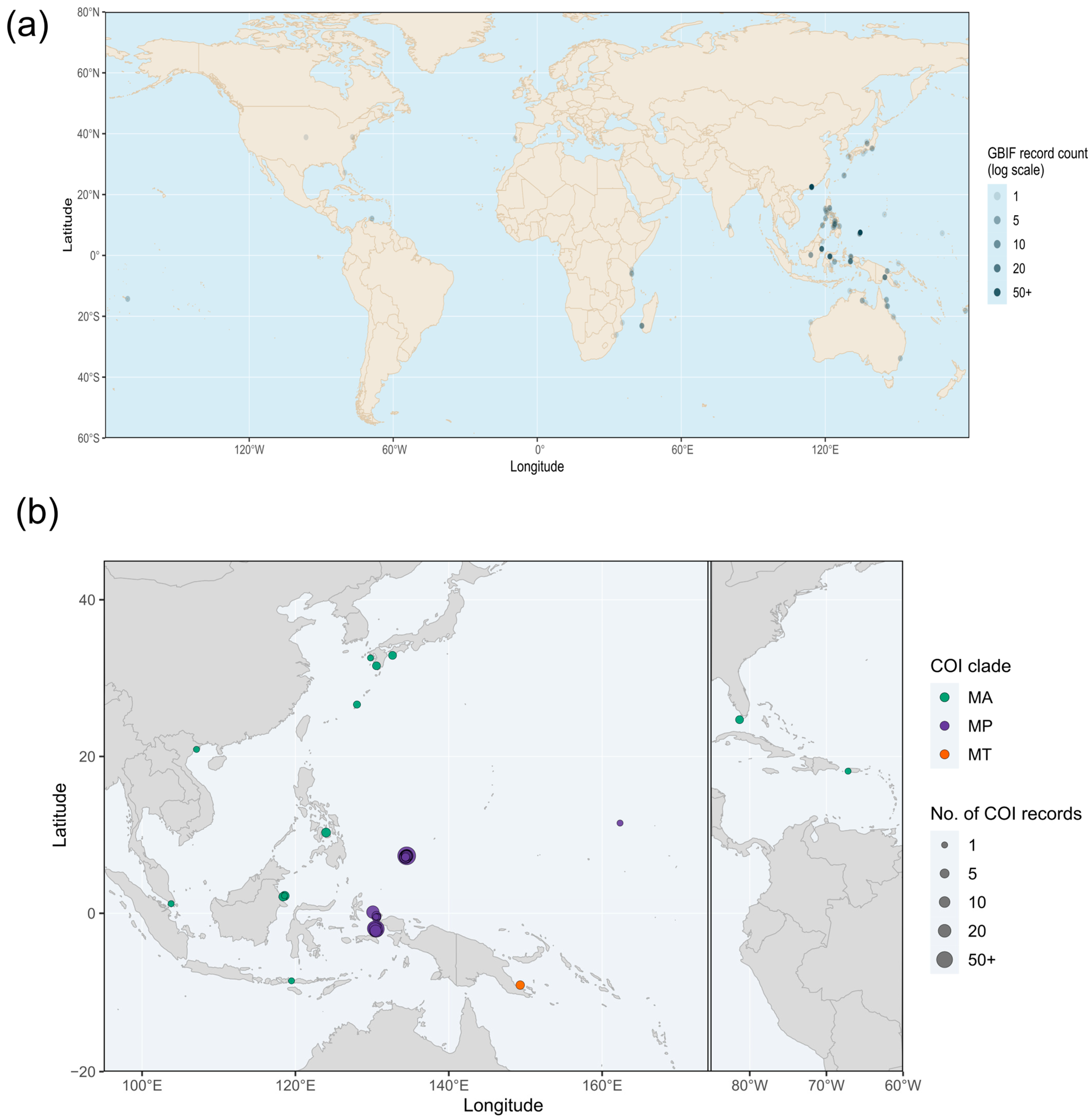
The spotted or golden jellyfish Mastigias Agassiz 1862 is one of two widely distributed genera in the family Mastigiidae (Order Rhizostomeae), the other being Phyllorhiza; a third currently valid genus, Mastigietta Stiasny, 1921, has been reported rarely and remains poorly known. Mastigias are nearshore jellyfishes whose distribution spans much of the tropical and subtropical Indo-Pacific from the Fijian Islands to the western Indian Ocean—with extra-limital reports in Florida and Hawaii—and ranges latitudinally between Japan and Australia [1,2] (Figure 1a). Historically, as many as 10 species have been listed for the genus Mastigias [3]—though for much of the past 50 years, in most of these localities the medusae were inferred to be Mastigias papua [1].
In Japan, Mastigias is called “tako kurage” or “octopus jellyfish” because its hemispherical umbrella and eight long oral arms with appendages give its silhouette the appearance of an octopus. Mastigias is commonly reported in littoral waters along the Pacific coast of Japan (Figure 1a), with historical records from the Ryukyu Islands (Okinawa) in the south and as far north as Mito (Ibaraki) [4,5]. Though originally identified in Japan as M. physophora (see Kishinouye [6]), phenotypic variation witnessed at different localities resulted in several new species assignments, leading to confusion about its identity in Japan [4]. Subsequently, M. physophora was synonymized with M. papua by Maas [7], which became the accepted species name for the ensuing ~110 years until the results of combined morphological and molecular analyses of Mastigias revealed at least three clades in the western Pacific [1]. Of these, one was redescribed as Mastigias papua [8]—the type species for the genus—with type locality Waigeo, West Papua. A second was identified as Mastigias albipunctata Stiasny 1920, originally described from the “Malaiischen Archipels” (Malay Archipelago) [9,10], based on morphological concordance and on the geographic distribution of genotyped specimens spanning Japan, Komodo, Berau and the Philippines. The third clade was identified tentatively as M. andersoni or M. ocellatus in Papua New Guinea [1].
Additional molecular analysis of Mastigias from Kagoshima and Nagasaki, Japan [11], corroborates the placement of these specimens in the clade with M. albipunctata sensu de Souza and Dawson [1]. By contrast, Hamaguchi et al.’s [12] study of umbrella colour and cytomorphology in specimens from Okinawa and Minami-Izu, Japan, retained M. papua sensu lato based on the specimens’ concordance with the morphological traits of that species. Uchida’s [4] extensive study on Mastigias reproduction, development and histomorphology similarly relied on morphology alone, and documented consistent morphological results for specimens collected from Kanagawa, Okinawa (as Loo-Choo Islands), Wakayama (as Kii), and Kyushu (as Kiushu). Here, we use COI phylogenetics, together with two new specimens from Okinawa, to verify whether Mastigias populations from four populations concentrated in southern and western Japan form a single well-supported molecular clade comprising M. albipunctata and map these clade assignments against historical distribution records for the genus to reveal the biogeographic range of the genus globally (Figure 1b). Accordingly, Figure S1 includes the same GBIF occurrence data shown in Figure 1a, while additionally overlaying these sequence-confirmed COI clade localities. Thus, a broader distribution of the genus can be deduced despite limited geographic coverage based on molecular data.

2. Materials and Methods

2.1. Sample Collection and Observation

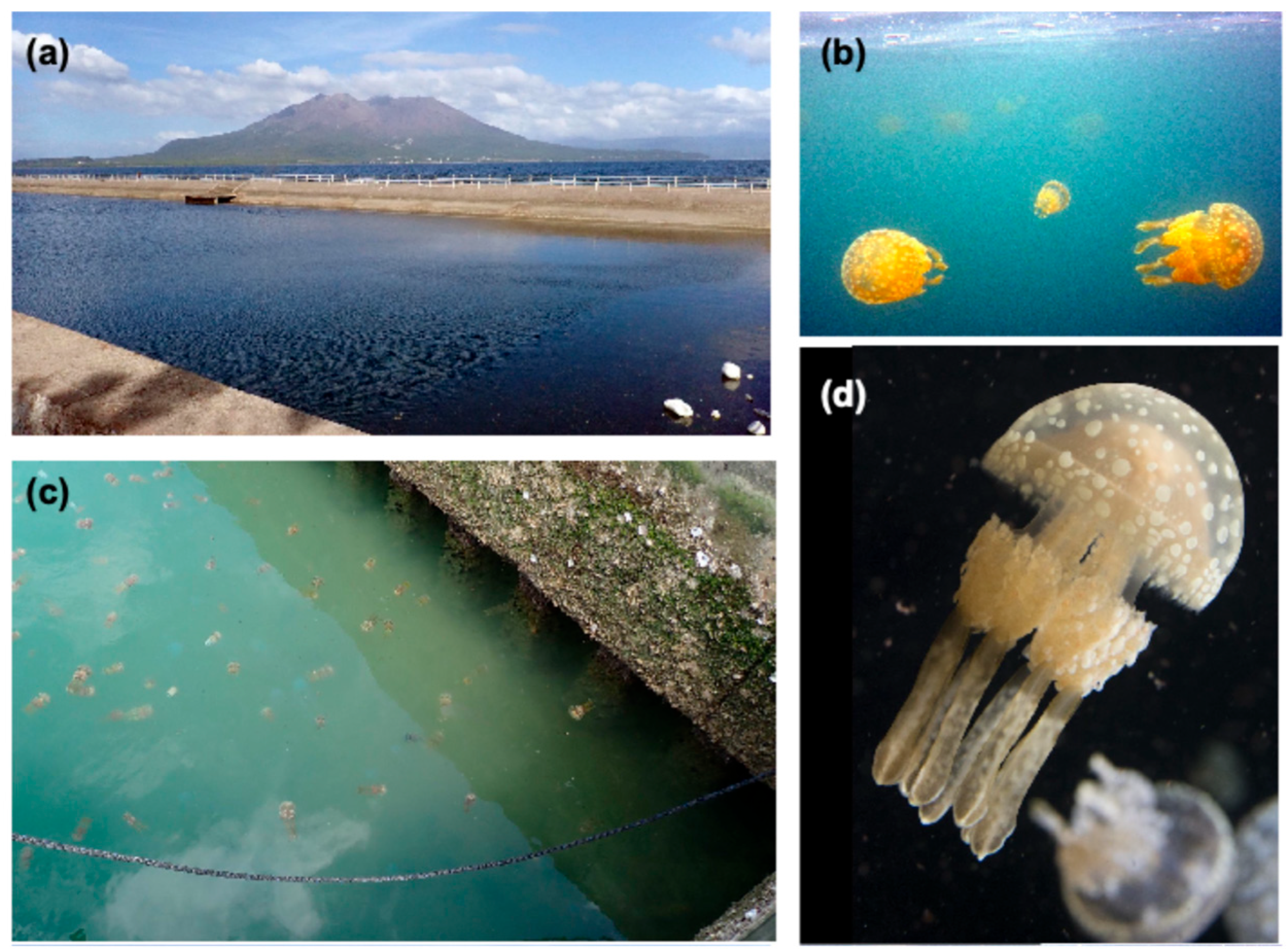
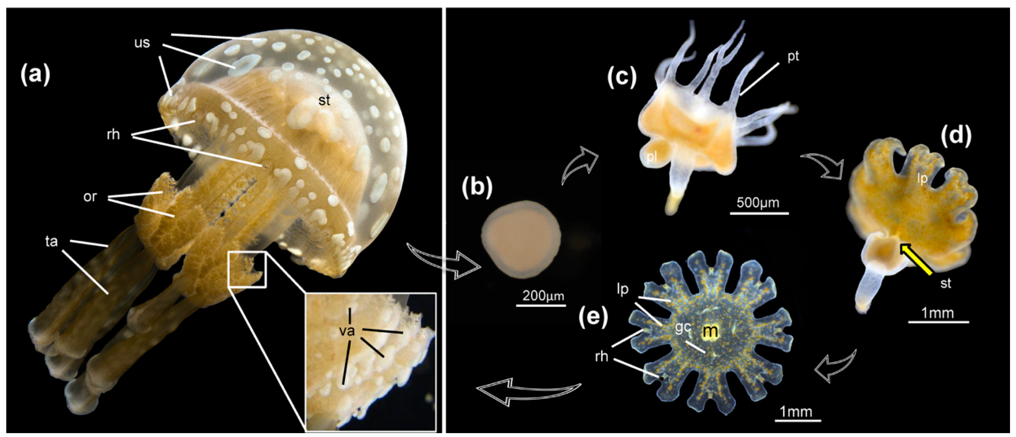
Kagoshima: Mastigias albipunctata medusae were collected on 17 November 2021 at around 10:00 a.m. in a nearshore waterway (aqueduct) of Yojiro, Kagoshima City (Kagoshima) (31.558469, 130.56741) (Figure 2a) using SCUBA and/or skin diving. M. albipunctata medusae swimming at the surface (Figure 2b) were collected in the vicinity at a depth of 2 m. Water temperature 20.9 °C. Live M. albipunctata medusae (Figure 2c) were exhibited at Kagoshima City Aquarium (Kagoshima) or transported to Kamo Aquarium (Tsuruoka, Japan) for public exhibition and rearing. M. albipunctata exhibits a typical rhizostome life cycle: starting as a planula (Figure 3b) that settles to a polyp (Figure 3c), followed by a mono-disc strobila (Figure 3d) metamorphosing into a free-swimming ephyra (Figure 3e) before maturation into a medusa (Figure 2 and Figure 3a).
Okinawa: Additional medusae were collected at Nakaoshi Port, Nago City, (Okinawa) (26.628350, 128.025703) (Figure 2d) on 24 July 2024. Mastigias medusae observed roughly 50 cm below the surface were captured using a bucket with an extended handle.
Tissue samples from the oral arm and umbrella were cut and placed in 1.5 mL Eppendorf tubes filled with 99.5% ethanol for molecular analysis. Samples were kept chilled during transport to the International Marine Sciences Laboratory (Tohoku University) for further processing.

2.2. Morphological Analysis

Medusa umbrella diameter of Mastigias specimens analysed herein varies from 5 to 10 cm and body length from exumbrella apex to tip of the terminal clubs varies from 15 to 20 cm. Additionally, 2 specimens of M. albipunctata of approximate umbrella width 80 mm, collected August 1997, at the mouth of Nasadagawa River, Nago City, Okinawa, by H. Mori, were accessioned into the Tohoku University Museum (accession numbers TUM 112973, TUM 112974) as morphological vouchers for the species.

2.3. Phylogenetic Analysis

DNA was extracted from tissues samples of two Mastigias specimens using the Qiagen DNeasy Blood and Tissue Kit (Qiagen, Hilden, Germany). The COI region was amplified using primers Jellyfish_CO1_F 5′-KKTCAACAAAYCATAAAGATATWGG-3′ and Jellyfish_CO1_R2 5′-GGAACTGCTATWATCATWGTWGC-3′ [13]. Amplification protocols were initial denaturation (95 °C for 5 min) followed by 38 cycles of denaturation (96 °C for 30 s), annealing (46 °C for 40 s), and extension (72 °C for 55 s), with a final extension of 72 °C for 7 min. Amplicon size was validated by electrophoresis on a 1.5% agarose gel and purified using ExoSAP-IT (Applied Biosystems, Waltham, MA, USA) prior to Sanger sequencing on a BigDyeTM Terminator v3.1 Cycle Sequencing Kit (Applied Biosystems, Thermo Fisher Scientific, Tokyo, Japan). All publicly available Mastigias COI sequences were retrieved from GenBank on 28 June 2026, including COI sequences extracted from whole mitochondrial genome assemblies previously deposited on GenBank and the two new samples from Okinawa (n = 515 sequences; accessions, organism names, localities, and coordinates listed in Supplementary Table S2). Two COI sequences of Phyllorhiza punctata were included as an outgroup (Figure 5a). Together with newly accessioned Okinawa sequences (PZ770286, PZ770287), all sequences were aligned using MAFFT [14] and trimmed using trimAl v1.5.1 [15] with the -automated1 heuristic. A maximum-likelihood phylogeny was inferred in IQ-TREE 3 [16], with the best-fit substitution model (HKY+F+I) selected using ModelFinder [17] and branch support assessed with 1000 ultrafast bootstrap replicates (UFBoot2 [18]) alongside SH-aLRT and aBayes support. Trees were visualised and edited on FigTree v1.4.4 [19], iTOL v5 [20] and Inkscape v1.4.4 [21]. To further assess support for the recovered topology, the same alignment was also analysed under Bayesian inference in MrBayes [22,23], under an HKY+I model (Nst = 2 with a proportion-of-invariable-sites rate correction). Two independent MCMC runs, each comprising four chains, were conducted for 10,000,000 generations. Trees and model parameters were sampled every 500 generations, and the first 25% of samples were discarded as burn-in.

2.4. Distribution Mapping

Global Mastigias occurrence records were downloaded from GBIF (Global Biodiversity Information Facility). For the distribution map, records were filtered during download to retain only coordinate-bearing records with present occurrence status and no GBIF-flagged geospatial issues, resulting in 418 mappable records used to construct Figure 1a (GBIF Occurrence Download. Available online: https://doi.org/10.15468/dL.qkcajr (accessed on 16 September 2026)). These GBIF records originated predominantly from direct human observations and lacked vouchers for genetical or morphological validation. They are provided herein exclusively for background context on the genus’s broader reported range. For the distribution mapping of molecular results, COI sequence records were plotted when locality information was informative enough to assign approximate coordinates. Records with country-level localities without a specific collection site were omitted from the map because they could not be placed reliably, which is indicated in Supplementary Table S1.
Maps were produced in R Studio v2026.07.1+147 [24] using ggplot2 v4.0.3 [25] for plotting. Basemap coastline and country polygons were sourced from rnaturalearth v1.2.0 [26] for the GBIF background map and from the maps package v3.4.3 [27] for the clade distribution map; coordinate reference system handling for the GBIF map used sf v1.1.2 [28,29] via coord_sf().

3. Results

Morphological diagnosis: in the genus Mastigias, the subumbrella comprises the gonads which are contiguous with the stomach; the exumbrella is covered in fine nematocysts and bears eight light- and gravity-sensing structures (rhopalia) along the margin. The umbrella is connected by pillars and canals to an oral disc and eight oral arms [12]; from each arm extends a terminal club. Mastigias albipunctata life history is typical of Scyphozoa, i.e., cycling through planula, polyp, strobila, ephyra and medusa stages (Figure 3). The medusae begin at ~1 cm (umbrella diameter) and grow to a maximum of ~20 cm.
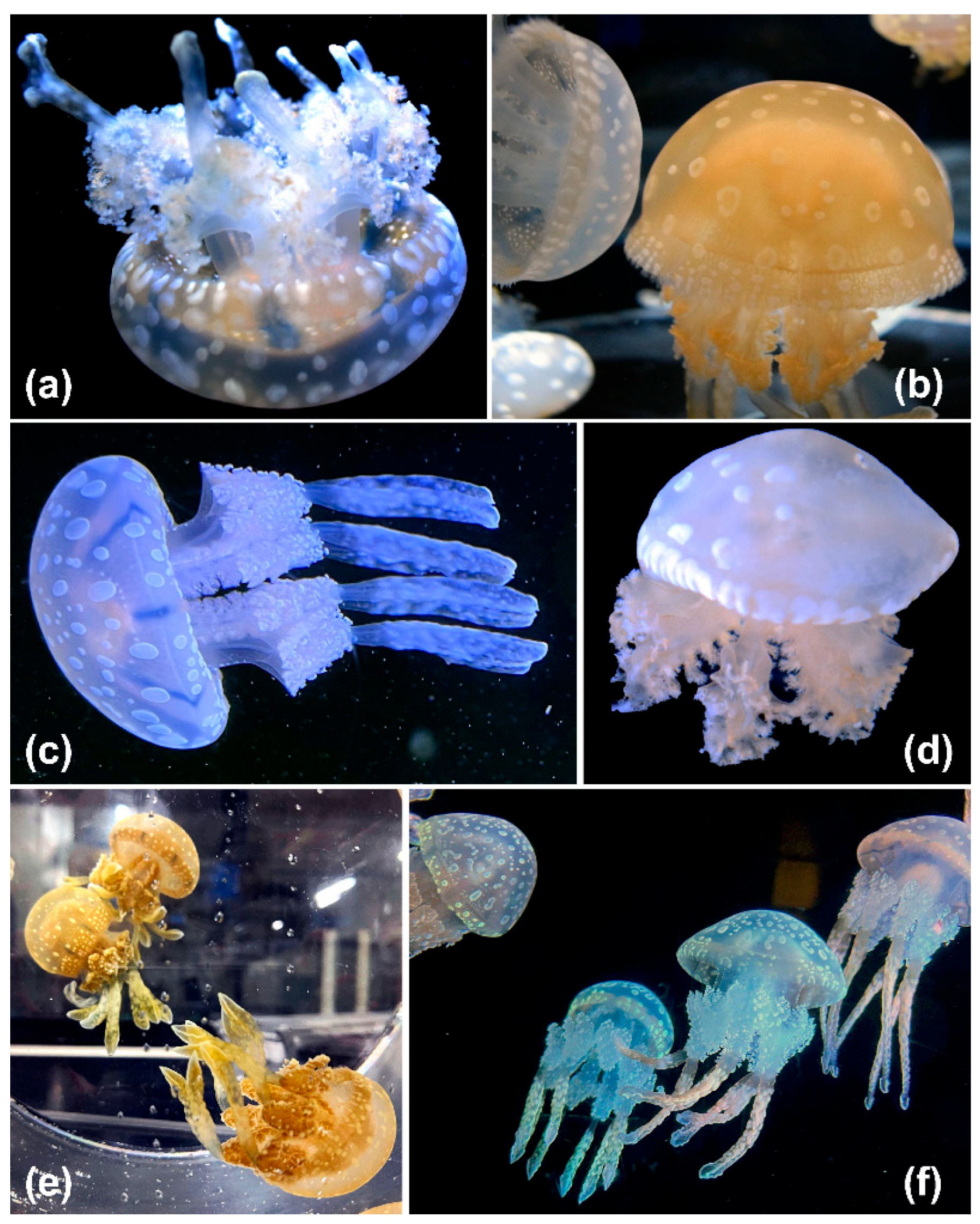
Detailed observation of Mastigias reveals several interesting anatomical features. (1) The oral arms bear “vesicular heads” or “vesiculated appendages”, as illustrated by Uchida [4] (a.k.a. ‘intermediate filaments’ sensu Dawson [30]) and appear to bear clusters of nematocysts which correspond to the motile cellular structures called cassiosomes identified in Mastigias and other rhizostomes [31]. (2) The underlying colour of Mastigias is typically beige but varies from light to dark brown (Figure 4) owing to endosymbiotic Symbiodiniaceae (a.k.a. zooxanthellae) colonising the gastrodermis and mesoglea [12]. (3) Histomorphology studies by Uchida [4] on Mastigias (as M. papua) from various locations in Japan, including Okinawa (as Loo-choo Islands) revealed that Symbiodiniaceae ingested by the medusa pass into the gastrovascular cavity where they are endocytosed by gastrodermal cells which, subsequently, may transport the endosymbionts into the mesoglea via “wandering cells”. (4) Conspicuous spots are found on the exumbrella arranged in various sizes from the apex to the umbrella margin, but no single pattern is prevalent (Figure 3 and Figure 4); umbrellar spots which may be white in juveniles become yellowish as the medusa grows and the spots acquire a ring-like appearance in larger medusae [4]. The exumbrella spots are formed by aggregations of free cells containing reflective granules (putative amoebocytes) in the mesoglea and may play a role in regulating solar radiation conditions for the endosymbionts [12]. (5) Additional colour variations, including blue pigmentation linked to production of rhizostomins pigments [32], may also be related to different stages in the life history of the medusa and its endosymbiotic algae [4] (Figure 3 and Figure 4), though all aforementioned colour differences in the spots and body also occur among populations or within the same population [30].
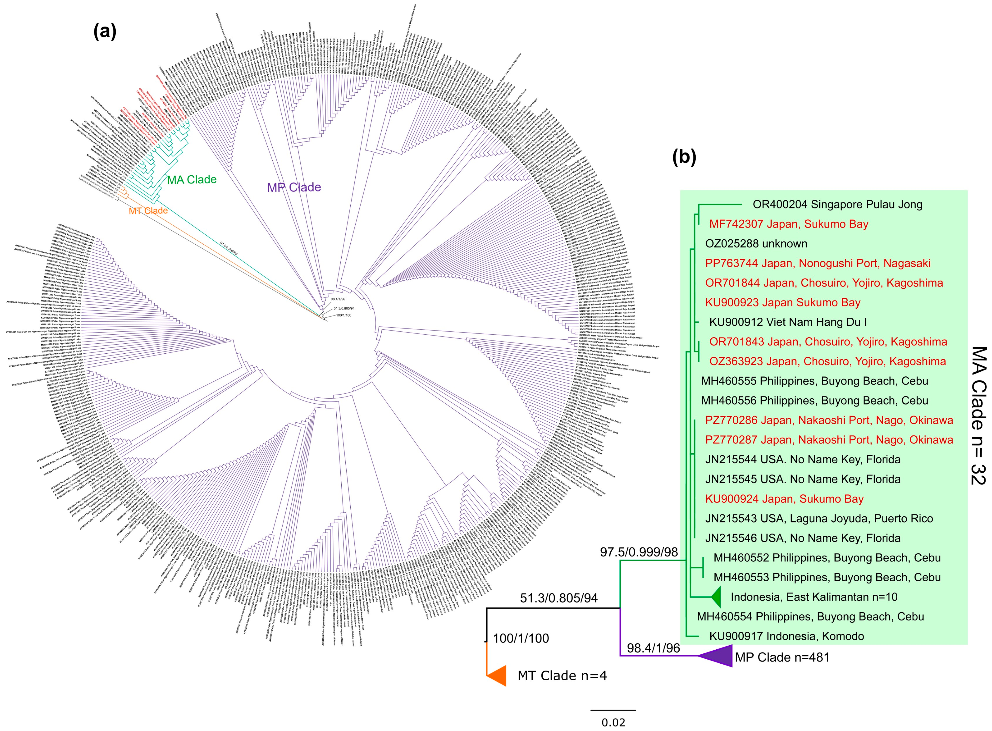
Phylogenetic reconstruction (Figure 5a,b) recovered three clades within Mastigias, consistent with de Souza and Dawson [1]: a strongly supported M. papua clade (MP; SH-aLRT/aBayes/UFBoot = 98.4/1.0/96, posterior probability = 1.0), a strongly supported M. albipunctata clade (MA; SH-aLRT/aBayes/UFBoot = 97.5/0.999/98; posterior probability = 1.0), and a weakly resolved third lineage (MT) corresponding to samples from Tufi, Papua New Guinea, consistent with the tentative M. andersoni/M. ocellatus assignment of de Souza and Dawson [1]; internal relationships among the Tufi/IDORTUF samples themselves were poorly resolved (SH-aLRT/aBayes/UFBoot as low as 0/0.33–0.38/28–41), mirroring the limited resolution de Souza and Dawson [1] also reported for this lineage. The split separating the MT clade from MA and MP was maximally supported (SH-aLRT/aBayes/UFBoot = 100/1.0/100; posterior probability = 0.9995). MA and MP were each recovered as strongly supported, reciprocally monophyletic sister clades, though support for their specific sister relationship was ambiguous (SH-aLRT/aBayes/UFBoot = 51.3/0.805/94). Of 517 Mastigias sequences analysed, 481 fell within MP, 32 within MA, and 4 within MT (Supplementary Table S1). All nine Japanese sequences—from Sukumo Bay (n = 3), Chosuiro/Yojiro, Kagoshima City (n = 3), Nonogushi Port, Nagasaki (n = 1), and Nakaoshi Port, Okinawa (n = 2, comprising the two new specimens collected in this study)—fell within the MA clade, extending the confirmed molecular range of M. albipunctata to the southernmost Ryukyu Islands.
Both MrBayes runs converged, reaching a final average standard deviation of split frequencies of 0.0176, an average potential scale reduction factor (PSRF) of 1.0002 (maximum PSRF = 1.0005), and estimated sample sizes (ESSs) for all model parameters exceeding 5300 (range: 5324–13,431). Together, both molecular results support the conclusion that Mastigias from southern to western Japan is M. albipunctata.

4. Discussion

The COI phylogeny recovered Japanese Mastigias, from Sukumo Bay in the north to Okinawa in the south, as a single, well-supported clade corresponding to M. albipunctata rather than M. papua, sister to the M. papua clade with high and near-maximal support. Historically, Japanese Mastigias was identified by morphology alone, first as a distinct species [6], then under the broader designation as M. papua for at least a century [4,7]. An additional study from Hamaguchi et al. [12] referred to specimens from Okinawa and Minami-Izu as M. papua based on arbitrary measures of umbrella hardness and cytomorphology. At the time of the previous studies, no reference molecular data existed for specimens from Japan, making it difficult to validate M. albipunctata as a species distinct form M. papua. Since de Souza and Dawson’s [1] molecular phylogenetic reconstruction of the genus, evidence began to accumulate suggesting that Japanese Mastigias belonged instead to the M. albipunctata clade: de Souza and Dawson [1] contributed a Japanese sequence (Sukumo Bay, Shikoku Island) within this clade, and Tan et al. [11] similarly placed Kagoshima and Nagasaki specimens there. The latter was part of a broader study of Symbiodiniaceae associations rather than a targeted phylogenetic assessment of Mastigias phylogeography. The present study extends this accumulating molecular evidence by adding the first molecular confirmation from Okinawa, which is the southernmost extent of the species’ historically reported range in Japan, using an extensive COI dataset reflecting the most recent publicly available sequences. These findings confidently confirm that Mastigias in western to southern Japan, from Sukumo Bay to Okinawa, corresponds consistently to M. albipunctata. Given the large geographic distance between the new Okinawa sampling site for M. albipunctata in this study and the Nagasaki sampling location reported in Tan et al. [11] the lack of molecular divergence supports a broad subtropical distribution range in Japan.
Beyond phylogenetic placement, morphological comparison lends partial support to this identification. Uchida [4] reported 80 total lappets per medusa (16 ephyral, 64 velar) in specimens from Kanagawa, Wakayama, Kyushu and Okinawa, equating to 8 velar lappets per octant—a value that aligns more closely with the historical diagnostic velar lappet count for M. albipunctatus (8 [9], as tabulated in de Souza and Dawson [1]) than with that of true M. papua (6 [8]). De Souza and Dawson’s [1] own measurements of a Japanese specimen from Sukumo Bay similarly yielded 7–9 velar lappets per octant, consistent with this pattern. However, the same specimen’s oral arm length (0.36–0.45 umbrella diameters) fell well short of the elongated oral arms considered diagnostic of M. albipunctata (~1 umbrella diameter), and its adradial gastrovascular canal origins (6–8) did not clearly align with the historical M. albipunctata value (12–14) either. Morphological support for M. albipunctata as the Japanese Mastigias (tako kurage) is partially character-dependent; however, de Souza and Dawson [1] reported that although Japanese sequences failed to cluster with all M. albipunctata specimens in their morphospace analysis, molecular phylogenetics served as a primary line of evidence supporting this identification, with morphology offering corroborating rather than independently sufficient support.
Independent molecular reference data for M. albipunctata reported the complete mitochondrial genome [33] of M. albipunctata from a polyp culture maintained at the Smithsonian National Museum of Natural History and further reassigned a previously mislabelled GenBank sequence (OZ025288, originally deposited as M. papua) to M. albipunctata based on phylogenetic analysis. However, as the culture’s original wild-collection locality cannot be ascertained, this mitogenome cannot be used to confirm the species’ geographic distribution within Japan.
Such observations of phenotypic attributes that may be important in the relationship Mastigias forms with endosymbiotic Symbiodiniaceae are of relevance to the Aquatic Symbiosis Genomics Project [34]. Additional details include that the single-celled algae are acquired anew each generation when a symbiont is engulfed by a polyp [35]. Uchida [4] reported that during mid-summer (July to August), ephyrae were witnessed swimming at the surface at midday with their oral side down, revealing their daily light requirement for carrying out photosynthesis. Initiating this photosymbiotic relationship is considered necessary for asexual reproduction via strobilation, along with specific temperature requirements [36]. Reports show that ephyrae strobilate from polyps at sea water temperatures of 22 °C in situ and 20–22 °C in the lab and that strobilation is induced within days of providing Symbiodiniaceae for polyps to ingest [36]. Though several genera of Symbiodiniaceae are known to form photosymbioses with rhizostome jellyfishes [37,38] preliminary sequencing suggests the primary photosymbiont of Mastigias is Cladocopium (a.k.a. Clade C [11,39]). Mastigias papua is known to be a prey item of the sea anemone Entacmaea medusivora, which retains the ingested endosymbionts Cladocopium (Symbiodiniaceae) for several days before releasing them intact within its faecal pellets [40].
Mastigias is regularly displayed in aquaria around the world due to its relative availability and the ease of maintaining the species in tanks; their lack of tentacles means there is no risk of entanglement damaging the medusae. For example, (1) in Kagoshima (Kyushu), Mastigias medusae occur in sufficient abundance during annual autumn blooms that news stories caution people about jellyfish stings [41], but it remains difficult to predict abundance from year to year given changes in environments inhabited by planulae, polyps, ephyrae and medusae respectively [42]. (2) Species of jellyfish bearing endosymbiotic algae may provide ecosystem services such as the early detection and mitigation of phytoplankton and zooplankton blooms in eutrophic areas by top-down food web control [43], while also serving as a valuable food source for some countries [44]. (3) Mastigias are commensal, e.g., with caridean shrimps in Aburatsubo Cove (Kanagawa) [4] and in Tanabe Bay (Wakayama) [5], possibly providing protection, shelter, food or mobility for the shrimp.

5. Conclusions

This combined molecular and morphological study validates the identity of Mastigias distributed in Japan from Sukumo Bay (Shikoku Island) to Okinawa Island definitively as M. albipunctata, extending confirmation to the southernmost limit of the species’ historical range in Japan. This conclusion rests primarily on COI phylogenetics, corroborated by partial morphological agreement with de Souza and Dawson’s [1] diagnostic characters. Though the findings rest on the analysis of one gene target, the mitochondrial COI gene is known as a rapidly evolving region in metazoans; as such, it consistently proves its utility in delineating Medusozoa species. In addition to accessioning the molecular sequences for M. albipuncatata into NCBI GenBank, we also accessioned two specimens collected from Okinawa into the Tohoku University Museum as morphological reference vouchers.
The northernmost reports in the literature and in GBIF document Mastigias in Japan are from 35° N and 37° N on the Pacific Ocean and Sea of Japan respectively. Future targeted sampling of specimens across different seasons and geographic regions would allow the full diagnostic character set to be assessed directly against other curated museum specimens. This study serves as the foundation for establishing an systematic approach to broaden understanding of the biogeography of M. albipunctata in Japan.

Supplementary Materials

The following supporting information can be downloaded at: https://doi.org/10.5281/zenodo.21793244. Figure S1. Combined distribution map presenting overlaying GBIF distribution records confirmed with COI sequence data. The composite map illustrates the limited geographic coverage of the genus Mastigias when relying exclusively on molecular sequence validation relative to the broader literature based on visual confirmation; Table S1: GBIF occurrence records used for Figure 1a; Table S2: GenBank accessions, organism names, localities, coordinates, voucher/sample IDs, and assigned COI clade for all sequences used in the phylogenetic analysis.

Author Contributions

Conceptualization, C.L.A., K.C.T.; methodology, C.L.A., K.C.T.; validation, C.L.A., K.C.T., M.C., S.I., R.T., K.O.; formal analysis, K.C.T.; investigation, C.L.A., K.C.T., M.C., S.I., R.T., K.O. resources, C.L.A., M.C., S.I., R.T., K.O.; data curation, K.C.T.; writing—original draft preparation, C.L.A., K.C.T.; writing—review and editing, C.L.A., K.C.T., M.C., S.I., R.T., K.O.; visualisation, C.L.A., K.C.T., M.C., S.I. and R.T.; supervision, C.L.A., K.O.; project administration, C.L.A.; funding acquisition, C.L.A. All authors have read and agreed to the published version of the manuscript.

Funding

This research was supported by the World Premier International Research Center Initiative–Advanced Institute for Marine Ecosystem Change (WPI-AIMEC), Tohoku University, funded by the Ministry of Education, Culture, Sports, Science and Technology (MEXT) of Japan.

Institutional Review Board Statement

Not applicable. This study did not involve human participants. Mastigias albipunctata Stiasny, 1920 is a scyphozoan jellyfish (Cnidaria), and is not listed as an endangered or protected species, hence, institutional ethical approval was therefore not required.

Informed Consent Statement

Not applicable.

Data Availability Statement

The COI sequences generated in this study are available in GenBank under accession numbers PZ770286 (OM01) and PZ770287 (OM02). All other COI sequences analysed were retrieved from GenBank as detailed in Table S1. GBIF Occurrence Download. Available online: https://doi.org/10.15468/dL.qkcajr (mappable records) and https://doi.org/10.15468/dL.zdb4fe (full download) (accessed on 16 September 2026). Sequence alignments, trimmed alignments, and phylogenetic tree files are available at https://doi.org/10.5281/zenodo.21793244.

Acknowledgments

We are grateful to two anonymous reviewers whose advice helped us improve this work.

Conflicts of Interest

Author Mitsuko Chikuchishin was employed by the company Kagoshima City Aquarium. Author Shuhei Ikeda was employed by the company Kamo Aquarium, Tsuruoka. The remaining authors declare that the research was conducted in the absence of any commercial or financial relationships that could be construed as a potential conflict of interest.

References

  1. Souza, M.R.D.; Dawson, M.N. Redescription of Mastigias papua (Scyphozoa, Rhizostomeae) with Designation of a Neotype and Recognition of Two Additional Species. Zootaxa 2018, 4457, 520–536. [Google Scholar] [CrossRef] [Scilit] [PubMed]
  2. Mohd Syazwan, W.; Low, L.B. Mohammed Rizman-Idid First Record in Peninsular Malaysia and Morphological Redescription of Lychnorhiza Malayensis (Scyphozoa: Rhizostomeae: Lychnorhizidae). Raffles Bull. Zool. 2020, 68, 3249. [Google Scholar] [CrossRef]
  3. Kramp, P.L. Synopsis of the Medusae of the World. J. Mar. Biol. Assoc. United Kingd. 1961, 40, 7–382. [Google Scholar] [CrossRef] [Scilit]
  4. Uchida, T. The Anatomy and Development of a Rhizostome Medusa, Mastigias papua L. Agassiz, with Observations on the Phylogeny of Rhizostomae. J. Fac. Sci. Univ. Tokyo 1926, 4, 45–95. [Google Scholar]
  5. Hayashi, K.; Miyake, S. Three Caridean Shrimps Associated With A Medusa From Tanabe Bay, Japan. Publ. SMBL 1968, 16, 11–19. [Google Scholar] [CrossRef] [Scilit] [PubMed][Green Version]
  6. Kishinouye, K. Description of a New Rhizostoma Mastigias physophora, Nov. sp. Zool. Mag. (Dobutsugaku Zasshi) 1895, 7, 86–88. [Google Scholar]
  7. Maas, O. Japanische Medusen. In Beiträge Zur Naturgeschichte Ostasiens; Abhandlungen Der Mathematisch-Physikalischen Klasse Der Königlich Bayerischen; Akademie der Wissenschaften: Munich, Germany, 1909; pp. 1–52. [Google Scholar]
  8. Lesson, R.P. Voyage Autour Du Monde: Exécuté Par Ordre Du Roi, Sur La Corvette de Sa Majesté, La Coquille, Pendant Les Années 1822, 1823, 1824, et 1825; Arthus Bertrand: Paris, France, 1830; Zoologie; Volume 2, Part 1. [Google Scholar]
  9. Stiasny, G. Die Scyphomedusen-Sammlung des Naturhistorischen Reichsmuseums in Leiden: III. Rhizostomae. Zool. Meded. 1920, 5, 213–230. [Google Scholar]
  10. Stiasny, G. Studien Über Rhizostomeen Mit Besonderer Berücksichtigung Der Fauna Des Malayischen Archipels Nebst Eine Revision Des Systems. Capita Zool. 1921, 1, 1–179. [Google Scholar]
  11. Tan, K.C.; Chikuchishin, M.; Ikeda, S.; Tamada, R.; Okuizumi, K.; Nishitani, G.; Ikeda, M.; Ames, C.L. A Comparative Molecular Study of Rhizostome Jellyfishes (Cnidaria, Scyphozoa, Rhizostomeae) from Japan Reveals Variability in Symbiodiniaceae Taxon Associations and Cassiosome Production. Front. Mar. Sci. 2026, 12, 1679299. [Google Scholar] [CrossRef] [Scilit]
  12. Hamaguchi, Y.; Iida, A.; Nishikawa, J.; Hirose, E. Umbrella of Mastigias papua (Scyphozoa: Rhizostomeae: Mastigiidae): Hardness and Cytomorphology with Remarks on Colors. Plankton Benthos Res. 2021, 16, 221–227. [Google Scholar] [CrossRef] [Scilit]
  13. Minamoto, T.; Fukuda, M.; Katsuhara, K.R.; Fujiwara, A.; Hidaka, S.; Yamamoto, S.; Takahashi, K.; Masuda, R. Environmental DNA Reflects Spatial and Temporal Jellyfish Distribution. PLoS ONE 2017, 12, e0173073. [Google Scholar] [CrossRef] [Scilit] [PubMed]
  14. Katoh, K.; Standley, D.M. MAFFT Multiple Sequence Alignment Software Version 7: Improvements in Performance and Usability. Mol. Biol. Evol. 2013, 30, 772–780. [Google Scholar] [CrossRef] [Scilit] [PubMed]
  15. Capella-Gutiérrez, S.; Silla-Martínez, J.M.; Gabaldón, T. trimAl: A Tool for Automated Alignment Trimming in Large-Scale Phylogenetic Analyses. Bioinformatics 2009, 25, 1972–1973. [Google Scholar] [CrossRef] [Scilit] [PubMed]
  16. Wong, T.K.F.; Ly-Trong, N.; Ren, H.; Demotte, P.; Baños, H.; Roger, A.J.; Susko, E.; Bielow, C.; Maio, N.D.; Goldman, N.; et al. IQ-TREE 3: Phylogenomic Inference Software Using Complex Evolutionary Models. Mol. Biol. Evol. 2026, 43, msag117. [Google Scholar] [CrossRef] [Scilit] [PubMed]
  17. Kalyaanamoorthy, S.; Minh, B.Q.; Wong, T.K.F.; von Haeseler, A.; Jermiin, L.S. ModelFinder: Fast Model Selection for Accurate Phylogenetic Estimates. Nat. Methods 2017, 14, 587–589. [Google Scholar] [CrossRef] [Scilit] [PubMed]
  18. Hoang, D.T.; Chernomor, O.; von Haeseler, A.; Minh, B.Q.; Vinh, L.S. UFBoot2: Improving the Ultrafast Bootstrap Approximation. Mol. Biol. Evol. 2018, 35, 518–522. [Google Scholar] [CrossRef] [Scilit] [PubMed]
  19. Rambaut, A. FigTree: Tree Figure Drawing Tool, v1.4.4; University of Edinburgh: Edinburgh, UK, 2018.
  20. Letunic, I.; Bork, P. Interactive Tree of Life (iTOL) v6: Recent Updates to the Phylogenetic Tree Display and Annotation Tool. Nucleic Acids Res. 2024, 52, W78–W82. [Google Scholar] [CrossRef] [Scilit] [PubMed]
  21. Inkscape Project. Inkscape, v.1.4.4; Inkscape Team: Brooklyn, NY, USA, 2026.
  22. Ronquist, F.; Huelsenbeck, J.P. MrBayes 3: Bayesian Phylogenetic Inference under Mixed Models. Bioinformatics 2003, 19, 1572–1574. [Google Scholar] [CrossRef] [Scilit] [PubMed]
  23. Ronquist, F.; Teslenko, M.; Van Der Mark, P.; Ayres, D.L.; Darling, A.; Höhna, S.; Larget, B.; Liu, L.; Suchard, M.A.; Huelsenbeck, J.P. MrBayes 3.2: Efficient Bayesian Phylogenetic Inference and Model Choice Across a Large Model Space. Syst. Biol. 2012, 61, 539–542. [Google Scholar] [CrossRef] [Scilit] [PubMed]
  24. R Core Team. R: A Language and Environment for Statistical Computing; R Foundation for Statistical Computing: Vienna, Austria, 2026. [Google Scholar]
  25. Wickham, H. Ggplot2: Elegant Graphics for Data Analysis; Springer International Publishing: Cham, Switzerland, 2016; ISBN 978-3-319-24275-0. [Google Scholar]
  26. Massicotte, P.; South, A. Rnatualearth: World Map Data from Natural Earth. 2026. Available online: https://cran.r-project.org/web/packages/rnaturalearth/index.html (accessed on 16 September 2026).
  27. Becker, R.A.; Wilks, A.R.; Brownrigg, R.; Minka, T.P.; Deckmyn, A. Maps: Draw Geographical Maps. 2025. Available online: https://cran.r-project.org/web/packages/maps/index.html (accessed on 16 September 2026).
  28. Pebesma, E. Simple Features for R: Standardized Support for Spatial Vector Data. R J. 2018, 10, 439. [Google Scholar] [CrossRef] [Scilit]
  29. Pebesma, E.; Bivand, R. Spatial Data Science: With Applications in R, 1st ed.; Chapman and Hall/CRC: Boca Raton, FL, USA, 2023; ISBN 978-0-429-45901-6. [Google Scholar]
  30. Dawson, M.N. Morphological Variation and Systematics in the Scyphozoa: Mastigias (Rhizostomeae, Mastigiidae)—A Golden Unstandard? Hydrobiologia 2005, 537, 185–206. [Google Scholar] [CrossRef] [Scilit]
  31. Ames, C.L.; Klompen, A.M.L.; Badhiwala, K.; Muffett, K.; Reft, A.J.; Kumar, M.; Janssen, J.D.; Schultzhaus, J.N.; Field, L.D.; Muroski, M.E.; et al. Cassiosomes Are Stinging-Cell Structures in the Mucus of the Upside-down Jellyfish Cassiopea xamachana. Commun. Biol. 2020, 3, 67. [Google Scholar] [CrossRef] [Scilit] [PubMed]
  32. Lawley, J.W.; Carroll, A.R.; McDougall, C. Rhizostomins: A Novel Pigment Family From Rhizostome Jellyfish (Cnidaria, Scyphozoa). Front. Mar. Sci. 2021, 8, 752949. [Google Scholar] [CrossRef] [Scilit]
  33. Tan, K.C.; Ames, C.L.; Collins, A.G. Complete Linear Mitochondrial Genomes for Cephea cephea and Mastigias albipunctata (Scyphozoa: Rhizostomeae), with an Analysis of Phylogenetic Relationships. Mitochondrial DNA Part B 2024, 9, 1544–1548. [Google Scholar] [CrossRef] [Scilit] [PubMed]
  34. McKenna, V.; Archibald, J.M. The Aquatic Symbiosis Genomics Project: Probing the Evolution of Symbiosis across the Tree of Life. Wellcome Open Res. 2021, 6, 254. [Google Scholar] [CrossRef] [Scilit]
  35. Sugiura, Y. On the Life-History of Rhizostome Medusae II. Indispensability of Zooxanthellae for Strobilation in Mastigias papua. Embryologia 1964, 8, 223–233. [Google Scholar] [CrossRef] [Scilit]
  36. Sugiura, Y. On the Life-History of Rhizostome Medusae. III. On the Effects of Temperature on the Strobilation of Mastigias papua. Biol. Bull. 1965, 128, 493–496. [Google Scholar] [CrossRef] [Scilit]
  37. LaJeunesse, T.C.; Parkinson, J.E.; Gabrielson, P.W.; Jeong, H.J.; Reimer, J.D.; Voolstra, C.R.; Santos, S.R. Systematic Revision of Symbiodiniaceae Highlights the Antiquity and Diversity of Coral Endosymbionts. Curr. Biol. 2018, 28, 2570–2580.e6. [Google Scholar] [CrossRef] [Scilit] [PubMed]
  38. Djeghri, N.; Pondaven, P.; Stibor, H.; Dawson, M.N. Review of the Diversity, Traits, and Ecology of Zooxanthellate Jellyfishes. Mar. Biol. 2019, 166, 147. [Google Scholar] [CrossRef] [Scilit]
  39. Santos, S.R.; Taylor, D.J.; Kinzie, I.; Hidaka, M.; Sakai, K.; Coffroth, M.A. Molecular Phylogeny of Symbiotic Dinoflagellates Inferred from Partial Chloroplast Large Subunit (23S)-rDNA Sequences. Mol. Phylogenetics Evol. 2002, 23, 97–111. [Google Scholar] [CrossRef] [Scilit] [PubMed]
  40. Vega de Luna, F.; Dang, K.-V.; Cardol, M.; Roberty, S.; Cardol, P. Photosynthetic Capacity of the Endosymbiotic Dinoflagellate Cladocopium sp. Is Preserved during Digestion of Its Jellyfish Host Mastigias papua by the Anemone Entacmaea medusivora. FEMS Microbiol. Ecol. 2019, 95, fiz141. [Google Scholar] [CrossRef] [Scilit] [PubMed]
  41. Shimazaki, A. Jellyfish Swarm in Kagoshima in Odd, out of Season Appearance. Asahi Shinbun, 8 November 2021.
  42. Fernández-Alías, A.; Marcos, C.; Pérez-Ruzafa, A. The Unpredictability of Scyphozoan Jellyfish Blooms. Front. Mar. Sci. 2024, 11, 1349956. [Google Scholar] [CrossRef] [Scilit]
  43. Pérez-Ruzafa, A.; Gilabert, J.; Gutiérrez, J.M.; Fernández, A.I.; Marcos, C.; Sabah, S. Evidence of a Planktonic Food Web Response to Changes in Nutrient Input Dynamics in the Mar Menor Coastal Lagoon, Spain. In Nutrients and Eutrophication in Estuaries and Coastal Waters; Orive, E., Elliott, M., De Jonge, V.N., Eds.; Springer Netherlands: Dordrecht, The Netherlands, 2002; pp. 359–369. ISBN 978-90-481-6123-2. [Google Scholar]
  44. Kitamura, M.; Omori, M. Synopsis of Edible Jellyfishes Collected from Southeast Asia, with Notes on Jellyfish Fisheries. Plankton Benthos Res. 2010, 5, 106–118. [Google Scholar] [CrossRef] [Scilit]
Figure 1. Geographic distribution of Mastigias relevant to this study. (a) Global distribution of Mastigias based on GBIF occurrence records. Only coordinate-bearing records with present occurrence status and no GBIF-flagged geospatial issues were mapped, resulting in 418 mappable records. Circles indicate GBIF occurrence records, with colour intensity showing log-scaled GBIF record count at the same rounded coordinate. (b) Geographic distribution of sequence-confirmed Mastigias COI clades included in this study. Circles are coloured according to COI clade assignment: MA (M. albipunctata; green), MP (M. papua; purple) and MT (cf. M. andersoni or M. ocellatus; orange). Circle size is scaled by the square-root-transformed number of COI records per locality/clade group to improve visibility of differences among localities. Records with overly broad locality information were shown in Supplementary Table S1 and plotted in Figure S1 allowing direct comparison between the broader reported distribution of the genus and the more limited coverage validated by molecular sequence data.
Oceans 07 00080 g001
Figure 2. Collection site in situ images of Mastigias albipunctata in this study. (a) Nearshore waterway (aqueduct) of Yojiro, Kagoshima City where medusae were collected. (b) M. albipunctata medusae swimming at the surface. (c) Nakaoshi Port, Nago City, Okinawa where medusae were collected. (d) M. albipunctata in this study on exhibit at Kamo Aquarium, Tsuruoka (Yamagata, Japan).
Oceans 07 00080 g002
Figure 3. Life cycle of Mastigias albipunctata photographed at Kamo Aquarium, Tsuruoka City (Yamagata, Japan). (a) Morphology of a M. albipunctata medusa. The magnified region shows the vesicular appendages (va; a.k.a. intermediate filaments) covering the oral arms. Abbreviations: us, umbrella spots (numerous); rh, rhopalia (8 total); or, oral arms (8 total); ta, terminal appendages (8 total); st, stomach. (b) A planula released into the water from the brood filaments on the oral disc of the female after fertilisation. (c) A polyp bearing multiple polyp tentacles (pt) orally, whose apical portion will undergo metamorphosis following ingestion of Symbiodiniaceae; a planuloid bud (pl) is released asexually by lateral fission from the base of the polyp that will swim away and settle on the substrate to become another polyp. (d) A mono-disc strobila bearing 8 terminally bifurcating lappets (lp) whose apical end will soon be released from the stalk (st) as a free-swimming ephyra. Colonised Symbiodiniaceae are visible as orange-brown spots throughout the strobila. (e) A free-swimming ephyra with 8 lappets (lp) and rhopalia (rh) at the terminal bifurcations, bearing Symbiodiniaceae throughout the body. Early development of the gastrovascular structures present in medusae are visible as the start of the central mouth (m) and one gastric cirrus per quadrant (gc). According to Uchida (1926) [4] this metagenetic life cycle of M. albipunctata begins in the summer to autumn season (mid-July to October) in Aburatsubo Cove at the tip of the Miura Peninsula (Kanagawa, Japan). The following summer, polyps modify into the strobila form, releasing a free-swimming ephyra that will develop into a juvenile medusa within about 20 days.
Oceans 07 00080 g003
Figure 4. Variation in colour and spot patterns of Mastigias albipunctata photographed on exhibit in Kamo Aquarium, Tsuruoka City (Yamagata, Japan). (a) A medusa (~6 cm umbrella diameter) with primarily beige coloration but bearing blue pigment along the radial canals that terminates at each marginal rhopalium. (b) Two medusae (~6 cm umbrella diameters), one with a white umbrella trimmed by beige along the margin bearing small spots on the umbrella apex and larger spots along the margin (left) reared together with a fully orange-beige medusae with ring-like spots on the apex and smaller patches along the margin (right). (c) A fully light blue medusa (~8 cm umbrella diameter) with pronounced darker blue along the radial canals. (d) A mostly white medusa (~6 cm) with some orange hue in the oral arms. (e) M. albipunctata from Nagasaki, reared for morphological observation (sequences were published previously as PV539587) and photographed under natural light at Tohoku University. (f) M. albipunctata from Okinawa, photographed in an aquarium under artificial light on display at Churaumi Aquarium.
Oceans 07 00080 g004
Figure 5. (a) Full and (b) collapsed COI Maximum Likelihood tree of Mastigias. Branch lengths represent substitutions per site, and coloured branches denote the major clades MP (M. papua; purple), MA (M. albipunctata; green) and MT (cf. M. andersoni or M. ocellatus; orange). Tip labels for samples of Japanese origin are highlighted in red. In (b), selected monophyletic groups containing closely related sequences from the same broad geographic region were collapsed for clarity and labelled with the region and the number of sequences represented. Uncollapsed tips are labelled with accession number and locality. Internal node labels indicate support values in the order SH-aLRT/aBayes/ultrafast bootstrap support. The tree was rooted using two Phyllorhiza punctata outgroup sequences, which were omitted from the displayed tree for clarity.
Oceans 07 00080 g005
Disclaimer/Publisher’s Note: The statements, opinions and data contained in all publications are solely those of the individual author(s) and contributor(s) and not of MDPI and/or the editor(s). MDPI and/or the editor(s) disclaim responsibility for any injury to people or property resulting from any ideas, methods, instructions or products referred to in the content.