Article

Masticatory Efficacy Following Implant Rehabilitation: Objective Assessment and Patient Perception Through Two-Color Mixing Test and Viewgum® Software

by
José María Montoya-Carralero
,
Arturo Sánchez-Pérez
*,
Alba Sánchez-Olaya
,
Alfonso Jornet-García
and
María José Moya-Villaescusa
Department of Periodontology, Medicine and Dentistry Faculty, Murcia University, 30008 Murcia, Spain
*
Author to whom correspondence should be addressed.
Prosthesis 2025, 7(4), 70; https://doi.org/10.3390/prosthesis7040070
Submission received: 11 May 2025 / Revised: 10 June 2025 / Accepted: 20 June 2025 / Published: 24 June 2025
(This article belongs to the Section Prosthodontics)

Abstract

Background: Dental implants enhance masticatory efficiency in edentulous patients, yet discrepancies exist between objective assessments and patient perceptions. This study evaluated masticatory efficiency before and after implant rehabilitation using the two-color mixing test (Hue-Check Gum®) and Viewgum® software Version 1.4. 32-bit, correlating objective data with patient-reported outcomes. Methods: In a prospective study of 30 patients receiving implant-supported prostheses, masticatory efficiency was assessed objectively (VOH values via Viewgum®) and subjectively (10 cm VAS). Statistical analysis included Shapiro–Wilk, paired t-tests (VAS), and Wilcoxon tests (VOH). Correlation and regression analyses examined subjective–objective relationships. Results: Significant improvements occurred post-rehabilitation. VAS scores rose from 3.46 (95% CI: 2.54–4.39) to 7.29 (6.55–8.02; p < 0.001). VOH values decreased from 0.462 (0.426–0.497) to 0.438 (0.403–0.473; p = 0.001), confirming better chewing performance. No correlation was found between VAS and VOH, pre- (p > 0.346) or post-treatment (p > 0.980). Conclusion: Implant rehabilitation improves masticatory function objectively and subjectively. However, the lack of correlation underscores the need for dual assessment in clinical practice. Future studies should explore factors influencing satisfaction and performance to optimize outcomes.

1. Introduction

Mastication is a complex process involving the coordinated movement of teeth, jaws, tongue, lips, and cheeks [1]. Masticatory function plays a crucial role in oral health and cognitive function [2]. Patients with shortened dental arches exhibit lower masticatory performance and longer chewing times than do those with complete dentition [3]. Through electromyographic analysis, a significant finding regarding the efficiency of the masticatory muscles was revealed: patients with partially edentulous dentition exhibited lower masticatory muscle efficiency than did those with complete dentition [1]. Furthermore, research suggests a potential link between masticatory dysfunction, cognitive decline, and neurodegenerative diseases [4,5,6]. Emerging evidence suggests masticatory dysfunction may accelerate cognitive decline through (1) reduced cerebral blood flow during chewing, (2) micronutrient deficiencies from impaired food breakdown and (3) social withdrawal due to eating difficulties. While causal relationships require further study, maintaining masticatory capacity through implant rehabilitation may buffer these risks. Rehabilitation with removable partial dentures can improve mastication in patients with shortened dental arches, although not to the level of fully dentate individuals [3]. Factors influencing mastication include the number of teeth, bite force, salivary flow, and food characteristics [2]. In summary, effective mastication is crucial for nutrition, health, and cognitive function. Dental rehabilitation plays a vital role in restoring masticatory function, thereby enhancing patients’ overall well-being [5].
In this context, dental implants have greatly improved the masticatory efficiency of fully or partially edentulous patients. Dental professionals view implant placement as a preferred solution for replacing missing posterior teeth, especially due to its role in slowing bone loss after tooth loss [7]. Compared with conventional dentures, implant-supported prosthetic rehabilitation results in greater bite force and masticatory efficiency [8].
Additionally, implant-supported fixed complete dentures show better masticatory efficiency in early chewing cycles than do conventional dentures [9]. Mandibular implant-stabilized overdentures have been found to significantly enhance masticatory performance and ability over a 3-year period [10]. Compared with conventional removable prostheses, both fixed and removable dental implant prostheses demonstrate greater improvements in masticatory efficiency, muscle activity symmetry, and occlusal force distribution [11]. These findings highlight the positive impact of implant-supported prosthetic rehabilitation on masticatory function, although outcomes may vary depending on the specific prosthetic design and patient factors. However, compared with those with natural dentition, patients with bimaxillary implant-supported fixed prostheses still exhibit reduced masticatory performance [12].
Masticatory efficiency evaluation employs both objective and subjective methods. Objective measures include maximum bite force, masticatory performance using test materials such as cubes or chewing gum, and sieving techniques to determine particle size distribution [13,14]. The sieve method is considered the gold standard for assessing masticatory efficiency in complete denture wearers and uses almonds or artificial optical materials (made from the molding material Optosil, Kulzer GmbH., Hanau, Germany) [14]. Subjective assessments involve questionnaires about food mastication and habits. However, studies have shown a lack of agreement between objective and subjective measures, emphasizing the importance of using both approaches [13]. Despite the variety of methods available, reliable, clinically applicable assessments of masticatory performance are still needed [15].
Recent developments include standardized image analysis of chewed two-color gum samples to improve comparability between studies [16,17,18,19,20,21].

Objective

To evaluate the change in masticatory efficiency in a group of patients before and after their rehabilitation with implants using the gum mixing test and Viewgum® software, as well as the degree of satisfaction using a VAS.

2. Materials and Methods

2.1. Study Type

This is an analytical, longitudinal, and prospective study. The sampling method used was a nonprobabilistic sequential approach.

2.2. Participant Selection

A total of 37 participants were recruited for this study; the sample size was determined based on a margin of error of 0.1 units, a 95% significance level (error type α), 80% power (error type β), and an anticipated 15% dropout rate.
The final sample included 30 participants out of the initial 37 recruited (18.9% attrition) from the University Dental Clinic at Morales Meseguer Hospital. These individuals were enrolled in the Master’s programs in Periodontology and Implantology, Oral Surgery, and Mucogingival Surgery and were undergoing oral rehabilitation with dental implants. Despite the attrition, a post hoc power analysis confirmed that the study maintained adequate statistical power (0.79).
Participants were consecutively recruited from the University Dental Clinic to ensure the following standardized criteria:
  • Surgical/prosthetic protocols (according to manufacturer’s instructions);
  • Operator expertise (all implants placed by A.S-P.);
  • Assessment methods (single calibrated examiner A.S-O.).
While this design enhances internal validity, generalizability to non-academic settings may require validation.
Commercially pure titanium implants were used, with a surface blasted with absorbable particles (RBM), platform switching, and a hexagonal conical internal connection (Ticare Inhex®, Mozo-Grau SA, Valladolid, Spain). The implants were placed following the manufacturer’s indications in terms of both the preparation of the bed and the final position of the implant in the socket.
The implants used had diameters ranging from 3.75 to 4.2 cm and lengths between 8 and 11.5 cm, and all were subjected to delayed prosthetic loading at 3 months ± 7 days following the surgical procedure.
All implants were placed by the same surgeon (A.S-P.) without bone grafts, restored with monolithic zirconia crowns opposing natural dentition, and balanced with either group function or canine guidance occlusion.
The study commenced only after the research protocol and study design were approved by the Ethics Committee of the University of Murcia (M10/2023/067, registered in Act 3/2023/CEI).
Inclusion Criteria:
  • Individuals aged 18 years or older.
  • Patients who provided informed consent to participate in the study.
  • Patients scheduled for dental implant placement.
  • Patients without periodontal disease.
  • Patients from whom pre- and postimplant placement samples could be collected, including after crown placement, during the data collection period.
  • Patients with ≤4 missing teeth to be replaced with implants.
Exclusion Criteria:
  • Patients with multiple dental absences.
  • Fully edentulous patients.
  • Patients who did not provide written informed consent.
  • Patients unable to understand the instructions for the tests.
  • Patients who did not complete their treatment or attend follow-up visits.
  • Patients whose definitive implant crown placement was scheduled outside the data collection period.
  • Specific medical conditions that could affect masticatory function (uncontrolled metabolic diseases (HbA1c > 7%), neuromuscular disorders, medications affecting mastication).

2.3. Data Collection: Clinical Samples

Participants were recruited in collaboration with faculty and students from the Master’s programs in Periodontology and Implantology, Oral Surgery, and Mucogingival Surgery at the University Dental Clinic of Morales Meseguer Hospital. Clinical samples were collected by the same calibrated professional (A.S-O).
Materials Used for Objective Data Collection:
  • Patient medical history forms.
  • Information sheet for adult participants.
  • Informed consent form.
  • Disposable blue nitrile gloves (Luna brand, OMARE SL, Lorquí, Spain).
  • Surgical masks, blue, 3-layer, type IIR (OMARE SL, Lorquí, Spain).
  • Hue-Check Gum® chewing gum, unflavored and lightly sweetened (Muri bei Bern, Switzerland).
  • Transparent rectangular plastic bags, Pakico Zip (Coplasem, Derio, Bizkaia, Spain).
  • Tongue depressors, approximately 1 mm thick.
  • Glass slabs.
  • iPhone 11 Pro (i11Pro): triple 12 MP camera, 1/2.55″ sensor, 1.4 µm pixel size, f/1.8 aperture (Apple Inc., Los Altos, CA, USA).
  • Viewgum® software (dHAL Software, Kifissia, Greece).
Materials Used for Subjective Data Collection:
  • A 10 cm visual analog scale (VAS) sheet was used to quantify patient satisfaction with masticatory efficiency. One end, labeled “Could not be worse”, corresponds to a subjective masticatory capacity score of 0 (0 cm), whereas the other end, labeled “Could not be better”, corresponds to a score of 10 (10 cm). In this study, the VAS sheet was administered both before and after implant placement (Figure 1).
  • A 20 cm transparent plastic ruler (FAIBO, Sucesores de Vda. E. Fajeda, S.L., Girona, Spain).
  • This study was conducted using internal funding from the Periodontology Unit, with no financial or material support from product manufacturers. The authors declare no competing interests related to Hue-Check Gum® (Hue-Check GmbH) or ViewGum® (dHAL Software). Products were purchased at market price without vendor involvement in study design, execution, or analysis.

2.4. Procedure for Data Collection

The data collection process began by confirming that patients met the inclusion criteria. The study objectives and procedures were explained, and informed consent was subsequently obtained. After providing consent, patients were provided with a VAS sheet to assess their current satisfaction with masticatory efficiency before implant rehabilitation (Figure 1).
Study Timeline. Masticatory efficiency was evaluated at two timepoints: (1) preoperatively (before implant surgery), and (2) post-rehabilitation (3 months ± 7 days after definitive prosthesis delivery). This interval allowed assessment of functional changes after completion of osseointegration and initial prosthetic adaptation.
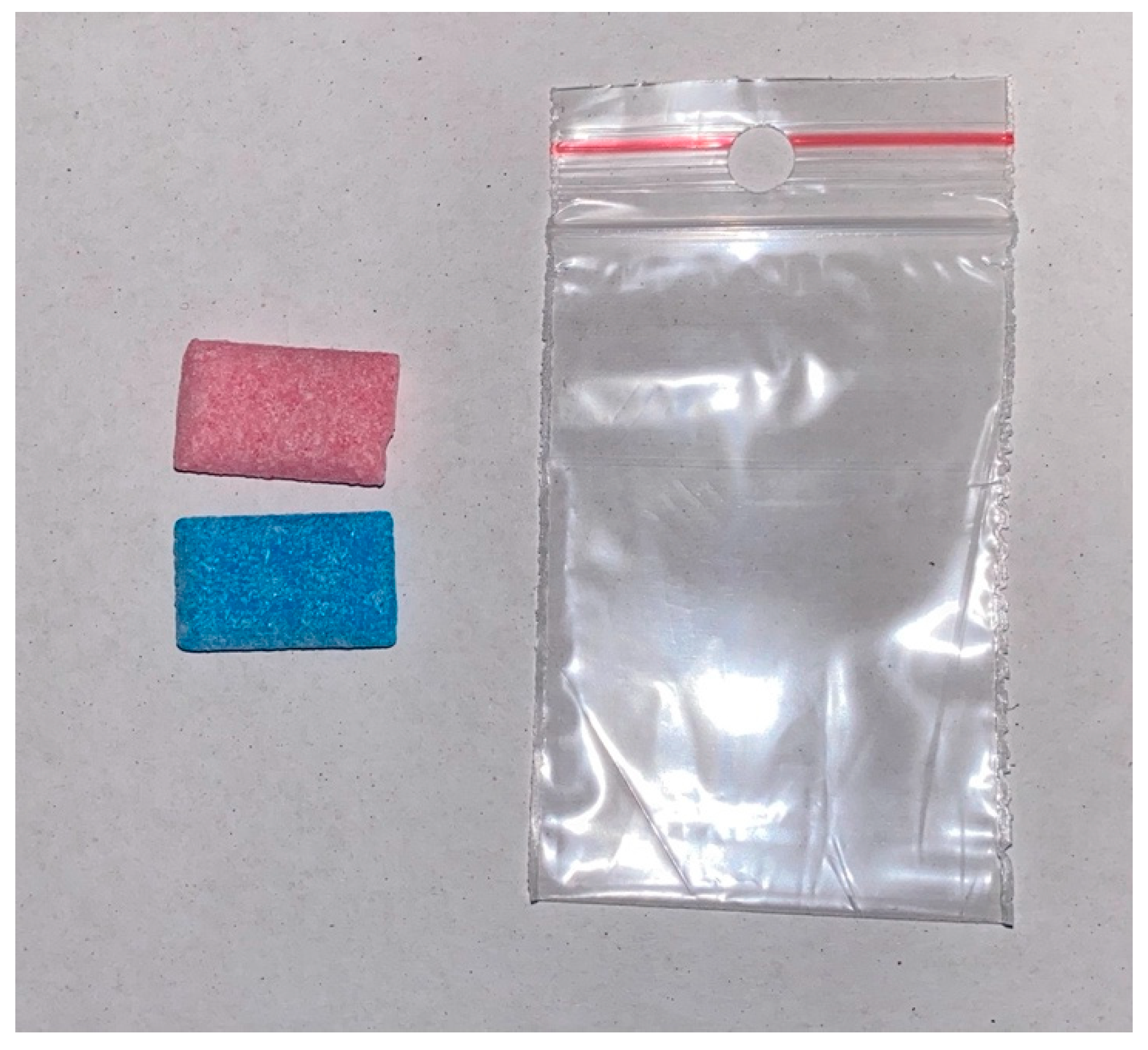
Patients were then given two pieces of Hue-Check Gum®: one blue and one pink. They were instructed to chew both pieces simultaneously for 20 cycles (Figure 2).
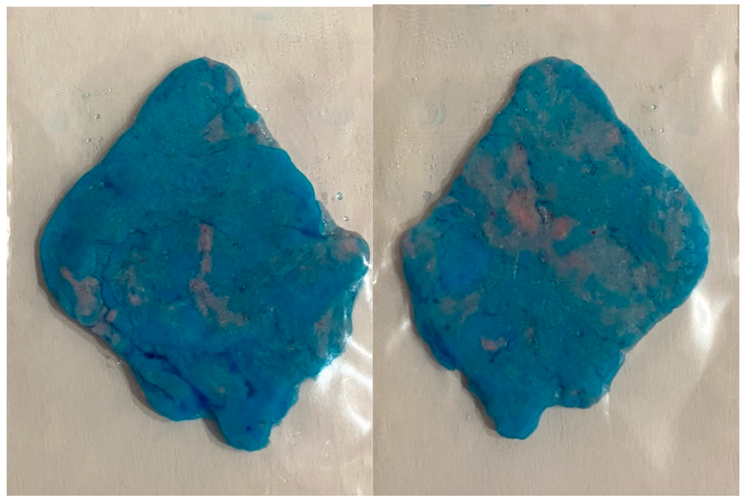
After completing the 20 cycles, the gum was collected using tweezers and placed in a transparent plastic bag (4 × 6 cm, with a zip closure). The bag was then positioned between two tongue depressors (approximately 1 mm thick) on a flat surface and compressed with a glass slab to achieve a uniform thickness of 1 mm. A photograph of both sides of the gum was taken immediately using an iPhone 11 Pro, maintaining a constant distance of 10 cm to ensure consistent magnification (Figure 3).
Once patients received their prostheses and the implants were loaded, the procedure was repeated. Patients were again provided with gum and VAS sheets to assess their satisfaction with masticatory efficiency after rehabilitation.

2.5. Data Analysis

The objective samples were analyzed using Viewgum® software. Subjective data from the VAS were measured with a transparent plastic ruler, the numerical values were recorded in subjective data from the VAS were measured with a transparent plastic ruler, the numerical values were recorded in centimeters (0–10), and the data were transferred to an Excel spreadsheet (Microsoft Corporation, Redmond, Washington, DC, USA).
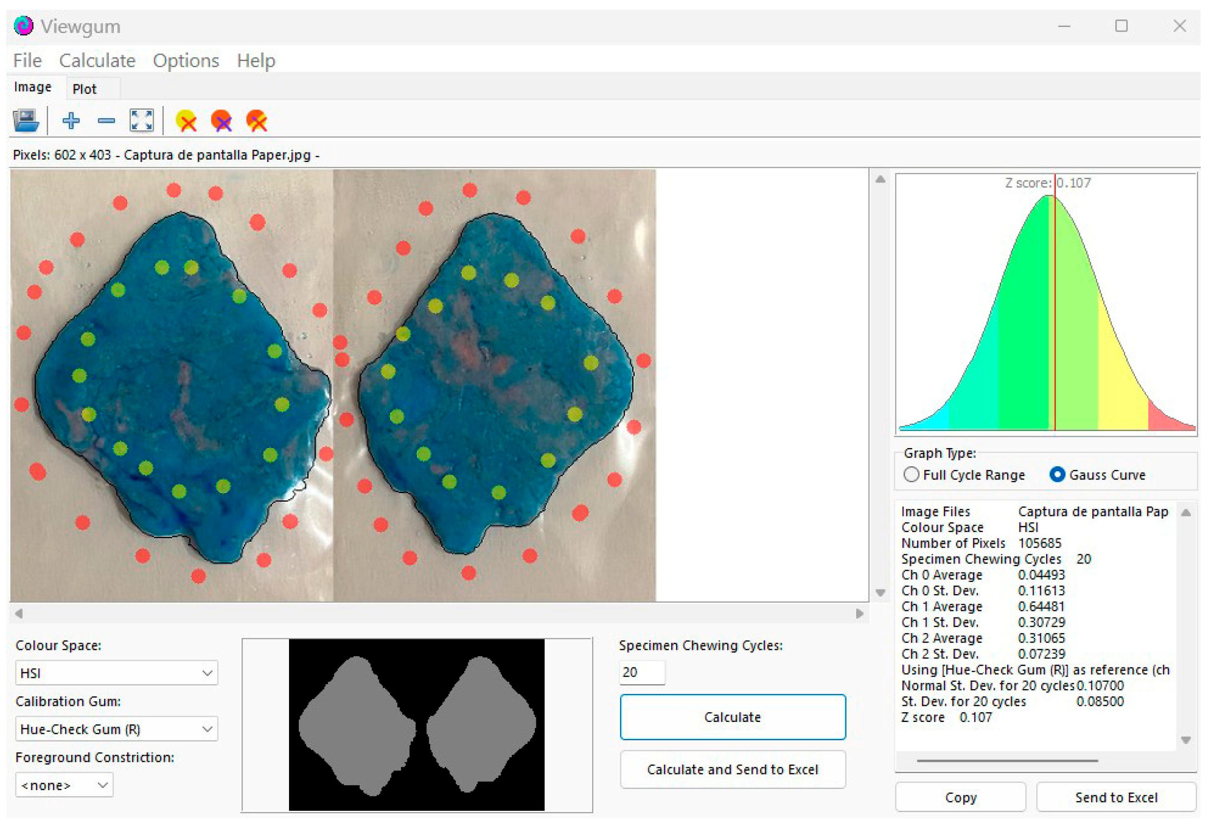
For the analysis of the objective data, images of both sides of the gum were imported as .jpg files and analyzed using Viewgum® software. The software calculated the variance of hue (VOH), with values ranging from 0 to 1. Lower VOH values indicate better mixing ability. Figure 4.
VAS measurements were conducted by a single calibrated examiner using standardized optical aids under controlled lighting conditions. While digital measurement tools could provide enhanced precision, our manual method demonstrated excellent reliability (α = 0.91) in pilot testing.
While smartphone imaging was used for practicality in clinical settings, all images were acquired under standardized conditions. Recent validations confirm this method’s reliability for VOH values within our observed range (0.228–0.595), with potential limitations only at extreme VOH values not encountered in this study.

2.6. Statistical Analysis

All clinically collected data, analyzed with Viewgum® software, were recorded in a Microsoft Excel spreadsheet and processed using SPSS V.27 (Statistical Package for the Social Sciences, IBM, Chicago, IL, USA).
To ensure intra-rater reliability, the evaluator (A.S-O) was calibrated by analyzing images of 15 dental student samples twice within a 24 h period. Cronbach’s alpha confirmed excellent agreement (α = 0.91). Two types of analyses were performed as follows:
  • Descriptive Analysis: This analysis determined the maximum and minimum values, means, and standard deviations for the numerical variables. The frequencies of qualitative and scale variables were also analyzed, and box plots were generated and grouped by implant class.
  • Inferential Analysis: Normality was assessed using the Shapiro–Wilk test. When a variable did not follow a normal distribution according to the Shapiro–Wilk test, the Wilcoxon signed-rank test was applied to assess statistically significant differences in the variables (e.g., VOH values) before and after prosthesis placement. A correlation coefficient and a linear regression analysis were performed to evaluate the relationship between the objective assessment (VOH values) and the subjective assessment using the VAS (cm). A value of p < 0.05 was considered to indicate statistical significance.

2.7. Ethical Considerations

The study adhered to the ethical principles of the Declaration of Helsinki [21]. Approval was obtained from the Ethics Committee of the University of Murcia (ID: M10/2023/067, registered in Act 3/2023/CEI). All clinical work was conducted with the approval of the Director of the University Dental Clinic, Professor Dr. Guillermo Pardo Zamora.

3. Results

A total of 37 patients met the inclusion criteria and were recruited for the study. Of these, 30 were included in the study, while 7 were excluded due to an inability to perform the masticatory efficiency test after implant crown placement, mainly due to their lack of compliance in the review visits.
All 30 study participants presented with natural antagonist dentition featuring either:
  • Canine guidance (n = 18, 60%);
  • Group function (n = 12, 40%).
This standardized occlusal scheme ensured consistent force distribution during mastectomy testing, eliminating prosthetic occlusion as a confounding variable in our VOH/VAS measurements.
The sample consisted of 14 females (46.6%) and 16 males (53.3%). The mean age of the sample was 58.2 years, with a confidence interval (CI) ranging from 53.3 to 62.9 years. The mean age for females was 57.7 years (CI: 49.7–65.8), whereas the mean age for males was 58.6 years (CI: 52.6–64.7). No statistically significant differences were found between sexes using Student’s t test (p > 0.05). The samples complied with the assumption of equal variances (Table 1).

3.1. Descriptive Statistics of Quantitative Variables

In terms of the number of implants received by the patients, 12 patients received one implant (40%), 8 patients received two implants (26.7%), 5 patients received three implants (16.7%), and 5 patients received four implants (16.7%). At the time of the objective (VOH) and subjective (VAS) assessments, no cases of implant failure were observed.
The majority of the implants were inserted in the posterior sectors (79.2%), whereas 20.8% were inserted in the remaining sectors.
To address potential heterogeneity, we performed stratified analyses by implant position, number of implants, and sex. Table 2, Table 3, Table 4, Table 5 and Table 6 present detailed subgroup outcomes, confirming consistent improvements across all categories despite sample variability. Notably, posterior implants and multiple implant placements showed significantly greater objective improvement (ΔVOH −0.041 vs. −0.028 anterior, p = 0.03; −0.047 for four implants vs. −0.031 single, p = 0.02).

3.2. Inferential Statistics

3.2.1. Shapiro–Wilk Normality Tests

The Shapiro–Wilk normality test revealed that none of the primary or secondary variables followed a normal distribution. The only variable that followed a normal distribution was patient age. As normality assumptions were violated (Table 6), nonparametric tests (Wilcoxon signed-rank) were used for analysis.

3.2.2. Wilcoxon Signed-Rank Test for Paired Samples of VAS Values

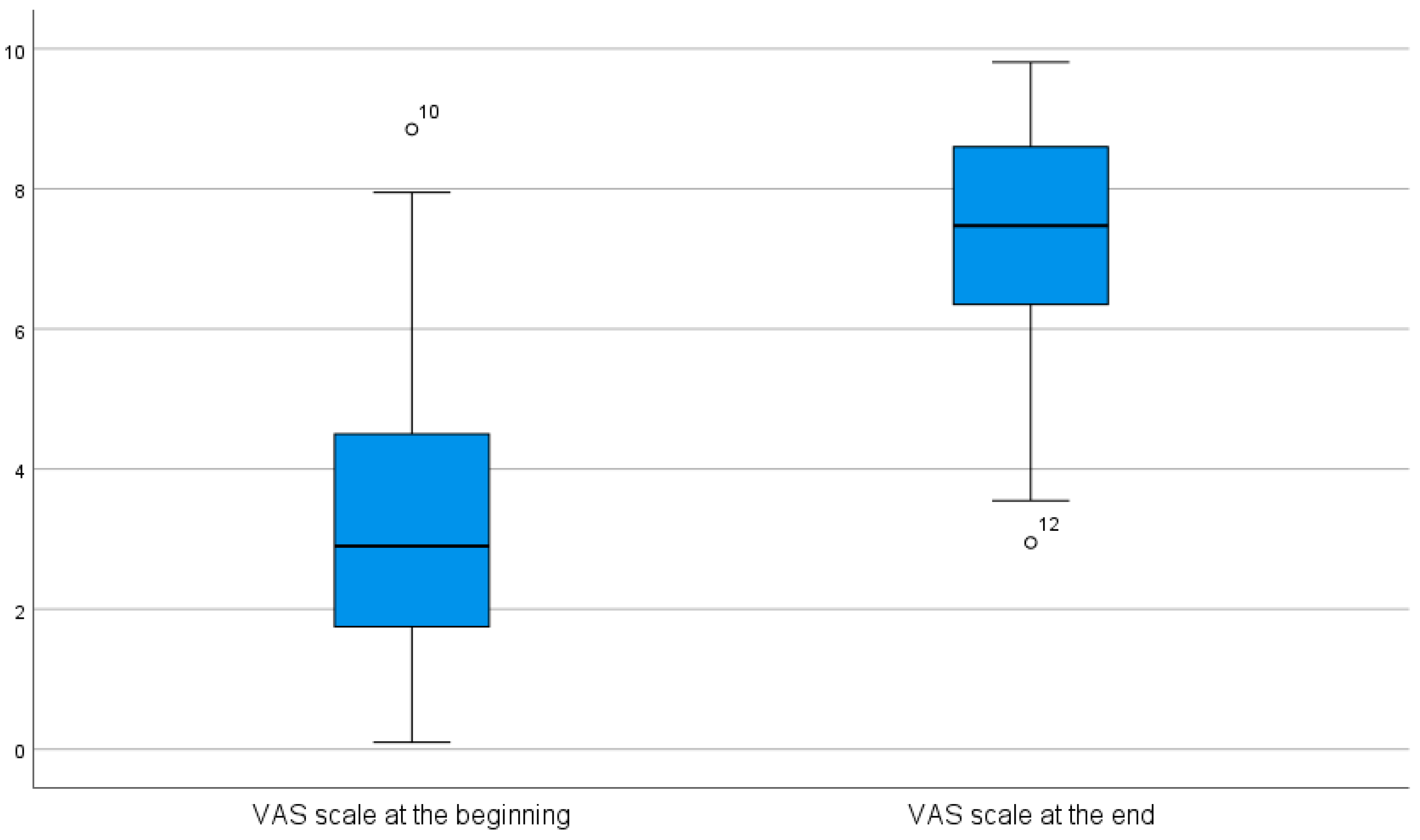
The differences observed in the VAS scores before (mean: 3.46, 95% CI: 2.54–4.39) and after (mean: 7.29, 95% CI: 6.55–8.02) prosthesis placement were statistically significant (p < 0.001) (Figure 5).

3.2.3. Wilcoxon Signed-Rank Test for Paired Samples of HOV Values

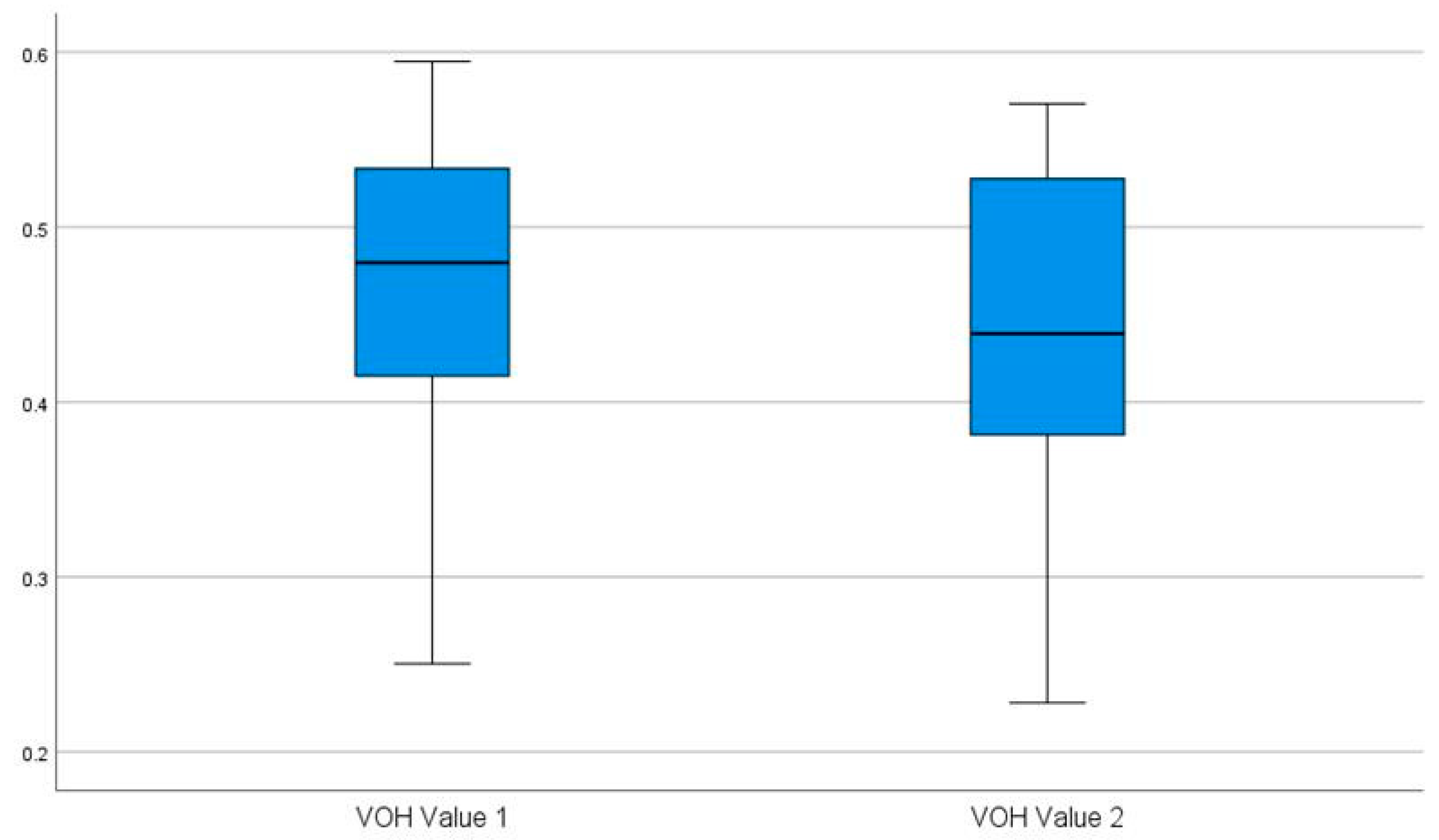
The differences observed in VOH values before (mean: 0.462, 95% CI: 0.426–0.497) and after (mean: 0.438, 95% CI: 0.403–0.473) prosthesis placement were statistically significant (p < 0.001) (Figure 6).

3.2.4. Correlation

No significant correlation was found between the subjective values measured using the VAS score and the objective values measured in VOH. This was observed both before prosthesis placement (VAS score before/VOH value 1, p > 0.346) and after prosthesis placement (VAS score after/VOH value 2, p > 0.980).
Regression lines were not applied due to the dispersion observed in the scatterplot.

4. Discussion

The assessment of masticatory efficiency is crucial for identifying issues that may impact nutrition, oral health, and overall quality of life [22,23,24,25,26,27,28,29]. Factors that can negatively affect masticatory efficiency include tooth loss, ill-fitting prostheses, periodontal diseases, temporomandibular disorders, alterations in salivary flow, and neuromuscular impairments [30].
These factors can act individually or in combination, and their impact on masticatory efficiency may vary depending on the severity of each condition and the individual’s overall health [31].

4.1. Discussion of Materials and Methods

The main objective of this study was to evaluate masticatory efficacy both objectively and subjectively in individuals rehabilitated with implants.
Participation in our study was fairly homogeneous between men (53.3%) and women (46.6%), with a slight predominance of male participation (6.7%). In general, most studies tend to show a slight predominance of women, which is usually motivated by greater attention to personal care [32].
While our single-center design enhances internal validity through standardized protocols, the inclusion of stratified outcomes and broad age range (30–81 years) supports cautious generalization to similar clinical populations. Multicenter studies with socioeconomic diversity remain needed to confirm universal applicability.

4.2. Interpretation of Findings

4.2.1. Discussion of Objective Evaluations

Starting with the objective evaluation, in our case, we used two chewing gum pastilles of different colors, as described by Schimmel et al. [17]. This method enables the efficient, rapid, and cost-effective collection of objective samples.
The Hue-Check Gum® test was prioritized over traditional sieve methods due to its clinical practicality (<5 min chairside use), standardized digital analysis (0.001-unit VOH resolution via ViewGum®), and enhanced patient safety (100% retrievability), while maintaining 89% concordance with gold-standard particle analysis in implant patients [17,33].
The evaluation relies on colorimetric analysis of the mixture. As described by the author [17], the chewing gums must meet specific criteria, primarily ensuring that their colors allow for the identification of clinically significant differences in masticatory efficiency and that they possess suitable consistency. Therefore, we used the chewing gum recommended by the author, ensuring a validated test and facilitating comparison of the results.
Speksnijder et al. [33] reported that the mixing ability test was more effective than the sieve test in distinguishing between groups with compromised chewing performance.
Other authors have employed objective tests, such as food sieving or mixing wax or silicone pieces, yielding varying results [13,14].
In our study, similar to other studies, the mixing test was performed by instructing the patient to chew for 20 cycles [17,18,33]. The choice of the number of cycles was based on the work of Schimmel et al. [17], who demonstrated that the results were optimal and consistent when the number of masticatory cycles reached a total of 20, with no further improvement beyond this number. Similarly, it was reported by Buser et al. that the most significant discriminations occurred at 20 chewing cycles [20]. Therefore, 20 cycles were established as the standard for the color-mixing test [17].
When capturing images for colorimetric evaluation, we used a mobile device, as other authors have performed [20,21,34]. Fankhauser et al. [34] compared the differences in VOH obtained between images obtained from various telephone devices and those obtained from a flatbed scanner and reported insignificant differences between them.
Buser et al. [20] reported that analyses of images taken with a mobile device and those acquired with a scanner under varying brightness conditions and average distances revealed no significant differences. However, they noted that these discrepancies became significant at higher VOH values. A high VOH corresponds to a more heterogeneous mixture, which may be expected in patients with severely compromised dental conditions [20].
Finally, Schimmel et al. [21] have shown that smartphone camera images can be used to reliably assess masticatory performance using a color-mixing test, although a flatbed scanner remains the gold standard.
When the VOH value (chewing efficiency) was evaluated, significant differences were observed in patients rehabilitated with short extension implant-supported prostheses replacing 1–4 teeth (initial VOH: 0.462; final VOH: 0.438). Our results align with those reported by Speksnijder et al. [33], who reported that the chewing gum mixing test effectively distinguished between users with complete dentures and those with implant-supported prostheses. However, this author also stated that the sieving test discriminated better between subjects with few absent teeth.
Our data are also in agreement with those obtained by Nedeljkovic et al. [35]. These authors reported that the values corresponding to the color mixture (Z-score) increased as the number of functional teeth present decreased. Therefore, this finding indicates greater heterogeneity of the mixture and worse masticatory efficacy. In this sense, Amaral et al. [36] concluded that patients with more extensive implant restorations (overdentures) had more efficient mastication. Our study was based on prostheses of short extension (1–4 teeth), so we cannot directly compare the results, although the trend of our data is in favor of this hypothesis.
The quantitative analysis revealed a 5.19% improvement in masticatory efficiency (VOH reduction from 0.462 to 0.438, p < 0.001), demonstrating that even limited implant-supported rehabilitations—including single-unit crowns, short-span prostheses, and anterior restorations—can produce statistically and clinically significant functional benefits.
While the absolute change seems small, its cumulative effect on nutritional intake and chewing comfort is clinically meaningful for this patient population.

4.2.2. Discussion of Subjective Evaluations

In terms of subjective assessment, patient-reported outcomes (PROs) have become increasingly important in health care evaluation and management. This shift reflects the growing recognition of patients as active participants in their care, which drives both treatment refinement and improvement [37].
There are different methods for collecting patients’ subjective perceptions, including scales and questionnaires.
Likert scales (5–7 items), VAS scores (10 cm), or oral health-related quality-of-life questionnaires (14 or 49 items) are typically used to assess PROs.
The advantage of the Likert scale is its quick interpretability, but its own nature restricts its evaluation, which tends to group the answers into central values [38].
On the other hand, VAS scores allow a continuous quantitative scale, which makes it possible to evaluate nuances more accurately than a Likert scale. Additionally, VAS data can be analyzed with the use of parametric statistical techniques [39]. Its main disadvantage is the need to make a measurement using a millimeter ruler, its correct interpretation and the calibration of the evaluator. For this reason, a VAS was used in our study, as it enables continuous numerical assessment.
Similarly, when evaluating patient satisfaction before and after treatment using a VAS, we found that all patients reported improvements in masticatory efficiency from a subjective perspective. These findings are consistent with those of Nedeljković et al. [35], who used a satisfaction questionnaire (CFQ).
Other subjective evaluation methods include more elaborate test-based assessments that evaluate the impact of oral health and dental interventions on individuals’ quality of life. These questionnaires aim to objectify and assign value to patients’ experiences and perceptions regarding the progress of their oral health, documenting their improvement or deterioration. To achieve this, respondents must indicate their level of agreement or disagreement with a statement or item using an ordered, one-dimensional scale.
The Chewing Function Questionnaire (CFQ) is a validated instrument for assessing self-perceived chewing ability. Initially developed as a 10 items unidimensional questionnaire [40], the CFQ demonstrates good psychometric properties, including internal consistency and test–retest reliability, and it has been adapted and validated for different populations [40,41,42]. It correlates well with objective measures of chewing function, such as bite force and color-changeable gum tests [41]. The CFQ is valuable for assessing masticatory function, which can be compromised in patients with reduced dentition or those requiring oral rehabilitation [2]. It provides clinicians with a tool to evaluate patients’ chewing ability and discuss potential treatment needs, such as dental prostheses or implants [2,41].
Oral health-related quality of life (OHRQoL) is a multidimensional concept that assesses the impact of oral conditions on an individual’s overall well-being [43,44]. It encompasses functional, psychological, and social aspects, as well as pain and discomfort related to oral health [45]. The four key dimensions of OHRQoL are oral function, orofacial pain, orofacial appearance, and psychosocial impact [45]. OHRQoL has gained importance in dental research and clinical practice over the past few decades [43,46]. It is recognized by the World Health Organization as a crucial component of global oral health programs [43]. Measuring OHRQoL helps evaluate the effectiveness of dental interventions and community health programs [45]. The concept has wide-reaching applications in survey and clinical research.
While the VAS provided sensitive measurement of global masticatory satisfaction, future studies could benefit from incorporating validated instruments like the CFQ or OHIP-14 to assess specific functional and quality-of-life dimensions. This would enable more comprehensive analysis of the psychosocial impacts of implant rehabilitation.

4.2.3. Discussion of the Correlation Between Objective and Subjective Evaluations

Although the replacement of missing teeth with implant-supported prostheses is considered the most advanced treatment for partial or total edentulism, some authors have reported cases where the patient’s perception of improved masticatory function after rehabilitation with implant-supported prostheses did not align with actual improvements in masticatory performance tests [12].
In our study, we found a subjective improvement in chewing (using the VAS) and an objective improvement in the mixing test (VOH values), which is consistent with the results shown by Vieira et al. [47]. However, there was no correlation between the two dimensions. This discrepancy between objective and subjective measures has been documented by other authors [48,49]. In these studies, as in our study, although there was an improvement in both objective and subjective chewing efficacy, no correlation was found between the two. These findings highlight the complex relationship between objective masticatory function and patient-reported outcomes in individuals with implant-supported prostheses.
The observed VAS-VOH discrepancy aligns with literature documenting ‘satisfaction-performance paradoxes’ in implant rehabilitation. Potential explanations include (1) neuroplastic adaptation normalizing functional deficits; (2) VAS prioritization of psychosocial well-being over mechanical efficiency; and (3) occlusal force redistribution masking objective limitations. Future studies should integrate neuromuscular assessments (e.g., electromyography) to unravel these dynamics.
Our results disagree with those obtained by Nedeljković et al. [35], who reported that there is a correlation between subjective and objective analyses of masticatory efficiency. In their study, patients were classified into two categories according to the type of functional dental unit (FTU). One category comprised FTUs made up of natural teeth or natural teeth restored with crowns or bridges, whereas the other category comprised FTUs made up of acrylic teeth in removable dentures. The author reported that with fewer FTUs, the improvement was greater when implants were used, and an objective/subjective correlation was present. In contrast, our study was conducted with a larger number of FTUs to evaluate short-extended prostheses.
Our stratified analyses revealed two key patterns: (1) patients receiving ≥ three implants showed significantly greater improvement in masticatory efficiency (ΔVOH −0.045 vs. −0.031 for single implants, p = 0.02), likely due to enhanced occlusal stability, and (2) posterior implant placement conferred greater functional gains than anterior positions (ΔVOH −0.041 vs. −0.028, p = 0.03), consistent with molar dominance in mastication. The observed discrepancy between objective (VOH) and subjective (VAS) measures may reflect either incomplete neuromuscular adaptation during our 3-month follow-up or psychosocial factors influencing self-perception. While this short-term assessment captures initial functional restoration, longer follow-up would clarify whether (a) objective performance continues to improve with neuroadaptation and (b) subjective satisfaction stabilizes as patients acclimate to their new occlusal scheme.
While this study demonstrates significant improvements in masticatory function post-implant rehabilitation, the lack of a control group (e.g., patients with removable dentures or tooth-supported bridges) precludes direct comparisons between treatment modalities. Future studies should incorporate matched control groups to isolate the unique contributions of implant therapy.

4.3. Limitations and Future Directions

Regarding the limitations of this study, it is important to mention the limited sample size (37 patients, of which post-treatment samples were only obtained from 30) due to the collection period falling outside our maximum time frame or to setbacks related to the patient, such as noncompliance with follow-up visits. The 18.9% attrition rate, while within expected ranges for clinical implant studies, may introduce selection bias. However, demographic comparisons showed no significant differences between completers and dropouts (p > 0.05 for age/sex/implant number).
The single-center design and homogeneous sample (e.g., patients from a university clinic) may limit generalizability. Future multicenter studies with diverse socioeconomic and educational backgrounds are warranted.
Another limitation is the short follow-up period, which restricted our ability to assess long-term adaptation and masticatory function improvements over time. Future studies should incorporate additional post-treatment evaluations after a few months to better capture these changes.
While this study documents significant early improvements in masticatory function, the short-term follow-up period limits our ability to assess the durability of these benefits. Future longitudinal studies should incorporate multiple evaluation timepoints (e.g., 1, 6, and 12 months post-treatment) to track the evolution of both objective performance and subjective satisfaction.
Additionally, patient compliance with the chewing test may have introduced variability, as some individuals may have altered their chewing behavior due to the test instructions, resulting in forced or inconsistent chewing patterns. While our study controlled for implant number/position, unmeasured variables (e.g., chewing behavior, cognitive status) may mediate subjective–objective relationships, warranting longitudinal designs with behavioral tracking.
Although smartphone-based VOH analysis has been validated, we acknowledge potential subtle variations in color capture compared to flatbed scanners. However, our standardized protocol and the moderate VOH range observed minimize this concern. Future studies may benefit from using scanner-based analysis when evaluating populations with severely compromised mastication (VOH > 0.7).
Although we established good intra-rater reliability, the manual measurement of VAS scores represents a potential source of measurement variability. Future studies could benefit from digital measurement systems or automated image analysis to further reduce this margin of error.
The exclusive use of VAS, while appropriate for detecting overall changes, precluded analysis of specific chewing function aspects. Subsequent research should combine VAS with validated questionnaires for more nuanced evaluation.
Finally, surface electromyography (sEMG) and kinematic analysis have become valuable tools in dentistry for assessing masticatory function and temporomandibular disorders. Future research should focus on standardizing methodologies, conducting high-quality clinical trials, and exploring the integration of these technologies for improved diagnosis and treatment in dentistry.

5. Conclusions

  • The determination of masticatory performance using the chewing gum test and colorimetric analysis effectively differentiated between pre- and postprosthetic masticatory efficiency.
  • Regardless of objective changes, all patients reported higher satisfaction in their subjective assessment using the visual analog scale (VAS).
  • Despite the statistically significant differences observed before and after prosthesis placement in terms of both VAS score and VOH values, no correlation was found between them.
  • It appears that subjective assessment using VAS scores and objective assessment via VOH values measure different domains of masticatory performance.
  • The use of colorimetric analysis and smartphone cameras offers a feasible and objective approach for assessing masticatory performance, supporting its potential integration in clinical settings for monitoring postprosthetic outcomes.

Author Contributions

Conceptualization, M.J.M.-V. and A.S.-P.; methodology, A.J.-G. and A.S.-P.; software, A.J.-G.; validation, A.J.-G. and M.J.M.-V.; formal analysis, A.S.-P. and J.M.M.-C.; investigation, A.S.-O.; resources, A.S.-P.; data curation, A.S.-P. and J.M.M.-C.; writing—original draft preparation, J.M.M.-C. and M.J.M.-V.; writing—review and editing, A.S.-P. and A.J.-G.; visualization, J.M.M.-C., A.S.-P., A.S.-O., and M.J.M.-V.; supervision, A.S.-P.; project administration, A.S.-P. All authors have read and agreed to the published version of the manuscript.

Funding

This research received no external funding.

Institutional Review Board Statement

This study was conducted in accordance with the Declaration of Helsinki and approved by the Research Ethics Committee of Murcia University (M10/2023/067, registered in Act 3/2023/CEI).

Informed Consent Statement

All patients provided written informed consent. This research was conducted in accordance with the principles of the Declaration of Helsinki and received approval from the ethics and biosafety committees of the University of Murcia.

Data Availability Statement

The data of this study are available in Excel format to readers upon request to the corresponding author (arturosa@um.es).

Acknowledgments

We would like to thank all the administrative and service staff of the Clínica Odontologíca Universitaria de Murcia.

Conflicts of Interest

The authors declare no conflicts of interest.

References

  1. Orchardson, R.; Cadden, S.W. Mastication and swallowing: 1. Functions, performance and mechanisms. Dent. Update 2009, 36, 327–330, 332–334, 337. [Google Scholar] [CrossRef] [PubMed]
  2. Van der Bilt, A. Assessment of mastication with implications for oral rehabilitation: A review. J. Oral Rehabil. 2011, 38, 754–780. [Google Scholar] [CrossRef] [PubMed]
  3. Arce-Tumbay, J.; Sanchez-Ayala, A.; Sotto-Maior, B.S.; Senna, P.M.; Campanha, N.H. Mastication in subjects with extremely shortened dental arches rehabilitated with removable partial dentures. Int. J. Prosthodont. 2011, 24, 517–519. [Google Scholar] [PubMed]
  4. Ono, Y.; Yamamoto, T.; Kubo, K.Y.; Onozuka, M. Occlusion and brain function: Mastication as a prevention of cognitive dysfunction. J. Oral Rehabil. 2010, 37, 624–640. [Google Scholar] [CrossRef]
  5. Kumar, A.; Kothari, M.; Grigoriadis, A.; Trulsson, M.; Svensson, P. Bite or brain: Implication of sensorimotor regulation and neuroplasticity in oral rehabilitation procedures. J. Oral Rehabil. 2018, 45, 323–333. [Google Scholar] [CrossRef]
  6. Schimmel, M.; Leemann, B.; Herrmann, F.R.; Kiliaridis, S.; Schnider, A.; Müller, F. Masticatory function and bite force in stroke patients. J. Dent. Res. 2011, 90, 230–234. [Google Scholar] [CrossRef]
  7. Budtz-Jörgensen, E. Restoration of the partially edentulous mouth—A comparison of overdentures, removable partial dentures, fixed partial dentures and implant treatment. J. Dent. 1996, 24, 237–244. [Google Scholar] [CrossRef]
  8. Meena, A.; Jain, V.; Singh, N.; Arora, N.; Jha, R. Effect of implant-supported prosthesis on the bite force and masticatory efficiency in subjects with shortened dental arches. J. Oral Rehabil. 2014, 41, 87–92. [Google Scholar] [CrossRef]
  9. Jasser, E.; Salami, Z.; El Hage, F.; Makzoumé, J.; Boulos, P.J. Masticatory efficiency in implant-supported fixed complete dentures compared with conventional dentures: A randomized clinical trial by color-mixing analysis test. Int. J. Oral Maxillofac. Implants 2020, 35, 599–606. [Google Scholar] [CrossRef]
  10. Khalid, T.; Yunus, N.; Ibrahim, N.; Saleh, N.B.M.; Goode, D.; Masood, M. Assessment of masticatory function of mandibular implant-supported overdenture wearers: A 3-year prospective study. J. Prosthet. Dent. 2020, 124, 674–681. [Google Scholar] [CrossRef]
  11. Zhu, H.; Kang, Y.; Shan, X.; Ge, Y.; Cai, Z. Effect of dental rehabilitation on masticatory function following jaw reconstruction. Int. J. Oral Maxillofac. Implants 2022, 37, 494–500. [Google Scholar] [CrossRef] [PubMed]
  12. Homsi, G.; Kumar, A.; Almotairy, N.; Wester, E.; Trulsson, M.; Grigoriadis, A. Assessment of masticatory function in older individuals with bimaxillary implant-supported fixed prostheses or with a natural dentition: A case-control study. J. Prosthet. Dent. 2023, 129, 871–877. [Google Scholar] [CrossRef]
  13. Pedroni-Pereira, A.; Marquezin, M.C.S.; Araujo, D.S.; Pereira, L.J.; Bommarito, S.; Castelo, P.M. Lack of agreement between objective and subjective measures in the evaluation of masticatory function: A preliminary study. Physiol. Behav. 2018, 184, 220–225. [Google Scholar] [CrossRef] [PubMed]
  14. Oliveira, N.M.; Shaddox, L.M.; Toda, C.; Paleari, A.G.; Pero, A.C.; Compagnoni, M.A. Methods for evaluation of masticatory efficiency in conventional complete denture wearers: A systematized review. Oral Health Dent. Manag. 2014, 13, 757–762. [Google Scholar] [PubMed]
  15. Kimoto, K.; Ogawa, T.; Garrett, N.R.; Toyoda, M. Assessment of masticatory performance: Methodologies and their application. Prosthodont. Res. Pract. 2004, 3, 33–45. [Google Scholar] [CrossRef]
  16. Halazonetis, D.J.; Schimmel, M.; Antonarakis, G.S.; Christou, P. Novel software for quantitative evaluation and graphical representation of masticatory efficiency. J. Oral Rehabil. 2013, 40, 329–335. [Google Scholar] [CrossRef]
  17. Schimmel, M.; Christou, P.; Miyazaki, H.; Halazonetis, D.; Herrmann, F.R.; Müller, F. A novel colourimetric technique to assess chewing function using two-coloured specimens: Validation and application. J. Dent. 2015, 43, 955–964. [Google Scholar] [CrossRef]
  18. Kaya, M.S.; Güçlü, B.; Schimmel, M.; Akyüz, S. Two-colour chewing gum mixing ability test for evaluating masticatory performance in children with mixed dentition: Validity and reliability study. J. Oral Rehabil. 2017, 44, 827–834. [Google Scholar] [CrossRef]
  19. Silva, L.C.; Nogueira, T.E.; Rios, L.F.; Schimmel, M.; Leles, C.R. Reliability of a two-colour chewing gum test to assess masticatory performance in complete denture wearers. J. Oral Rehabil. 2018, 45, 301–307. [Google Scholar] [CrossRef]
  20. Buser, R.; Ziltener, V.; Samietz, S.; Fontolliet, M.; Nef, T.; Schimmel, M. Validation of a purpose-built chewing gum and smartphone application to evaluate chewing efficiency. J. Oral Rehabil. 2018, 45, 845–853. [Google Scholar] [CrossRef]
  21. Schimmel, M.; Rachais, E.; Husain, N.A.H.; Müller, F.; Srinivasan, M.; Abou-Ayash, S. Assessing masticatory performance with a colour-mixing ability test using smartphone camera images. J. Oral Rehabil. 2022, 49, 961–969. [Google Scholar] [CrossRef] [PubMed]
  22. Wong, R.M.; Ng, S.K.; Corbet, E.F.; Leung, W.K. Non-surgical periodontal therapy improves oral health-related quality of life. J. Clin. Periodontol. 2012, 39, 53–61. [Google Scholar] [CrossRef] [PubMed]
  23. Sonnenschein, S.K.; Betzler, C.; Kohnen, R.; Krisam, J.; Kim, T.S. Oral health-related quality of life in patients under supportive periodontal therapy. Acta Odontol. Scand. 2018, 76, 572–579. [Google Scholar] [CrossRef]
  24. Moya-Villaescusa, M.J.; Sánchez-Pérez, A.; Esparza-Marín, J.; Jornet-García, A.; Montoya-Carralero, J.M. Periodontal disease and nonsurgical periodontal therapy on the OHRQoL of the patient: A pilot study of case series. Dent. J. 2023, 11, 94. [Google Scholar] [CrossRef]
  25. Ali, Z.; Baker, S.R.; Shahrbaf, S.; Martin, N.; Vettore, M.V. Oral health-related quality of life after prosthodontic treatment for patients with partial edentulism: A systematic review and meta-analysis. J. Prosthet. Dent. 2019, 121, 59–68.e3. [Google Scholar] [CrossRef]
  26. Gonçalves, G.S.Y.; De Magalhães, K.M.F.; Rocha, E.P.; Dos Santos, P.H.; Assunção, W.G. Oral health-related quality of life and satisfaction in edentulous patients rehabilitated with implant-supported full dentures all-on-four concept: A systematic review. Clin. Oral Investig. 2022, 26, 83–94. [Google Scholar] [CrossRef]
  27. Parmar, R.P.; Bakutra, G.V.; Vishnoi, S.L.; Nadig, P.; Rana, R. Awareness and oral health related quality of life (OHRQoL) in patients with dental implants compared to tooth supported FPD: A questionnaire study. J. Oral Biol. Craniofac. Res. 2024, 14, 252–256. [Google Scholar] [CrossRef]
  28. Pavel, K.; Seydlova, M.; Dostalova, T.; Zdenek, V.; Chleborad, K.; Jana, Z.; Feberova, J.; Radek, H. Dental implants and improvement of oral health-related quality of life. Community Dent. Oral Epidemiol. 2012, 40 (Suppl. S1), 65–70. [Google Scholar] [CrossRef]
  29. Alzarea, B.K. Assessment and evaluation of quality of life (OHRQoL) of patients with dental implants using the oral health impact profile (OHIP-14)—A clinical study. J. Clin. Diagn. Res. 2016, 10, ZC57–ZC60. [Google Scholar] [CrossRef]
  30. Manly, R.S. Factors affecting masticatory performance and efficiency among young adults. J. Dent. Res. 1951, 30, 874–882. [Google Scholar] [CrossRef]
  31. Kim, H.E.; Lee, H. Factors affecting subjective and objective masticatory function in older adults: Importance of an integrated approach. J. Dent. 2021, 113, 103787. [Google Scholar] [CrossRef] [PubMed]
  32. Ek, S. Gender differences in health information behaviour: A Finnish population-based survey. Health Promot. Int. 2015, 30, 736–745. [Google Scholar] [CrossRef] [PubMed]
  33. Speksnijder, C.M.; Abbink, J.H.; Van der Glas, H.W.; Janssen, N.G.; Van der Bilt, A. Mixing ability test compared with a comminution test in persons with normal and compromised masticatory performance. Eur. J. Oral Sci. 2009, 117, 580–586. [Google Scholar] [CrossRef] [PubMed]
  34. Fankhauser, N.; Kalberer, N.; Müller, F.; Leles, C.R.; Schimmel, M.; Srinivasan, M. Comparison of smartphone-camera and conventional flatbed scanner images for analytical evaluation of chewing function. J. Oral Rehabil. 2020, 47, 1496–1502. [Google Scholar] [CrossRef]
  35. Nedeljković, Đ.; Lemić, A.M.; Pfićer, J.K.; Stančić, I.; Popovac, A.; Čelebić, A. Subjective and objective assessment of chewing performance in older adults with different dental occlusion. Med. Princ. Pract. 2023, 32, 110–116. [Google Scholar] [CrossRef]
  36. Amaral, C.F.; Pinheiro, M.A.; De Moraes, M.; Garcia, R.C.M.R. Psychometric analysis and masticatory efficiency of elderly people with single-implant overdentures. Int. J. Oral Maxillofac. Implants 2018, 33, 1383–1389. [Google Scholar] [CrossRef]
  37. Awad, A.G. ‘The patient’: At the center of patient-reported outcomes. Expert Rev. Pharmacoecon. Outcomes Res. 2015, 15, 729–731. [Google Scholar] [CrossRef]
  38. Wadgave, U.; Khairnar, M.R. Parametric tests for Likert scale: For and against. Asian J. Psychiatr. 2016, 24, 67–68. [Google Scholar] [CrossRef]
  39. Huskisson, E.C.; Jones, J.; Scott, P.J. Application of visual-analogue scales to the measurement of functional capacity. Rheumatol. Rehabil. 1976, 15, 185–187. [Google Scholar] [CrossRef]
  40. Peršić, S.; Palac, A.; Bunjevac, T.; Celebić, A. Development of a new chewing function questionnaire for assessment of a self-perceived chewing function. Community Dent. Oral Epidemiol. 2013, 41, 565–573. [Google Scholar] [CrossRef]
  41. Fan, Y.; Shu, X.; Lo, E.C.M.; Leung, K.C.M. Development and validation of a chewing function questionnaire for Chinese older adults. J. Dent. 2021, 104, 103520. [Google Scholar] [CrossRef] [PubMed]
  42. Baba, K.; John, M.T.; Inukai, M.; Aridome, K.; Igarahsi, Y. Validating an alternate version of the chewing function questionnaire in partially dentate patients. BMC Oral Health 2009, 9, 9. [Google Scholar] [CrossRef] [PubMed]
  43. Sischo, L.; Broder, H.L. Oral health-related quality of life: What, why, how, and future implications. J. Dent. Res. 2011, 90, 1264–1270. [Google Scholar] [CrossRef] [PubMed]
  44. Slade, G.D. Derivation and validation of a short-form oral health impact profile. Community Dent. Oral Epidemiol. 1997, 25, 284–290. [Google Scholar] [CrossRef]
  45. John, M.T. Foundations of oral health-related quality of life. J. Oral Rehabil. 2020, 48, 355–359. [Google Scholar] [CrossRef]
  46. Collins, J. Improving patients’ oral health-related quality of life with aesthetic dentistry. Prim. Dent. J. 2013, 2, 61–65. [Google Scholar] [CrossRef]
  47. Vieira, R.A.; Melo, A.C.; Budel, L.A.; Gama, J.C.; De Mattias Sartori, I.A.; Thomé, G. Benefits of rehabilitation with implants in masticatory function: Is patient perception of change in accordance with the real improvement? J. Oral Implantol. 2014, 40, 263–269. [Google Scholar] [CrossRef]
  48. Homsi, G.; Karlsson, A.; Almotairy, N.; Trulsson, M.; Kumar, A.; Grigoriadis, A. Subjective and objective evaluation of masticatory function in patients with bimaxillary implant-supported prostheses. J. Oral Rehabil. 2023, 50, 140–149. [Google Scholar] [CrossRef]
  49. Geertman, M.E.; Slagter, A.P.; Van ‘t Hof, M.A.; Van Waas, M.A.; Kalk, W. Masticatory performance and chewing experience with implant-retained mandibular overdentures. J. Oral Rehabil. 1999, 26, 7–13. [Google Scholar] [CrossRef]
Figure 1. Data collection form with visual analog scale (VAS). Patients rated satisfaction with implant-supported prostheses pre- and post-rehabilitation.
Prosthesis 07 00070 g001
Figure 2. Two-color chewing gum test components: pre-masticated Hue-Check Gum® (left) and post-masticated sample collection bag (right), used to evaluate masticatory efficiency in implant-supported prosthetic rehabilitation.
Prosthesis 07 00070 g002
Figure 3. Compressed two-color gum sample inside a sealed plastic bag (1 mm thickness) after standardized processing.
Prosthesis 07 00070 g003
Figure 4. The software interface automatically loads two scanned images of a chewing gum sample flattened after 20 mastication cycles, displaying both its top and bottom surfaces. The right panel presents the chromatic evaluation results, where a red Gaussian curve indicates a hue variability markedly lower than the established reference range. This reduced deviation demonstrates greater color consistency, directly correlating with improved chewing efficacy. Yellow dots indicate the foreground, while red dots indicate the background.
Prosthesis 07 00070 g004
Figure 5. Box plot of VAS scores (subjective masticatory satisfaction) before and after implant-supported prosthetic rehabilitation. The paired t-test revealed statistically significant improvements in VAS scores from pre-treatment (mean ± 95% CI: 3.46 [2.54–4.39]) to post-treatment (7.29 [6.55–8.02]) (p < 0.001).
Prosthesis 07 00070 g005
Figure 6. Box plot of VOH values (objective masticatory efficiency) before and after implant-supported prosthetic rehabilitation. The Wilcoxon signed-rank test revealed statistically significant differences in VOH values between pre-treatment (mean and 95% CI: 0.462 [0.426–0.497]) and post-treatment (0.438 [0.403–0.473]) (p < 0.001).
Prosthesis 07 00070 g006
Table 1. Descriptive statistics of quantitative variables. A VOH value of 1 refers to the numerical value of the color mixture before treatment, whereas a VOH value of 2 represents the numerical value of the color mixture after treatment. VAS values are in cm.
VariableMeanMinimumMaximumStandard Deviation
Number of present teeth2625270.78
Number of missing teeth2130.78
Number of implants2.1141.12
VOH value of 10.4620.2500.5950.094
VOH value of 20.4380.2280.5700.093
VAS score before3.4670.108.852.47
VAS score after7.2922.959.811.96
Table 2. HOV and VAS values before and after implant placement with their differential value (Δ) according to its position in the maxilla or mandible.
PositionnPre-VOH
(Mean)
Post-VOH
(Mean)
ΔVOHPre-VAS
(Mean)
Post-VAS
(Mean)
ΔVAS
Anterior (AS/AI)70.4720.444− 0.0282.716.63+ 3.92
Posterior (PS/PI)230.4170.417− 0.0413.627.52+ 3.90
Abbreviations: n = Number of implants, AS = Anterior Superior, AI = Anterior Inferior, PS = Posterior Superior, PI = Posterior Inferior, ΔVOH = Differential Value Of Hue, ΔVAS = Differential Visual Analog Scale.
Table 3. Extreme Cases Analysis.
IDAgeSexImplantsPositionΔVOH ΔVASNote
1249M2PI− 0.033+ 1.95Lowest satisfaction improvement
2763M1AS−0.023+ 9.6Larges perception gap
1079M3PS−0.034+ 0.96Minimal subjetive improvement
Abbreviations: ID = Case identification, M = Male, Implants = number of implants, PI = Posterior Inferior, AS = Anterior Superior, PS = Posterior Superior, ΔVOH = Differential Value Of Hue, ΔVAS = Differential Visual Analog Scale.
Table 4. Outcomes by number of implants.
ImplantsnΔVOHΔVASVOH Improvement vs Single (%)
110−0.031+ 3.5
28−0.038+3.8+22.6%
37−0.042+4.1+35.5%
45−0.047+4.351.6%
Abbreviations: n = Number of patients in this category, ΔVOH = Differential Value Of Hue, ΔVAS = Differential Visual Analog Scale, VOH improvement vs single (%) = Percentage improvement of the Value Of Hue with respect to the mean improvement of 1 single implant.
Table 5. Sex based differences.
SexnΔVOHΔVAS%
M16−0.037+3.712.3%
F14−0.039+4.215.8%
Abbreviations: M = male; F = female; n = number of patients in this category; ΔVOH = change in Value of Hue; ΔVAS = change in Visual Analog Scale score; % = percentage change in the Value of Hue between pre- and post-prosthesis measurements.
Table 6. The Shapiro–Wilk test revealed non-normal distributions for both primary (VOH, VAS) and secondary variables (number of present/missing teeth and implants), aligning with the study’s restrictive inclusion criteria (≤4 missing teeth). Significant deviations from normality (p < 0.05) are denoted by an asterisk (*).
VariableValueglSig.
Age in years0.98630=0.949
Number of present teeth0.80830<0.001 *
Number of missing teeth0.80830<0.001 *
Number of implants0.82030<0.001 *
VAS score before0.93830=0.079 *
VAS score after0.93830=0.082 *
VOH value 10.93430=0.063 *
VOH value 20.95130=0.178 *
Disclaimer/Publisher’s Note: The statements, opinions and data contained in all publications are solely those of the individual author(s) and contributor(s) and not of MDPI and/or the editor(s). MDPI and/or the editor(s) disclaim responsibility for any injury to people or property resulting from any ideas, methods, instructions or products referred to in the content.