Article

Hip Replacement Following Intertrochanteric Osteosynthesis Failure: Is It Possible to Restore Normal Hip Biomechanics?

by
Davide Bizzoca
*,
Giorgio Giannini
,
Francesco Domenico Cannito
,
Giulia Colasuonno
,
Giuseppe De Giosa
and
Giuseppe Solarino
Orthopaedic & Trauma Unit, UOSD Vertebral Surgery, Department DiBraiN, University of Bari Aldo Moro, AOU Consorziale Policlinico di Bari, 70124 Bari, Italy
*
Author to whom correspondence should be addressed.
Prosthesis 2025, 7(3), 50; https://doi.org/10.3390/prosthesis7030050
Submission received: 28 March 2025 / Revised: 1 May 2025 / Accepted: 3 May 2025 / Published: 8 May 2025
(This article belongs to the Special Issue State of Art in Hip, Knee and Shoulder Replacement (Volume 2))

Abstract

Introduction: Intertrochanteric femoral fractures (IFFs) are the most common traumatic injuries in elderly people and significantly impact the patient’s health status. The current evidence indicates that short intramedullary nails may be a better choice than dynamic hip screws in IFF management, being less invasive and biomechanically superior, providing a buttress to limit fracture collapse. On the other hand, an unstable fracture may collapse even after adequate reduction and fixation. This paper aims to describe the surgical complexity of the nail-to-total hip arthroplasty (THA) conversion, focusing on the restoration of normal hip geometry. Material and Methods: Patients referred to our level I trauma center with failed cephalomedullary nailing following IFFs and managed with the nail-to-THA conversion were retrospectively recruited. The anteroposterior postoperative pelvis radiographs were analyzed to establish whether the normal biomechanics of the involved hip were restored. The following radiographic parameters were recorded and compared to the contralateral unaffected side: hip offset, cervical–diaphyseal angle, and limb length discrepancy. Clinical assessment was performed using the following scores: the Harris hip score (HHS) and the visual analog scale for pain (VAS). The independent samples t-test and the Pearson correlation test were performed. The tests were two-tailed; a p < 0.05 was considered significant. Results: A total of 31 patients met the inclusion and exclusion criteria (10 males and 21 females; mean age: 76.2 years; range: 66–90 years) and were included in this study. The modes of trochanteric nail failure included the following: cut-out in 22 cases (70.97%), non-union in 4 cases (12.9%), peri-implant fracture in 1 case (3.23%), cut-through in 2 cases (6.45%), and femoral head avascular necrosis (HAN) in 2 cases (6.45%). Long stems were used in 21 patients out of 31 (67.74%), while dual-mobility cups were implanted in 24 patients out of 31 (77.41%). A significant mean neck shaft angle (NSA) increase (p < 0.001) and a significant mean femoral offset reduction (FO, p 0.001) compared to the contralateral hip were recorded; a mean limb length discrepancy (LLD) of 8.35 mm was observed. A significant correlation between HHS and ∆NSA (p = 0.01) and ∆FO (p = 0.003) was recorded. Conclusions: Conversion from a cephalomedullary nail to THA is a complex procedure that should be considered a revision surgery, rather than a primary surgery. Surgeons must be aware that normal hip geometry may not be obtained during this surgical procedure; thus, a patient undergoing the nail-to-THA conversion for intertrochanteric fixation failure may have an increased risk of implant-related complications.

1. Introduction

Proximal femur fractures (PFFs), which include both intracapsular fractures (femoral neck fractures, FNFs) and extracapsular fractures (intertrochanteric femoral fractures, IFFs), affect approximately 1.5 million people globally each year, representing a significant public health issue [1].
PFFs are the most common traumatic injuries among the elderly and can severely impact patients’ health, leading to reduced quality of life, increased disability, and higher mortality rates [1,2].
In Italy, there are about 78,000 cases of PFFs annually, with 90% occurring in individuals over 65 [3]. As life expectancy continues to rise, this number is projected to increase, reaching 8.2 million fractures worldwide by 2050 [3,4,5,6].
IFFs, which account for over 60% of PFFs, have an annual mortality rate of 15–20% [4]. Additionally, the incidence of IFFs is growing more rapidly than that of PFFs in the elderly population [7]. A prospective study by Fox et al. [7], involving 923 elderly patients, found that those with IFFs were older and had worse pre-injury health status. Moreover, they were less likely to recover to their pre-fracture level of function after 2 months of follow-up [7].
Surgical management is required for IFFs, which can be treated with intramedullary or extramedullary implants [1,3]. A recent meta-analysis by Zhang et al. found that short intramedullary nails (INs) might be a better option than dynamic hip screws (DHS) for treating IFFs [8]. Intramedullary nailing offers potential advantages over DHS, such as being less invasive and biomechanically superior, providing a stabilizing effect to prevent fracture collapse [1]. Additionally, patients treated with cephalomedullary nails have shown improved recovery to their pre-injury activity levels, less blood loss, and fewer complications [1,3,8].
The choice of surgical approach and implant depends heavily on the stability of the fracture pattern [9]. Stable fractures, which resist displacement after proper reduction and fixation, typically involve two fragments with an intact posteromedial cortical calcar [9]. The intact medial buttress helps redistribute stress in the proximal femur and resists compressive forces [10]. In contrast, unstable fractures, which may collapse despite proper reduction and fixation, include those with compromised medial calcar (e.g., comminuted fractures or those with large posteromedial fragments), fractures with subtrochanteric extension, reverse obliquity fractures, or intertrochanteric fractures involving the lateral cortical wall [9,10].
The management of unstable fractures remains challenging, with several factors influencing the surgical technique and implant choice [9,10]. Cephalomedullary nailing has shown biomechanical superiority over extramedullary fixation for unstable fracture patterns, as it can withstand higher loading forces and provide greater stability, reducing the bending moment across the implant and the fracture site [11,12,13,14,15].
Fixation failure following surgery for IFF is a serious complication, with several factors affecting its stability during follow-up, including the patient’s age, gender, bone quality, device choice, fracture reduction quality, and the presence of neurological comorbidities that may impact rehabilitation [16,17,18].
Conversion from a proximal femur nail to total hip arthroplasty (THA) following an IFF fixation failure is a complex procedure aiming to restore hip biomechanics, alleviate pain, and improve functionality [19]. Patients undergoing this conversion surgery generally experience significant improvements in the hip function and the quality of life [20]. However, complication rates can vary depending on the initial fixation quality, bone quality, and the patient’s preoperative comorbidities [19,20].
This study aimed to explore the complexity of surgical conversion from an intramedullary trochanteric nail to a total hip arthroplasty (THA), describe the postoperative biomechanical parameters of the prothesized hip and their correlations with clinical scores.

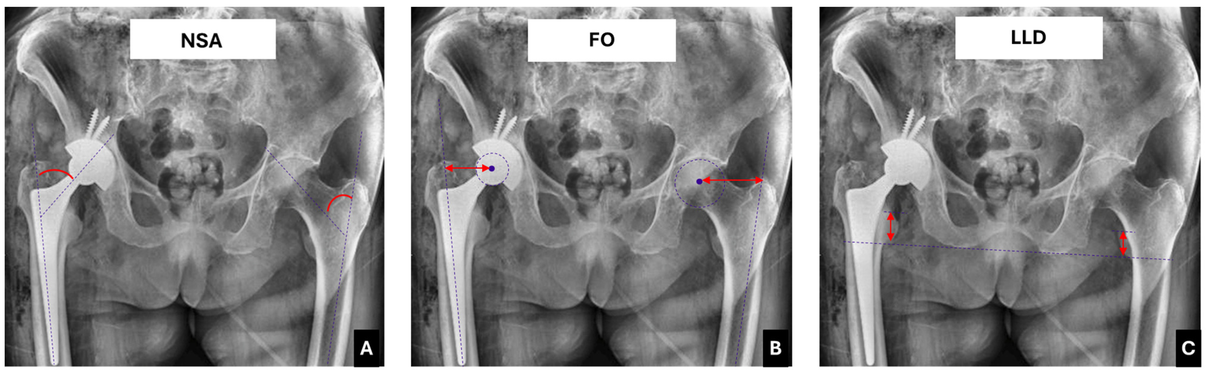
2. Materials and Methods

Patients undergoing THA following a failed trochanteric intramedullary nailing at our level I trauma center between January 2021 and June 2024 were retrospectively recruited based on specific inclusion and exclusion criteria. Ethical approval was obtained from our clinical research ethics board following the 1964 Declaration of Helsinki, and all the patients provided informed consent before participating in the study.
Inclusion criteria: age over 65 years; good cognitive state; osteoporosis, defined as a lumbar T-score ≤ −2.5 (all the patients underwent a DEXA scan during hospitalization).
Exclusion criteria: moderate cognitive impairment (Mini-Mental State Examination score < 19); history of malignant neoplasms; hemoglobin (Hb) < 10 g/dL at baseline; morbid obesity (body mass index (BMI) ≥ 35 kg/m2); medical conditions or cognitive disorders preventing follow-up participation; concomitant lower limb fractures; traumatic head injury.
Patient demographics, including age, sex, BMI, and comorbidities (atrial fibrillation, previous myocardial ischemia, diabetes mellitus, and chronic kidney disease) were recorded at recruitment. All the patients underwent total hip arthroplasty using the Hardinge approach [18], with the patient positioned in the lateral decubitus. Adhesions around scar tissue were cleared to fully expose the trochanter, allowing assessment of the greater and lesser trochanters and the calcar femorale.
The same surgical and anesthesiology team performed all the procedures under spinal anesthesia. Cefazolin (2 g intravenously) or, if contraindicated, clindamycin (600 mg intravenously) was administered 60 min before incision. For antithrombotic prophylaxis, subcutaneous injections of low-molecular-weight heparin (enoxaparin, 4000 UI once daily, starting 12 h after surgery) were given until patients could bear full weight.
The postoperative pelvis radiographs were analyzed to determine whether the normal biomechanical geometry of the operated hip had been restored. The following parameters were recorded: the femoral neck shaft angle (NSA) and the femoral offset (FO), which were compared with the unaffected contralateral side, and the limb length discrepancy (LLD), assessed as the difference between the bisichial line and the lesser trochanter (Figure 1).
Clinical assessment was performed using the following scores: the Harris hip score (HHS) and the visual analog scale for pain (VAS). All the patients underwent a minimum 24-month follow-up, and orthopedic complications at the final follow-up were recorded.
Statistical analysis was conducted using STATA/MP 14 for Windows (StataCorp LP, College Station, TX, USA), and all the data were described as the means and standard deviation. The Kolmogorov–Smirnov test showed a normal distribution of the data. The independent samples t-test and the Pearson correlation test were performed. The tests were two-tailed; a p-value equal to or less than 0.05 was considered significant.

3. Results

The main data of the study are presented in Table 1. A total of 31 patients met the inclusion and exclusion criteria (10 males and 21 females; mean age: 76.2 years, range: 66–90 years) and were included in this study. The patients were initially referred to our orthopedic unit with the following fracture patterns: 31-A1.2 (15 out of 31; 48.39%), 31-A1.3 (8 out of 31; 25.8%), 31-A2.2 (5 out of 31; 16.13%), and 31-A2.3 (3 out of 31; 9.7%).
The modes of trochanteric nail failure included the following: cut-out in 22 cases (70.97%), non-union in 4 cases (12.9%), peri-implant fracture in 1 case (3.23%), cut-through in 2 cases (6.45%), and femoral head avascular necrosis (HAN) in 2 cases (6.45%).
Regarding preoperative comorbidities, 9 patients out of 31 (29%) suffered from atrial fibrillation; previous myocardial ischemia was noted in 3 patients out of 31 (9.7%), diabetes mellitus was observed in 17 patients out of 31 (54.84%) and chronic kidney disease was found in 12 patients out of 31 (38.71%).
The average operating time was 102 min (88–191 min). The average blood loss was 464 mL (range: 380–1320 mL)—a total of 17 patients (54.84%) required transfusion of 3 or more blood units.
Table 2 shows the features of the implanted hip prosthesis. All the patients underwent the nail-to-THA conversion. Long stems were used in 21 patients out of 31 (67.74%), while dual-mobility cups were implanted in 24 patients out of 31 (77.41%).
The restoration of the normal hip geometry parameters was seldom obtained during the nail-to-THA conversion surgical procedure. Hence, a significant mean neck shaft angle (NSA) increase of 7.4° (p < 0.001) and a significant mean femoral offset reduction (FO, mean = 9 mm; p ≤ 0.001) compared to the contralateral native hip were recorded. Moreover, a postoperative limb length discrepancy (LLD) of 8.35 mm was observed (Table 2).
Table 3 shows the correlation between the radiographic parameters and the clinical scores. A significant correlation between HHS and ∆NSA (p = 0.01) and ∆FO (p = 0.003) was observed.
Table 4 summarizes the orthopedic complications observed at the final follow-up.

4. Discussion

Intertrochanteric femur fractures (IFFs) are common fragility fractures affecting the elderly [1]. IFFs require surgical management; hence the following surgical options are available: cephalomedullary nails, compression hip screws, and the paracortical proximal plate [2,3]. However, despite advancements in surgical techniques, internal fixation still faces a failure rate of 3–12% [3,4]. The factors contributing to fixation failure include poor bone quality, fracture comminution, biomechanical instability, inadequate reduction, and suboptimal fixation [3,4,5].
Even though significant technological advancements have been observed in the past two decades, mechanical and biological complications still occur, leading to potentially serious outcomes. Mechanical complications include issues such as cut-outs, cut-throughs, peri-implant fractures, and implant fractures.
In the present study, we recruited patients referred to our level I trauma center with failed trochanteric intramedullary nailing and managed with surgical conversion from a cephalomedullary nail to THA.
Several factors contribute to nail failure, such as a tip-to-apex distance (TAD) greater than 25 mm, unstable fracture patterns, poor fracture reduction leading to varus deformity, and improper positioning of the lag screw in the femoral neck [5,6,7].
Medial mobilization of the screw or blade toward the hip joint, known as cut-through, is another complication [6,7,8].
For failed IFF internal fixation, salvage procedures such as revision osteosynthesis or conversion to hip arthroplasty are typically considered [5]. These procedures aim to reduce pain and improve functional recovery [5].
Revision osteosynthesis is often performed in patients under 50 years of age with good bone quality and high life expectancy [9]. It involves femoral head preservation and re-fixation through hardware exchange, bone graft augmentation, and re-insertion of a blade or screw, either cemented or uncemented [10].
Conversely, conversion to total hip arthroplasty (THA) is usually preferred for elderly patients with severely compromised joint surfaces, as it allows for earlier weight-bearing and mobilization [11]. However, the nail-to-THA conversion is a complex procedure, often hindered by challenges such as bone loss, deformity, poor bone quality, and the presence of non-union or malunion, which can affect surgical outcomes [12].
In the present study, we described the complexity of this salvage procedure, focusing on the postoperative analysis of the prosthesized joint compared to the contralateral native hip. Hence, 31 patients (10 males and 21 females; mean age: 76.2 years, range: 66–90 years) undergoing the nail-to-THA conversion were included in this study.
Postoperative radiographic analysis of the prosthesized hip showed a significant mean NSA increase (p < 0.001) and a significant mean FO reduction (p ≤ 0.001) compared to the contralateral hip. Moreover, a limb length discrepancy (LLD) of 8.35 mm was recorded (Table 2). A significant correlation between HHS and ∆NSA (p = 0.01) and ∆FO (p = 0.003) was also observed (Table 3).
Nonetheless, ∆NSA, ∆FO, and LLD had no impact on postoperative hip pain, as no significant correlation was found between these radiographic parameters and the postoperative VAS. These findings highlight that radiologic parameters affect the postoperative hip function and, consequently, the quality of life, but a direct connection with the postoperative pain was not identified. Pain is a subjective perception influenced by various biopsychosocial factors. The recruited patients experienced acute pain when reporting the PPF, postoperative pain after femoral nailing, and still had continuous pain from nailing failure until the nail-to-THA conversion. All these subjective experiences influenced the patients’ perception, leading to significant pain improvement after THA surgery.
Dual-mobility cups were used in 24 patients out of 31 (77.41%) to avoid postoperative THA dislocation and/or implant instability. The two cases of THA dislocation and implant instability were observed in the patients who did not receive dual-mobility cups. These patients underwent further THA revision surgery.
Long stems were used in 21 patients out of 31 (67.74%); the choice of this kind of stem relied on the quality of the femoral metaphysis bone. In the patients with impaired metaphyseal bone quality, long stems were preferred to reduce the risk of periprosthetic fractures and stem mobilization. Three cases of periprosthetic femoral fractures were observed in the patients who received short stems.
The two cases of late periprosthetic joint infections were observed in two diabetic patients in the present study. The patients needed a further hip revision surgery, together with a targeted antibiotic therapy.
These findings highlight that the nail-to-THA conversion surgical procedures, performed for IFF fixation failure, are complex surgeries, during which the biomechanical restoration of the native hip biomechanics is rarely obtained [17]. These biomechanical findings may negatively impact the postoperative clinical outcome, thus predisposing to postoperative pain and implant-related complications [17].
Based on the findings of the current study, it is essential to carefully plan surgical treatment for patients presenting with PFFs to reduce the risk of femoral nailing failure. Revision surgery in these cases should be viewed as a salvage procedure, and this aspect should be detailed in the informed consent to correctly inform the patient about the expected postoperative outcome.
The limitations of the present study could not be overcome. Although the study sample was relatively small, the applied inclusion and exclusion criteria enhanced patient selection. Additionally, the retrospective nature of the study presented another limitation. However, the risk of bias was mitigated by standardizing the study protocol, with all the nail-to-THA conversion procedures performed by the same orthopedic and anesthesiology team.

5. Conclusions

Hip prosthesis implantation after failed femoral intramedullary fixation is a challenging surgery and should be considered a revision procedure.
Orthopedic surgeons should be aware that normal hip geometry may not be achieved during the nail-to-THA conversion, and this may lead to an increased risk of implant-related complications.
Future research in this area may support the adoption of emerging technologies, such as preoperative three-dimensional (3D) planning, navigation surgery, and robotic surgery, in the context of the nail-to-THA conversion. These modalities may offer valuable assistance to hip surgeons in optimizing the restoration of native hip geometry and biomechanics during revision procedures.

Author Contributions

Conceptualization, G.S. and D.B.; methodology, G.S.; formal analysis, D.B.; investigation, G.G., F.D.C., G.C. and G.D.G.; data curation, G.G.; writing—original draft preparation, G.G. and D.B.; writing—review and editing, D.B.; project administration, G.S. All authors have read and agreed to the published version of the manuscript.

Funding

This research received no external funding.

Institutional Review Board Statement

The study was conducted under the Declaration of Helsinki and approved by the Ethics Committee of Policlinico di Bari (protocol code 5559/2018).

Informed Consent Statement

Informed consent was obtained from all the subjects involved in the study.

Data Availability Statement

The data that support the findings of this study are available from the corresponding author upon reasonable request.

Conflicts of Interest

The authors declare no conflicts of interest.

References

  1. Kanis, J.A.; Oden, A.; McCloskey, E.V.; Johansson, H.; Wahl, D.A.; Cooper, C.; IOF Working Group on Epidemiology and Quality of Life. A systematic review of hip fracture incidence and probability of fracture worldwide. Osteoporos. Int. 2012, 23, 2239–2256. [Google Scholar] [CrossRef] [PubMed]
  2. Mattisson, L.; Bojan, A.; Enocson, A. Epidemiology, treatment and mortality of trochanteric and subtrochanteric hip fractures: Data from the Swedish fracture register. BMC Musculoskelet. Disord. 2018, 19, 369. [Google Scholar] [CrossRef] [PubMed]
  3. Innocenti, M.; Civinini, R.; Carulli, C.; Matassi, F. Proximal femoral fractures: Epidemiology. Clin. Cases Miner. Bone Metab. 2009, 6, 117–119. [Google Scholar] [PubMed]
  4. Sambrook, P.; Cooper, C. Osteoporosis. Lancet 2006, 367, 2010–2018, Erratum in Lancet 2006, 368, 28. [Google Scholar] [CrossRef] [PubMed]
  5. Piazzolla, A.; Bizzoca, D.; Solarino, G.; Moretti, L.; Moretti, B. Vertebral fragility fractures: Clinical and radiological results of augmentation and fixation—A systematic review of randomized controlled clinical trials. Aging Clin. Exp. Res. 2020, 32, 1219–1232. [Google Scholar] [CrossRef] [PubMed]
  6. Veronese, N.; Maggi, S. Epidemiology and social costs of hip fracture. Injury 2018, 49, 1458–1460. [Google Scholar] [CrossRef] [PubMed]
  7. Fox, K.M.; Magaziner, J.; Hebel, J.R.; Kenzora, J.E.; Kashner, T.M. Intertrochanteric versus femoral neck hip fractures: Differential characteristics, treatment, and sequelae. J. Gerontol. Ser. A Biomed. Sci. Med. Sci. 1999, 54, M635–M640. [Google Scholar] [CrossRef] [PubMed]
  8. Zhang, K.; Zhang, S.; Yang, J.; Dong, W.; Wang, S.; Cheng, Y.; Al-Qwbani, M.; Wang, Q.; Yu, B. Proximal femoral nail vs. dynamic hip screw in treatment of intertrochanteric fractures: A meta-analysis. Med. Sci. Monit. 2014, 20, 1628–1633. [Google Scholar] [CrossRef] [PubMed]
  9. Tawari, A.A.; Kempegowda, H.; Suk, M.; Horwitz, D.S. What made an intertrochanteric fracture unstable in 2015? Does the lateral wall play a role in the decision matrix? J. Orthop. Trauma 2015, 29 (Suppl. 4), S4–S9. [Google Scholar] [CrossRef] [PubMed]
  10. Lu, Y.; Uppal, H.S. Hip Fractures: Relevant Anatomy, Classification, and Biomechanics of Fracture and Fixation. Geriatr. Orthop. Surg. Rehabil. 2019, 10, 2151459319859139. [Google Scholar] [CrossRef] [PubMed]
  11. Marsillo, E.; Pintore, A.; Asparago, G.; Oliva, F.; Maffulli, N. Cephalomedullary nailing for reverse oblique intertrochanteric fractures 31A3 (AO/OTA). Orthop. Rev. 2022, 14, 38560. [Google Scholar] [CrossRef] [PubMed]
  12. Queally, J.M.; Harris, E.; Handoll, H.H.; Parker, M.J. Intramedullary nails for extracapsular hip fractures in adults. Cochrane Database Syst. Rev. 2014, 9, CD004961. [Google Scholar] [CrossRef] [PubMed]
  13. Bizzoca, D.; Bortone, I.; Vicenti, G.; Caringella, N.; Rifino, F.; Moretti, B. Gait analysis in the postoperative assessment of intertrochanteric femur fractures. J. Biol. Regul. Homeost. Agents 2020, 34 (Suppl. 3), 345–351. [Google Scholar] [PubMed]
  14. Méndez-Ojeda, M.M.; Herrera-Rodríguez, A.; Álvarez-Benito, N.; González-Pacheco, H.; García-Bello, M.A.; Álvarez-de la Cruz, J.; Pais-Brito, J.L. Treatment of Trochanteric Hip Fractures with Cephalomedullary Nails: Single Head Screw vs. Dual Integrated Compression Screw Systems. J. Clin. Med. 2023, 12, 3411. [Google Scholar] [CrossRef]
  15. Zeelenberg, M.L.; Nugteren, L.H.T.; Plaisier, A.C.; Loggers, S.A.I.; Joosse, P.; Den Hartog, D.; Verhofstad, M.H.J.; van Lieshout, E.M.M.; STABLE-HIP Study Group. Extramedullary versus intramedullary fixation of stable trochanteric femoral fractures: A systematic review and meta-analysis. Arch. Orthop. Trauma Surg. 2023, 143, 5065–5083. [Google Scholar] [CrossRef] [PubMed]
  16. Haidukewych, G.J.; Berry, D.J. Hip arthroplasty for salvage of failed treatment of intertrochanteric hip fractures. J. Bone Jt. Surg. Am. 2003, 85, 899–904. [Google Scholar] [CrossRef]
  17. Solarino, G.; Bizzoca, D.; Dramisino, P.; Vicenti, G.; Moretti, L.; Moretti, B.; Piazzolla, A. Total hip arthroplasty following the failure of intertrochanteric nailing: First implant or salvage surgery? World J. Orthop. 2023, 14, 763–770. [Google Scholar] [CrossRef] [PubMed]
  18. Hardinge, K. The direct lateral approach to the hip. J. Bone Jt. Surg. Br. 1982, 64, 17–19. [Google Scholar] [CrossRef] [PubMed]
  19. Zeng, X.; Zhan, K.; Zhang, L.; Zeng, D.; Yu, W.; Zhang, X.; Zhao, M. Conversion to total hip arthroplasty after failed proximal femoral nail antirotations or dynamic hip screw fixations for stable intertrochanteric femur fractures: A retrospective study with a minimum follow-up of 3 years. BMC Musculoskelet. Disord. 2017, 18, 38. [Google Scholar] [CrossRef] [PubMed]
  20. Yu, W.; Han, X.; Chen, W.; Mao, S.; Zhao, M.; Zhang, X.; Han, G.; Ye, J.; Chen, M.; Zhuang, J. Conversion from a failed proximal femoral nail anti-rotation to a cemented or uncemented total hip arthroplasty device: A retrospective review of 198 hips with previous intertrochanteric femur fractures. BMC Musculoskelet. Disord. 2020, 21, 791. [Google Scholar] [CrossRef] [PubMed]
Figure 1. The figure shows the radiographic parameters recorded. (A) The femoral neck shaft angle (NSA) is shown in red on both sides; (B) the femoral offset (FO) is shown in red on both sides; (C) the limb length discrepancy (LLD), assessed as the difference between the bis-ischial line and the lesser trochanter, is the difference between the two red lines.
Prosthesis 07 00050 g001
Table 1. Main data of the study.
Patients (n = 31)
Age
 Mean ± SD76.2 ± 6.84
Gender
 Female, n (%)21 (67.74%)
BMI (kg/m2)
 Mean ± SD27.8 ± 4.76
Side
 Right, n (%)16 (51.61%)
Timing from nailing to THA
(mean ± SD, months)
11.86 ± 10.3
Mean operating time
(mean ± SD, minutes)
101 ± 48.73
Patients undergoing ≥3
blood transfusions, n (%)
17 (54.84%)
VAS
(mean ± SD, at the final follow-up)
2.3 ± 3.45
Note: SD, standard deviation; BMI, body mass index; THA, total hip arthroplasty; VAS, visual analog scale for pain.
Table 2. Radiographic study findings.
Healthy HipAffected Hipp
Neck shaft angle (NSA)127.7 ± 5.6°135.1 ± 2.4°7.4 ± 2.6°<0.001 *
Femoral offset (FO)42.4 ± 2.7 mm51.4 ± 7.42 mm9 ± 3.2 mm<0.001 *
Limb length discrepancy (LLD)--8.35 ± 2.25 mm-
Note: * significant p-value (independent samples t-test).
Table 3. Pearson correlation test between the radiographic parameters and the clinical scores.
HHSVAS
RpRp
∆NSA−0.610.01 *0.410.095
∆FO−0.560.03 *0.240.176
∆LLD−0.210.1120.130.223
Note: * significant p-value (Pearson correlation test); HHS, Harris hip score; VAS, visual analog scale for pain.
Table 4. Orthopedic complications at the final follow-up.
Patients (n = 31)
NPercentage
Periprosthetic fractures39.6%
Implant instability13.23%
THA dislocation13.23%
Periprosthetic Joint Infections26.45%
Disclaimer/Publisher’s Note: The statements, opinions and data contained in all publications are solely those of the individual author(s) and contributor(s) and not of MDPI and/or the editor(s). MDPI and/or the editor(s) disclaim responsibility for any injury to people or property resulting from any ideas, methods, instructions or products referred to in the content.

