Article

Cordia Myxa Fruit Extract Antibacterial Efficacy and Its Effect on the Surface Roughness of Heat-Cured Acrylic Denture Base Material

by
Noor Riadh Taha
and
Shorouq Majid Abass
*
Department of Prosthodontics, College of Dentistry, University of Baghdad, Baghdad 1417, Iraq
*
Author to whom correspondence should be addressed.
Prosthesis 2025, 7(3), 48; https://doi.org/10.3390/prosthesis7030048
Submission received: 7 April 2025 / Revised: 30 April 2025 / Accepted: 6 May 2025 / Published: 8 May 2025

Abstract

Background/Objectives: Using chemical disinfectants to clean the base of heat-cured acrylic dentures has several negative effects, including toxicity. On the other hand, therapeutic herbs have fewer adverse effects. This study intended to determine how the antibacterial efficacy and the surface roughness of the heat-cured acrylic material used to fabricate dentures were affected by the use of Cordia myxa fruit (CMF) extract as a disinfection solution for dentures using the immersion technique. Methods: Heat-cured acrylic specimens were prepared (N = 110; 55 specimens) for each test; each group contained five specimens. Three CMF extract concentrations (50, 100, and 150 mg/mL) were made and examined for three immersion times (5, 10, and 15 min). The results were compared to the first control group, which used distilled water, and the second group, which used 2% glutaraldehyde for ten minutes, in accordance with the guidelines. One way analysis of variance ANOVA and Games–Howell post hoc test were employed in SPSS (Statistical Package for the Social Sciences) program for statistical analysis. Results: The results for the antibacterial test revealed that CMF solutions had a statistically significant difference in all test groups in comparison with the first control group and non-significant differences with (H p = 0.92; J p = 0.278; K p = 0.303) groups in comparison with the second control group (Glutaraldehyde 2%). For the surface roughness test, the effect was not statistically significant for all groups compared to the first and second control groups. Conclusions: It can be concluded that immersing the heat-cure acrylic samples in a solution of 100 mg/mL CMF extract for 15 min, and 150 mg/mL for 10 and 15 min, has an antibacterial effect similar to that of the Glutaraldehyde 2% antiseptic and no negative effect on surface roughness.

1. Introduction

Heat-polymerized acrylic resin has been the most widely used material for dentures for over 60 years [1]. The fabrication of dentures involves a multi-step process that includes both laboratory and clinical procedures. During these stages, dentures can harbor pathogenic and opportunistic microorganisms, posing a risk of cross-contamination [2]. Inadequate disinfection may allow microorganisms to enter the dental laboratory environment, threatening the safety of patients, dental laboratory personnel, and clinicians. Therefore, implementing effective infection control measures is essential to break one or more of the links in the chain of infection and prevent the spread of disease [3].
According to the Centers for Disease Control and Prevention (CDC) guidelines, dental prostheses, impressions, and other prosthodontic materials should be thoroughly cleaned, disinfected using an Environmental Protection Agency (EPA)-registered hospital disinfectant with a tuberculocidal claim, and then appropriately rinsed. For heat-sensitive semi-critical objects, the CDC recommends using glutaraldehyde, a high-level disinfectant. However, glutaraldehyde, despite its potent sporicidal properties, is highly toxic. Even with proper precautions, such as the use of closed containers to limit vapor release, chemically resistant gloves and aprons, goggles, and face shields, there have been reports of skin sensitivity, respiratory issues, dermatological effects, and eye irritation. Additionally, standard medical gloves do not offer adequate protection, as they are not chemically resistant to glutaraldehydes [4].
Medicinal plants offer accessible and cost-effective therapy alternatives, particularly in areas with restricted access to contemporary medicine, and they also support the formulation of numerous pharmaceutical medications [5].
Cordia myxa fruit (CMF), belonging to the Boraginaceae family, is a flowering plant that grows in tropical and subtropical regions of Africa, Australia, and Asia [6]. The CMF tree is referred to as “Bumber” in Iraq. It has pharmaceutical and medicinal applications and shows antibacterial activity and antioxidant qualities in vitro [7]. Studies have shown that CMF extract exhibits antimicrobial activity against Gram-positive and Gram-negative bacteria, as well as various fungal species [8,9].
Alyamani et al. [10] manufactured an antibacterial wound dressing using CMF extract incorporated into polycaprolactone/chitosan (PCL/CH) nanofibers as an antimicrobial agent. The agent demonstrated significant antibacterial activity against both Gram-positive and Gram-negative bacteria.
Similarly, Zahra et al. [5] evaluated the antibacterial activity of CMF extract and reported the inhibition of bacterial growth against Pseudomonas aeruginosa, Staphylococcus aureus, and Klebsiella pneumoniae.
To the best of the authors’ knowledge, no other research has investigated the use of CMF extract as a disinfectant for heat-cured acrylic materials or its effective immersion duration. As CMF fruit is safe, readily available in the region, cost-effective, and holds cultural significance due to its therapeutic properties, it presents a promising candidate for such applications.
The study proposes two null hypotheses:
  • CMF extract has an antibacterial effect when used via the immersion technique on heat-cured acrylic denture base material.
  • The immersion of heat-cured acrylic in CMF extract does not adversely affect its surface roughness.
Correspondingly, the alternative hypotheses are:
  • CMF extract does not have an antibacterial effect when used via the immersion technique on heat-cured acrylic.
  • The immersion of heat-cured acrylic in CMF extract adversely affects its surface roughness.
The aim of this study was to evaluate the antibacterial efficiency of CMF extract at different concentrations (50, 100, and 150 mg/mL) when used as a disinfectant solution via the immersion technique with varying durations (5, 10, and 15 min). Additionally, the study aimed to investigate the effect of CMF extract immersion on the surface roughness of heat-cured acrylic denture base materials

2. Materials and Methods

2.1. Preparation of Disinfectants

This study was conducted using the following two types of disinfectants: glutaraldehyde 2% (as a control disinfectant agent) and CMF alcoholic extract solutions.
Glutaraldehyde 2% was obtained ready-to-use from the National Health Factory for the Production Of Disinfectants (M.P.C., Baghdad, Iraq).

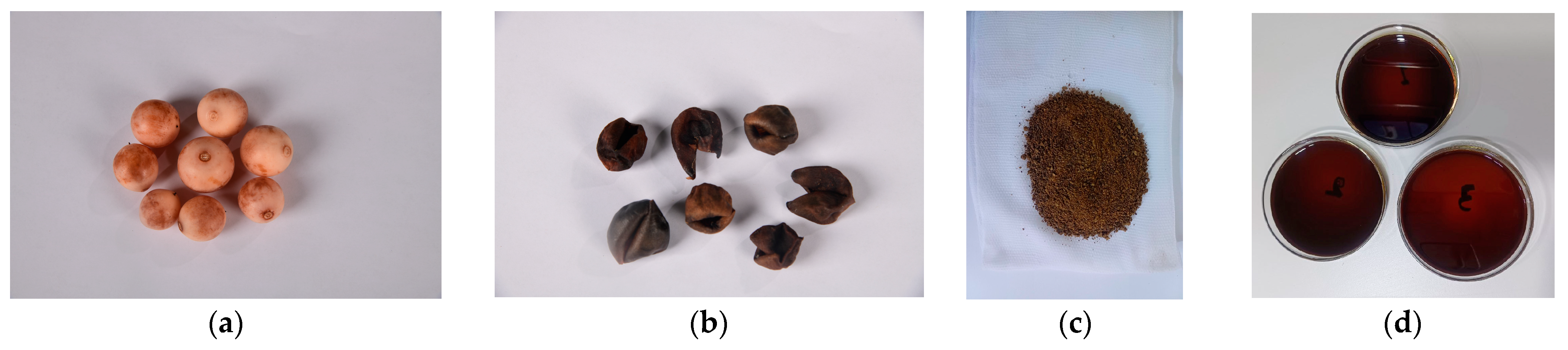
2.2. CMF Collection, Identification, Extraction, and Solution Preparation

Ripe fruits of Cordia myxa (Boraginacea family) were collected from a tree in AL Jamaa Neighborhood in Baghdad city, Iraq (33°19′18.0″ N, 44°18′41.9″ E). The Iraqi National Herbarium, Baghdad, Iraq, identified the fruit with certificate no.1743 on 18 March 2025. Only physically intact fruits were selected. The cleaning procedure involved removing dirt and extra genus materials and washing the fruits three times with running tap water and once with distilled water [7].
The fruits’ seeds were extracted by pressing them [11], and they were then allowed to dry for two weeks in the shade. A thermometer (THLMT, China) was used to measure the temperature and humidity three times a day. The average was 40 °C with 25% relative humidity. Lastly, an electric blender (Kenwood FDM 30, China) was used to grind the dried fruit into powder [7,11].
Petroleum ether (40:60) was used to defatten the powdered substance [12,13]. Extraction was carried out with 70% ethanol [12,13] and the Soxhlet technique [14]. Whatman filter sheets No. 1 were then used for filtering [7]. To make sure the solvent was completely removed, the extract was concentrated under vacuum in a rotary evaporator at 40–50 °C until a viscous, black residue was left behind [13,14]. The yield percentage was 47%. Phytochemical screening tests were carried out to check the quality of the extract before the experimental test [15]. The extract was kept in the refrigerator in a non-transparent glass container to prevent decomposition [14]. The extract was caramel-like in consistency and color, as shown in Figure 1.
At the time of the experimental tests, concentrations of 50, 100, and 150 mg/mL were prepared through reconstituting the crude extract in nonionized distilled water (AL-Rafidain Environment Office, Iraq) [16].

2.3. Specimen Preparation

A disk-shaped specimen measuring 5 mm in diameter and 2 mm in thickness was created for the antibacterial test based on the measurements of the test tubes [17].
For the test of surface roughness, a rectangular specimen with dimensions of 65 × 10 × 2.5 mm for length, width, and thickness was created, in compliance with American Dental Association Specification No.12 [18].
Molds were prepared using type III dental stone (Zhermack, Italy) in accordance with the traditional flasking process for removable dentures [19].
According to the manufacturer’s instructions, heat-cured acrylic resin was mixed by adding 14 milliliters of liquid for every 34 g of powder. The packing process began when the mixture reached the dough stage. After the mold was filled with acrylic rolls, a polyethylene sheet was placed on top of the acrylic dough [20]. Polyester sheets were used for surface roughness specimens. The flask portions were then collected and placed under a hydraulic press, which applied a gradual pressure of up to 20 bar to ensure that the acrylic was distributed evenly in the mold space. The flask was then reopened to remove the polyethylene/polyester sheet and remove any excess material using a sharp blade.
By applying 100 bar of pressure to the flask for five minutes, the following experiments were carried out without the use of polyethylene sheets (the polyester sheet was then placed back over the resin for the surface roughness specimens) [20].
For the curing process, the flask clamp was submerged in a digital water bath, in compliance with the guidelines provided by the manufacturer. Figure 2 shows the specimens after curing and before removal from the flasks.
Specimens were finished through using a stone bur to remove excess material, and then they were polished (with the exception of the surface roughness specimens, which were left unpolished) through hand abrasion with 400 grit emery paper, used ten times in a figure-of-eight motion. They were then polished for 60 s using a brush disk and pumice slurry [21]. After that, the specimens underwent a 20 min ultrasonic cleaning [22].
Before the test was established, the specimens were stored in distilled water for 48 h [23].

2.4. Test Groups

A total of 110 heat-cured acrylic specimens (SMD, Turkey) were prepared, which included 55 specimens for the antibacterial test and 55 specimens for the surface roughness test. Five specimens were used for each test group; sample size was determined according to previous studies [24,25]. The test groups were as follows:
Control group (A): Acrylic specimens were immersed in distilled water. This test group served as the negative control group.
Glutaraldehyde 2% test group (B): Acrylic specimens were immersed in glutaraldehyde solution for 10 min. This test group served as the positive control group.
CMF 50a test group (C): Acrylic specimens were immersed in 50 mg\mL CMF extract for 5 min.
CMF 50b test group (D): Acrylic specimens were immersed in 50 mg\mL CMF extract for 10 min.
CMF 50c test group (E): Acrylic specimens were immersed in 50 mg\mL CMF extract for 15 min.
CMF 100a test group (F): Acrylic specimens were immersed in 100 mg\mL CMF extract for 5 min.
CMF 100b test group (G): Acrylic specimens were immersed in 100 mg\mL CMF extract for 10 min.
CMF 100c test group (H): Acrylic specimens were immersed in 100 mg\mL CMF extract for 15 min.
CMF 150a test group (I): Acrylic specimens were immersed in 150 mg\mL CMF extract for 5 min.
CMF 150b test group (J): Acrylic specimens were immersed in 150 mg\mL CMF extract for 10 min.
CMF 150c test group (K): Acrylic specimens were immersed in 150 mg\mL CMF extract for 15 min. Table 1 shows the group coding.

2.5. Antibacterial Efficiency Test

By the use of the colony-forming unit (CFU) method with CMF extract on the Bacillus subtilis-colonized bacteria present on acrylic specimens after immersion for various time intervals (5, 10, and 15 min) for each test group, this test was carried out to assess the antibacterial efficiency.
All specimens were kept in sterile containers after being disinfected for 15 min at 121 °C and 15 Psi in an autoclave [25].
According to the guidelines of the Centers for Disease Control and Prevention (2016), Bacillus subtilis was employed as a biological indicator of successful disinfection [3].
These bacteria were obtained from the Scientific Research Commission/Center for Environment, Water, and Renewable Energy/Food Contamination Research Lab. They were identified via PCR. A small number of Bacillus subtilis colonies were individually injected into 10 mL of sterile Tryptic Soy Broth (TSB) on the first day, and they were then incubated aerobically for 24 h at 37 degrees Celsius.
On the second day, the tubes’ turbidity was adjusted to resemble that of McFarland tube No. 5, which is equivalent to 107 organisms/mL. Under sterile conditions, 100 μL of each prior test tube was inoculated into 10 mL of TSB. Following that, each specimen was placed separately into the inoculation tubes and incubated for 24 h at 37 °C in an aerobic environment. Following that, each specimen was subjected to a distinct disinfection immersion period based on the test groups, as described previously in Table 1 and shown in Figure 3.
Following the completion of the disinfection solution and control treatments, the broth was thrown away, and each specimen was put into a test tube with 10 mL of sterile saline [25]. The quantity of microorganisms in the serial dilution was determined after the tubes were subjected to a brief 10 W vortex for 20 s [26]. For this, 1 mL of normal saline from the first tube, containing the specimens, was added to 9 mL of sterile normal saline. This resulted in a total amount of 10 mL, which formed the first dilution (10-1). Subsequent dilutions were made using the same technique [25]. Nutrient agar plates were filled with 100 µL of the second dilution [27], and the plates were incubated aerobically for 24 h at 37 °C [25]. Colonies were counted using the colony-counter device (Gallenkamb, Cambridge, England) with help of a blinded operator. All five replicates were counted the same way for each group. The following formula was used to determine the final count (number of bacteria per milliliter) during plate incubation [28]:
( C F U / m L ) = c o l o n y   n o d i l u t i o n   f a c t o r v o l u m   o f   c u l t u r e   p l a t e    

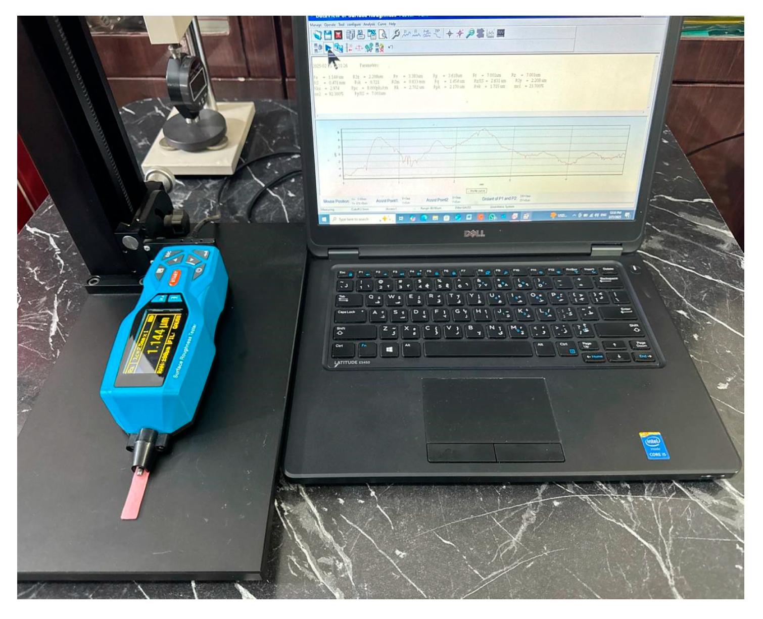
2.6. Surface Roughness Test

The profilometer (VTSYIQI, China) was used after calibration to calculate the surface roughness (Ra, µm). Ra, as determined from a mean line within a predetermined specimen length, on the unpolished surface (where the polyester sheets were used), is the mathematical mean of the absolute values of the measured profile height of surface imperfections [29], as shown in Figure 4.

2.7. Statistical Analysis

The data were evaluated using SPSS (Statistical Package for the Social Sciences) program version 27 (IBM Corp., NY, USA).
Descriptive statistics, one-way analysis of variance ANOVA, and Games–Howell post hoc tests were conducted for all test groups with significance levels set at p < 0.05.

3. Results

3.1. Antibacterial Efficiency (CFU) Test

Table 2 and Figure 5 show the means of the CFU for all test groups; the highest mean was obtained for control group A (202.00).
The one-way ANOVA analysis of CFU showed a significant difference between all test groups, p = 0.000, of <0.05, as shown in Table 3.
The analysis with the Games–Howell post hoc test between control group A and the test groups showed a significant difference (p < 0.05) with all test groups p = 0.00. The comparison of the B group with all test groups was significant (p < 0.05), except for the H p = 0.925, J p = 0.278, and K p= 0.303 groups, for which the comparison was insignificant (p > 0.05), as shown in Table 4.

3.2. Surface Roughness Test

Table 5 and Figure 6 present the results of the surface roughness changes; the I group had a higher mean (1.77).
The one-way ANOVA analysis showed that the difference was insignificant, p = 0.107, between all the test groups (p value < 0.05), as shown in Table 6.

4. Discussion

The current investigation demonstrated that the alcoholic extract of Cordia myxa fruit (CMF) exhibited an antibacterial effect against Gram-positive Bacillus subtilis bacteria, with the effect increasing as both the concentration and duration of immersion rose.
The antibacterial activity is attributed primarily to the phenolic compounds, especially flavonoids, which act through several mechanisms, including the adsorption and disruption of microbial membranes, ion chelation, enzyme inhibition, and interference with membrane transporters [7,30]. It is well established that the bioactive compounds in Cordia myxa exert three major effects on bacteria: they inhibit bacterial attachment, suppress bacterial enzymes, and lead to alterations in bacterial cell surface proteins [5].
Almusawi et al. [9] evaluated the bioactive components of CMF ethanol extract using GC-MS and HPLC analyses. GC-MS identified 19 major compounds, notably α-D-glucopyranoside and O-α-D-glucopyranosyl-(1→3)-β-D-fructofuranosyl, which were present in significant amounts and have been associated with antimicrobial activity. HPLC analysis further detected gallic acid, ferulic acid, chlorogenic acid, caffeic acid, and fumaric acid in the CMF extract, with gallic acid being notably abundant and recognized for its antibacterial properties.
In the present study, the most effective concentration against B. subtilis, determined via the mean differences, was 100 mg/mL. This finding aligns with the results of Almusawi et al. [7] Similarly, Alyamani et al. [10] demonstrated that CMF fruit extract-loaded polycaprolactone/chitosan (PCL/CH) nanofibrous mats exhibited antibacterial efficacy against B. subtilis at the same concentration of 100 mg/mL.
The surface roughness analysis revealed no statistically significant increase (p > 0.05) following immersion in CMF solutions at various concentrations and durations.
These results are consistent with those of Abed [31], who found that immersion in electrolyzed water did not significantly alter acrylic roughness.
Comparable findings were reported by Heidrich et al. [32], who observed that propolis extract, castor oil, and rosemary extract did not negatively affect acrylic resin surface roughness until after four months. Similarly, Salman et al. [33] found that the surface roughness of heat-polymerized acrylic resin remained unchanged after immersion in several denture-cleaning solutions. Noori et al. [24] also noted insignificant changes in surface roughness following a 10 min immersion in Tea Tree Oil, which was used as a denture cleanser.
However, these findings contrast with those of Porwal et al. [29], who reported significant surface roughness changes in heat-cured acrylic after 180 days of immersion in sodium perborate and sodium hypochlorite denture cleansers.
Thus, CMF extract solution appears to be safe for use as a natural antibacterial denture disinfectant. It offers a cost-effective and culturally significant alternative, avoiding the hazards and higher costs associated with chemical disinfectants, and does not significantly degrade the surface quality of the acrylic material.
Accordingly, the following conclusions were derived:
  • Null Hypothesis 1 was accepted, and Alternative Hypothesis 1 was rejected.
  • Null Hypothesis 2 was accepted, and Alternative Hypothesis 2 was rejected.
This study was limited by the small sample size in each group, the testing of only one bacterial strain (Bacillus subtilis), and the short-term nature of the exposure.
We suggest that future studies could involve larger sample sizes, the use of multiple bacterial strains, longer exposure times, and an investigation into the effects of CMF extract on other properties of heat-cured acrylic resin.

5. Clinical Implications

The findings suggest that CMF extract can be utilized in dental laboratories and clinics as a natural disinfectant for a heat-cured denture base material. Immersion at a concentration of 100 mg/mL for 15 min or 150 mg/mL for 10–15 min demonstrated antibacterial efficacy comparable to 2% glutaraldehyde.
This could enhance infection control practices in prosthodontics, while minimizing chemical exposure risks to patients and healthcare workers.

6. Conclusions

Cordia myxa fruit extract solutions, at concentrations of 100 mg/mL for 15 min or 150 mg/mL for 10 and 15 min, can effectively disinfect heat-cured acrylic denture base resin via the immersion method, providing antibacterial effects without adversely affecting the material’s surface roughness.

Author Contributions

Conceptualization, N.R.T. and S.M.A.; methodology, N.R.T. and S.M.A.; validation, S.M.A.; formal analysis, N.R.T. and S.M.A.; investigation, N.R.T. and S.M.A.; resources N.R.T.; data curation, S.M.A.; writing—original draft preparation, N.R.T.; writing—review and editing, N.R.T. and S.M.A.; visualization; supervision, S.M.A.; project administration, S.M.A.; funding acquisition, N.R.T. All authors have read and agreed to the published version of the manuscript.

Funding

This research received no external funding.

Institutional Review Board Statement

Not applicable.

Informed Consent Statement

Not applicable.

Data Availability Statement

The data presented in this study are available on request from the corresponding author.

Acknowledgments

The authors would like to acknowledge the support of the University of Baghdad.

Conflicts of Interest

The authors declare no conflict of interest.

References

  1. Heidari, B.; Firouz, F.; Izadi, A.; Ahmadvand, S.; Radan, P. Flexural strength of cold and heat cure acrylic resins reinforced with different materials. J. Dent. 2015, 12, 316. [Google Scholar]
  2. Glass, R.T.; Conrad, R.S.; Bullard, J.W.; Goodson, L.B.; Mehta, N.; Lech, S.J.; Loewy, Z.G. Evaluation of microbial flora found in previously worn prostheses from the Northeast and Southwest regions of the United States. J. Prosthet. Dent. 2010, 103, 384–389. [Google Scholar] [CrossRef]
  3. Centers for Disease Control; GA: Centers for Disease Control Prevention. Summary of infection prevention practices in dental settings: Basic expectations for safe care. J. Atlanta US Dept. Health Prev. Hum. Serv. 2016, 6–15. Available online: https://www.google.com.hk/url?sa=t&source=web&rct=j&opi=89978449&url=https://www.cdc.gov/dental-infection-control/media/pdfs/2024/07/safe-care2.pdf&ved=2ahUKEwj7-PfKr5ONAxWesVYBHQmkBIcQFnoECBUQAQ&usg=AOvVaw3z5FtBelPx5DbwUuWOHzYC (accessed on 5 May 2025).
  4. Kohn, W.G.; Collins, A.S.; Cleveland, J.L.; Harte, J.A.; Eklund, K.J.; Malvitz, D.M. Guidelines for infection control in dental health-care settings-2003. MMWR Recomm. Rep. 2003, 52, 1–61. [Google Scholar]
  5. Zahra, K.; Khan, A.S.; Saeed, A.; Gul, H. Studies on analysis of minerals, phytochemicals, proximate composition, and in vitro biological activities of methanolic extracts from the fruits of Cordia dichotoma Forst and Cordia myxa L. Biol. Sci. 2024, 4, 575–584. [Google Scholar] [CrossRef]
  6. Sivalingam, P.; Singh, D.; Chauhan, S. Morphological and molecular diversity of an underutilized fruit crop-Cordia myxa L. germplasm from the arid region of Rajasthan, India. Genet. Resour. Crop Evol. 2012, 59, 305–316. [Google Scholar] [CrossRef]
  7. Al-Musawi, M.H.; Ibrahim, K.M.; Albukhaty, S. In vitro study of antioxidant, antibacterial, and cytotoxicity properties of Cordia myxa fruit extract. Iran. J. Microbiol. 2022, 14, 97. [Google Scholar] [CrossRef]
  8. Hadi, A.M.A. The effect of methanol and water plant extracts of Cordia myxa on Some pathogenitic bacteria and Candida albicans. Baghdad Sci. J. 2011, 8, 400–405. [Google Scholar] [CrossRef]
  9. Al-Musawi, M.H.; Ibrahim, K.M.; Albukhaty, S. Phytochemical analysis, and anti-microbial activities of ethanol extract of Cordia myxa fruit: In vitro study. Res. J. Pharm. Technol. 2022, 15, 2871–2876. [Google Scholar] [CrossRef]
  10. Alyamani, A.A.; Al-Musawi, M.H.; Albukhaty, S.; Sulaiman, G.M.; Ibrahim, K.M.; Ahmed, E.M.; Jabir, M.S.; Al-Karagoly, H.; Aljahmany, A.A.; Mohammed, M.K. Electrospun polycaprolactone/chitosan nanofibers containing Cordia myxa fruit extract as potential biocompatible antibacterial wound dressings. Molecules 2023, 28, 2501. [Google Scholar] [CrossRef]
  11. Hamdia, M.; Al-Faraji, A. Evaluation of inhibitory activity of Cordia myxa fruit extract on microorganisms that causes spoilage of food and its role in the treatment of certain disease states. Evaluation 2017, 7, 2224–3208. [Google Scholar]
  12. El-Massry, K.F.; Farouk, A.; Mahmoud, K.F.; El-Ghorab, A.H.; Musa, A.; Mostafa, E.M.; Ghoneim, M.M.; Naguib, I.A.; Abdelgawad, M.A. Chemical characteristics and targeted encapsulated Cordia myxa fruits extracts nanoparticles for antioxidant and cytotoxicity potentials. Saudi J. Biol. Sci. 2021, 28, 5349–5358. [Google Scholar] [CrossRef]
  13. Ibrahim, A.Y.; El-Newary, S.A.; Ibrahim, G.E. Antioxidant, cytotoxicity and anti-tumor activity of Cordia dichotoma fruits accompanied with its volatile and sugar composition. Ann. Agric. Sci. 2019, 64, 29–37. [Google Scholar] [CrossRef]
  14. Larki, A.; Shakiba Maram, N.; Zarei Ahmady, A.; Mohtasham, N.; Mafakher, L.; Khelghati, N.; Hedayati, E. Comparing different extraction methods for oral syrup formulation of major bioactive compounds from Cordia myxa fruit. Eurasian Chem. Commun. 2020, 2, 953–960. [Google Scholar]
  15. Jasiem, T.M.; Al-mugdadi, S.F.H.; Aljubory, I.S.; Latef, Q.N. Phytochemical study and antibacterial activity of crude alkaloids and mucilage of Cordia myxa in Iraq. Int. J. Pharm. Sci. Rev. Res. 2016, 39, 232–236. [Google Scholar]
  16. Abdul-Ameer, F.M.H. Effect of plant-extract disinfectant solutions on the specific properties of reinforced maxillofacial silicone elastomers with nanofiller and intrinsic pigment. Eur. J. Gen. Dent. 2020, 9, 55–61. [Google Scholar] [CrossRef]
  17. Shakir, T.; Abass, S. The effect of Magnesium Oxide (MgO) nano-fillers on the antibacterial activity and some properties of heat cured acrylic resin. Int. J. Sci. Res. 2018, 7. [Google Scholar] [CrossRef]
  18. Jasim, S.; Abass, S.M. Effect of alum disinfectant solutions on some properties of a heat-cured acrylic resin. J. Res. Med. Dent. Sci. 2021, 9, 42–47. [Google Scholar]
  19. Mohammed, D.; Mudhaffar, M. Effect of modified zirconium oxide nano-fillers addition on some properties of heat cure acrylic denture base material. J. Baghdad Coll. Dentistry 2012, 24. [Google Scholar]
  20. Al-Rubaie, N.A.; Al-Khafaji, A.M. Investigation the Effect of Piranha Solution Surface Treatment on Poly-Methylmethacrylate. Adv. J. Chem. Sect. A 2025, 8, 178–193. [Google Scholar]
  21. Karakis, D.; Akay, C.; Oncul, B.; Rad, A.Y.; Dogan, A. Effectiveness of disinfectants on the adherence of Candida albicans to denture base resins with different surface textures. J. Oral Sci. 2016, 58, 431–437. [Google Scholar] [CrossRef]
  22. Cavalcanti, Y.W.; Bertolini, M.M.; Cury, A.A.D.B.; da Silva, W.J. The effect of poly (methyl methacrylate) surface treatments on the adhesion of silicone-based resilient denture liners. J. Prosthet. Dent. 2014, 112, 1539–1544. [Google Scholar] [CrossRef]
  23. Zoccolotti, J.d.O.; Tasso, C.O.; Arbeláez, M.I.A.; Malavolta, I.F.; Pereira, E.C.d.S.; Esteves, C.S.G.; Jorge, J.H. Properties of an acrylic resin after immersion in antiseptic soaps: Low-cost, easy-access procedure for the prevention of denture stomatitis. PLoS ONE 2018, 13, e0203187. [Google Scholar] [CrossRef]
  24. Noori, Z.S.; Al-Khafaji, A.M.; Dabaghi, F. Effect of tea tree oil on candida adherence and surface roughness of heat cure acrylic resin. J. Baghdad Coll. Dent. 2023, 35, 46–54. [Google Scholar] [CrossRef]
  25. Mahmood, M.A.; Khalaf, B.S.; Abass, S.M. Efficiency of different denture disinfection methods. Global J. Bio-sci. Biotech. 2017, 6, 439–444. [Google Scholar]
  26. Pourhajibagher, M.; Noroozian, M.; Ahmad Akhoundi, M.S.; Bahador, A. Antimicrobial effects and mechanical properties of poly (methyl methacrylate) as an orthodontic acrylic resin containing Curcumin-Nisin-poly (L-lactic acid) nanoparticle: An in vitro study. BMC Oral Health 2022, 22, 158. [Google Scholar] [CrossRef]
  27. Yahya, Y.K.; Al-Khafaji, A.M. The Impact of Tea Tree Oil on Bacillus subtilis and the Surface Roughness of Type III Dental Stone. J. Int. Dent. Med. Res. 2024, 17, 498–506. [Google Scholar]
  28. Khalaf, B.S.; Abass, S.M.; Al-Khafaji, A.M.; Issa, M.I. Antimicrobial efficiency of hypochlorous acid and its effect on some properties of alginate impression material. Int. J. Dent. 2023, 2023, 8584875. [Google Scholar] [CrossRef]
  29. Porwal, A.; Khandelwal, M.; Punia, V.; Sharma, V. Effect of denture cleansers on color stability, surface roughness, and hardness of different denture base resins. J. Indian Prosthodont. Soc. 2017, 17, 61–67. [Google Scholar] [CrossRef]
  30. Khan, A.U.; Yuan, Q.; Wei, Y.; Tahir, K.; Khan, S.U.; Ahmad, A.; Khan, S.; Nazir, S.; Khan, F.U. Ultra-efficient photocatalytic deprivation of methylene blue and biological activities of biogenic silver nanoparticles. J. Photochem. Photobiol. B Biol. 2016, 159, 49–58. [Google Scholar] [CrossRef]
  31. Abed, A.A.; Al-Khafaji, A.M. Examining how PMMA and polyamide denture base materials’ physical characteristics are affected by electrolyzed water used as a denture cleaner. Bionatura 2021, 13, 1–6. [Google Scholar]
  32. Heidrich, D.; Fortes, C.B.B.; Mallmann, A.T.; Vargas, C.M.; Arndt, P.B.; Scroferneker, M.L. Rosemary, castor oils, and propolis extract: Activity against Candida albicans and alterations on properties of dental acrylic resins. J. Prosthodont. 2019, 28, e863–e868. [Google Scholar] [CrossRef]
  33. Salman, M.; Saleem, S. Effect of different denture cleanser solutions on some mechanical and physical properties of nylon and acrylic denture base materials. J. Baghdad Coll. Dentistry 2011, 23. [Google Scholar]
Figure 1. (a) Cordia myxa fruit; (b) fruit after drying; (c) fruit after being ground into powder; (d) evaporated extract.
Prosthesis 07 00048 g001
Figure 2. (a) Surface roughness specimens after curing with polyester sheet. (b) Antibacterial test specimens after curing.
Prosthesis 07 00048 g002
Figure 3. (a) CFU test for all test groups. (b) Immersion of acrylic specimens for each group.
Prosthesis 07 00048 g003
Figure 4. Surface roughness test with profilometer.
Prosthesis 07 00048 g004
Figure 5. Bar chart for CFU test groups means.
Prosthesis 07 00048 g005
Figure 6. Line chart for surface roughness test groups means.
Prosthesis 07 00048 g006
Table 1. Group coding.
TreatmentConcntrations
Mg/mL
Immersion Time in MinutesGroup Code
Distilled water--A
Glutaraldehyde 2%-10B
CMF * extract505C
CMF extract5010D
CMF extract5015E
CMF extract1005F
CMF extract10010G
CMF extract10015H
CMF extract1505I
CMF extract15010J
CMF extract15015K
* Cordia myxa fruit.
Table 2. Descriptive statistics for all test groups of colony-forming unit CFU counts/ML × 103.
Test GroupNMeanStandard. DeviationMinimumMaximum
A5202.0037.683160260
B51.602.60806
C567.4012.8765082
D560.2011.7565078
E532.404.3362838
F561.207.5635070
G585.005.0008090
H513.602.0741116
I533.603.5073038
J523.002.1212025
K522.602.5102025
Total5554.7854.2820260
Table 3. ANOVA table for the CFU test.
Sum of SquaresDegree of FreedomMean SquareFSignificance
Between Groups151,678.5821015,167.85889.7410.000 *
Within Groups7436.80044169.018
Total159,115.38254
* The mean difference is significant at p < 0.05.
Table 4. Games–Howell post hoc test for the CFU test.
Test GroupMean Difference Significance95% Confidence Interval
Lower BoundUpper Bound
AB200.400 *0.000172.50228.30
C134.600 *0.000106.70162.50
D141.800 *0.000113.90169.70
E169.600 *0.000141.70197.50
F140.800 *0.000112.90168.70
G117.000 *0.00089.10144.90
H188.400 *0.000160.50216.30
I168.400 *0.000140.50196.30
J179.000 *0.000151.10206.90
K179.400 *0.000151.50207.30
BA−200.400 *0.000−228.30−172.50
C−65.800 *0.000−93.70−37.90
D−58.600 *0.000−86.50−30.70
E−30.800 *0.020−58.70−2.90
F−59.600 *0.000−87.50−31.70
G−83.400 *0.000−111.30−55.50
H−12.0000.925−39.9015.90
I−32.000 *0.013−59.90−4.10
J−21.4000.278−49.306.50
K−21.0000.303−48.906.90
* The mean difference is significant at p < 0.05.
Table 5. Descriptive statistics of surface roughness test for all test groups.
Test GroupN MeanStandard. DeviationMinimumMaximum
A51.260.047221.201.31
B51.420.055681.351.47
C51.330.503540.832.18
D51.400.073961.341.52
E51.410.108771.241.53
F51.310.127001.161.47
G51.340.413100.932.02
H51.510.064191.421.58
I51.770.534681.042.53
J51.610.335341.252.13
K51.680.071621.561.74
Total551.460.299370.832.53
Table 6. ANOVA table for surface roughness test.
Sum of SquaresDegree of FreedomMean SquareFSignificance
Between Groups1.357100.1361.7150.107
Within Groups3.482440.079
Total4.84054
Disclaimer/Publisher’s Note: The statements, opinions and data contained in all publications are solely those of the individual author(s) and contributor(s) and not of MDPI and/or the editor(s). MDPI and/or the editor(s) disclaim responsibility for any injury to people or property resulting from any ideas, methods, instructions or products referred to in the content.

