Article

Overlay Preparation Accuracy: An In Vitro Study on the Influence of Magnification and Operator Expertise

by
Giuseppe Tafuri
1,2,*,
Gianmaria D’Addazio
1,2,†,
Manlio Santilli
1,2,†,
Giulio Argentieri
1,2,
Giovanna Murmura
1,2,*,
Sergio Caputi
1,2,‡ and
Bruna Sinjari
1,2,‡
1
Unit of Prosthodontics, Department of Innovative Technologies in Medicine and Dentistry, University “G. d’Annunzio” of Chieti-Pescara, 66100 Chieti, Italy
2
Hi-Tech Dental Materials Laboratory, University “G. d’Annunzio” of Chieti-Pescara, 66100 Chieti, Italy
*
Authors to whom correspondence should be addressed.
These authors contributed equally to this work and share second authorship.
These authors contributed equally to this work and share last authorship.
Prosthesis 2025, 7(1), 5; https://doi.org/10.3390/prosthesis7010005
Submission received: 5 September 2024 / Revised: 2 December 2024 / Accepted: 3 December 2024 / Published: 6 January 2025

Abstract

:
This study aims to analyze the precision and amount of dental tissue removed during overlay preparation by experienced and less experienced operators, with and without magnification systems. Methods: Sixty-four first upper molar Frasaco Typodonts were divided into four groups: experienced operators with magnification (Group 3, EXP+LOU), experienced operators without magnification (Group 1, EXP), dental students with magnification (Group 4, STU+LOU), and dental students without magnification (Group 2, STU). Preparations including an occlusal reduction of 1.5 mm and an interproximal box of 1 mm width. The prepared teeth were scanned and analyzed using reverse engineering software (Geomagic Control X, Oqton, San Francisco, CA, USA) to evaluate preparation accuracy, dental substance removal, and comparison to a control unprepared tooth. Results: The analysis showed that the average reduction was 32.19%. Group 4 (STU+LOU) showed significantly improved accuracy with magnification compared to Group 2 (STU). Group 3 (EXP+LOU) achieved the best result with an average reduction of 23.96%, while Group 2 (STU) had the worst result with 41.28%. Conclusions: Conservative indirect restorations, such as overlays, effectively preserve dental tissue. Operator experience and magnification systems are crucial for preparation accuracy. Magnification improves precision for less experienced operators, reducing tooth volume loss by 9.1%, and enhances cavity design accuracy, ensuring a better restoration fit and minimizing the marginal gap.

1. Introduction

Dentists often use tooth preparation to replace missing parts of the tooth and enhance resistance to occlusal force by applying a full-coverage crown [1]. However, these parameters necessitate the removal of the entire coronal surface, which might be excessively invasive for teeth with intact structural tissues. The standard approach could be too aggressive for teeth without significant damage [2]. To minimize the removal of natural tooth structures, a practice known as conservative treatment, dental bonded restorations have been developed. This technique aims to preserve as much of the intact tooth structure as possible, provided it is still in suitable condition for restorations [3]. An overlay is an alternative treatment with the preparation of only part of the tooth for total coverage of all cusps, especially in cases of bruxism-induced wear [4]. Traditional full coverage crowns often require the unnecessary removal of the intact tooth structure [5]. Non-retentive adhesive occlusal overlay designs have been proven to be useful for less invasive indirect occlusal restorations [6,7,8]. Bondable restorative materials, typically dental ceramics and resin-based materials are recommended for this type of restoration, in conjunction with a dental adhesive system and resin cement [5]. However, there are few clear recommendations for conservative tooth preparation before a ceramic overlay. In the interests of preserving as much of the tooth structure as possible while balancing the need for high fracture resistance to chewing forces, the full occlusal coverage of overlay restorations has been shown to enhance the fracture resistance of root canal-treated teeth, which are generally weaker and more prone to fractures [9,10,11]. Overlays are often an excellent conservative option for root canal-treated teeth that have lost a significant amount of the original tooth structure. These teeth typically exhibit reduced cuspal flexure and decreased fracture resistance in the remaining tooth structure [12,13,14]. Overlay restorations effectively distribute biting forces evenly across the tooth and possess a favorable cavity configuration (C-factor), leading to low polymerization shrinkage stresses at the adhesive interface [15]. Additionally, overlays only require occlusal reduction, unlike full crowns, which necessitate the removal of all surrounding tooth surfaces [16].
One of the main factors affecting the longevity of indirect posterior restorations is the quality of marginal adaptation [17]. Marginal inaccuracy can result in recurrent caries, luting cement degradation, microleakage, and restoration failure [18,19]. There is limited evidence in the literature on overlay ceramic adhesive replacement in the posterior teeth in terms of the design of preparation performed, and whether these variable influences marginal adaption [7]. The conservative preparation had a significantly higher marginal adaptation both before and after cementation [7]. This study aims to analyze the different precision and removal of dental tissue after the preparation of an overlay performed by experienced and less experienced operators, with or without the use of magnification systems. The comparison is carried out using reverse engineering software for 3D analysis (Geomagic, Control X, Oqton). The preservation of hard dental structures is a primary goal in modern prosthodontics, particularly when preparing teeth for restorations. Traditional approaches often require the significant removal of healthy dental tissue, which can compromise the tooth’s long-term integrity. In response, conservative techniques, such as overlay preparations, have been developed to minimize tissue removal while achieving the necessary functional and esthetic outcomes [5,6,7,8].
This study aims to evaluate the effects of operator experience and the use of magnification tools on the accuracy of overlay preparations. Specifically, we hypothesize that (1) experienced operators will achieve more conservative volume reduction compared to students, (2) the use of magnification will enhance preparation precision, and (3) there will be a significant interaction effect between experience and magnification on preparation outcomes.

Research Hypotheses

Hypothesis 1:
There is a significant difference in both volume reduction and interproximal box width between experienced practitioners (EXP) and students (STU).
Hypothesis 2:
The use of dental loupes significantly affects both volume reduction and interproximal box width, compared to procedures performed without magnification.
Hypothesis 3:
There is a significant interaction effect between operator experience and the use of dental loupes on both volume reduction and interproximal box width.

2. Materials and Methods

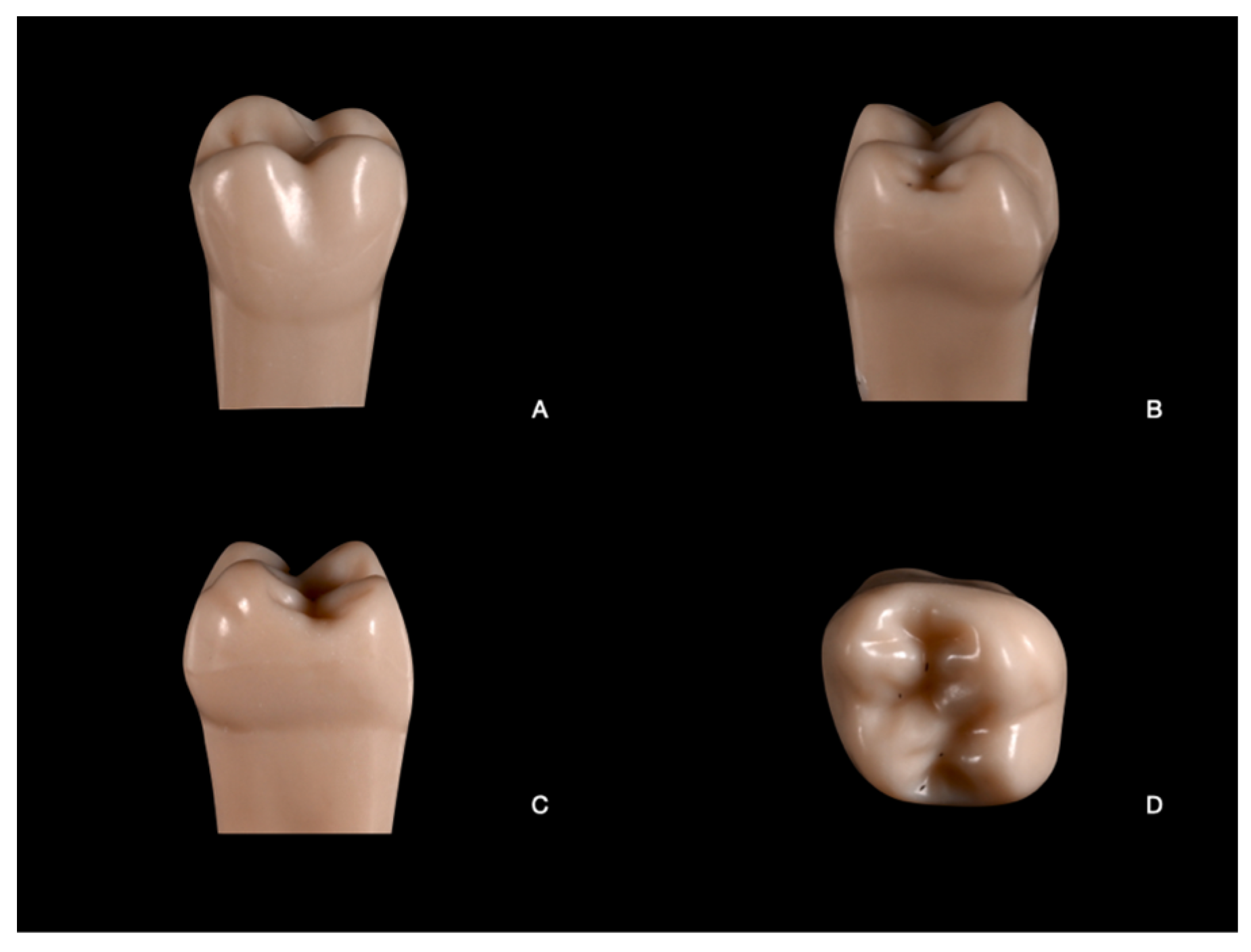
The study included 64 first upper human molars in acrylic resin (Typodont, Frasaco, Tettnang, Germany) (n = 64). Sample size adequacy was confirmed by a power analysis (GPower version 3.0.10) with parameters set at a power of 0.95, an alpha of 0.05, and an effect size of 0.8 (Figure 1 and Figure 2).
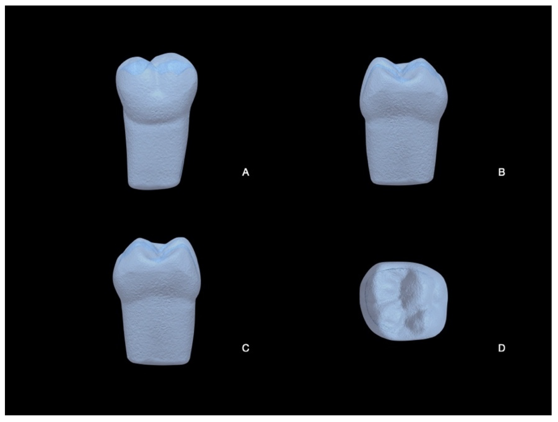
The 64 teeth were divided into the following four groups of different preparation designs (n= 16 each): Group 1 (experienced practitioners without loupes, EXP), Group 2 (students without loupes, STU), Group 3 (experienced practitioners with loupes, EXP+LOU), and Group 4 (students with loupes, STU+LOU) (Figure 1). Four operators participated in the study: two expert practitioners and two dental students. The expert operators included a full professor in prosthodontics (S.C.) and an associate professor in prosthodontics (G.M.), each with over twenty years of clinical and academic experience in the field. The two student operators had received specific and comprehensive training in prosthetic preparations, with a particular focus on overlay techniques, and performed the preparations assigned to their respective groups under the supervision of the experts. The teeth were mounted on training models (Typodont, Frasaco) and on a training manikin (Frasaco) to replicate positions and clinical preparation challenges. An unprepared tooth, used as a control, was scanned (AmannGirbach, Ceramill Map 600+) to assess the volume reduction of prepared teeth (Figure 3). The scanner used had an high accuracy (up to 4 microns) with a high-resolution camera. Using structured blue-light technology, it provided precise 3D models essential for the accurate measurement of preparation details.
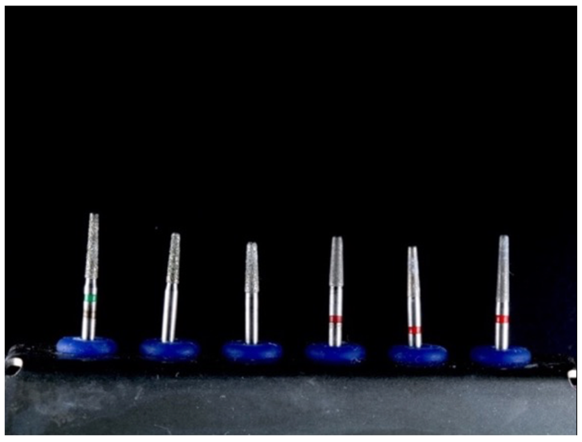
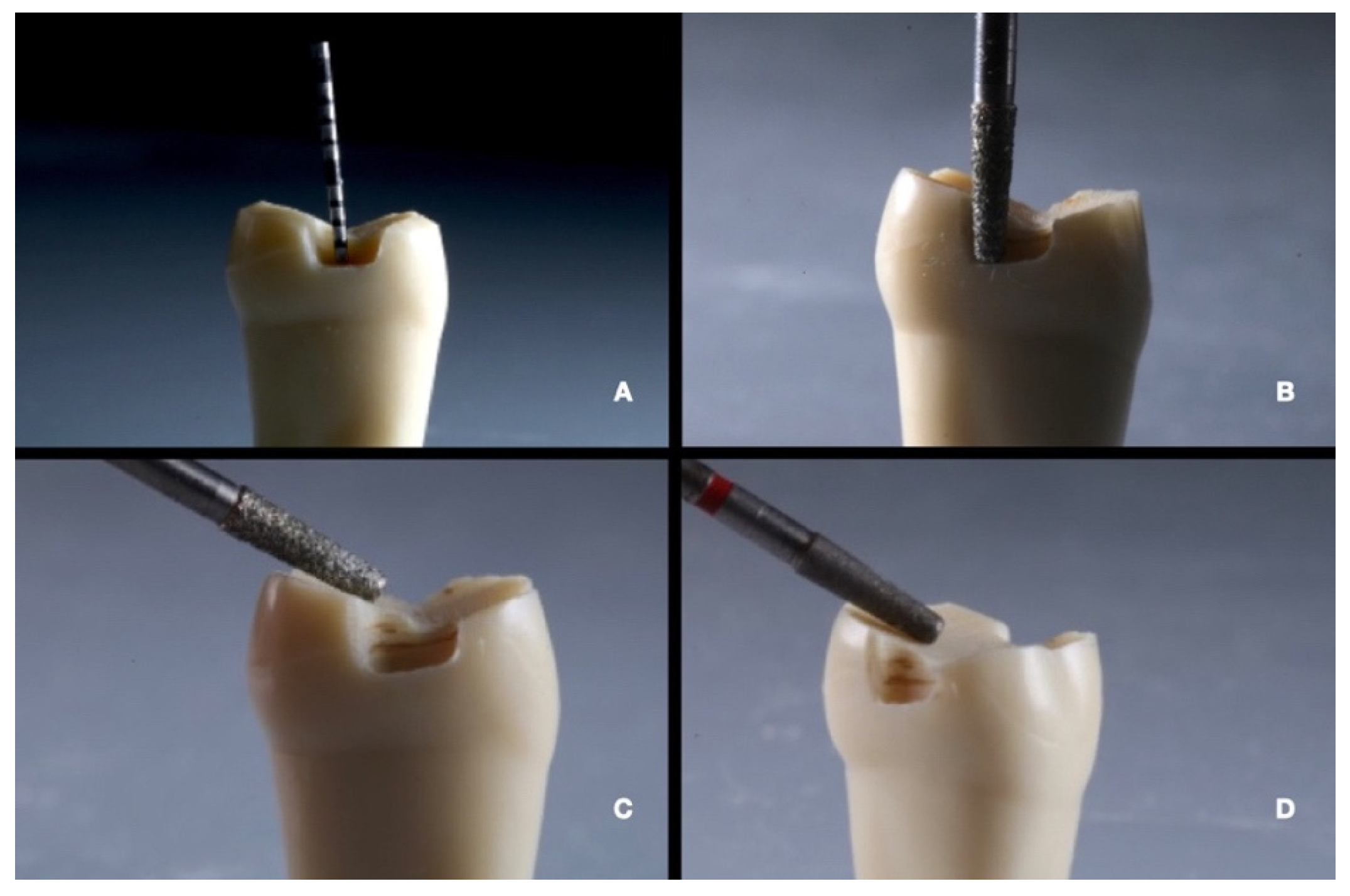
The other teeth were prepared using a high-speed handpiece (NSK, Kanuma, Japan) without magnification in Groups 1 and 2, and with a magnification tool (4X Eyemag PRO F, Zeiss, Oberkochen, Germany) in Groups 3 and 4. For all teeth, according to Veneziani et al. [8] a bur (N°. 8845KR 314 012, Komet, Lemgo, Germany) was used for the anatomical occlusal reduction of 1.5 mm. A flat-end diamond bur (N°. 8959 KR 314 014, Komet, Lemgo, Germany) was used to prepare a proximal box (butt joint) with rounded internal angles, 1 mm width, 1 mm depth, located 1 mm above the CEJ, at the maximum contour line of the tooth. The hollow chamfer finish line was obtained using a cylinder chamfer bur (N°. 8847 KR 314 014, Komet) with 0.8 mm depth. An interproximal box of 1.2 mm depth was prepared using a tapered diamond bur (N°. 8959 314 012, Komet) (Figure 4 and Figure 5). Each preparation was performed using a new set of diamond burs to ensure consistency and avoid variability due to bur wear.
Once the tooth was prepared, it was scanned using the same scanner (AmannGirbach, Ceramill Map 600+, Mäder, Austria) (Figure 6), and subsequently, the Standard Tessellation Language (STL) file was processed with Geomagic, (Control X, Oqton, Valencia, CA, USA, software version 2024.2.0).
The analyses performed with Geomagic (Control X, Oqton, Valencia, CA, USA, software version 2024.2.0) involved the volume analysis of the prepared tooth compared to the original tooth’s volume (Figure 7) and the linear analysis regarding the preparation of the interproximal box (width) (Figure 8).
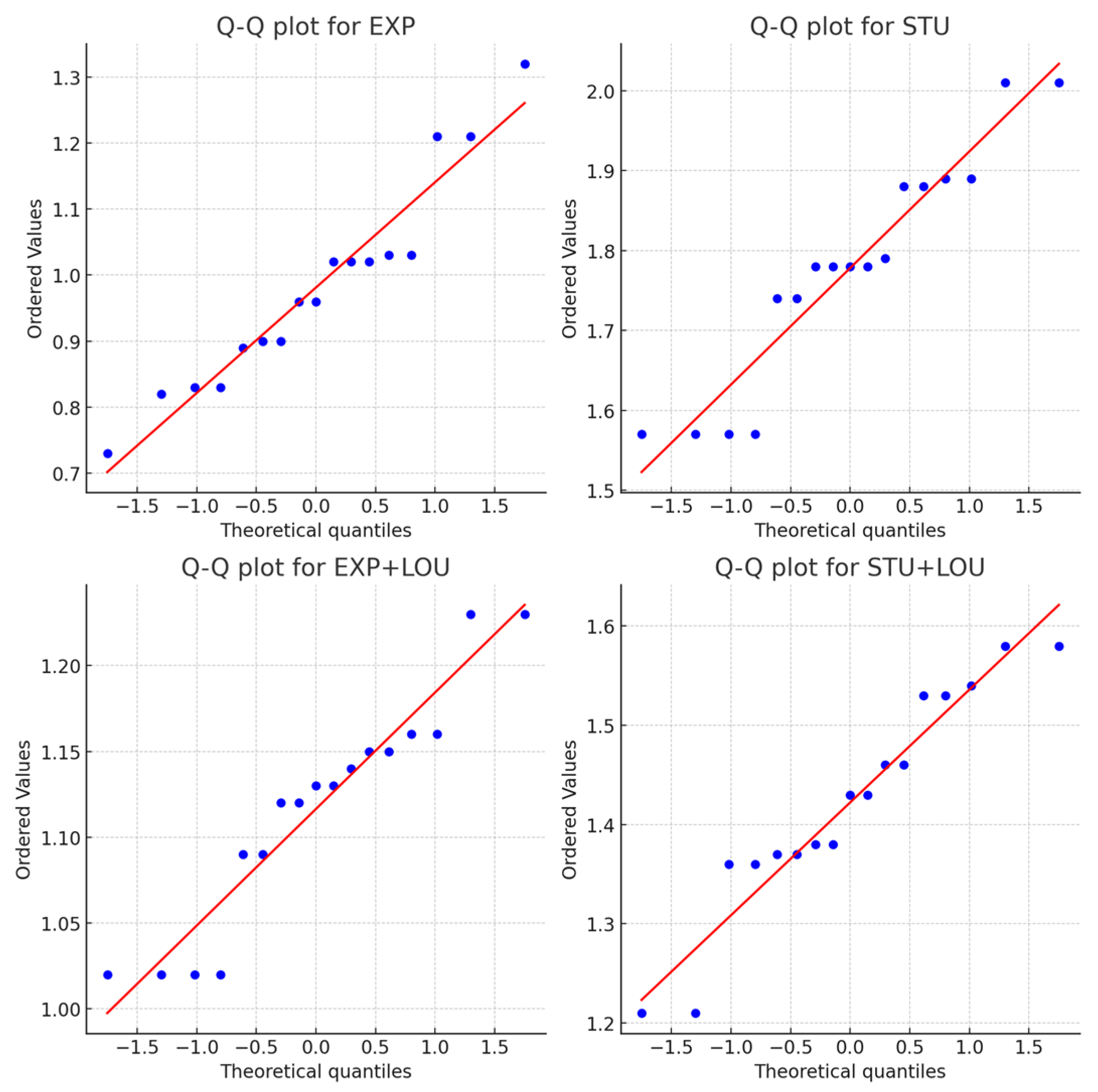
The data were statistically analyzed using two-way ANOVA and post hoc Tukey tests (GraphPad Prism, version 10.3.0). The results are presented as mean ± standard deviation (SD), 95% confidence interval (C.I.), or percentage. Two-way ANOVA was used to assess if there were differences in dental preparation for the expert and student groups with and without dental magnification. The Shapiro–Wilk normality test was applied for each of the variables considered to check whether the data were normally distributed. Additionally, Q-Q plots were analyzed for each group to visually confirm the normality of the data distributions, further supporting the use of parametric analyses (Figure 9 and Figure 10).

3. Results

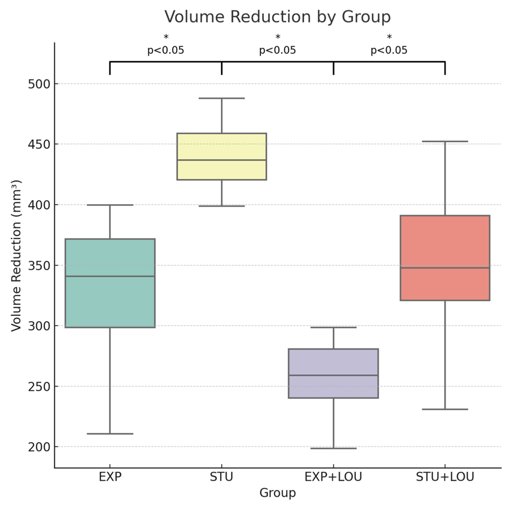
The data for volume reduction and interproximal box width were approximately normally distributed across all groups, as confirmed by the Shapiro–Wilk test. The results indicated a significant effect of both operator experience and the use of magnification on volume reduction (Figure 11). Specifically, experienced operators with magnification (EXP+LOU) achieved the lowest mean volume reduction (256.739 ± 31.979 mm3), while students without magnification (STU) exhibited the highest (442.236 ± 28.213 mm3). Detailed statistical values, including means and standard deviations for each group, are presented in Table 1. The interaction effect between experience and the use of loupes was also significant (p < 0.05), suggesting that these factors significantly influenced the accuracy of the interproximal box preparation (Table 2, Figure 12). The results clearly demonstrate that both operator experience and the use of magnification systems significantly improve the accuracy of dental preparations. Specifically, the use of loupes resulted in a significant reduction in the volume of tooth structure removed and improved the accuracy of the interproximal box width, particularly for less experienced operators (Table 1 and Table 2). The best results were achieved by experienced operators using loupes (EXP+LOU), while the least accurate preparations were performed by students without loupes (STU). The Q-Q plots for both volume reduction and interproximal box width supported the normality assumptions of the data, validating the use of parametric analyses. The data for volume reduction and interproximal box width are summarized in Table 1 and Table 2 respectively. These tables present the mean values, standard deviations, and 95% confidence intervals for each group, indicating statistically significant differences across groups with p-values for all major experimental factors and interaction terms being less than 0.05. The Q-Q plots for volume reduction and interproximal box width (Figure 9 and Figure 10) confirm the normality of the data distributions, supporting the validity of the parametric analyses used in this study. These results underscore the importance of both operator experience and magnification systems in achieving precise and conservative dental preparations, ultimately improving the outcomes of indirect restorations.

4. Discussion

The findings of this study demonstrate that all null hypotheses are rejected, as statistically significant differences were observed between Groups 1–2 and Groups 3–4 (p < 0.05). Operator experience and the use of magnification significantly influenced the conservative nature of tooth preparation, with experienced operators and those using loupes achieving greater accuracy and less dental volume reduction. Similarly, interproximal box reduction showed significant improvements with both operator experience and magnification (p < 0.05), further supporting the critical role of these factors. The precision of dental preparation is essential for ensuring adequate marginal adaptation, which is a key determinant of the long-term clinical success of ceramic restorations. These findings underscore the importance of combining operator expertise with magnification tools to optimize the accuracy and outcomes of indirect restorations. Additionally, there is a need to be increasingly conservative, removing as little dental tissue as possible. This study objectively evaluated how much substance was removed. The study found statistically significant differences in removal among different groups. There is a statistically significant difference between the group of students without magnification (Group 1) and the group of experienced operators with magnification (Group 4). The preparations of the overlays in this study were based on the following preparation design protocol guided by tooth morphology according to Veneziani et al. [8]. An interproximal box was used for the proximal preparation in all groups, as it is a standard described for preparation after the removal of interproximal caries [8]. A 4× magnification tool was used to enhance the accuracy of the preparation design. The use of magnification was therefore a determining factor in improving preparation accuracy, as widely demonstrated in the literature [20,21]. Magnification systems particularly facilitated less experienced operators, but still improved results [20]. They are therefore an indispensable means to ensure accuracy in dentistry. It was useful for less experienced operators who had a better gain in accuracy in preparation. Even in experienced operators, the analyzed preparations are more precise if performed with magnification, thanks to the better finishing and polishing that can be achieved by working under magnification. This study corroborates evidence that magnification tools significantly enhance preparation precision and conservativeness. Microscopes and loupes reduce marginal gaps and improve restoration outcomes [20,22]. Additionally, loupes offer ergonomic and visual benefits, particularly for less experienced operators, supporting their role in enhancing preparation quality [23]. These findings highlight the clinical importance of magnification in achieving accurate and tissue-preserving restorative procedures. The analysis of the results showed that Groups 3 and 4 presented significantly higher design accuracy compared to Groups 1 and 2. The good results obtained by Group 1 and 3 can be attributed to the simple characteristics of the preparation design, including a smooth and flat occlusal reduction, the absence of retention features, and fewer internal angles. These characteristics like smooth and flat occlusal reduction, the absence of retention features, and fewer internal angles also seem to facilitate digital flow procedures [7,15,24]. Further evidence highlights the importance of precision in preparation design. Minimally invasive CAD/CAM lithium disilicate restorations have demonstrated superior in vitro fatigue performance compared to full crowns, emphasizing the benefits of conservative preparation [25]. The choice of finishing line and luting material also significantly influences the seating accuracy and success of CAD/CAM restorations, reinforcing the need for meticulous preparation protocols [25,26]. The analyses obtained in this study do not present variables such as the type of teeth or anatomical variations, which could be limitations in the evaluation of the preparation design [7]. After evaluating the individual research hypotheses, it was evident that both operator experience and the use of magnification systems significantly impacted the accuracy and conservativeness of dental preparations. The findings consistently showed that magnification improved the precision of less experienced operators, reducing dental tissue removal [21]. Experienced operators using loupes achieved the best results, highlighting the importance of magnification tools in clinical practice. These findings have significant clinical implications, as magnification systems and operator experience can optimize restorative procedures, particularly by preserving dental tissue and ensuring the longevity of indirect restorations [25,26,27]. The improved precision observed in less experienced operators using loupes suggests that these tools can bridge the gap between student and expert performance. Despite these promising results, this study has several limitations. The sample size, while determined by power analysis, could be larger to further validate the findings. The study was conducted in vitro, which may not fully replicate clinical conditions. Patient-specific factors were not considered, which could influence the results. Additionally, the study focused on a single type of preparation, and results may vary with different preparation designs. Future research should aim to address these limitations by conducting larger-scale in vivo studies to confirm the findings in clinical settings. Exploring the impact of magnification systems on different types of dental preparations and across various tooth anatomies would provide a more comprehensive understanding of their benefits. Additionally, longitudinal studies assessing the long-term outcomes of restorations performed with and without magnification could further substantiate the clinical advantages observed in this study.

5. Conclusions

In conclusion, both operator experience and the use of magnification significantly enhance the accuracy of overlay preparations, particularly in terms of volume reduction and interproximal box width. The combination of experience and magnification yielded the best results, highlighting their importance in achieving precise and conservative dental preparations. These findings underscore the value of magnification tools, especially for less experienced operators, in improving preparation quality.

Author Contributions

Conceptualization, B.S. and G.M.; methodology, G.D.; software, M.S.; validation, S.C., B.S. and G.D.; formal analysis, M.S.; investigation, B.S.; resources, G.A.; data curation, G.T.; writing—original draft preparation, G.T. and G.M.; writing—review and editing, B.S.; visualization, B.S. and M.S.; supervision, S.C.; project administration, B.S.; funding acquisition, B.S. All authors have read and agreed to the published version of the manuscript.

Funding

GT has a PhD fellowship (code n. DOT20A93TY-1) in the framework of PONRI 2014/2020, DM1061, 10/08/2021, XXXVII cycle, Action IV.4 “Ph.D programs and research contracts on innovation topics”, funded by the Ministry of Education, University and Research (MIUR), Italy, FSE-FESR. This work has been partially funded by the European Union-NextGenerationEU under the Italian Ministry of University and Research (MUR) National Innovation Ecosystem grant ECS00000041-VITALITY-CUP D73C22000840006. This work has been partially funded by the “737 Project” CUP D75F21003210001.

Institutional Review Board Statement

Not applicable.

Informed Consent Statement

Not applicable.

Data Availability Statement

Data are contained within the article.

Conflicts of Interest

The authors declare no conflicts of interest. The funders had no role in the design of the study; in the collection, analyses, or interpretation of data; in the writing of the manuscript; or in the decision to publish the results.

References

  1. Tiu, J.; Al-Amleh, B.; Waddell, J.N.; Duncan, W.J. Reporting numeric values of complete crowns. Part 2: Retention and resistance theories. J. Prosthet. Dent. 2015, 114, 75–80. [Google Scholar] [CrossRef] [PubMed]
  2. Yang, Y.; Yang, Z.; Zhou, J.; Chen, L.; Tan, J. Effect of tooth preparation design on marginal adaptation of composite resin CAD-CAM onlays. J. Prosthet. Dent. 2020, 124, 88–93. [Google Scholar] [CrossRef] [PubMed]
  3. Wayakanon, K. Partially Coverage Restoration: An Esthetically Conservative Treatment for a Complex Cavity Restoration. Open J. Stomatol. 2017, 7, 234–241. [Google Scholar] [CrossRef]
  4. Bandéca, M.C.; Malheiros, A.S.; de Jesus Tavarez, R.R.; Firoozmand, L.M.; Silva, M.B. Overlays or ceramic fragments for tooth restoration: An analysis of fracture resistance. J. Contemp. Dent. Pract. 2014, 15, 56–60. [Google Scholar] [CrossRef] [PubMed]
  5. Magne, P. Composite resins and bonded porcelain: The postamalgam era? J. Calif. Dent. Assoc. 2006, 34, 135–147. [Google Scholar] [CrossRef]
  6. Ferraris, F. Posterior indirect adhesive restorations (PIAR): Preparation designs and adhesthetics clinical protocol. Int. J. Esthet. Dent. 2017, 12, 482–502. [Google Scholar]
  7. Ferraris, F.; Mascetti, T.; Tognini, M.; Testori, M.; Colledani, A.; Marchesi, G. Comparison of posterior indirect adhesive restorations (PIAR) with different preparations designs according to the adhesthetics classification. Int. J. Esthet. Dent. 2021, 16, 262–279. [Google Scholar]
  8. Veneziani, M. Posterior indirect adhesive restorations: Updated indications and the Morphology Driven Preparation Technique. Int. J. Esthet. Dent. 2017, 12, 204–230. [Google Scholar]
  9. Adolphi, G.; Zehnder, M.; Bachmann, L.M.; Göhring, T.N. Direct resin composite restorations in vital versus root-filled posterior teeth: A controlled comparative long-term follow-up. Oper. Dent. 2007, 32, 437–442. [Google Scholar] [CrossRef] [PubMed]
  10. Lin, C.-L.; Chang, Y.-H.; Pai, C.-A. Evaluation of failure risks in ceramic restorations for endodontically treated premolar with MOD preparation. Dent. Mater. 2011, 27, 431–438. [Google Scholar] [CrossRef] [PubMed]
  11. Bitter, K.; Meyer-Lueckel, H.; Fotiadis, N.; Blunck, U.; Neumann, K.; Kielbassa, A.M.; Paris, S. Influence of endodontic treatment, post insertion, and ceramic restoration on the fracture resistance of maxillary premolars. Int. Endod. J. 2010, 43, 469–477. [Google Scholar] [CrossRef]
  12. Leoney, A.; Balasubramanian, R. Angulated Post used for Esthetic makeover—A Case Report. Indian J. Multidiscip. Dent. 2013, 3, 711. [Google Scholar]
  13. Dietschi, D.; Duc, O.; Krejci, I.; Sadan, A. Biomechanical considerations for the restoration of endodontically treated teeth: A systematic review of the literature, Part II (Evaluation of fatigue behavior, interfaces, and in vivo studies). Quintessence Int. 2008, 39, 117–129. [Google Scholar]
  14. Burke, F.J.; Wilson, N.H.; Watts, D.C. The effect of cuspal coverage on the fracture resistance of teeth restored with indirect composite resin restorations. Quintessence Int. 1993, 24, 875–880. [Google Scholar]
  15. Panahandeh, N.; Johar, N. Effect of Different Cusp Coverage Patterns on Fracture Resistance of Maxillary Premolar Teeth in MOD Composite Restorations. J. Islam. Dent. Assoc. Iran. 2014, 25, 228–232. [Google Scholar]
  16. Ioannidis, A.; Mühlemann, S.; Özcan, M.; Hüsler, J.; Hämmerle, C.H.; Benic, G.I. Ultra-thin occlusal veneers bonded to enamel and made of ceramic or hybrid materials exhibit load-bearing capacities not different from conventional restorations. J. Mech. Behav. Biomed. Mater. 2019, 90, 433–440. [Google Scholar] [CrossRef] [PubMed]
  17. Abduo, J.; Sambrook, R.J. Longevity of ceramic onlays: A systematic review. J. Esthet. Restor. Dent. 2018, 30, 193–215. [Google Scholar] [CrossRef]
  18. Kim, J.-H.; Cho, B.-H.; Lee, J.-H.; Kwon, S.-J.; Yi, Y.-A.; Shin, Y.; Roh, B.-D.; Seo, D.-G. Influence of preparation design on fit and ceramic thickness of CEREC 3 partial ceramic crowns after cementation. Acta Odontol. Scand. 2014, 73, 107–113. [Google Scholar] [CrossRef]
  19. Cunali, R.S.; Saab, R.C.; Correr, G.M.; Da Cunha, L.F.; Ornaghi, B.P.; Gonzaga, C.C.; Ritter, A.V. Marginal and internal adaptation of zirconia crowns: A comparative study of assessment methods. Braz. Dent. J. 2017, 28, 467–473. [Google Scholar] [CrossRef] [PubMed]
  20. Eichenberger, M.; Biner, N.; Amato, M.; Lussi, A.; Perrin, P. Effect of magnification on the precision of tooth preparation in dentistry. Oper. Dent. 2018, 43, 501–507. [Google Scholar] [CrossRef]
  21. Sorrentino, R.; Ruggiero, G.; Borelli, B.; Barlattani, A.; Zarone, F. Dentin Exposure after Tooth Preparation for Laminate Veneers: A Microscopical Analysis to Evaluate the Influence of Operators’ Expertise. Materials 2022, 15, 1763. [Google Scholar] [CrossRef]
  22. Atlas, A.M.; Janyavula, S.; Elsabee, R.; Alper, E.; Isleem, W.F.; Bergler, M.; Setzer, F.C. Comparison of loupes versus microscope-enhanced CAD-CAM crown preparations: A microcomputed tomography analysis of marginal gaps. J. Prosthet. Dent. 2022, 131, 643–651. [Google Scholar] [CrossRef] [PubMed]
  23. Nóbrega, C.; Manso, M.C.; Herrero-Climent, M.; Gil, J.; Ribeiro, P. Impact of Magnifying Loupes on the Finish Lines of Fixed Prosthesis Preparations. Prosthesis 2024, 6, 631–642. [Google Scholar] [CrossRef]
  24. Spitznagel, F.A.; Prott, L.S.; Hoppe, J.S.; Manitckaia, T.; Blatz, M.B.; Zhang, Y.; Langner, R.; Gierthmuehlen, P.C. Minimally invasive CAD/CAM lithium disilicate partial-coverage restorations show superior in-vitro fatigue performance than single crowns. J. Esthet. Restor. Dent. 2024, 36, 94–106. [Google Scholar] [CrossRef]
  25. Mancuso, E.; Gasperini, T.; Maravic, T.; Mazzitelli, C.; Josic, U.; Forte, A.; Pitta, J.; Mazzoni, A.; Fehmer, V.; Breschi, L.; et al. The influence of finishing line and luting material selection on the seating accuracy of CAD/CAM indirect composite restorations. J. Dent. 2024, 148, 105231. [Google Scholar] [CrossRef] [PubMed]
  26. Jánosi, K.M.; Cerghizan, D.; Rétyi, Z.; Kovács, A.; Szász, A.; Mureșan, I.; Albu, A.I.; Hănțoiu, L.G. Influence of the Operator`s Experience, Working Time, and Working Position on the Quality of the Margin Width: In Vitro Study. Medicina 2023, 59, 244. [Google Scholar] [CrossRef]
  27. D’addazio, G.; Amoroso, F.; Tafuri, G.; Baima, G.; Santilli, M.; Mussano, F.; Sinjari, B. Impact of the Luting Technique on the Positioning of CAD-CAM Porcelain Laminate Veneers: An In Vitro Study. Prosthesis 2024, 6, 1095–1105. [Google Scholar] [CrossRef]
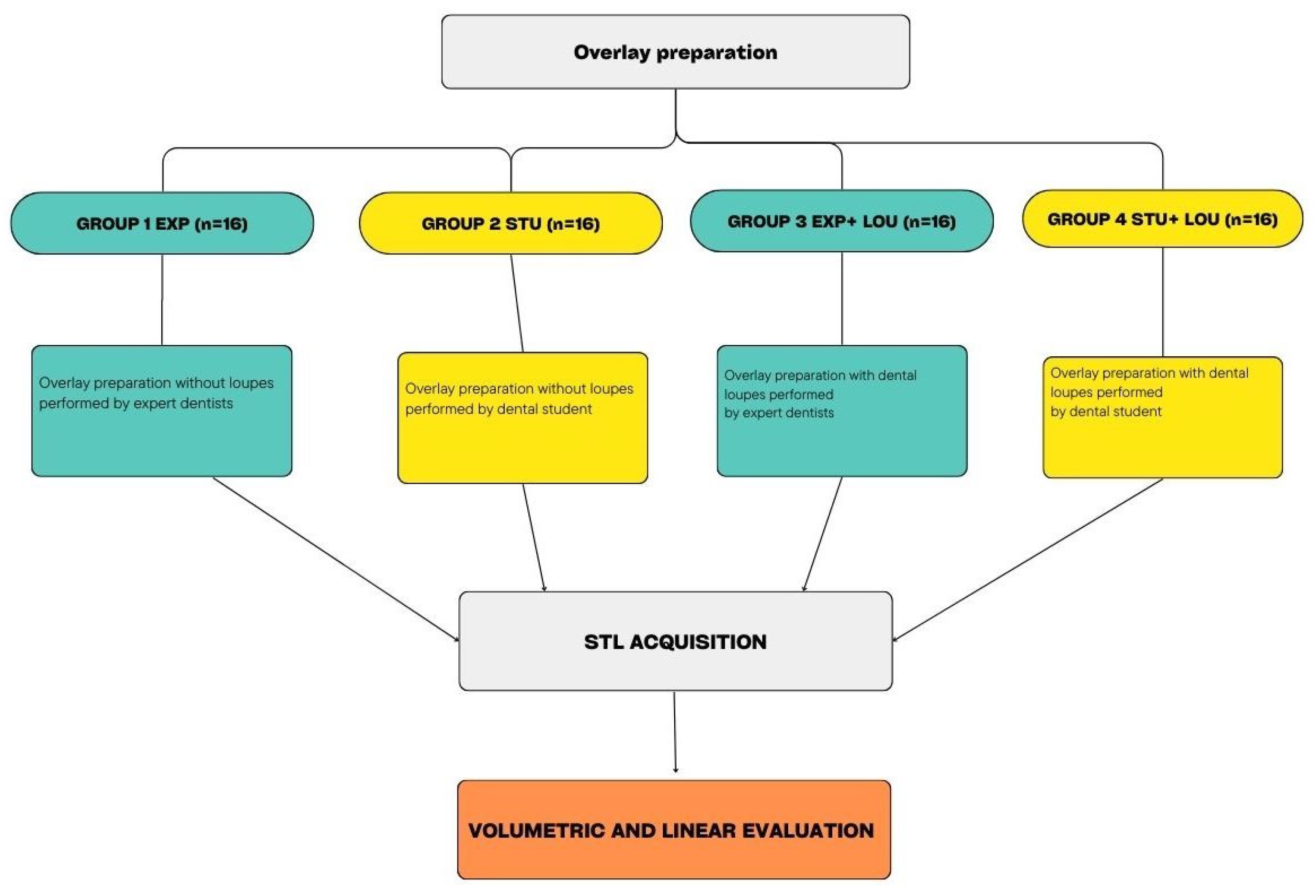
Figure 1. Flowchart of the study design.
Prosthesis 07 00005 g001
Figure 2. Tooth 1.6 (Frasaco, Germany) used in the study. Buccal view (subfigure (A)), distal view (subfigure (B)), mesial view (subfigure (C)), and occlusal view (subfigure (D)).
Prosthesis 07 00005 g002
Figure 3. Tooth 1.6 (Frasaco, Germany) after scanning. Buccal view (subfigure (A)), distal view (subfigure (B)), mesial view (subfigure (C)), and occlusal view (subfigure (D)).
Prosthesis 07 00005 g003
Figure 4. Burs used in the study to make overlay preparation.
Prosthesis 07 00005 g004
Figure 5. Overlay preparation technique. Measurement with a periodontal probe of the interproximal box (subfigure (A)), interproximal box (subfigure (B)), occlusal reduction (subfigure (C)), occlusal reduction with fine-grain bur (subfigure (D)).
Prosthesis 07 00005 g005
Figure 6. Tooth 1.6 (Frasaco, Germany) after scanning. Buccal view (subfigure (A)), distal view (subfigure (B)), mesial view (subfigure (C)), and occlusal view (subfigure (D)).
Prosthesis 07 00005 g006
Figure 7. Superimposition through Geomagic software (Control X, USA).
Prosthesis 07 00005 g007
Figure 8. Linear measurement of interproximal box with Geomagic software (Control X, USA).
Prosthesis 07 00005 g008
Figure 9. Q-Q plot for volume reduction by group.
Prosthesis 07 00005 g009
Figure 10. Q-Q plot for interproximal box reduction by group.
Prosthesis 07 00005 g010
Figure 11. Boxplot of volume reduction (mm3).
Prosthesis 07 00005 g011
Figure 12. Boxplot of interproximal box reduction (mm3).
Prosthesis 07 00005 g012
Table 1. Volume reduction.
VOLUME
REDUCTION
(mm3)
EXPSTUEXP+LOUSTU+LOU
Mean
* 329,012* 442,236* 256,739* 351,338
SD
55,62728,21331,97959,776
CI (95%)
223.241, 434.783195.932, 317.546388.589, 495.882237.676, 464.998
p-Value
Factor A (experience) < 0.05 (*)
Factor B (dental loupes) < 0.05 (*)
Table 2. Interproximal box reduction.
INTERPROXIMAL BOX
width (mm)
EXPSTUEXP+LOUSTU+LOU
Mean
* 0.945* 1.793* 1.129* 1.447
SD
0.1750.1420.0720.122
CI (95%)
0.669, 1.3211.528, 2.0580.994, 1.2631.219, 1.674
p-Value
Factor A (experience) < 0.05 (*)
Factor B (dental loupes) < 0.05 (*)
Disclaimer/Publisher’s Note: The statements, opinions and data contained in all publications are solely those of the individual author(s) and contributor(s) and not of MDPI and/or the editor(s). MDPI and/or the editor(s) disclaim responsibility for any injury to people or property resulting from any ideas, methods, instructions or products referred to in the content.