Article

Wearing Effect of Implant Steel Drills and Tappers for the Preparation of the Bone Osteotomies: An Infrared Thermal Analysis and Energy Dispersive Spectroscopy-Scanning Electron Microscopy (EDS-SEM) Study

by
Felice Lorusso
1,
Sergio Alexandre Gehrke
2,3,
Felice Festa
1 and
Antonio Scarano
1,2,4,*
1
Department of Innovative Technologies in Medicine & Dentistry, University of Chieti-Pescara, 66100 Chieti, Italy
2
Department of Research, Bioface/PgO/UCAM, Montevideo 11300, Uruguay
3
Department of Biotechnology, Universidad Católica de Murcia (UCAM), 30001 Murcia, Spain
4
Director of Specialization School of Oral Surgery, Department of Innovative Technologies in Medicine & Dentistry, University of Chieti-Pescara, 66100 Chieti, Italy
*
Author to whom correspondence should be addressed.
Prosthesis 2022, 4(4), 679-694; https://doi.org/10.3390/prosthesis4040054
Submission received: 6 November 2022 / Revised: 23 November 2022 / Accepted: 26 November 2022 / Published: 30 November 2022
(This article belongs to the Special Issue Bioactive Materials for Dental and Maxillofacial Repair)

Abstract

:
Background: The thermal effect correlated with implant osteotomy could produce significant effects on the healing process and fixture osseointegration. The aim of the present investigation was to assess the heat generation and surface wearing of dental implant drills and manual tappers during simulated osteotomies on animal ribs. Methods: Steel drills (20 units per type) and tappers (20 units per type) were evaluated for a total of 30 osteotomies. The infrared thermal analysis was performed at the first and thirtieth osteotomy. The surface alteration and wearing was assessed by energy dispersive spectroscopy–scanning electron microscopy (EDS-SEM) prior to and after use. Conclusions: The drill material produced a non-significant temperature change during bone osteotomy. Lower heating was reported for manual tappers in favor of a manual osteotomy instead rotary instruments.

1. Introduction

The osseointegration process represents an interface relationship determined by the intimate contact between an implant and the surrounding bone tissue [1]. This interface is essential for the long-term prognosis of implant-supported rehabilitation [1]. The failure of dental implant osteointegration is frequently produced during the early healing phase and it is commonly produced by surgical trauma on the bone osteotomy preparation [2,3]. The current scientific literature on drills and tappers in clinical practice concerns to in vitro studies and in vivo investigations oriented to measure the effect on bone tissue of different variables such as implant microgeometry, diameter, rotation speed, strength and drills/tappers insertion angle [4,5,6,7,8]. These effects mainly concern the temperature increase that, for decades, has been associated with bone tissue necrosis, for temperature >47 °C for at least one-minute drilling [6]. Many studies have also evaluated the effect of air- and water-based irrigation solution on the surgical site during the drilling phase to limit temperature increase [9,10]. Some have observed that when cooling is not implemented, there is rapid overheating that can reach critical temperatures within a few seconds at the conventional rotation velocity [8]. Ozcan et al. tested a low rotation speed technique/no cooling (~50 rpm) compared to the high speed (> 400 rpm) with saline irrigation in an animal study [9]. The low-speed technique is associated with higher primary stability and cortical bone temperature, while this technique is useful in several clinical protocols and guided surgery involved with bone milling in absence of cooling [11,12,13]. The highest secondary stability observed by micro-CT evaluation was reported with the 1200 rpm with saline irrigation procedure at 8 weeks implant healing [9]. In the literature, it has been demonstrated that the drill geometry could also produce a significant variation in the osteotomy site heat [4]. Scarano et al. (1) reported that the use of a cylindrical drilling system instead of the conical geometry in vitro on bovine femoral cortical bone could produce a significant increase of ~2 °C of the bone calculated at the level of the coronal and apical part of the drill. Moreover, through infrared thermal assessment, Scarano et al. [5] reported a significant increase in steel drill heat after >120 osteotomies in very dense bovine cortical bone. The temperature increase observed at the level of the implant osteotomies with the steel drill was probably due to the resistance to wear compared to zirconia drills. In addition, the drills disinfection and sterilization procedures could affect the heat during the osteotomies through an alteration of the surface characteristics and roughness [14]. Scarano et al. reported that the disinfection agents produced a weak increase in the heat produced during the bone osteotomies [14]. The heat sterilization produced no significant changes in the temperature findings during the drilling. Moreover, the drilling time could represent a key factor for heat generation during the site osteotomy. In the literature, it was demonstrated that ultrasonic device is correlated to a significant increase in the drilling duration compared to the implant site preparation through stainless steel drills [15]. The wearing of the implant drills and tappers determined by repeated use produces an increase in the clutch and total cutting energy, thus implying potential bone damage. This process could impair or decrease the bone healing process around the dental implant [4,5,6,7,8]. The aim of the present investigation was to compare the heat generation and the wear between the dental implant steel drills and bone tappers on bovine ribs through infrared thermal evaluation and EDS–Scanning electron microscopy (SEM).

2. Materials and Methods

The in vitro evaluation was conducted comparing the thermal effect and wear produced by the different burs of the implant system protocol for the preparation of the implant site simulating two different implant lengths (10 mm/13 mm) and diameters. The experimentation was performed on bovine ribs maintained at the level of the lower portion in a saline bath of physiological solution at electronically controlled temperature (26.0 °C). The site preparation was produced when the bone temperature, measured by infrared thermography, reached the bath temperature of 37.0 ± 0.1 °C. A saline solution at the same temperature was used to irrigate the drilling site and maintained continuously at a rate of 40 mL/min. A series of new drills and tappers for each group were evaluated. The bone osteotomies were obtained applying a speed of 800 rpm/30 Ncm with a surgical motor (NSK, Surgipro, Japan). An axial load of 19.61 N was applied by a surgical motor calibrated by the Universal Testing Machine (Lloyd Instruments, AMETEK, Berwyn, PE, USA). The drilling depth was controlled electronically. The tappers were used according to a speed of 30 rpm. The drill and tappers wearing were assessed after a total of 30 osteotomies, cleaning and sterilizations of the drills and tampers according to the implant system protocol.

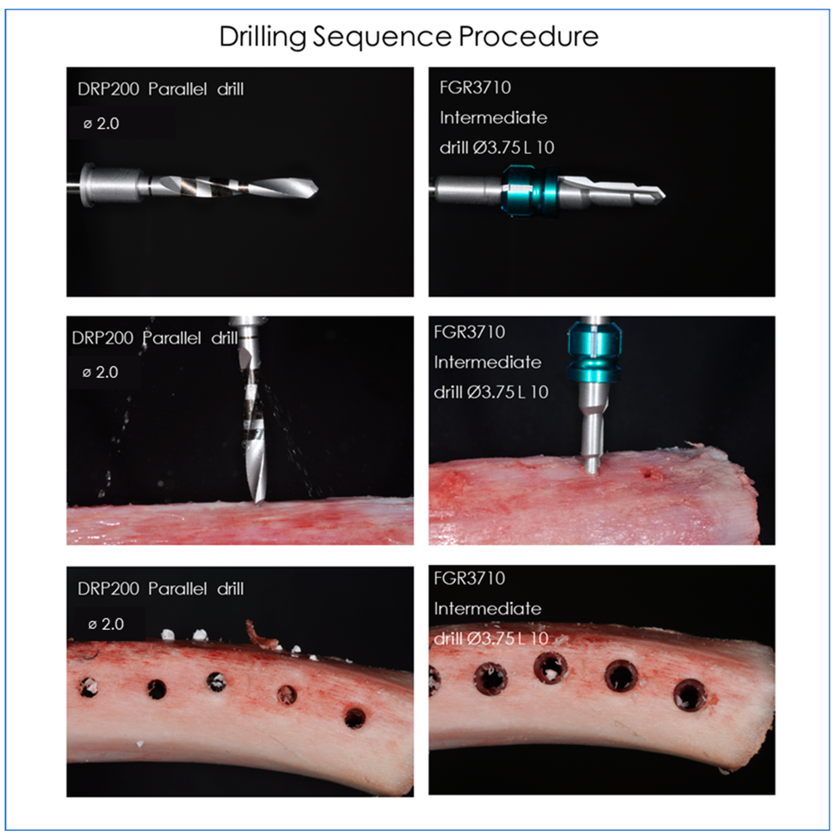
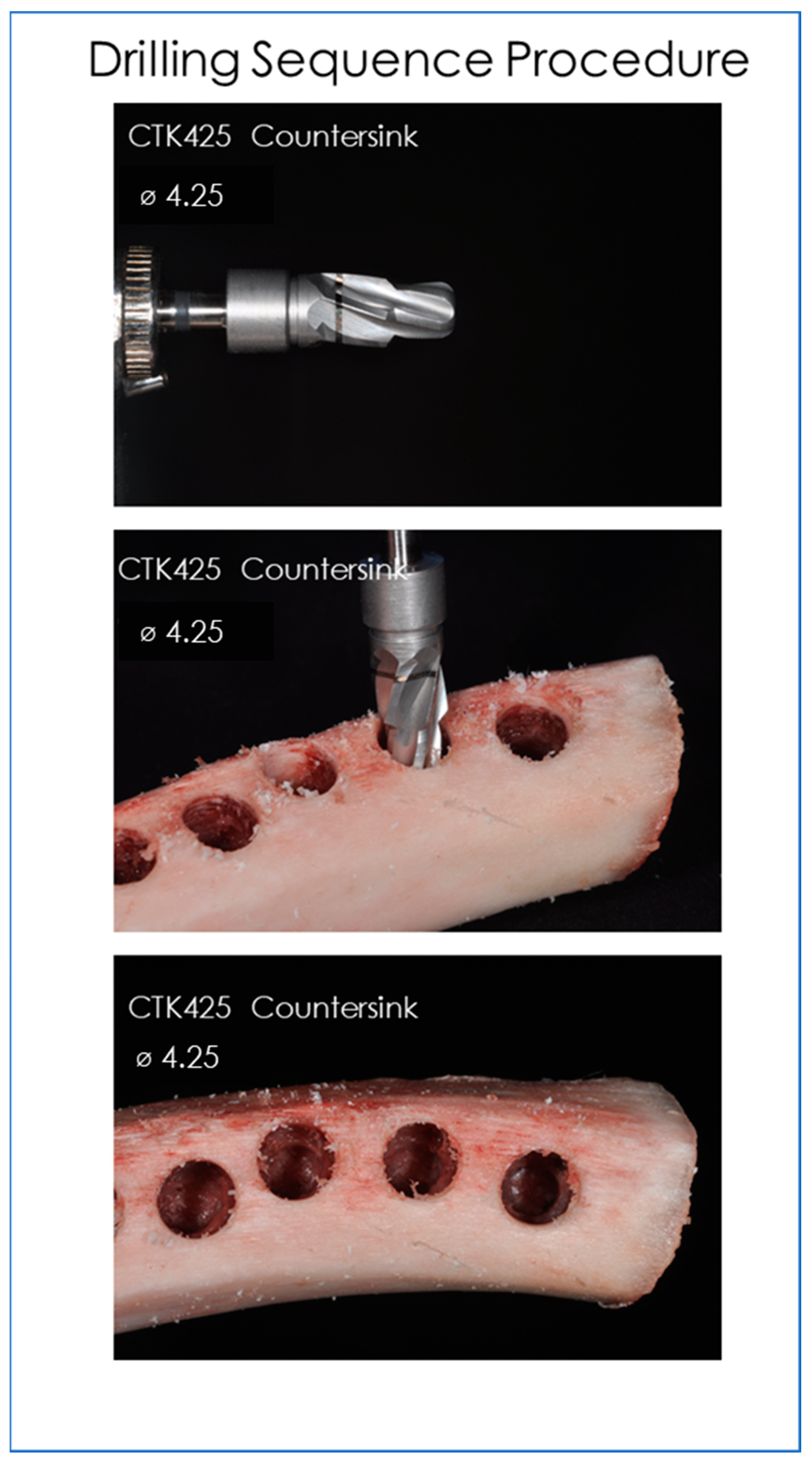
2.1. Drills and Bone Tappers Evaluated

For the drills concerned, the tests were performed on the following codes, following the implant system protocol Figure 1, Figure 2, Figure 3, Figure 4:
  • FGR3710 Intermediate drill Ø3.75 L 10 (Dental Tech, Misinto MB, Italy).
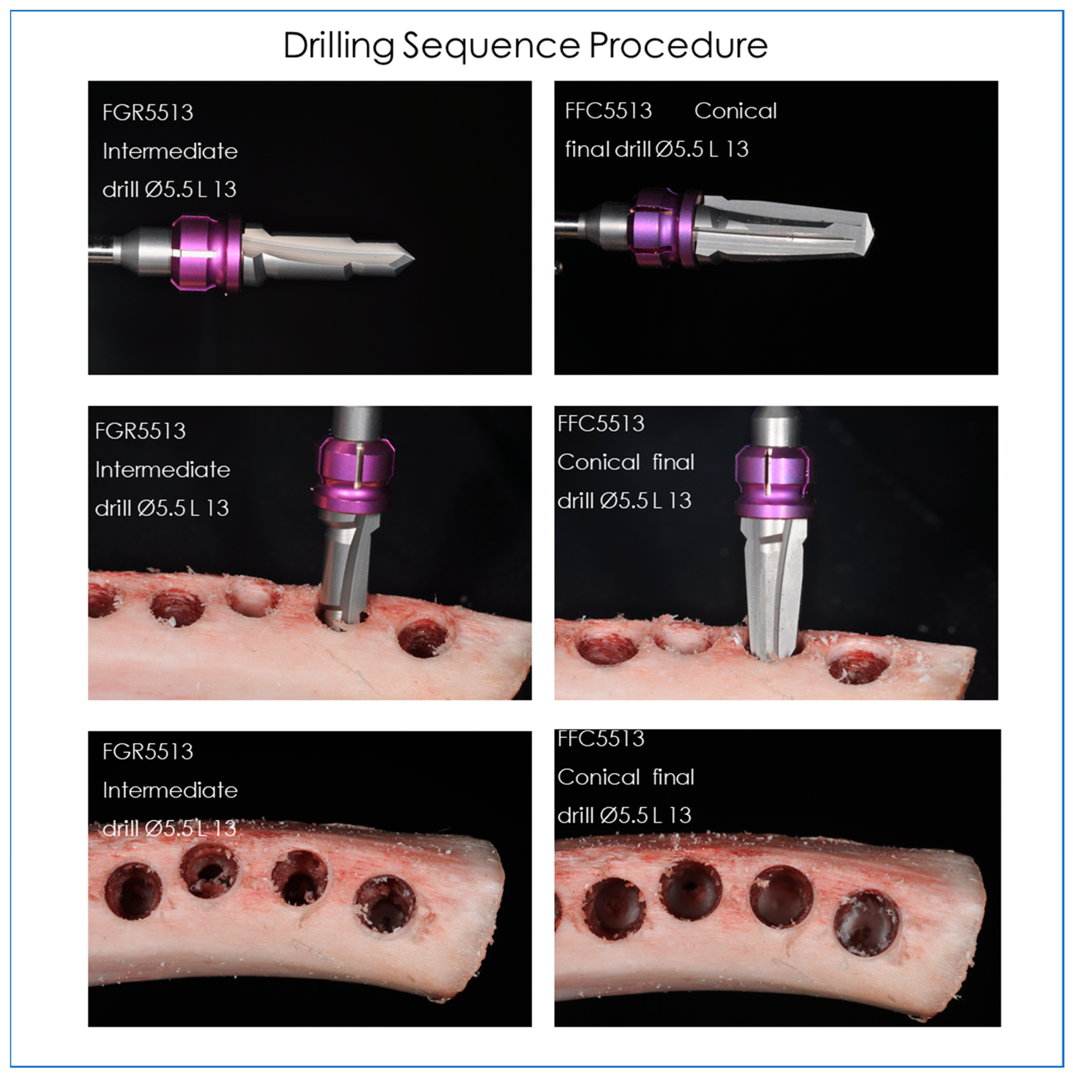
  • FGR5513 Intermediate drill Ø5.5 L 13 (Dental Tech, Misinto MB, Italy).
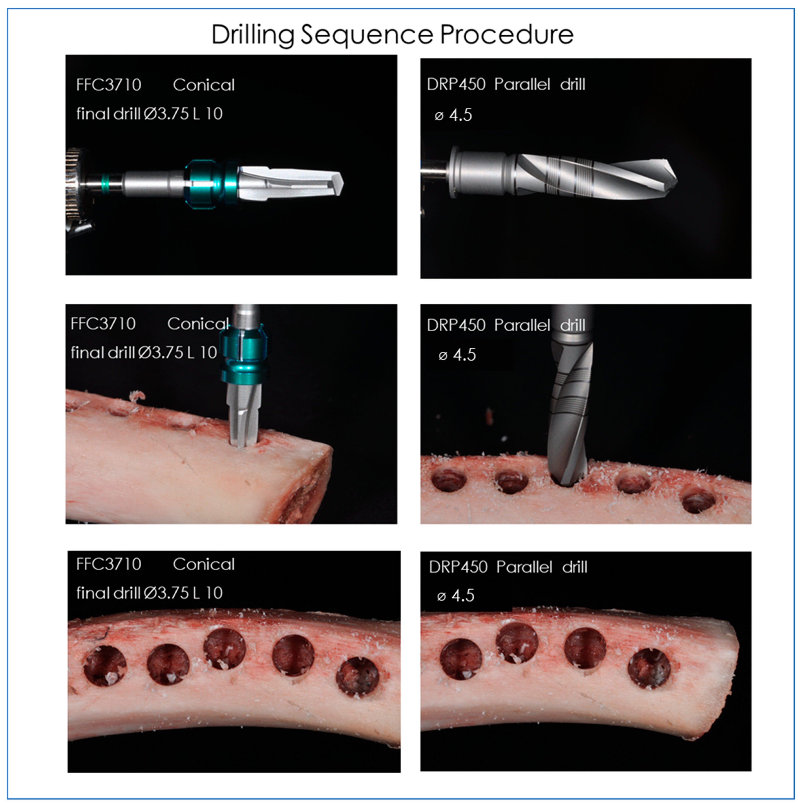
  • FFC3710 Conical final drill Ø3.75 L 10 (Dental Tech, Misinto MB, Italy).
  • FFC5513 Conical final drill Ø5.5 L 13 (Dental Tech, Misinto MB, Italy).
  • DRP200 Parallel drill Ø2,0 (Dental Tech, Misinto MB, Italy).
  • DRP450 Parallel drill Ø4,5 (Dental Tech, Misinto MB, Italy).
  • CTK425 Countersink Ø4,25 (Dental Tech, Misinto MB, Italy) (Figure 1, Figure 2, Figure 3)
Concerning the titanium tappers, the following devices were tested at different lengths and diameters (Figure 5 and Figure 6):
  • MKN3710 Conical tapper Ø3.75 L 10 (Dental Tech, Misinto MB, Italy).
  • MKN3711 Conical tapper Ø3.75 L 11.5 (Dental Tech, Misinto MB, Italy).
  • MKN3713 Conical tapper Ø3.75 L 13 (Dental Tech, Misinto MB, Italy).
  • MKN3714 Conical tapper Ø3.75 L 14.5 (Dental Tech, Misinto MB, Italy).
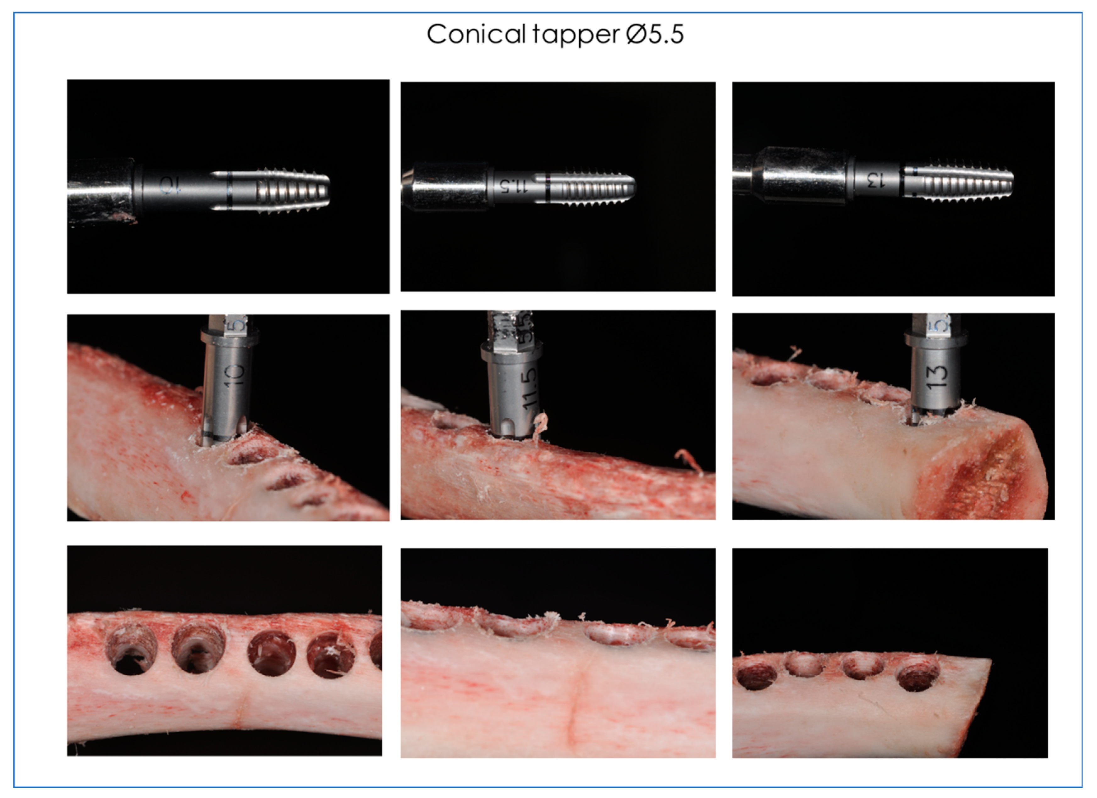
  • MSL5510 Conical tapper Ø5.5 L 10 (Dental Tech, Misinto MB, Italy).
  • MSL5511 Conical tapper Ø5.5 L 11.5 (Dental Tech, Misinto MB, Italy).
  • MSL5513 Conical tapper Ø5.5 L 13 (Dental Tech, Misinto MB, Italy).

2.2. Infrared Thermographic Measurements

Thermal image scans were obtained using a 14-bit digital infrared camera (FLIR SC3000 QWIP, FLIR Systems, Danderyd, Sweden). The acquisition parameters were: 320 × 240 focal plane array; 8–9 µm spectral range; 0.02 K noise equivalent temperature differences (NETD); 50 Hz sampling rate; optics: germanium lens; f 20; and f/1.5). The images were acquired at a rate of 10 images per second and subsequently re-aligned using an edge-detection-based method implemented with in-house software. A video was obtained, and the photos were extrapolated via dedicated software (FLIR Reporter, Danderyd, Sweden). The infrared thermographic system was positioned at a focal distance of 1 m from the specimens. The implant bed was positioned in a way that it was perpendicular to the surface from which the thermal image system measured any observed temperature change. To avoid the interference of water with infrared radiation emitted from the specimens, a plastic screen was applied that protected the flat bone surface of interest from the irrigant.

2.3. EDS-SEM Observations

The surface alterations and changes were evaluated by scanning electron microscope (SEM, Zeiss; Oberkochen, Germany), by a solid-state backscattered detector operated at a 20 kV accelerating voltage. Each sample was attached to an aluminum stub with sticky conductive carbon tape, and the images were taken in both secondary and backscattered electrons. The pictures were recorded for each specimen to characterize the surface and presence of the corrosion area. The drills were examined before and after use by a scanning electron microscope/Energy Dispersive X-ray Spectrometry (SEM/EDS) using scanning electron microscopy (SEM, Zeiss; Germany) with the EDS QUANTAX-200 probe (Bruker Nano GmbH, Berlin, Germany).

2.4. Statistical Evaluation

The sample power analysis was performed using clinical software to determine the number of drills needed to obtain statistical significance for quantitative temperature analyses. A calculation model has been adopted for dichotomous variables (yes/no effect) with alpha = 0.05 and power = 90%. The optimal number of samples for analysis is 20 drills per group. The Kolmogorov–Smirnov test was applied to evaluate the distribution of data, and the temperature differences will be analyzed statistically using the Mann–Whitney Test. Statistically significant differences will be considered with a value of p < 0.05.

3. Results

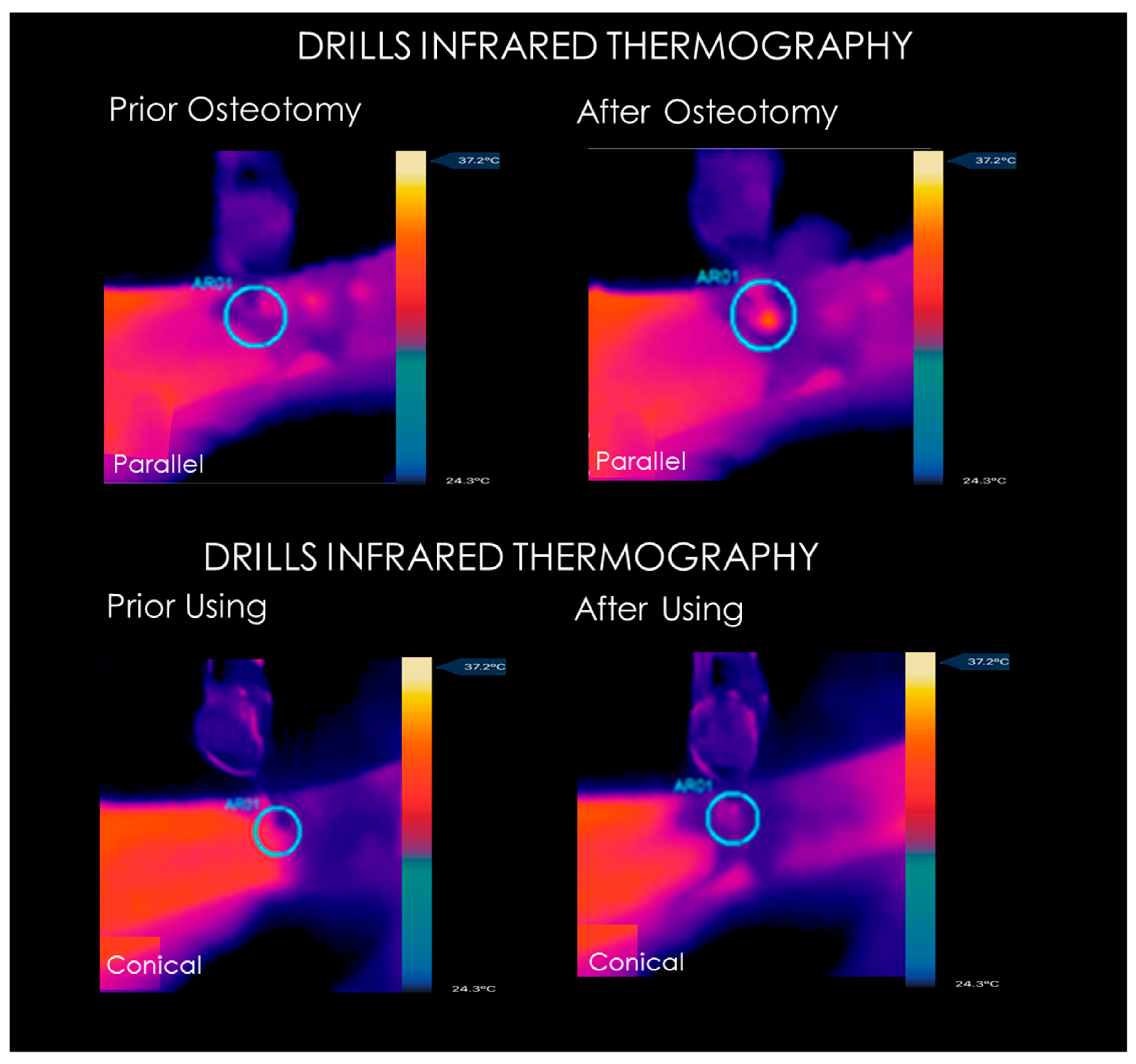
3.1. Infrared Thermography Measurements

The mean temperatures produced at the level of the cortical bone during the implant preparation are shown in Table 1. The increase in bone temperature was statistically higher when over 30 osteotomies were made for drill groups if compared to the baseline (p < 0.05). No statistical difference was detected for all the drill groups after 30 osteotomies compared to the first one (p > 0.05) (Figure 7). The mean temperatures generated during the conical tapper application are shown in Table 2. The mean temperature produced in the apical portion of the drill during conical tapper using was not significant for all study groups if compared to the baseline. A significant difference was detected between the drills osteotomies and the manual bone tappers application in all comparisons (p < 0.05).

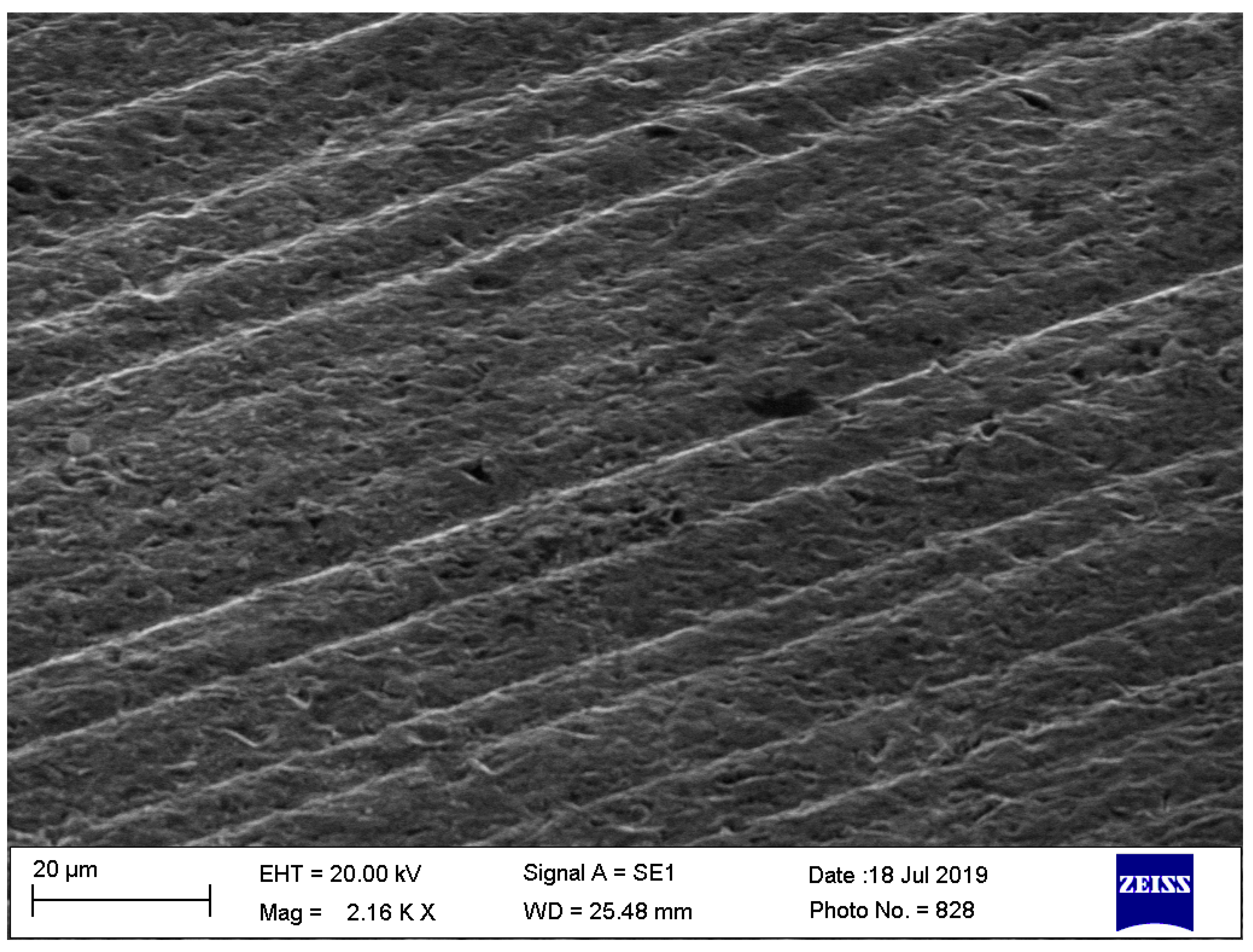
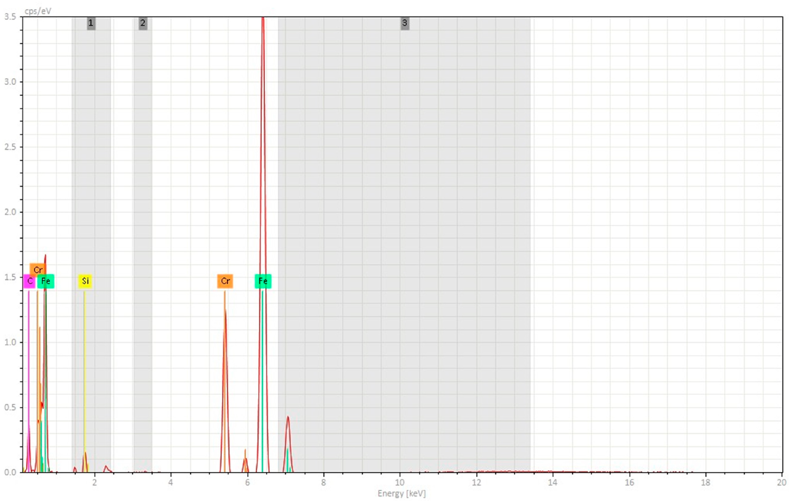
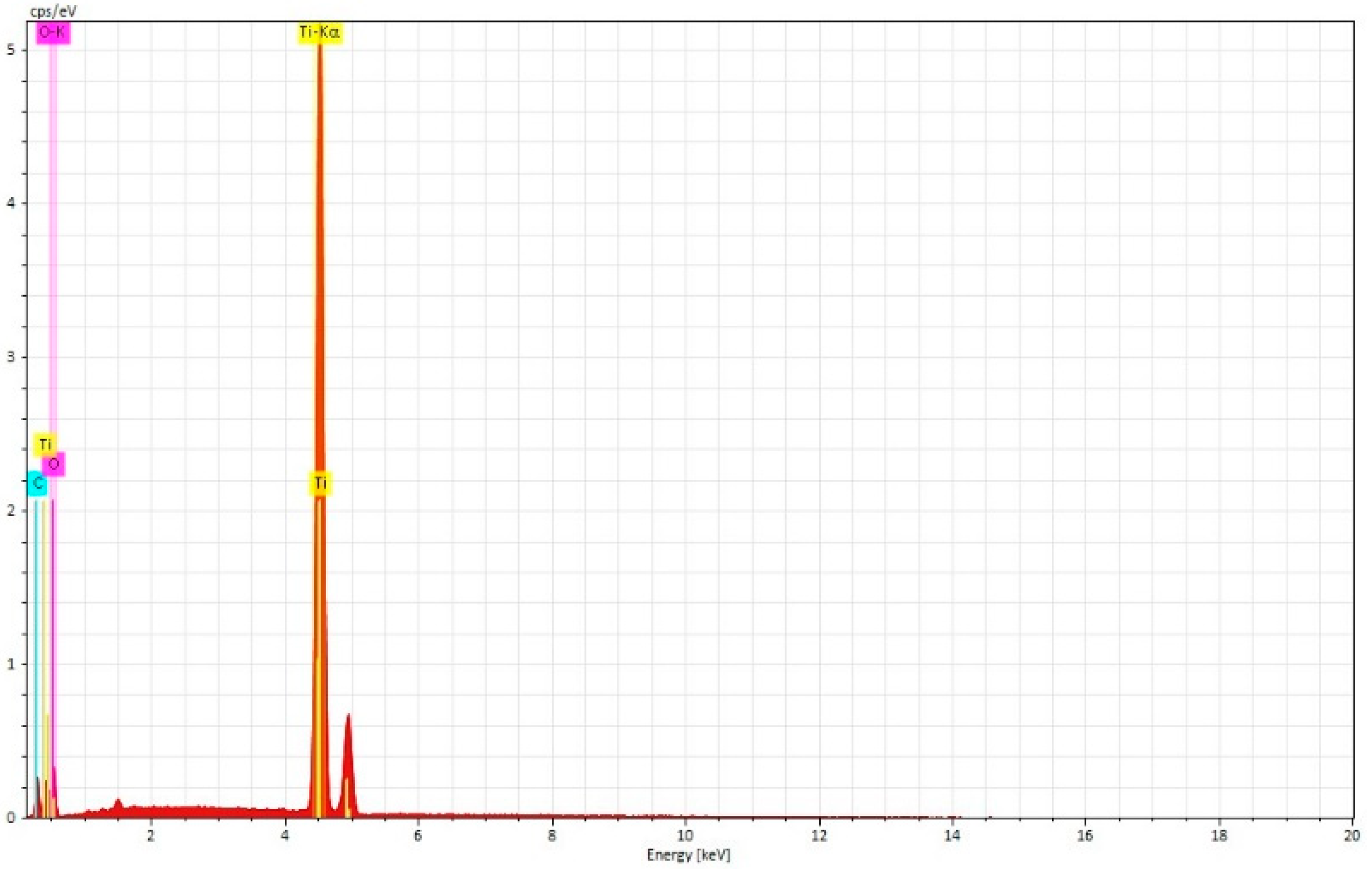
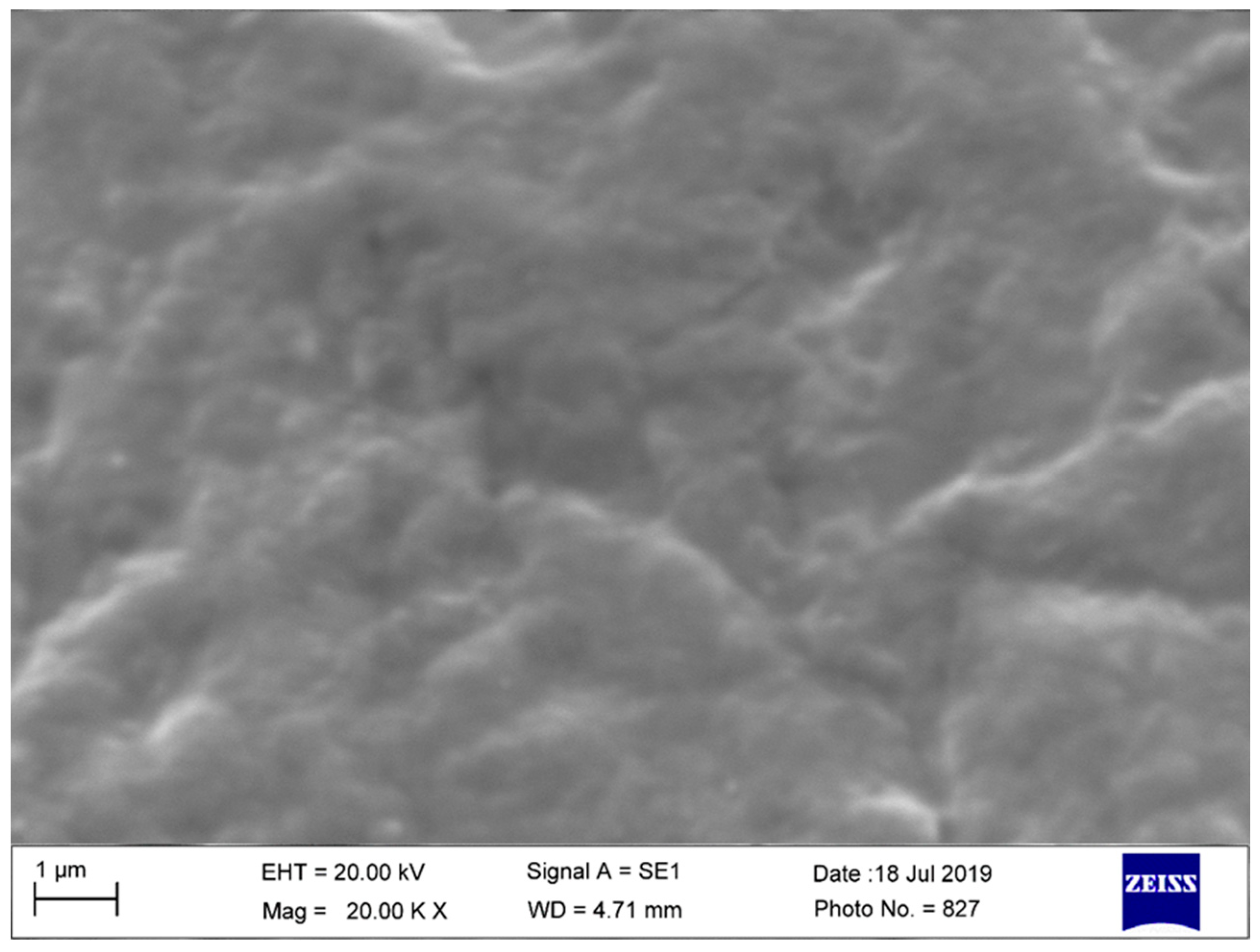
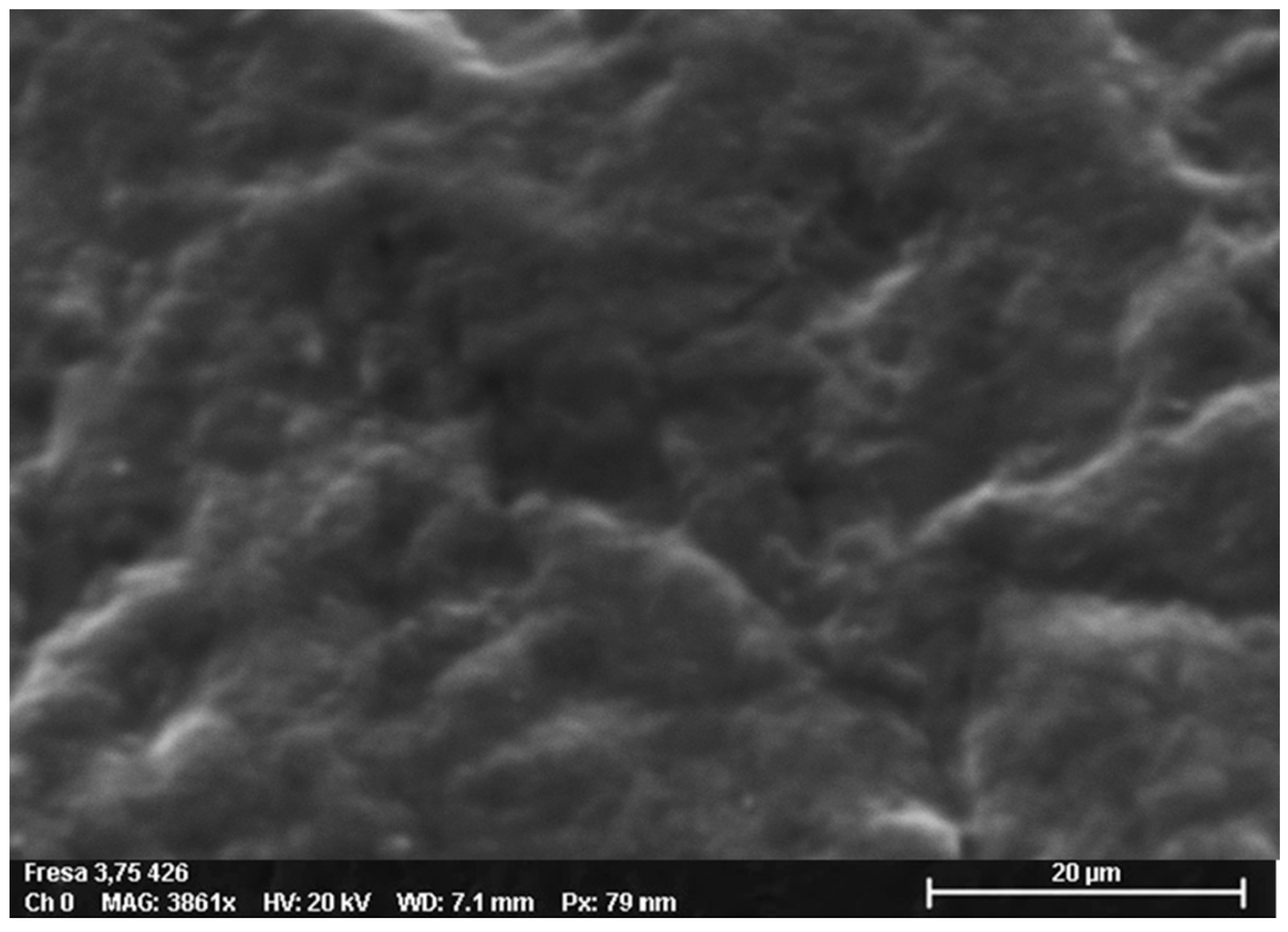
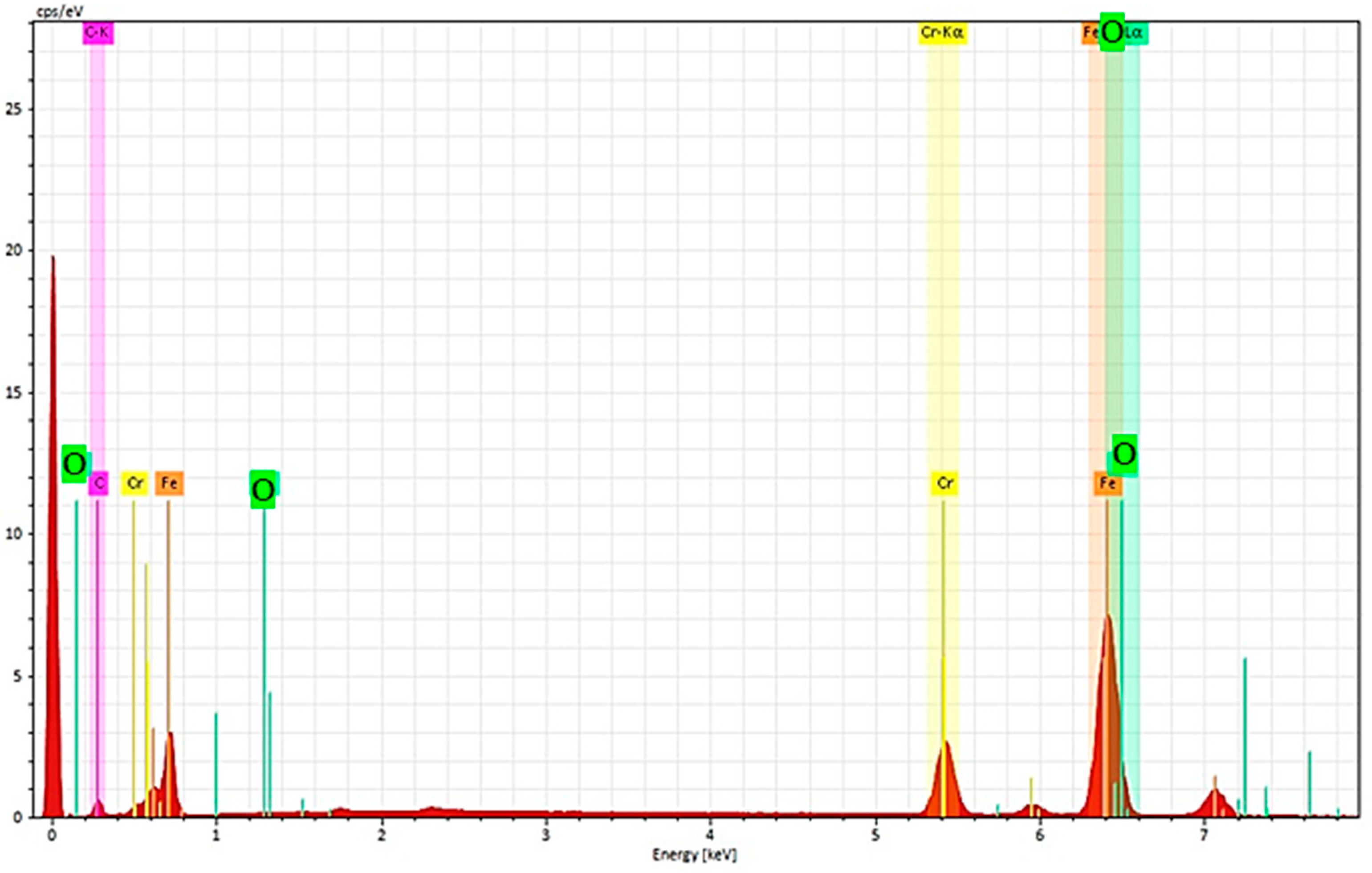
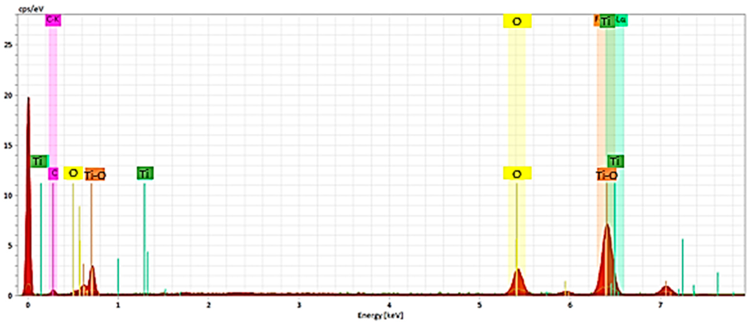
3.2. Scanning Electron Microscopy-EDS Analysis

3.2.1. Non-Sterilized Drill and Conical Tapper

The spectroscopic analysis showed that the flat area of implant drills was composed of stainless-steel alloys with a high content of iron, nickel and chromium. No microscopical surface alteration or microcracks have been detected (Figure 8, Figure 9, Figure 10 and Figure 11).

3.2.2. Used and Sterilized Drill and Conical Tapper

After a total of 30 osteotomies and sterilization processes, the drills and conical tappers showed macroscopical damages, but the damaged surface areas were topographically smaller and the laser depth marks remained clearly visible. A total of 9 ± 0.5% of the drill surface was covered by damage. No drills or conical tappers showed damage greater than 20% of the surface. The spectroscopic analysis of the drill surface showed a decrease in iron levels and a comparable increase in oxygen (Figure 12, Figure 13, Figure 14 and Figure 15).

4. Discussion

It is well known that the amount of osteotomies and the disinfection/sterilization procedures play a key role in drill cutting efficiency and bone heating, with significant clinical relevance [5,14]. Bone necrosis caused by excessive surgical trauma could affect the optimal healing and it is one of the most common causes of excessive early bone resorption, which could impair the implant stability, facilitate bacterial infiltration and lead to peri-implantitis induction [16]. The temperature increase is also correlated with bone density, and the drilling strength resistance, which depends on local bone composition, precisely by the cortical and spongious bone ratio [17,18,19]. A thicker cortical layer corresponds to a harder bone and a higher risk of generating high temperatures. Hence, the general advice is to use different protocols to create the implant site based on bone tissue density. These protocols envisage differences in terms of type and number of drills used and rotation velocity (number of revolutions per minute). It has been noticed that sharp drills, combined with high rotation velocity, allow us to rapidly create the implant site, reducing the risk of heat build-up [20,21,22,23]. On other hands, blunt drills find it more difficult to create a path, with a subsequently longer period of exposure to an increase in heat and a higher risk of bone necrosis [24]. A recent study assessed the changes in bone temperature during drilling performed repeatedly using the same drills (>100 times), comparing stainless steel drills or ceramic drills, with rotation velocity of 800 rpm and constant irrigation with saline solution at 21° and 50 mL/min [25]. The study concluded that both drill types could be used up to 50 times without producing hazardous temperatures for bone tissue and without showing evident signs of wear and deformation [25]. A recent systematic review of literature evaluated in vitro and in vivo studies conducted to measure a temperature increase generated during the creation of the implant site, to identify factors that can minimize damages [26]. The review identified 41 papers, of which 27 were correlated with a temperature increase during drilling. Many studies were conducted in vivo on non-vital bovine or porcine bone. The heat was measured in real time by thermocouples in 18 cases and infrared thermography in 7 cases. Three studies used immunohistochemical investigations to assess instant vitality of the cells. The highest temperature reached 64.4 °C in cortical areas, and the lowest was 28.4 °C. The drill deterioration was observed after 50 cycles of use, which corresponded to a significant increase in temperature and a moderate variation in the physiological homeostasis of bone cell proteins. Moreover, though bone cell vitality is maintained, one should not exceed this frequency of use for each drill. The use of irrigation, either internal or external, has provided contrasting results, and the studies included have rarely reported that temperature exceeds 47 °C. The authors conclude that the effect of irrigation requires further studies that should possibly be standardized. There is also evidence that the temperature increase seems to be correlated with the time required to prepare the implant site, which depends on the type of drill, on rotation velocity and on the load applied. With the same load, spiral drills generate the lowest temperature increase, while the greatest increase is recorded with piezoelectric drills [27]. Several authors have investigated the increase in heat generated by drills made of various materials, such as steel, steel-coated zirconium, titanium nitride, tungsten carbide or various types of ceramic [24,25,28,29,30]. Differences at various drilling depths have also been assessed (e.g., 3, 6, 9 mm). Significant differences have been observed only in the levels closest to the surface. However, broadly speaking, these studies have not observed significant differences in the increase in heat generated between drills made of different materials. The review generally found a low level of evidence and recommends greater standardization of future investigations on this topic, particularly regarding the axial load (2 kg), rotation velocity of the drills (1500 rpm), irrigation and type of bone to be drilled—most likely blocks of artificial bone. Finally, the use of infrared thermography is recommended, as it has proven to be more reliable and less invasive than thermocouples, which require additional perforation of bone tissue near the implant site to physically implant the thermocouple [26]. Moreover, infrared thermography has the great benefit of being able to study the temperature increase in vivo, thus closely approaching the actual situation in which most of the heat is dissipated by blood circulation of bone tissue. On the contrary, the limit of this technique could be determined by the thermal assessment of the sole external bone surface, with no direct measurements concerning the internal part of the implant site in contact with the drill milling. A recent in vitro study evaluated the influence of bone density on temperature and on the diameter of drills using blocks of artificial bone. Ten drills were used with diameters of 2.2, 2.8, 3.5 and 4.2 mm, as well as four blocks of artificial bone with density I-IV, constant velocity and external irrigation. The study observed that the temperature increase depends on bone density with an increase in density that causes a temperature increase. However, this effect diminishes as the drill diameter increases. This leads us to suggest that, when possible, precisely if there is sufficient bone volume available, large diameter implants and drills should be chosen, and preparation of the site should preferably limit the use of small-diameter drills [31]. An ex vivo study by Gehrke et al. evaluated the influence of the length of the drill (10 to 16 mm) and of the irrigation system (internal + external to prepare sites for 4.0 mm conical implants or only external for 4.1 or 4.3 mm cylindrical implants) on the production of heat during drilling of bovine ribs [32]. The drills were made of stainless steel. The heat measurements were recorded with three thermocouples positioned at three different vertical heights (2-7-12 mm from the bone crest), adjacent to the osteotomy site. The highest temperatures were recorded during removal of the drill from the osteotomy site. The lowest temperatures were recorded in the deepest sites, regardless of the drill length or of the irrigation system. However, the double irrigation system was significantly associated with lower temperatures compared to external irrigation systems. The conclusion is that, where possible, in multiple drilling systems, double irrigation systems should be used to maintain drilling temperature at its lowest levels, especially as the length of the drill increases [32]. A study conducted partly confuted this concept, showing that, in the artificial bone with density D1, the preparation of the implant site with drilling without irrigation at 50 rpm and drilling with irrigation at 1500 rpm produced similar temperatures (40.9 °C and 39.7 °C, respectively), though the study reported a significant difference, given the high number of repetitions and the low variability [33]. However, these conditions are hardly applicable to an actual situation in daily clinical practice, in addition to the fact that the properties of natural bone differ from those of artificial bone. In fact, in the former, besides the cortical region, there is the bone trabeculation, which gives different biomechanical responses; moreover, there is the bone vascularization system that contributes to the dissipation of the heat produced. Hence, in this case, a reduction in the drills’ speed corresponds to a reduction in the heating produced, especially in case of high-density bone, in which would be hard to create an appropriate implant site in a reasonable time and with appropriate precision. Another recent study by Gehrke et al. compared the insertion torque, implant stability and quality of the drilling site in the various drill systems [34]. The samples comprised blocks of high-density synthetic bone (Type I, similar to the anterior part of the mandible). Group 1 evaluated a single drill for a 4.2 mm conical implant; Group 2 assessed three consecutive drills for a 4.1 mm cylindrical implant, and Group 3 evaluated three consecutive drills for a 4.3 mm conical implant. The quality of the hole was evaluated with an automatic industrial device used to estimate cylindricity/circularity. The study showed that the single drill allows us to obtain greater insertion torque, and better quality of the implant site, which allows the implant to reach greater stability compared to the multiple drilling systems. Hence, the number of consecutive drilling procedures should be reduced, where possible, because each one contributes a series of imperfections in and changes to cylindricity, since it is hard to precisely maintain the same drilling axis (there is a tendency to ovalize implant sites), thus impairing the precision of the implant site and influencing the stability of the implant itself [34]. Generally, if the manufacturer’s recommendations are followed by maintaining an appropriate number of revolutions based on the type of bone, and if the drills are used a number of times that does not exceed the one indicated to avoid their loss of sharpness, the risks of generating bone necrosis and a subsequent high degree of resorption are extremely reduced. A randomized clinical study conducted in two centers compared the preparation of the implant site with a newly designed single cylindrical–conical drill, compared to a standard multi-drill preparation [35]. Participants were 40 subjects with single missing teeth and residual bone side of at least 10 mm in height and 5 mm in thickness. The implants were left to heal submerged for 3 months, and then loaded fixed crowns prosthesis. The variables measured were implant failure, complications, preference of the operator, time required from the start of the preparation to positioning of the implant, variations in the marginal bone measured after 4 months of load, pain, post-surgical swelling and number of analgesics administered. No implant had failed after 4 months, and no statistically significant differences were observed in terms of bone loss (mean 0.54 mm and 0.41 mm in the single drill and multi-drill group, respectively). Instead, there were statistically significant differences in favor of the single drill in terms of duration of surgery, operator’s preference, degree of pain, persistence of swelling and number of analgesics taken [35]. The outcome of the present investigation was that no statistically significant temperature increase was detected after 30 bone osteotomies of the implant bed sites prepared with steel drills and titanium tappers. An increase in temperature difference between the baseline and the experimental drill was about 1.5 °C, and lower heat levels were detected after the use of the manual conical tapper. The detected temperature differences were never critical to the health of preimplant bone. No evidence was detected about the influence of disinfection and sterilization or the extent of drill use. No damage was observed with SEM/EDS analysis for the steel drills and conical tappers after experimental osteotomies and sterilization cycles with no evidence of severe corrosion aspects, and no remarkable change in composition of the devices was detected. The results of the present study showed that after the bone osteotomies and sterilization procedure, the experimental drills and tappers conserved the cutting efficiency by preventing metal release and overheating of the bone, which represent potential factors for early implant failure.

5. Conclusions

The present investigation demonstrated that the implant site osteotomies in standardized in vitro condition and sterilization processes produced no effect on steel drill and manual conical tapper that showed inertness, structural stability, durability and high-level cutting performance.

Author Contributions

Conceptualization, A.S., F.L.; methodology, A.S., F.L.; software, F.L.; validation, A.S., S.A.G., F.L.; formal analysis, A.S., F.L.; investigation, A.S., F.L.; writing—original draft preparation, A.S., F.L.; writing—review and editing, A.S., F.L.; visualization, A.S., F.L., S.A.G., F.F.; All authors have read and agreed to the published version of the manuscript.

Funding

This research received no external funding.

Institutional Review Board Statement

Not applicable.

Informed Consent Statement

Not applicable.

Data Availability Statement

All experimental data to support the findings of this study are available contacting the corresponding author upon request. The authors have annotated the entire data building process and empirical techniques presented in the paper.

Conflicts of Interest

The authors declare no conflict of interest.

References

  1. Albrektsson, T.; Berglundh, T.; Lindhe, J. Osseointegration: Historic Background and Current Concepts. Clin. Periodontol. Implant. Dent. 2003, 4, 809–820. [Google Scholar]
  2. Bilhan, H.; Geckili, O.; Mumcu, E.; Bozdag, E.; Sünbüloğlu, E.; Kutay, O. Influence of Surgical Technique, Implant Shape and Diameter on the Primary Stability in Cancellous Bone: What Influences Primary Stability in Cancellous Bone. J. Oral Rehabil. 2010, 37, 900–907. [Google Scholar] [CrossRef] [PubMed]
  3. Arys, A.; Philippart, C.; Dourov, N.; He, Y.; Le, Q.T.; Pireaux, J.J. Analysis of Titanium Dental Implants after Failure of Osseointegration: Combined Histological, Electron Microscopy, and X-ray Photoelectron Spectroscopy Approach. J. Biomed. Mater. Res. 1998, 43, 300–312. [Google Scholar] [CrossRef]
  4. Scarano, A.; Piattelli, A.; Assenza, B.; Carinci, F.; Di Donato, L.; Romani, G.L.; Merla, A. Infrared Thermographic Evaluation of Temperature Modifications Induced during Implant Site Preparation with Cylindrical versus Conical Drills. Clin. Implant Dent. Relat. Res. 2011, 13, 319–323. [Google Scholar] [CrossRef] [PubMed]
  5. Scarano, A.; Lorusso, F.; Noumbissi, S. Infrared Thermographic Evaluation of Temperature Modifications Induced during Implant Site Preparation with Steel vs. Zirconia Implant Drill. J. Clin. Med. 2020, 9, 148. [Google Scholar] [CrossRef] [Green Version]
  6. Sener, B.C.; Dergin, G.; Gursoy, B.; Kelesoglu, E.; Slih, I. Effects of Irrigation Temperature on Heat Control in Vitro at Different Drilling Depths. Clin. Oral Implant. Res. 2009, 20, 294–298. [Google Scholar] [CrossRef]
  7. Benington, I.C.; Biagioni, P.A.; Briggs, J.; Sheridan, S.; Lamey, P.-J. Thermal Changes Observed at Implant Sites during Internal and External Irrigation: Thermal Changes at Implant Sites during Irrigation. Clin. Oral Implant. Res. 2002, 13, 293–297. [Google Scholar] [CrossRef]
  8. Lucchiari, N.; Frigo, A.C.; Stellini, E.; Coppe, M.; Berengo, M.; Bacci, C. In Vitro Assessment with the Infrared Thermometer of Temperature Differences Generated During Implant Site Preparation: The Traditional Technique Versus the Single-Drill Technique: Heat with Traditional and Single-Bur Drilling. Clin. Implant. Dent. Relat. Res. 2016, 18, 182–191. [Google Scholar] [CrossRef]
  9. Ozcan, M.; Salimov, F.; Temmerman, A.; Turer, O.U.; Alkaya, B.; Haytac, M.C. The Evaluation of Different Osteotomy Drilling Speed Protocols on Cortical Bone Temperature, Implant Stability and Bone Healing: An Experimental Study in an Animal Model. J. Oral Implant. 2022, 48, 3–8. [Google Scholar] [CrossRef]
  10. Tur, D.; Giannis, K.; Unger, E.; Mittlböck, M.; Rausch-Fan, X.; Strbac, G.D. Thermal Effects of Various Drill Materials during Implant Site Preparation—Ceramic vs. Stainless Steel Drills: A Comparative in Vitro Study in a Standardised Bovine Bone Model. Clin. Oral Implant. Res. 2021, 32, 154–166. [Google Scholar] [CrossRef]
  11. Cattoni, F.; Merlone, A.; Broggi, R.; Manacorda, M.; Vinci, R. Computer-Assisted Prosthetic Planning and Implant Design with Integrated Digital Bite Registration: A Treatment Protocol. J. Biol. Regul. Homeost. Agents 2021, 35, 11–29. [Google Scholar] [CrossRef] [PubMed]
  12. Merlone, A.; Tetè, G.; Cantile, N.; Manacorda, M.; Cattoni, F. Minimally Invasive Digital Implant-Prosthetic Procedure in “All on 4” Rehabilitation in Patients with Special Needs: A Three-Year Follow-Up. J. Biol. Regul. Homeost. Agents 2021, 35, 71–85. [Google Scholar] [CrossRef] [PubMed]
  13. Cattoni, F.; Chirico, L.; Merlone, A.; Manacorda, M.; Vinci, R.; Gherlone, E.F. Digital Smile Designed Computer-Aided Surgery versus Traditional Workflow in “All on Four” Rehabilitations: A Randomized Clinical Trial with 4-Years Follow-Up. Int. J. Environ. Res. Public Health 2021, 18, 3449. [Google Scholar] [CrossRef] [PubMed]
  14. Scarano, A.; Petrini, M.; Mastrangelo, F.; Noumbissi, S.; Lorusso, F. The Effects of Liquid Disinfection and Heat Sterilization Processes on Implant Drill Roughness: Energy Dispersion X-ray Microanalysis and Infrared Thermography. J. Clin. Med. 2020, 9, 1019. [Google Scholar] [CrossRef] [Green Version]
  15. Scarano, A.; Carinci, F.; Lorusso, F.; Festa, F.; Bevilacqua, L.; Santos de Oliveira, P.; Maglione, M. Ultrasonic vs Drill Implant Site Preparation: Post-Operative Pain Measurement Through VAS, Swelling and Crestal Bone Remodeling: A Randomized Clinical Study. Materials 2018, 11, 2516. [Google Scholar] [CrossRef] [Green Version]
  16. Oh, T.-J.; Yoon, J.; Misch, C.E.; Wang, H.-L. The Causes of Early Implant Bone Loss: Myth or Science? J. Periodontol. 2002, 73, 322–333. [Google Scholar] [CrossRef] [PubMed]
  17. Misch, C.E.; Dietsh-Misch, F.; Hoar, J.; Beck, G.; Hazen, R.; Misch, C.M. A Bone Quality–Based Implant System: First Year of Prosthetic Loading. J. Oral Implant. 1999, 25, 185–197. [Google Scholar] [CrossRef]
  18. Misch, C.E.; Qu, Z.; Bidez, M.W. Mechanical Properties of Trabecular Bone in the Human Mandible: Implications for Dental Implant Treatment Planning and Surgical Placement. J. Oral Maxillofac. Surg. 1999, 57, 700–706. [Google Scholar] [CrossRef]
  19. Norton, M.R.; Gamble, C. Bone Classification: An Objective Scale of Bone Density Using the Computerized Tomography Scan: Bone Classification: An Objective Scale of Bone Density Using the Computerized Tomography Scan. Clin. Oral Implant. Res. 2001, 12, 79–84. [Google Scholar] [CrossRef]
  20. Bubeck, K.A.; García-López, J.; Maranda, L.S. In Vitro Comparison of Cortical Bone Temperature Generation between Traditional Sequential Drilling and a Newly Designed Step Drill in the Equine Third Metacarpal Bone. Veter-Comp. Orthop. Traumatol. 2009, 22, 442–447. [Google Scholar] [CrossRef]
  21. Chacon, G.E.; Bower, D.L.; Larsen, P.E.; McGlumphy, E.A.; Beck, F.M. Heat Production by 3 Implant Drill Systems after Repeated Drilling and Sterilization. J. Oral Maxillofac. Surg. 2006, 64, 265–269. [Google Scholar] [CrossRef] [PubMed]
  22. Sharawy, M.; Misch, C.E.; Weller, N.; Tehemar, S. Heat Generation during Implant Drilling: The Significance of Motor Speed. J. Oral Maxillofac. Surg. 2002, 60, 1160–1169. [Google Scholar] [CrossRef] [Green Version]
  23. Gronkiewicz, K.; Majewski, P.; Wisniewska, G.; Pihut, M.; Loster, B.W.; Majewski, S. Experimental Research on the Possibilities of Maintaining Thermal Conditions within the Limits of the Physiological Conditions during Intraoral Preparation of Dental Implants. J. Physiol. Pharmacol. 2009, 60 (Suppl. 8), 123–127. [Google Scholar] [PubMed]
  24. Ercoli, C.; Funkenbusch, P.D.; Lee, H.-J.; Moss, M.E.; Graser, G.N. The Influence of Drill Wear on Cutting Efficiency and Heat Production during Osteotomy Preparation for Dental Implants: A Study of Drill Durability. Int. J. Oral Maxillofac. Implant. 2004, 19, 335–349. [Google Scholar]
  25. Oliveira, N.; Alaejos-Algarra, F.; Mareque-Bueno, J.; Ferrés-Padró, E.; Hernández-Alfaro, F. Thermal Changes and Drill Wear in Bovine Bone during Implant Site Preparation. A Comparative in Vitro Study: Twisted Stainless Steel and Ceramic Drills: Thermal Changes and Drill Wear in Bovine Bone. Clin. Oral Implant. Res. 2012, 23, 963–969. [Google Scholar] [CrossRef] [PubMed]
  26. Möhlhenrich, S.C.; Modabber, A.; Steiner, T.; Mitchell, D.A.; Hölzle, F. Heat Generation and Drill Wear during Dental Implant Site Preparation: Systematic Review. Br. J. Oral Maxillofac. Surg. 2015, 53, 679–689. [Google Scholar] [CrossRef]
  27. Stelzle, F.; Frenkel, C.; Riemann, M.; Knipfer, C.; Stockmann, P.; Nkenke, E. The Effect of Load on Heat Production, Thermal Effects and Expenditure of Time during Implant Site Preparation—An Experimental Ex Vivo Comparison between Piezosurgery and Conventional Drilling. Clin. Oral Implant. Res. 2014, 25, e140–e148. [Google Scholar] [CrossRef]
  28. Sumer, M.; Misir, A.F.; Telcioglu, N.T.; Guler, A.U.; Yenisey, M. Comparison of Heat Generation during Implant Drilling Using Stainless Steel and Ceramic Drills. J. Oral Maxillofac. Surg. 2011, 69, 1350–1354. [Google Scholar] [CrossRef]
  29. Harder, S.; Egert, C.; Wenz, H.J.; Jochens, A.; Kern, M. Influence of the Drill Material and Method of Cooling on the Development of Intrabony Temperature during Preparation of the Site of an Implant. Br. J. Oral Maxillofac. Surg. 2013, 51, 74–78. [Google Scholar] [CrossRef]
  30. Koo, K.-T.; Kim, M.-H.; Kim, H.-Y.; Wikesjö, U.M.E.; Yang, J.-H.; Yeo, I.-S. Effects of Implant Drill Wear, Irrigation, and Drill Materials on Heat Generation in Osteotomy Sites. J. Oral Implant. 2015, 41, e19–e23. [Google Scholar] [CrossRef]
  31. Möhlhenrich, S.C.; Abouridouane, M.; Heussen, N.; Modabber, A.; Klocke, F.; Hölzle, F. Influence of Bone Density and Implant Drill Diameter on the Resulting Axial Force and Temperature Development in Implant Burs and Artificial Bone: An in Vitro Study. Oral Maxillofac. Surg. 2016, 20, 135–142. [Google Scholar] [CrossRef] [PubMed]
  32. Gehrke, S.A.; Aramburú Júnior, J.S.; Pérez-Albacete Martínez, C.; Ramirez Fernandez, M.P.; Maté Sánchez de Val, J.E.; Calvo-Guirado, J.L. The Influence of Drill Length and Irrigation System on Heat Production during Osteotomy Preparation for Dental Implants: An Ex Vivo Study. Clin. Oral Implant. Res. 2018, 29, 772–778. [Google Scholar] [CrossRef] [PubMed]
  33. Oh, J.-H.; Fang, Y.; Jeong, S.-M.; Choi, B.-H. The Effect of Low-Speed Drilling without Irrigation on Heat Generation: An Experimental Study. J. Korean Assoc. Oral Maxillofac. Surg. 2016, 42, 9. [Google Scholar] [CrossRef] [Green Version]
  34. Gehrke, S.A.; Guirado, J.L.C.; Bettach, R.; Fabbro, M.D.; Martínez, C.P.-A.; Shibli, J.A. Evaluation of the Insertion Torque, Implant Stability Quotient and Drilled Hole Quality for Different Drill Design: An in Vitro Investigation. Clin. Oral Implant. Res. 2018, 29, 656–662. [Google Scholar] [CrossRef]
  35. Guazzi, P.; Grandi, T.; Grandi, G. Implant Site Preparation Using a Single Bur versus Multiple Drilling Steps: 4-Month Post-Loading Results of a Multicenter Randomised Controlled Trial. Eur. J. Oral Implant. 2015, 8, 283–290. [Google Scholar]
Figure 1. Details of the drilling sequence.
Prosthesis 04 00054 g001
Figure 2. Details of the drilling sequence (continued).
Prosthesis 04 00054 g002
Figure 3. Details of the drilling sequence (continued).
Prosthesis 04 00054 g003
Figure 4. Details of the countersink drilling sequence (continued).
Prosthesis 04 00054 g004
Figure 5. Details of the conical tappers preparation.
Prosthesis 04 00054 g005
Figure 6. Details of the conical tappers (continued).
Prosthesis 04 00054 g006
Figure 7. Details of the infrared thermal measurements.
Prosthesis 04 00054 g007
Figure 8. Details of the SEM analysis of the drills used in the present experiment.
Prosthesis 04 00054 g008
Figure 9. Details of the SEM analysis of the drills used in the present experiment (Mag. 2.16 Kx).
Prosthesis 04 00054 g009
Figure 10. EDS-SEM analysis composition of the non-used drills.
Prosthesis 04 00054 g010
Figure 11. EDS-SEM analysis composition of the non-used tappers.
Prosthesis 04 00054 g011
Figure 12. SEM analysis composition of the used drills.
Prosthesis 04 00054 g012
Figure 13. SEM analysis composition of the used drills (Mag. 3861x).
Prosthesis 04 00054 g013
Figure 14. EDS-SEM analysis composition of the non-used drills.
Prosthesis 04 00054 g014
Figure 15. EDS-SEM analysis composition of the non-used tappers.
Prosthesis 04 00054 g015
Table 1. Summary of the statistical analysis of the drills’ temperatures [Mann–Whitney Test; * p < 0.05].
Drill Bone Temperature (°C).BaselineFGR3710FGR5513FFC3710FFC5513DRP200DRP450CTK425
1st osteotomy37.0 ± 0.10 °C38.6 ± 0.6640.96 ± 0.5939.01 ± 0.5141.12 ± 0.4738.59 ± 0.7339.37 ± 0.6339.25 ± 0.55
After 30th osteotomy37.0 ± 0.13 °C39.01 ± 0.5641.83 ± 0.5639.37 ± 1.1241.97 ± 0.5139.01 ± 0.5639.89 ± 0.7139.71 ± 0.63
p-value/* p > 0.05* p > 0.05* p > 0.05* p > 0.05* p > 0.05* p > 0.05* p > 0.05
Table 2. Summary of the statistical analysis of the bone tappers temperature [Mann–Whitney Test; * p < 0.05].
TapperBone Temperature (°C)BaselineMKN3710MKN3711MKN3713MKN3714MSL5510MSL5511MSL5513
1st osteotomy37.0 ± 0.14 °C38.1 ± 0.538.3 ± 0.4738.3 ± 0.5038.5 ± 0.4538.2 ± 0.4638.4 ± 0.5738. 8± 0.66
After 30th osteotomy37.0 ± 0.12 °C38.1 ± 0.538.3 ± 0.4738.3 ± 0.5038.5 ± 0.4538.2 ± 0.4638.4 ± 0.5738.8 ± 0.66
p-value/* p > 0.05* p > 0.05* p > 0.05* p > 0.05* p > 0.05* p > 0.05* p > 0.05
Publisher’s Note: MDPI stays neutral with regard to jurisdictional claims in published maps and institutional affiliations.

