Article

Comparison of the Accuracy between Denture Bases Produced by Subtractive and Additive Manufacturing Methods: A Pilot Study

by
Francesco Grande
1,2,*,
Fabio Tesini
1,
Mario Cesare Pozzan
1,
Edoardo Mochi Zamperoli
1,
Massimo Carossa
2 and
Santo Catapano
1
1
Department of Prosthodontics, University of Ferrara, Via Luigi Borsari 46, 44121 Ferrara, Italy
2
Department of Surgical Sciences, CIR Dental School, University of Turin, 10126 Turin, Italy
*
Author to whom correspondence should be addressed.
Prosthesis 2022, 4(2), 151-159; https://doi.org/10.3390/prosthesis4020015
Submission received: 18 February 2022 / Revised: 17 March 2022 / Accepted: 25 March 2022 / Published: 28 March 2022

Abstract

:
Today, two different types of CAD-CAM fabrication methods for complete denture bases are available besides the conventional protocols: a subtractive milling process from a prepolymerized block of polymethylmethacrylate and an additive manufacturing process that built the denture base using a light-cured liquid in a VAT-polymerization process. The aim of this study was to evaluate and to compare the accuracy and precision of denture prosthetic bases made with subtractive and additive manufacturing technologies and to compare them with a denture base with the conventional method in muffle. From the results obtained, 3D printing dentures show a statistically significant higher accuracy than milled prosthetic bases. Milled prosthetic bases have similar accuracy than conventional fabricated dentures.

1. Introduction

A fundamental factor that determines the quality of removable dentures is the denture fit [1]. A high primary wearing comfort and a low or null occurrence of traumatic ulcers could be achieved only with a well-fitting prosthesis [2,3,4]. Furthermore, factors such as denture retention, masticatory performance and speaking ability depend on the tissue-congruent denture fit which then has a strong impact on the patients’ satisfaction and quality of life [5].
Conventional protocols for the fabrication of complete dentures (CDs) are well established and constitute a part of contemporary clinical practice. In recent years, CAD-CAM technologies have been successfully introduced in prosthetic dentistry and have also been applied to removable prostheses [6,7]. Two CAD-CAM techniques are nowadays available to fabricate complete dentures: a subtractive milling process starting from a pre-polymerized block of polymethylmethacrylate (PMMA) and an additive manufacturing or rapid prototyping process (RP), also known as 3D printing, that built the denture base using a light-cured liquid in a VAT-polymerization process [8,9].
Some important advantages have been brought by these new technologies: in case of loss or fracture of the denture, digital storing of patients’ data is useful to rapidly fabricate a new and identical prosthesis without having to make new clinical records [10,11]. A reduction in patient appointments and time also for the dentist and the technician has been advocated [8,12]. For subtractive manufacturing where the denture bases are milled from fully polymerized acrylic resin pucks, the avoidance of resin polymerization shrinkage could result in an improved fitting of CAD/CAM dentures than conventional ones [13]. However, the milling process requires large quantities of material, and 3D printing techniques can represent more sustainable methods reducing resin consumption [14].
However, the denture base adaptation to the denture bearing tissues remains essential for the retention and stability of the complete denture. In literature, several studies using different methodologies and materials try to establish which method results in the best denture adaptation. In order to understand the degree of accuracy of each method, trueness and precision must be taken into consideration. According to ISO standard, accuracy is defined as the closeness of a measurement to the true value while precision is the degree to which repeated measurements under unchanged conditions show the same results [15]. In this case, the accuracy is related to the discrepancy of the intaglio surface of the CDs produced with the plaster model while the precision is referred to the reproducibility of the same intaglio surface on each production method.
The aim of this study was to evaluate and to compare the accuracy and precision of denture prosthetic bases made with subtractive and additive manufacturing technologies.

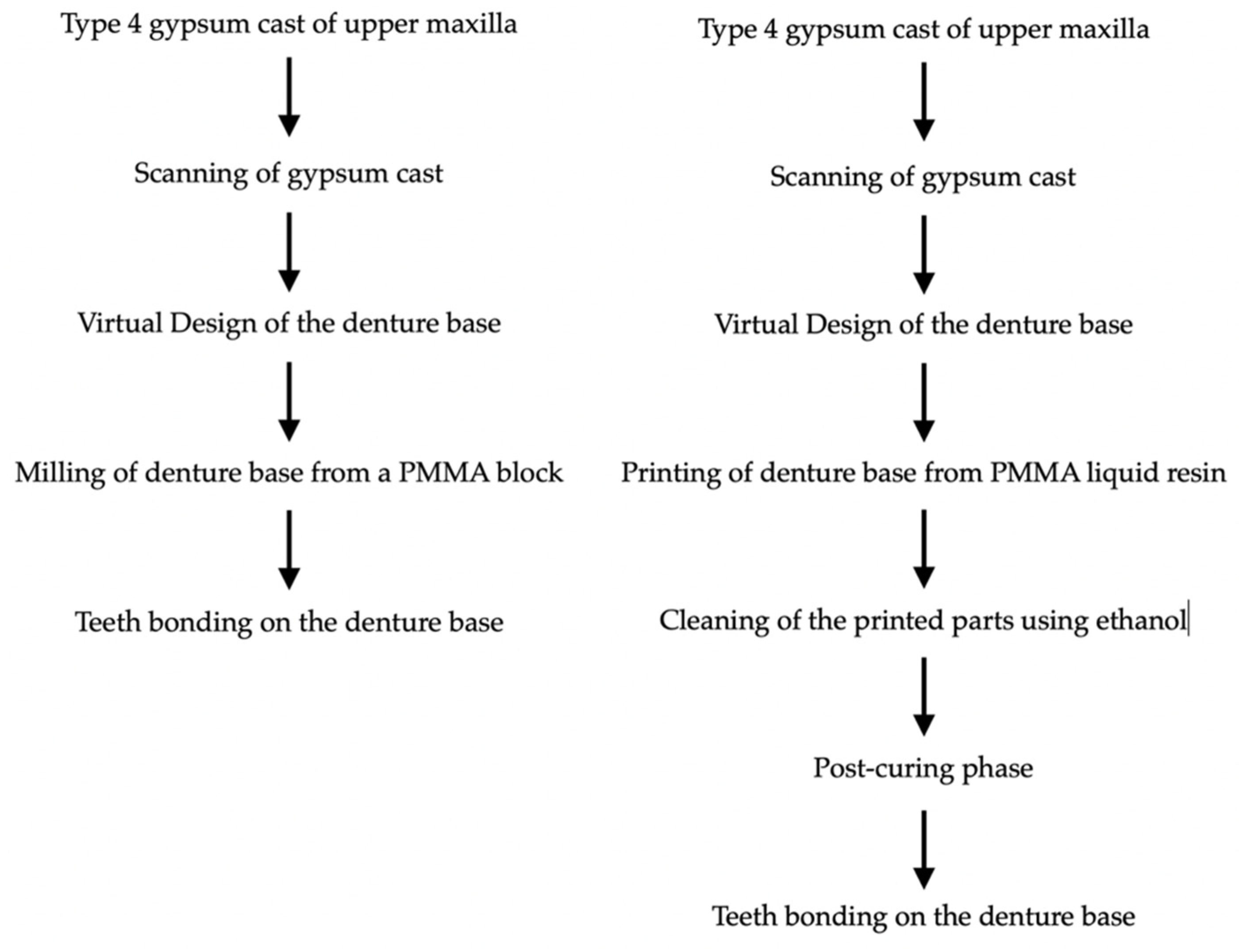
2. Materials and Methods

A type 4 gypsum cast was initially poured from a secondary impression of the upper arch of an edentulous patient, using a vacuum mixing machine. The gypsum model, declared scannable by the manufacturer (Elite Master, Zhermack Spa, Badia Polesine, Italy), was scanned using a desktop scanner (D810 laboratory scanner, 3Shape, Copenhagen, Denmark) to obtain a three-dimensional STL file of the upper edentulous jaw. Then, a virtual denture base was digitally designed on the model using a CAD software (Exocad software, Exocad GmbH, Darmstadt, Germany). The denture base was digitally designed following the conventional analogue technique: the postdam was positioned 2 mm posterior to the palatine fovee while the peripheral seal was placed 1.5 mm from the highest point of the vestibule. The anterior and lateral frenulum were avoided. The insertion axis of the prosthesis was highlighted, and the undercuts of the model were considered and avoided in the virtual design of the baseplate. Other denture properties were set; a base thickness of 2 mm, a cervical thickness of 0.8 mm, a smoothing of 5 mm, and an under teeth minimum thickness of 0.8 mm. Sockets for teeth bonding were digitally designed, based on the teeth library file chosen (PhysioSelect TCR, Candulor AG, Glattpark, Switzerland).
The STL file was used to fabricate six PMMA denture bases; three of them were milled by a five-axis milling machine (DMG Saver, DMG Mori Seiki, Nagoya, Japan), and the other three were additively manufactured by a 3D printer machine (NextDent 5100 3D, NextDent B.V., Soesterberg, The Netherlands) with a Digital Light Processing technology. The milled bases were made of a PMMA resin (PMMA CAD-CAM Ruthinium Monocromatic Disc A3, Ruthinium, Badia Polesine, Italy) while the 3D printer used a light-curing base resin (NextDent Denture 3D+, NextDent B.V., Soesterberg, The Netherlands) (Figure 1). In the CAM software of the 3D printer, the orientation of the denture base was vertical with a slight angle of the intaglio surface toward the build platform while the buccal flange was maintained opposite to the building platform. The support touchpoints were positioned around the perimeter edge of the denture and at the center of the palate in the intaglio surface, in order to facilitate the supports removal after the printing procedure. Parameters when using slicing software are 47 μm resolution and a layer thickness of 50 μm.
In the CAM software for the milling procedure, the baseplate originated from the center of the prepolimerized PMMA disc. Each baseplate was fabricated from 1 resin plate that have a diameter of 95 mm and a thickness of 16 mm.
After receiving the six resin denture bases from the milling center, they were manually positioned on the master model to exclude possible important distortions and to verify the presence undercuts that could prevent the correct accommodation of the baseplate on the model. None of the artifacts were excluded. Then, the same teeth (PhysioSelect TCR, Candulor AG, Glattpark, Switzerland) were mounted on the six models with the aid of plaster keys, previously obtained from the patient’s correct mounting. This was possible because the initial model was firstly duplicated using a duplication silicon, and all the clinical and technical steps for the fabrication of the upper complete denture were carried out also on the patient with a conventional technique. In this workflow, after the secondary impression, a record base was adjusted according to vertical dimension and centric relation measurement of the patient. After that, try-in of anterior and posterior teeth was performed, and then, the final mounting was registered with plaster keys. The liquid resin of the final prosthesis obtained with that conventional technique was pressed in muffle and then in water at 100 °C for 30 min.
Bonding of the artificial acrylic teeth to CAD-CAM denture bases was executed after the milling and printing procedure and after the post-curing, for 3D printed. For this procedure, for 3D printed prosthesis, a self-curing resin (Vertex Castavaria, Vertex Dental, Soesterberg, The Netherlands) compatible with the 3D printed resin, as indicated by the manufacturer, was used. For the milled baseplates, teeth were attached using a cold resin (Acry Self, Ruthinium, Badia Polesine, Italy) normally used for milled dentures.
Then, in order to quantify the discrepancies between the CAD-CAM denture bases and the master model, a Vinyl Polyether material (Fit-checker Advanced, GC Corporation, Tokio, Japan) was placed between them. A known equal quantity of material mixed with cartridges was used and applied every time in the same position of the denture base. After the fit-checker appliance, each base was positioned on the master model, and then, the entire system was mounted on a verticulator (Vertysystem, Vicenza, Italy) using a the previously prepared plaster keys. The verticulator, locked in its closed position, allows to maintain the same pressure conditions for all the denture bases in contact with the lower gypsum key by using every time the same vertical dimension. (Figure 2).
Each denture base was kept in contact with the master model for 3 min allowing the complete setting of the material. After opening the verticulator and removing the bases from the models, a thin film of the material was obtained for each denture base. Each polymerized silicone-ether film was mapped with a blue marker, identifying three different transverse regions:
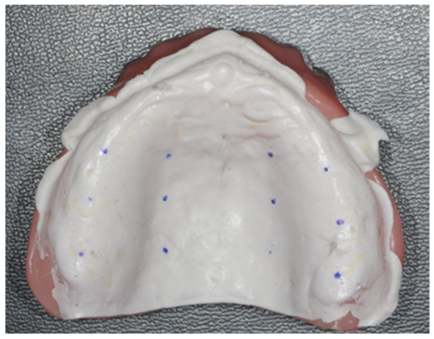
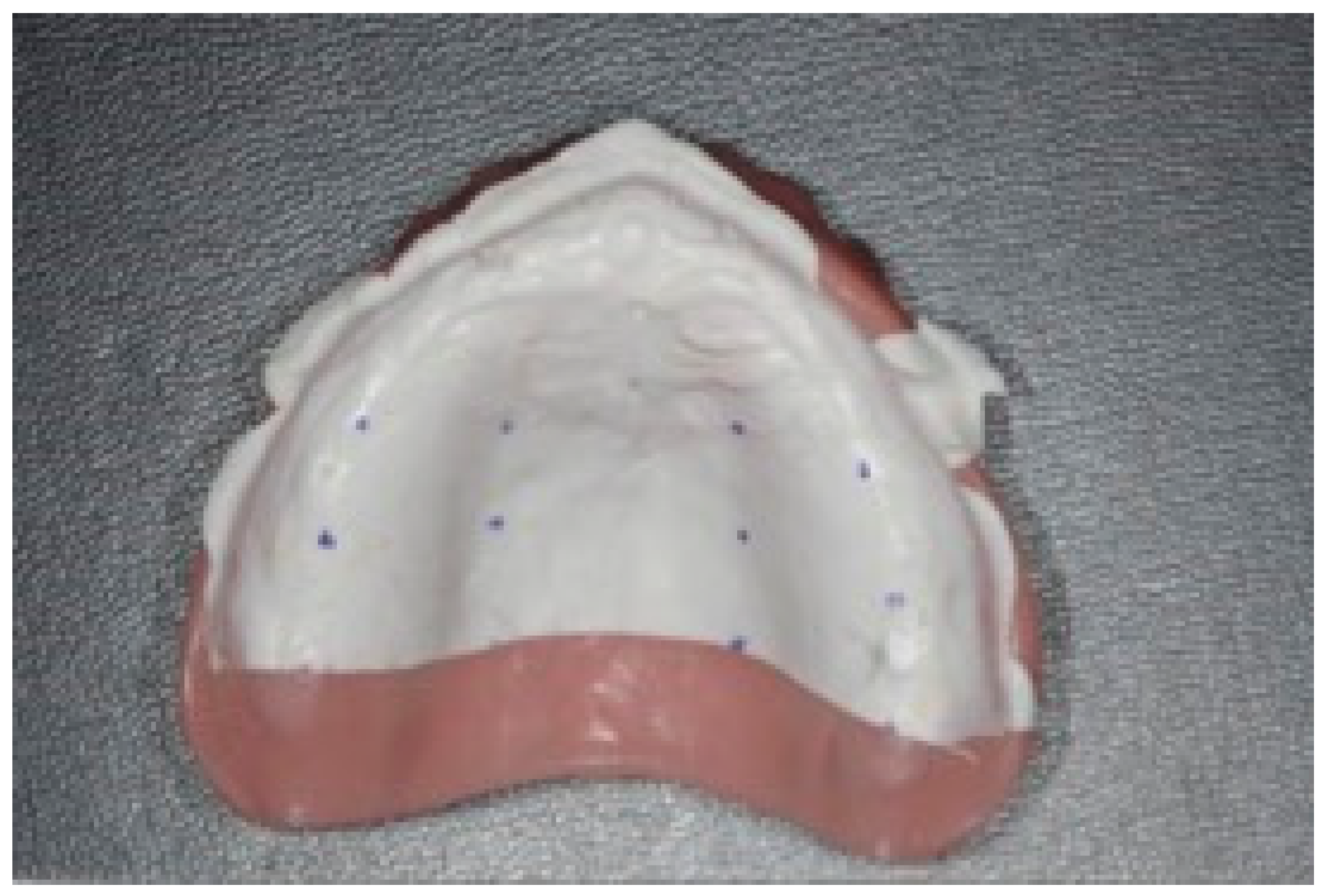
  • Zone 1: one centimeters from the post-dam;
  • Zone 2: two centimeters from the post-dam;
  • Zone 3: three centimeters from the post-dam.
For each region, two points on the hard palate and two points in the center of the alveolar ridge were identified (Figure 3).
The Fit-checker films were then cut with a No. 15 Hu-Friedy blade into transverse sections at the three identified zones and taken out from the intaglio surface of the denture (Figure 4). Then, thickness measurements at the identified points were performed. A highly accurate digital thickness gauge (Holex, Hoffmann SE, Munich, Germany) with a 10 µm tolerance was used for thickness measurements by F.T.

3. Statistical Analysis

A descriptive statistics was performed based on the data obtained from the measurements. For each group, the medium and the standard deviation were calculated. Then, the values were simply compared.

4. Results

For this study, a total of 84 measurements were performed (Table 1).

5. Discussion

The success of dental prostheses is dependent upon several factors [16,17,18,19,20]. In removable prosthodontics, the denture fit is very important for a satisfying prosthetic rehabilitation [21,22,23]. Nowadays, CAD-CAM technologies for prosthetic dentures could be classified into 2 different categories: additive manufacturing and subtractive manufacturing [6,7,8,9,10]. Subtractive manufacturing is based on computerized numerical control (CNC) machines that operate under the control of software, realizing the object previously designed in the virtual environment (CAD program) [8]. In this way, the desired geometry is obtained by physically removing the extra materials via machining (i.e., cutting or milling) according to the digital model [24]. The use of subtractive procedures has been extended to produce fixed prostheses, such as crowns and fixed bridges but also inlays, onlays, and veneers. Moreover, subtractive manufacturing has been applied to many different materials such as ceramics, titanium, precious and nonprecious metals, and composite resins. Complete denture baseplates can be obtained by milling a prepolymerized resin block. Then, depending on the system, prefabricated or milled denture teeth can be subsequently bonded on the base [6].
Additive manufacturing (AM), also called 3D printing or rapid prototyping (RP), is a technology that builds the object layer by layer without using molds or cutting tools. Despite its relative recent introduction, technologies of rapid prototyping have shown a great potential in many engineering and medical fields. This is due to the fact that, compared with the milling method, the 3D printing has no limitations regarding the design of geometric shapes [25], and there is no consumption of material in addition to the quantity required for the object construction. Dentistry is not excluded from this context, and in the prosthodontic field, removable prostheses are today manufacturable with this methodology [6,7,9]. However, the advent of this technology, combined with limited resolution and reproducibility of the printers along with their technical constraints [6], has hindered the process of manufacturing dental restorations for some years. In the last few years, progress in 3D printing techniques, especially with the increase in the use of digital light processing (DLP) machines for dental purposes has led to more predictable results [6,7,8,9,25].
Then, in this pilot study, we evaluated and compared the accuracy and precision of denture prosthetic bases made with subtractive and additive manufacturing technologies (DLP).
From the data analysis, the prosthetic bases obtained with 3D printed techniques showed less discrepancies from the reference cast model than the denture bases obtained with the traditional method and milling process. The reasons explaining discrepancies in complete denture fitting between the conventional technique and the 3D printed CAD-CAM fabrication method are probably linked to the volumetric distortion of the resin that occurs during the polymerization process [26,27,28]. This is determined by the linear and volumetric shrinkage of the resin material during the cooling phase, which can result in a volume up to three times less than that before the polymerization [26,27,28]. Both 3D printed resin using the VAT-polymerization process and resin polymerization in muffles are subjected to shrinkage; however, it is possible that the contraction of each simple layer of the 3D printed resin results in a lower volume reduction than that in the muffle where the shrinkage happens contemporary for all the resin bulk [29].
Regarding the thicknesses of the Fit-Checker films relative to CAD-CAM procedures, the milling process produced more similar dentures to the muffle model than the printed method. That difference could be explained considering the tool radius and the size of the bur used for the milling process [30,31]. The diameter of the bur used in our study is 2 mm; then, it is possible that this size does not allow an accurate reproduction of the denture base as additive manufacturing do, compromising, in this way, a future intimate contact with the oral mucosa. However, in the oral environment, it is also possible that, due to the viscoelasticity of the soft tissues and the presence of oral fluids, the milling dentures wear as comfortably as 3D-printed and muffole-resinated prostheses. In this sense, the maintenance of the peripheral seal is considered more important since low discrepancies between denture body base and oral mucosa can be occupied by saliva [32].
In this study, an analog method was used to measure the accuracy of the prosthetic bases against the reference model. This method has already been used in other studies in the literature regarding the fixed prosthesis on natural teeth [33,34,35]. We used this material in self-mixing cartridges to standardize the procedure. In many studies on CAD-CAM prostheses, the comparison method is performed by superimposing the scan of the master model with the prosthetic base using a best-fitting algorithm [10,36]. However, this method is prone to errors because the best-fit-alignment forces the two objects to align as much as possible resulting in a lower average deviation [36]. In this way, the comprehension of the real denture base fitting may be unrealistic. In addition, with the digital comparison, the prostheses’ deformation capacity when pushed against the arch of the patient is totally neglected. Then, we still considered the use of a vinyl-polyether material between the prosthesis and the model inserted into a verticulator system, a more reliable verification method of denture base fitting. Fit-checker, thanks to its very thin consistency, high flowability, short curing time, and high stability, guarantees realistic thickness measurements of the discrepancies between denture base and model that could be simply measured by using a digital caliper.
However, this study has several limitations. First, given the in vitro nature of the study, the absence of saliva and of patient muscles and movements leads us to read these results with caution. The pressure on an unmodifiable gypsum cast may also result differently than that on soft compressible tissues. In addition, the intraoral mucosa of an edentulous upper arch has different levels of resilience that are also different from patient to patient, and this makes the evaluation of the in vivo pressure very difficult. Furthermore, the verticulator, even if it allows a pressure control, may result in unrealistic results due to the rigidity of the system. Moreover, the number of samples is limited resulting in possible different results with other sample dimensions. The denture’s fit on the labial anterior part was not evaluated in this study because during the digital prosthesis design, we tried to avoid the major undercuts and relieved the portion of the denture base near the undercuts. Hence, we excluded from consideration the anterior labial part of the prosthesis where the major undercuts are usually located. Last but not least, in our study, the cast is a representation of the patient’s anatomy, and it is based on the clinician’s impression-making ability and the characteristics of the impression material used and of the gypsum. Then, it is possible that little imperfections during those processes occurred. However, no experimental protocol exists that accurately reproduces the soft tissue compression. Then, future in vitro and in vivo studies with new and more accurate comparison methods and larger samples are needed to compare the different technologies of production of the denture bases.

6. Conclusions

In this study, the prosthetic bases obtained with the digital light processing (3D printing) technique showed less discrepancies from the reference cast model than the denture bases obtained with the traditional method and milling process. In this way, it seems that this 3D printed technique results in more accurate results than milling process and that the volumetric distortion of the resin occurring during the photopolymerization does not influence the accuracy of the baseplate as the diameter of the milling machine does. However, these results must be read with caution because of the limited number of samples and of the absence of a statistical analysis. Further studies with a larger sample are needed to confirm these data.

Author Contributions

Conceptualization, S.C.; methodology, F.G.; validation, S.C., M.C., and E.M.Z.; formal analysis, M.C.P.; investigation, F.T.; resources, E.M.Z.; data curation, M.C.; writing—original draft preparation, F.T. and F.G.; writing—review and editing, F.G.; supervision, S.C.; project administration, S.C. All authors have read and agreed to the published version of the manuscript.

Funding

This research received no external funding.

Institutional Review Board Statement

Not applicable.

Informed Consent Statement

Not applicable.

Data Availability Statement

Not applicable.

Acknowledgments

The authors thank Mauro Serlini for the aid in the design and manufacturing procedures.

Conflicts of Interest

The authors declare no conflict of interest.

References

  1. De Baat, C.; van Aken, A.A.; Kalk, W. “Prosthetic condition” and patients’ judgment of complete dentures. J. Prosthet. Dent. 1997, 78, 472–478. [Google Scholar] [CrossRef] [Green Version]
  2. Al-Ansari, A.; Tantawi, M.E. Patient-reported outcomes and efficiency of complete dentures made with simplified methods: A meta-analysis. Dent. Med. Probl. 2019, 56, 411–418. [Google Scholar] [CrossRef] [PubMed] [Green Version]
  3. Ceraulo, S.; Leonida, A.; Lauritano, D.; Baldoni, A.; Longoni, S.; Baldoni, M.; Caccianiga, G. Proposal for a Clinical Approach to Geriatric Patients with Anchor Need on Implant for Removable Denture: New Technique. Prosthesis 2020, 2, 185–195. [Google Scholar] [CrossRef]
  4. Ortensi, L.; Ortensi, M.; Minghelli, A.; Grande, F. Implant-Supported Prosthetic Therapy of an Edentulous Patient: Clinical and Technical Aspects. Prosthesis 2020, 2, 140–152. [Google Scholar] [CrossRef]
  5. Fueki, K.; Yoshida, E.; Igarashi, Y. A structural equation model relating objective and subjective masticatory function and oral health-related quality of life in patients with removable partial dentures: Mastication and oral health-related quality of life. J. Oral Rehabil. 2011, 38, 86–94. [Google Scholar] [CrossRef]
  6. Anadioti, E.; Musharbash, L.; Blatz, M.B.; Papavasiliou, G.; Kamposiora, P. 3D printed complete removable dental prostheses: A narrative review. BMC Oral Health 2020, 20, 343. [Google Scholar] [CrossRef]
  7. Wang, C.; Shi, Y.-F.; Xie, P.-J.; Wu, J.-H. Accuracy of digital complete dentures: A systematic review of in vitro studies. J. Prosthet. Dent. 2021, 125, 249–256. [Google Scholar] [CrossRef]
  8. Srinivasan, M.; Schimmel, M.; Naharro, M.; O’ Neill, C.; McKenna, G.; Müller, F. CAD/CAM milled removable complete dentures: Time and cost estimation study. J. Dent. 2019, 80, 75–79. [Google Scholar] [CrossRef] [Green Version]
  9. Wagner, S.A.; Kreyer, R. Digitally Fabricated Removable Complete Denture Clinical Workflows using Additive Manufacturing Techniques. J. Prosthodont. 2021, 30, 133–138. [Google Scholar] [CrossRef]
  10. Goodacre, C.J.; Garbacea, A.; Naylor, W.P.; Daher, T.; Marchack, C.B.; Lowry, J. CAD/CAM fabricated complete dentures: Concepts and clinical methods of obtaining required morphological data. J. Prosthet. Dent. 2012, 107, 34–46. [Google Scholar] [CrossRef]
  11. Kattadiyil, M.T.; Goodacre, C.J.; Baba, N.Z. CAD/CAM complete dentures: A review of two commercial fabrication systems. J. Calif. Dent. Assoc. 2013, 41, 407–416. [Google Scholar] [PubMed]
  12. Bidra, A.S.; Taylor, T.D.; Agar, J.R. Computer-aided technology for fabricating complete dentures: Systematic review of historical background, current status, and future perspectives. J. Prosthet. Dent. 2013, 109, 361–366. [Google Scholar] [CrossRef]
  13. Steinmassl, O.; Dumfahrt, H.; Grunert, I.; Steinmassl, P.-A. CAD/CAM produces dentures with improved fit. Clin. Oral. Investig. 2018, 22, 2829–2835. [Google Scholar] [CrossRef] [PubMed] [Green Version]
  14. Kalberer, N.; Mehl, A.; Schimmel, M.; Müller, F.; Srinivasan, M. CAD-CAM milled versus rapidly prototyped (3D-printed) complete dentures: An in vitro evaluation of trueness. J. Prosthet. Dent. 2019, 121, 637–643. [Google Scholar] [CrossRef]
  15. ISO 5725. Accuracy (Trueness and Precision) of Measuring Methods and Results. Part-I: General Principles and Definitions. Available online: https://www.iso.org/obp/ui/#iso:std:iso:5725:-1:ed-1:v1:en (accessed on 1 August 2019).
  16. Mobilio, N.; Fasiol, A.; Catapano, S. Survival Rates of Lithium Disilicate Single Restorations: A Retrospective Study. Int. J. Prosthodont. 2018, 31, 283–286. [Google Scholar] [CrossRef]
  17. Mobilio, N.; Catapano, S. The use of monolithic lithium disilicate for posterior screw-retained implant crowns. J. Prosthet. Dent. 2017, 118, 703–705. [Google Scholar] [CrossRef]
  18. Mobilio, N.; Stefanoni, F.; Contiero, P.; Mollica, F.; Catapano, S. Experimental and numeric stress analysis of titanium and zirconia one-piece dental implants. Int. J. Oral. Maxillofac. Implant. 2013, 28, e135-42. [Google Scholar] [CrossRef] [Green Version]
  19. Mobilio, N.; Borelli, B.; Sorrentino, R.; Catapano, S. Effect of fiber post length and bone level on the fracture resistance of endodontically treated teeth. Dent. Mater. J. 2013, 32, 816–821. [Google Scholar] [CrossRef] [PubMed] [Green Version]
  20. Mobilio, N.; Fasiol, A.; Franceschetti, G.; Catapano, S. Marginal Vertical Fit along the Implant-Abutment Interface: A Microscope Qualitative Analysis. Dent. J. 2016, 4, 31. [Google Scholar] [CrossRef] [Green Version]
  21. Ceruti, P.; Mobilio, N.; Bellia, E.; Borracchini, A.; Catapano, S.; Gassino, G. Simplified edentulous treatment: A multicenter randomized controlled trial to evaluate the timing and clinical outcomes of the technique. J. Prosthet. Dent. 2017, 118, 462–467. [Google Scholar] [CrossRef]
  22. Corsalini, M.; Barile, G.; Catapano, S.; Ciocia, A.; Casorelli, A.; Siciliani, R.; Di Venere, D.; Capodiferro, S. Obturator Prosthesis Rehabilitation after Maxillectomy: Functional and Aesthetical Analysis in 25 Patients. Int. J. Environ. Res. Public Health 2021, 18, 12524. [Google Scholar] [CrossRef] [PubMed]
  23. Corsalini, M.; Di Venere, D.; Pettini, F.; Stefanachi, G.; Catapano, S.; Boccaccio, A.; Lamberti, L.; Pappalettere, C.; Carossa, S. A comparison of shear bond strength of ceramic and resin denture teeth on different acrylic resin bases. Open Dent. J. 2014, 8, 241–250. [Google Scholar] [CrossRef] [PubMed] [Green Version]
  24. Han, W.; Li, Y.; Zhang, Y.; Lv, Y.; Zhang, Y.; Hu, P.; Liu, H.; Ma, Z.; Shen, Y. Design and fabrication of complete dentures using CAD/CAM technology. Medicine 2017, 96, e5435. [Google Scholar] [CrossRef] [PubMed]
  25. Hwang, H.-J.; Lee, S.J.; Park, E.-J.; Yoon, H.-I. Assessment of the trueness and tissue surface adaptation of CAD-CAM maxillary denture bases manufactured using digital light processing. J. Prosthet. Dent. 2019, 121, 110–117. [Google Scholar] [CrossRef]
  26. Gibbs, S.B.; Versluis, A.; Tantbirojn, D.; Ahuja, S. Comparison of polymerization shrinkage of pattern resins. J. Prosthet. Dent. 2014, 112, 293–298, Erratum in J. Prosthet. Dent. 2015, 114, 872. [Google Scholar] [CrossRef]
  27. Mojon, P.; Oberholzer, J.P.; Meyer, J.M.; Belser, U.C. Polymerization shrinkage of index and pattern acrylic resins. J. Prosthet. Dent. 1990, 64, 684–688. [Google Scholar] [CrossRef]
  28. Collard, S.M.; Karimzadeh, A.; Smith, L.T.; Parikh, U. Polymerization shrinkage, impact strength and roughness of montmorillonite-modified denture base resins. Am. J. Dent. 1991, 4, 285–290. [Google Scholar]
  29. Ling, L.; Taremi, N.; Malyala, R. A Novel Low-Shrinkage Resin for 3D Printing. J. Dent. 2022, 118, 103957. [Google Scholar] [CrossRef]
  30. Chavali, R.; Nejat, A.H.; Lawson, N.C. Machinability of CAD-CAM materials. J. Prosthet. Dent. 2017, 118, 194–199. [Google Scholar] [CrossRef]
  31. Molinero-Mourelle, P.; Gómez-Polo, M.; Gómez-Polo, C.; Ortega, R.; del Río Highsmith, J.; Celemín-Viñuela, A. Preliminary Study on the Assessment of the Marginal Fit of Three-Dimensional Methacrylate Oligomer Phosphine Oxide Provisional Fixed Dental Prostheses Made by Digital Light Processing. Prosthesis 2020, 2, 240–245. [Google Scholar] [CrossRef]
  32. D’Arienzo, L.F.; D’Arienzo, A.; Borracchini, A. Comparison of the suitability of intra-oral scanning with conventional impression of edentulous maxilla in vivo. A preliminary study. J. Osseointegr. 2018, 10, 115–120. [Google Scholar]
  33. Quante, K.; Ludwig, K.; Kern, M. Marginal and internal fit of metal-ceramic crowns fabricated with a new laser melting technology. Dent. Mater. 2008, 24, 1311–1315. [Google Scholar] [CrossRef] [PubMed]
  34. Jahangiri, L.; Estafan, D. A method of verifying and improving internal fit of all-ceramic restorations. J. Prosthet. Dent. 2006, 95, 82–83. [Google Scholar] [CrossRef] [PubMed]
  35. Habib, S.R.; Ansari, A.S.; Bajunaid, S.O.; Alshahrani, A.; Javed, M.Q. Evaluation of Film Thickness of Crown Disclosing Agents and Their Comparison with Cement Film Thickness after Final Cementation. Eur. J. Dent. 2020, 14, 224–232. [Google Scholar] [CrossRef] [Green Version]
  36. Nagy, Z.; Simon, B.; Mennito, A.; Evans, Z.; Renne, W.; Vág, J. Comparing the trueness of seven intraoral scanners and a physical impression on dentate human maxilla by a novel method. BMC Oral Health 2020, 20, 97. [Google Scholar] [CrossRef] [PubMed] [Green Version]
Figure 1. Flow chart for the 2 types of CAD-CAM techniques.
Prosthesis 04 00015 g001
Figure 2. Denture model placed on the verticulator using plaster keys.
Prosthesis 04 00015 g002
Figure 3. Fit-checker film mapping.
Prosthesis 04 00015 g003
Figure 4. Fit-checker film sectioning.
Prosthesis 04 00015 g004
Table 1. Measurements of Fit-checker thickness expressed in mm. The statistical analysis shows several differences between the tested models. In particular, both the milled and printed prostheses showed relevant differences from the denture base obtained using the conventional method; however, the printed models demonstrate a greater accuracy than the milled ones. Models D and E (printed) represent the most accurate models with the least thickness of Fit-Checker film indicating the best-fitting models. Models A and F do not show any significant difference compared to the control test.
ControlMilled
DMG Saver
Printed
NextDent 5100 3D
SectionMappingPointCandulor Aesthetic HotModel (A)Model (B)Model (C)Model (D)Model (E)Model (F)
Zone 1Hard palate10.360.250.190.220.240.140.31
20.310.290.140.160.190.130.36
Alveolar Ridge30.350.290.150.210.130.160.20
40.270.500.140.160.170.170.39
Zone 2Hard palate10.300.190.190.230.190.120.17
20.240.200.110.160.140.110.22
Alveolar Ridge30.320.240.200.240.120.180.18
40.280.340.160.150.120.100.17
Zone 3Hard palate10.240.050.160.160.120.010.08
20.140.100.060.040.020.030.12
Alveolar Ridge30.380.310.270.320.110.200.18
40.340.350.150.240.150.150.17
Mean 0.290.260.160.190.140.130.21
Standard deviation 0.070.120.050.070.050.060.09
Publisher’s Note: MDPI stays neutral with regard to jurisdictional claims in published maps and institutional affiliations.

