1. Introduction
A pleasant appearance is ever more aspired to in the daily life of everyone, at any age. When adverse changes occur to a visible part of the body, the social and psychological impact can be negative for the individual [
1]. Among the least well-tolerated changes is edentulism; which, in addition to causing significant functional deficits (chewing, phonetics), involves visible changes of facial esthetics, because with the loss of teeth and the resulting reabsorption of the alveolar crests, there is naturally less support for the soft tissues of the face, which takes on an unpleasant look, regardless of the person’s age. Although the total removable prosthetic is doable in a short timeframe and is economical and efficient, it is not easy to make to enhance the function and esthetics of the totally edentulous patient. These two aspects are certainly an advantage, but that is not enough to consider removable prosthesis for the ideal prosthetic therapy. In fact, not even this type of state-of-the-art prosthetic can completely restore chewing capacity and strength in some types of patients [
2]. In elderly patients with atrophic maxilla, the implant-supported removable prosthesis (overdenture) is considered a feasible option in the prosthetic treatment plan [
3]. When a severe loss of supporting bone structure has already happened, it would be necessary to optimize the soft tissues aspect of the lower third of the face, facilitating, at the same time, home oral hygiene procedures and patient comfort [
4,
5]. Stabilization of the removable prosthesis with a reduced number of implants may present multiple advantages. The biological and economic costs could be contained, the time for oral rehabilitation is shorter, and the long-term success rates are over 90%. In the mandible, two implants seem to be sufficient to obtain good stabilization of the removable prosthesis and the literature does not show statistically significant differences in terms of survival rate and comfort for the patient between insertion of two or four implants, solidified by a bar or with connections not constraining the implants (i.e., ball-attachment or other individual attachment type) [
6]. On the other hand, for the upper maxilla the insertion of only two implants is not considered an ideal option if long cantilevers are expected; more predictable results could be achieved with the insertion of at least four implants connected by a metal or titanium bar, considering also the shape of the maxillary arch [
7]. In this specific therapy, the prosthesis could have a limited palate, improving the patient’s comfort and flavor perception [
8].
However, the treatment plan of atrophic patients is a challenging procedure for the clinician and also the technician. Several factors should be considered during the treatment planning in order to realize an appropriate oral rehabilitation.
Recently modern prosthetics makes use of digital technologies to support both the diagnostic and the therapeutic phases of patient rehabilitation, facilitating also the communication between the clinician and the lab [
9,
10,
11].
Traditional removable prosthetics and those supported by implants have benefitted from these innovations both in virtual planning of clinical cases and as a support during the construction phase [
12,
13,
14,
15].
The purpose of this article is to illustrate, through detailed step-by-step description of a complex clinical case, the construction of an inferior overdenture with implant support, applying the new digital technologies to every phase of the diagnostic and prosthetic therapy, reducing implementation time and analogical phases.
2. Materials and Methods
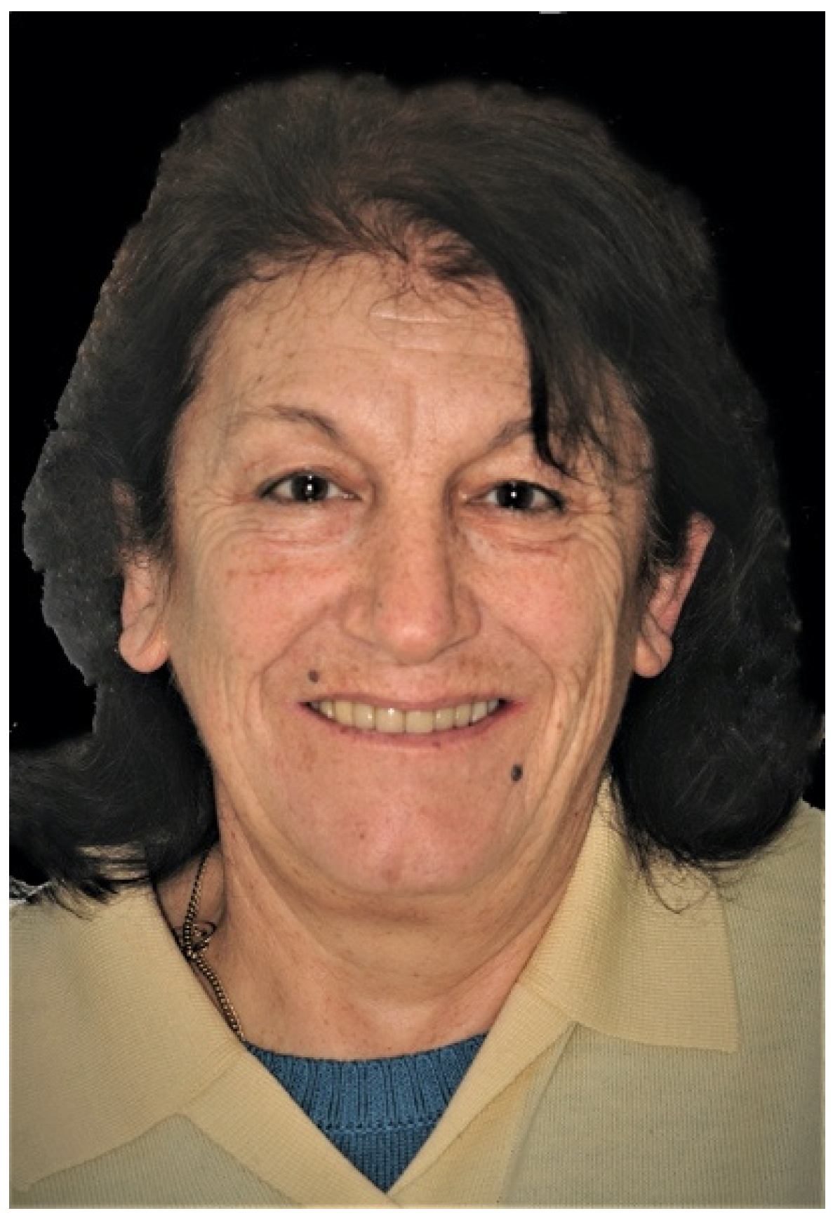
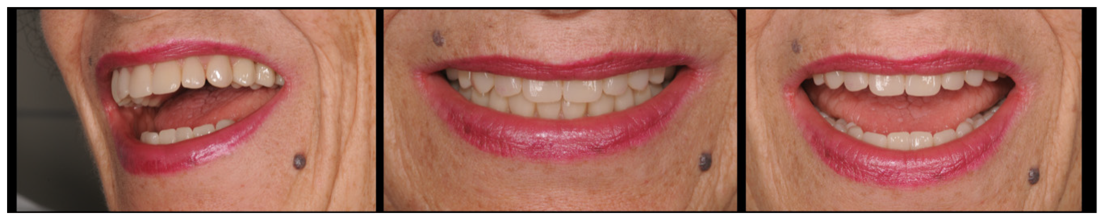
A 70-year old patient came to the dental office complaining of diminished masticatory capacity and loss of retention of both removable dentures. She wanted to improve her smile and facial esthetics, stating she was dissatisfied with the color and poor visibility of her teeth, no matter how big she smiled. The patient also asked to avoid multiple-steps therapies and desired to have a new prosthetic solution in the shortest time as possible. The patient’s smile seemed non-harmonious due to dental wear and the inclination of the occlusal planes, which affected her general appearance (
Figure 1). The patient gave her written consent to publish photos of her clinical case, including photos of her face.
Her history did not show any pathology incompatible with dental treatment and demonstrated that she was in good general health and classified as ASA1. The facial examination showed a reduction of the vertical dimension, with a widening of the nasolabial folds, and diminished tone of the perioral soft tissue, with a generalized deterioration of all facial esthetic parameters (
Figure 2). During the intraoral clinical examination, incongruous prosthesis in both arches have been observed (
Figure 3).
The upper arch had a full-removable denture, while the lower jaw had a fixed prosthesis supported by few periodontally compromised teeth as shown by the periapical mandibular radiographs, which also show several periodontal pockets, different carious lesions, and bone resorption around the natural abutments (
Figure 4).
During the first visit, several instrumental analyses were done, such as lateral cephalometric and electromyography. Lateral cephalometry is a valuable diagnostic tool that the authors consider pivotal for formulating a proper treatment plan in a complex prosthetic rehabilitation of an atrophic patient [
16]. This x-ray examination enables the study of the hard and soft tissues of the patient’s face; in particular, the relationship between the maxilla as well as the spatial position of the upper central incisor and the philtrum. It is also possible to identify the musculoskeletal classification with an appropriate and simple cephalometric analysis [
17]. The study of the patient’s latero–lateral radiography highlighted meso-facial musculoskeletal typology with reduced occlusal risk. Surface electromyography, using electrodes placed on the masticatory muscles, will allow the clinician to evaluate masticatory activity and to understand whether the occlusal load is adequate or excessive [
18]. A measurement of chewing loads produced by the patient is not a secondary element; on the contrary it represents an important aspect of comparison for the whole working group, and in particular with the dental technician who will have to take into consideration the extent of these values during the design and construction of the prosthesis.
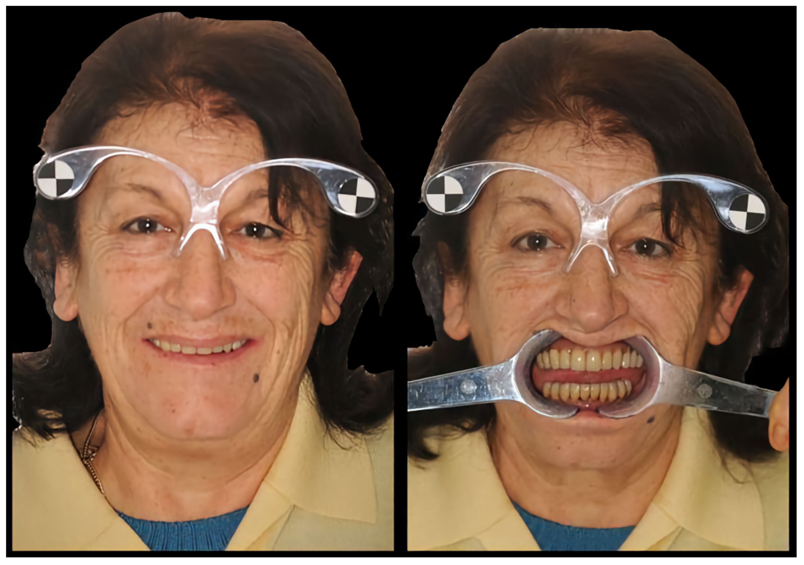
In the first appointment two photos were taken of the patient’s face according to a coded technique for the DSS (Digital Smile System, Bologna, Italy) software [
19]. It is important to take photos of the face keeping the patient in a position that is stable and repeatable over time, trying not to change the enlargement ratio between shots. For this purpose, the patient is asked to sit comfortably keeping her back straight while the operator used a camera set on a tripod to stabilize its position in relation to the patient being photographed. The subject had to be positioned so that their Frankfurt Plane (the line that joins the Porion and the Orbital Point) was parallel to the horizon. Once the spatial position of the head is identified, it must remain unchanged with respect to the camera-tripod complex. The patient may wear dedicated glasses used to calibrate the digital pre-rendering software (DSS;
Figure 5).
The glasses represent a true measuring system that differentiates this software from other similar systems. Thanks to their shape and the presence of calibration markers used as a reference, the glasses facilitate maintenance of the perpendicular position of the patient and the camera. The first facial photo was taken asking the patient to smile and show as much teeth as possible. The second facial photograph was taken with cheek retractors to better highlight the teeth of the patient.
The photographic status is completed with the profile shots of her face and with the intraoral photographs that allow us to make further diagnostic assessments regarding the overall esthetics of her face and its physiognomic characteristics on which we can proceed with our prosthetic therapy [
20]. After this phase, the two extraoral photos taken during the first visit were inserted in a dedicated 2D software (Digital Smile System, Bologna, Italy) useful for the final esthetic of the patient.
The Digital Smile System approach using digital techniques for the esthetic preview was only considered a tool for communication with the patient and for the entire dental team. DSS not only allowed the patient to see the esthetic future appearance, but it also enabled production of a prototype for the functional check of the digital project carried out [
21]. The fact that the patient can see the possible future esthetic results through digital rendering, including the possibility of changes if desired, reduces overall clinical practice time. The procedure requires that the photographs taken be imported into the DSS and then an esthetic preview of the future prosthetic therapy is developed (
Figure 6), which consists of a virtual arrangement of commercial teeth present in the software database. The database consists of upper and lower teeth of various shapes and sizes. Anterior and posterior teeth are positioned using the occlusal rim, previously suitably adapted in the oral cavity as a guide. Teeth are chosen according to esthetic and functional parameters and can be replaced with others of different shape or color, if necessary. This allows us to show the patient the possible final esthetic so that she can participate in the therapeutic project in collaboration with the whole clinical–technical team.
In a dedicated appointment, all the compromised dental elements in the lower arch were extracted except the lateral incisor which serves as a provisional anchor of the new provisional lower denture (
Figure 7).
After tissue healing, preliminary impressions of the soft tissue and edentulous arches were made. The authors did not use an intraoral scanner (IOS) because there is no consensus in the literature regarding real efficacy in edentulous subjects [
22]. We usually use a high-precision alginate with long setting time useful for tissue functionalization applied in two stages, where possible (only in edentulous subjects): a first impression is made with the alginate mixed with a high consistency (Neocolloid, Zhermack, Badia Polesine, Ro, Italy), it is then dried and modified by removing the undercut parts with a sharp tool and relined with the same material but in a more fluid form to read all the details of the anatomical tissues. The model obtained is scanned with a laboratory scanner (Sinergia Scan, Nobil Metal, Asti, Italy) and a resin occlusion base is built on it by sending a dedicated file to the 3D printer (Asiga MaxUV, Australia), coating it with wax for registration of the centric relationship, esthetic, functional determinants, and vertical dimension [
23].
It is important in the upper occlusal rim, during functionalization in the oral cavity, to mark some landmarks; the midline, the canine line, and the smile line. These reference lines will serve in the alignment phase of the occlusal rims in the 3D software (Exocad software, Exocad GmbH, Darmstadt, Germany).
Transfer of Data from the Dental Office to the Laboratory
Once the virtual teeth arrangement was obtained—and approved by the patient—the file containing the patient’s information, the photographic alignments, the libraries chosen, and the work process was transferred to the dental technician laboratory where the file was imported into a 3D software program (Exocad® software, Exocad GmbH). The information file exported from DSS consisted of a PDF format and individual photographs of the patient’s face with a customized two-dimensional (2D) virtual smile design.
The files from DSS were then superimposed onto scanned images of the denture. The dental technician used the outline of the virtual smile obtained to place a tooth from the library or to create customized teeth with tools from Freeform (Exocad
® software, Exocad GmbH) to convert the virtual 2D teeth arrangement into a 3D teeth arrangement [
24].
Simply put, “coupling” of the data, thinking that the final image of the mounting, obtained in the 2D version, represents a face of the volumetric solid corresponding to what is in the 3D version (
Figure 8).
The teeth used from the 3D database represent the volume of the solid, and the 2D teeth are the anterior face of this solid. The dental technician completes the 3D phase, improving the occlusal ratio between the arches (according to the literature) and producing the prosthetic base that will sustain the dental elements. After this phase, CAD (Computer-Aided Design) work can produce a prototype that corresponds entirely to the project made with DSS and processed in a 3D environment. The file obtained is sent to a 3D printer (Asiga MaxUV, Australia) and transformed into a prototype to be tested in the oral cavity, verifying the intraoral adaptation, the cranio–mandibular ratio, the esthetics of the smile and face. These prototypes represent the final volume of the final prosthesis. The clinician can make changes without impacting the protocol, in the prototypes, thus modified, can be scanned again by the technician, and overlapped digitally to the original project.
Subsequently the prototype is used as a radiological stent with which the CBCT(cone beam computed tomography) is done, using a dedicated device (Evobite with 3D marker, 3Diemme, Italy), which was adapted to the item with radiotransparent silicon (Elite Glass, Zhermack, Badia Polesine, Ro, Italy;
Figure 9).
The Dicom data resulting from the X-ray and the STL (Standard Triangulation Language) files relative to the anatomical and prosthetic parts obtained from the intraoral scan are imported in a specific implant planning software (Realguide 5.0, 3Diemme, Italy) where, thanks to a dedicated algorithm, they are overlapped using a replicable and controlled procedure. Through use of the implant line database (Thommen Medical AG, Grenchen, Switzerland), the number and position of the implant screws to be inserted via guided surgery are planned. After careful functional and esthetic evaluation and final verification, the prosthetic-driven plan was approved, and a stereolithographic surgical template was made using a newer rapid prototyping technology (New Ancorvis, Bargellino, Italy). Subsequently two prosthetic-driven implants with a diameter of 4.0 mm and a length of 9.5 mm (SPI ® CONTACT RC INICELL®, Thommen Medical AG) were placed, with a dedicated burr kit (Thommen Medical Guided Surgery Kit), in the lower jaw, taking into account the bone quality and quantity, soft-tissue thickness, anatomical landmarks, and the type, volume, and shape of the final restoration. The lateral incisor that acts as provisional anchor for the lower denture, in this phase was maintained in the mouth of the patient for the time necessary for implant loading. Healing screws are positioned directly post-surgically, and the prosthesis is readapted with resilient material (Coe-Soft™ GC America Inc., Alsip, IL, USA).
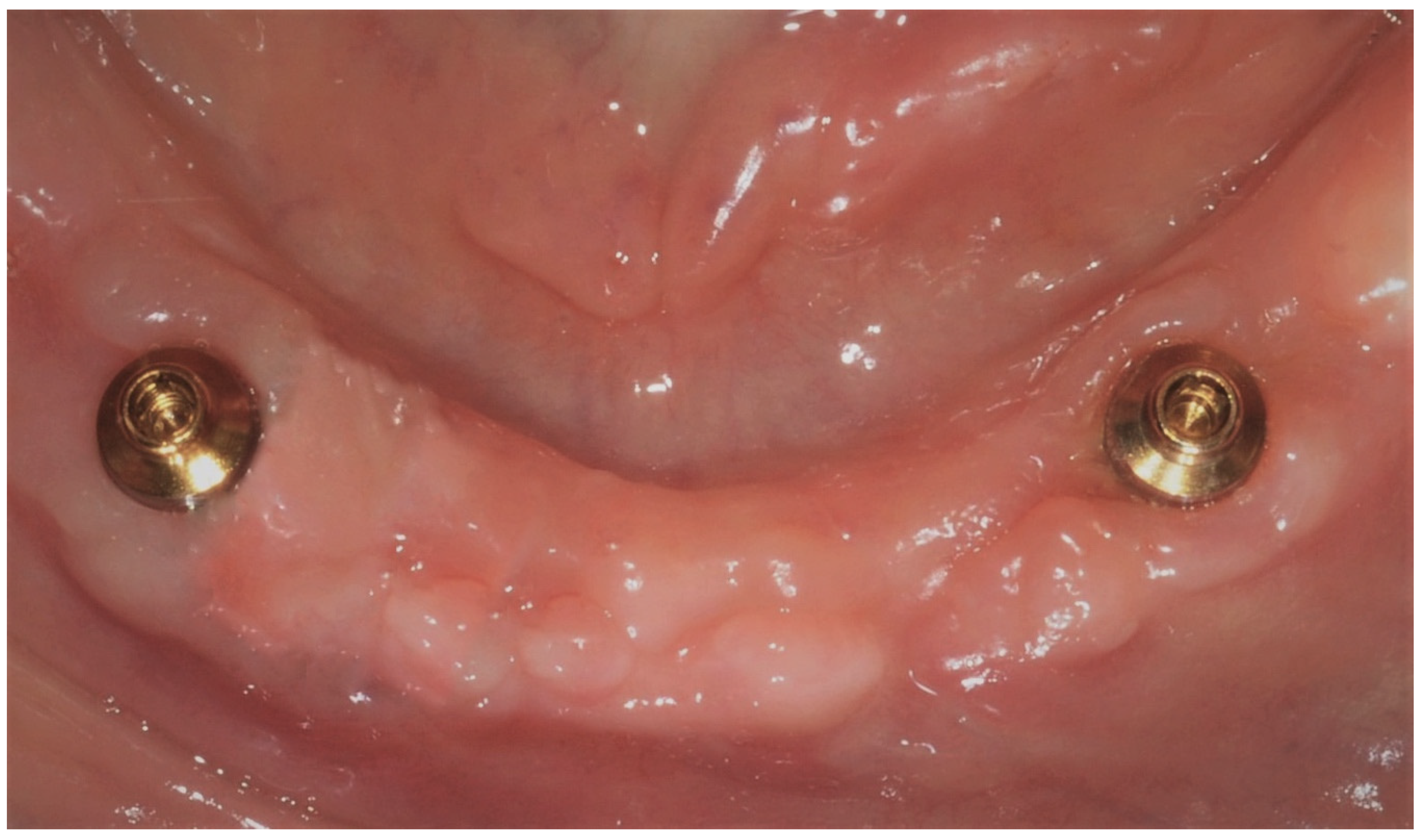
About four weeks later [
25], after tissue healing, with a dedicated tool (Cuff Height Measuring Tool, Rhein 83, Bologna, Italy), measurements of the transmucosal paths were performed and the most suitable retention systems means were chosen (OT Equator, Rhein 83, Bologna, Italy;
Figure 10) and were screwed to the implant fixtures with a preset force, with the corresponding retentive copings. The provisional prosthesis is readapted to make it suitable to receive the stainless-steel retentive cap housing nylon retentive inserts, improving the stability and hold. The general rule to apply for the choice of this type of attachments is the retentive part, which must extend beyond the transmucosal path by at least one millimeter [
26].
Thereafter the dental technician, using a 3D software (Exocad
®, Exocad GmbH), plans the counter-bar [
27], inserting retentive pins into the project for the mechanical hold of the teeth and preparing the area of the OT Equator attachment components (Rhein 83, Bologna, Italy).
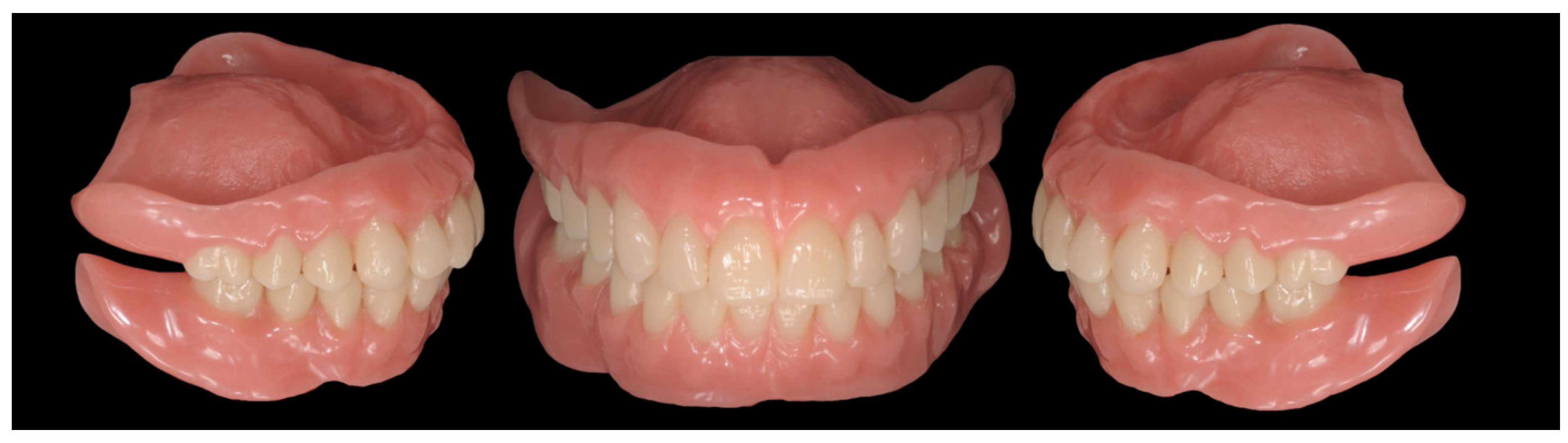
The project is sent to the milling center (New Ancorvis, Bologna, Italia) indicating the type of metal to be used and the type of construction (laser melting technology). After being checked in the dental laboratory and sent to dentist for clinical testing, the precision and passivity of the piece is checked. The commercial teeth are then mounted, taking advantage of the prototype as a positioning guide. The perfectly polished prostheses are sent to the dental office (
Figure 11,
Figure 12 and
Figure 13), making sure there are no compression areas on the soft tissues. The patient is provided guidelines for prosthesis hygiene maintenance procedures.
Once the prosthetic therapy was completed, the patient’s face improved greatly from an esthetic viewpoint (
Figure 14). The soft tissue of the face looked firm and toned. A reduction could be seen in the nasolabial folds and perilabial wrinkles (
Figure 15), both frontally and laterally. The vertical dimension, which was slightly increased, appeared adequate and well-tolerated esthetically. During phonation and smiling dynamics the patient displayed natural looking teeth that were perfectly integrated with his face (
Figure 16).
3. Discussion
During the prosthetic therapy, for both fixed and removable prostheses, communication with the patient is a vital part of the treatment. Effective digital previewing is the ideal way to explain esthetic changes to a patient and to receive their approval. Until now, many digital previewing methods have been used in dentistry solely for this purpose. The authors deem that digital previewing of the smile must be inserted into a more complex workflow [
28].
In this article, the Digital Smile System approach was introduced into a complex digital workflow. DSS (Digital Smile System, Bologna, Italy) not only allowed the patient to see their future appearance, but it also enabled production of a prototype for the functional check of the digital project carried out. The fact that the patient can see the possible future esthetic results through digital rendering, including the possibility of changes if desired, reduces overall clinical practice time. Additionally, the construction of a prototype, based on the virtual assembly, minimizes the number of errors in the manufacture of the final product and becomes a fundamental instrument for prosthetic-driven surgery. The decision-making process of using an implant overdenture prosthesis or a Toronto or hybrid prosthetic devices depends on different factors. The selection and extent of artificial tissues are related to facial support, emergence profile of the artificial tooth, the tooth angulation and the use of full flange extension is mentioned for esthetic advantages. However, the application of gingival prostheses may be limited to certain clinical situations where oral hygiene is manageable, function proper and esthetics acceptable. With a removable design, a larger volume of tissue can be replaced, and proper cleaning is still feasible. An instrument that could suggest a correct therapeutic choice is the latero–lateral radiography of the patient, which provides some important parameters: acrylic flange height, mucosal coverage, crown-Implant distance, and buccal prosthesis profile [
16]. According to Avrampou et al., when the mucosal coverage is less than 5 mm and the prosthetic profile is up to 30 degrees, a fixed prosthesis whit hybrid or crown design can be selected. On the other hand, when the mucosal coverage is more than 5 mm and the prosthetic profile is less than 30 degrees, a removable prosthesis with a buccal flange is advised.
The use of overdenture involves different advantages. Compared to complete removable dentures, implant supported dentures have better stability and retention, improving function, esthetic, and satisfaction for the patient [
29,
30]. Furthermore, some data indicate that after receiving implants, patients with overdenture eat a diet with more fiber and hard foods than with conventional dentures, even if this does not improve nutritional intakes of essential micronutrients and macronutrients [
31]. Furthermore, phonetic problems have been reported more often with fixed prostheses than with overdentures, probably because impaired phonetics appears to depend also on the palatal design of the prosthesis [
32,
33].
Use of the prototype as a radiological stent during the examination of the CBCT, and its transformation into a surgery-driven guide make it possible to position implants according to the digital study done with the DSS and approved by the patient. Some phases of the described workflow require a learning curve by the clinical operator and technician. For example, the photos taken by the clinician for the DSS, must be taken in the exact manner as previously described to facilitate superimposing of the photo of the patient’s face, with the scan of the model and the old denture. Another important stage is ensuring the teeth from the database of the 3D software are matched with the outlines obtained by digital previewing with the DSS. In this case, if the match is not precise, the prototype will not correspond perfectly with that approved by the patient [
34].


