Article

Prostaglandin F2α Affects the Cycle of Clock Gene Expression and Mouse Behavior

1
Department of Pharmaceutics, Faculty of Pharmaceutical Sciences, Kyushu University, 3-1-1 Maidashi, Higashi-ku, Fukuoka 812-8582, Japan
2
Department of Clinical Pharmacokinetics, Faculty of Pharmaceutical Sciences, Kyushu University, 3-1-1 Maidashi, Higashi-ku, Fukuoka 812-8582, Japan
3
Regulation in Metabolism and Behavior, Faculty of Agriculture, Kyushu University, 744 Motooka, Nishi-ku, Fukuoka 819-0395, Japan
4
SOUSEIKAI Fukuoka Mirai Hospital Clinical Research Center, 3-5-1 Kashiiteriha, Higashi-ku, Fukuoka 813-0017, Japan
5
Department of Pharmaceutical Biochemistry, Graduate School of Pharmaceutical Sciences, Kumamoto University, 5-1, Oe-Honmachi, Chuo-ku, Kumamoto 862-0973, Japan
6
Division of Reproductive Engineering, Center for Animal Resources and Development (CARD), Institute of Resource Development and Analysis, Kumamoto University, 2-2-1 Honjo, Chuo-ku, Kumamoto 860-0811, Japan
7
Division of Reproductive Biotechnology and Innovation, Center for Animal Resources and Development (CARD), Institute of Resource Development and Analysis, Kumamoto University, 2-2-1 Honjo, Chuo-ku, Kumamoto 860-0811, Japan
8
Department of Human Life Design and Science, Faculty of Design, Kyushu University, 4-9-1 Shiobaru, Minami-ku, Fukuoka 815-8540, Japan
9
Department of Glocal Healthcare Science, Faculty of Pharmaceutical Sciences, Kyushu University, 3-1-1 Maidashi, Higashi-ku, Fukuoka 812-8582, Japan
*
Authors to whom correspondence should be addressed.
These authors contributed equally to this work.
Int. J. Mol. Sci. 2024, 25(3), 1841; https://doi.org/10.3390/ijms25031841
Submission received: 20 December 2023 / Revised: 26 January 2024 / Accepted: 1 February 2024 / Published: 2 February 2024
(This article belongs to the Section Molecular Biology)

Abstract

:
Prostaglandins are bioactive compounds, and the activation of their receptors affects the expression of clock genes. However, the prostaglandin F receptor (Ptgfr) has no known relationship with biological rhythms. Here, we first measured the locomotor period lengths of Ptgfr-KO (B6.129-Ptgfrtm1Sna) mice and found that they were longer under constant dark conditions (DD) than those of wild-type (C57BL/6J) mice. We then investigated the clock gene patterns within the suprachiasmatic nucleus in Ptgfr-KO mice under DD and observed a decrease in the expression of the clock gene cryptochrome 1 (Cry1), which is related to the circadian cycle. Moreover, the expression of Cry1, Cry2, and Period2 (Per2) mRNA were significantly altered in the mouse liver in Ptgfr-KO mice under DD. In the wild-type mouse, the plasma prostaglandin F (PGF) levels showed a circadian rhythm under a 12 h cycle of light–dark conditions. In addition, in vitro experiments showed that the addition of PTGFR agonists altered the amplitude of Per2::luc activity, and this alteration differed with the timing of the agonist addition. These results lead us to hypothesize that the plasma rhythm of PGF is important for driving clock genes, thus suggesting the involvement of PGF- and Ptgfr-targeting drugs in the biological clock cycle.

1. Introduction

Various physiological and biological functions show a 24 h rhythmic cycle in synchronization with the external light environment. Periodic fluctuations are driven by an endogenous molecular system, known as the circadian clock [1]. The core of the circadian clock constitutes a transcription–translation feedback loop. Heterodimers consisting of CLOCK and BMAL1 promote the transcription of period (PER) and cryptochrome (CRY) genes by binding to E-box elements [2,3]. Upon reaching critical amounts, PER and CRY proteins dampen transcriptional activation via CLOCK/BMAL1, thereby generating circadian oscillations in their own transcription [4]. Clock genes, responsible for core oscillation loops, regulate the circadian expressions of clock-controlled output genes, such as albumin D-site binding protein (DBP), E4BP4 (NFIL3), and peroxisome proliferator-activated receptor-α (PPARα) [5]. DBP and E4BP4 regulate circadian gene expression by competitively binding to the D-site of the same DNA sequence [6]. Likewise, PPAR response elements (PPREs) are required for the circadian expression of PPARα target genes [7]. These mechanisms ultimately regulate various downstream events in the transcription, translation, and degradation processes [8,9].
The suprachiasmatic nucleus (SCN) of the brain is the master circadian clock that synchronizes the circadian clock mechanisms of individual cells [10]. Neurally active signals associated with photoreception in the retina activate the cAMP response element-binding protein (CREB) in the SCN, which, in turn, activates the transcription of the Per gene [11]. Increased Per expression triggers the transcription rhythm of clock genes in the SCN, evoking glucocorticoid secretion and sympathetic activation [12]. Secreted hormones and cytokines synchronize the clock genes of each cell in the peripheral organs under an external light environment [13].
Prostaglandins are endogenous, biologically active substances synthesized via arachidonic acid. Receptors corresponding to each prostaglandin are expressed in organs throughout the body, performing numerous physiological activities by mediating downstream signaling [14]. The plasma concentrations of some prostaglandins follow a 24 h cycle [15]. Although they are known to affect the expression of clock genes [16], no prostaglandins have been reported to affect the periodicity of clock genes or mouse circadian behavior.
Prostaglandin F (PGF) is a subtype of prostaglandin that acts on blood vessels around the glomus body and retinal arteries [17]. The prostaglandin F receptor (PTGFR) is localized in the eye and uterus, where its ligand binding activates PKC and AMPK [18]. Latanoprost is a highly selective agonist of PTGFR and a strong PTGFR agonist with weak prostaglandin E receptor effects [19]. Whether PTGFR signaling and latanoprost affect the expression cycle of clock genes or mouse behavior remains unclear. Here, we aimed to clarify the relationship between PGF and the circadian clock mechanism by analyzing the locomotor activity rhythm and expression of clock genes in Ptgfr-KO mice.

2. Results

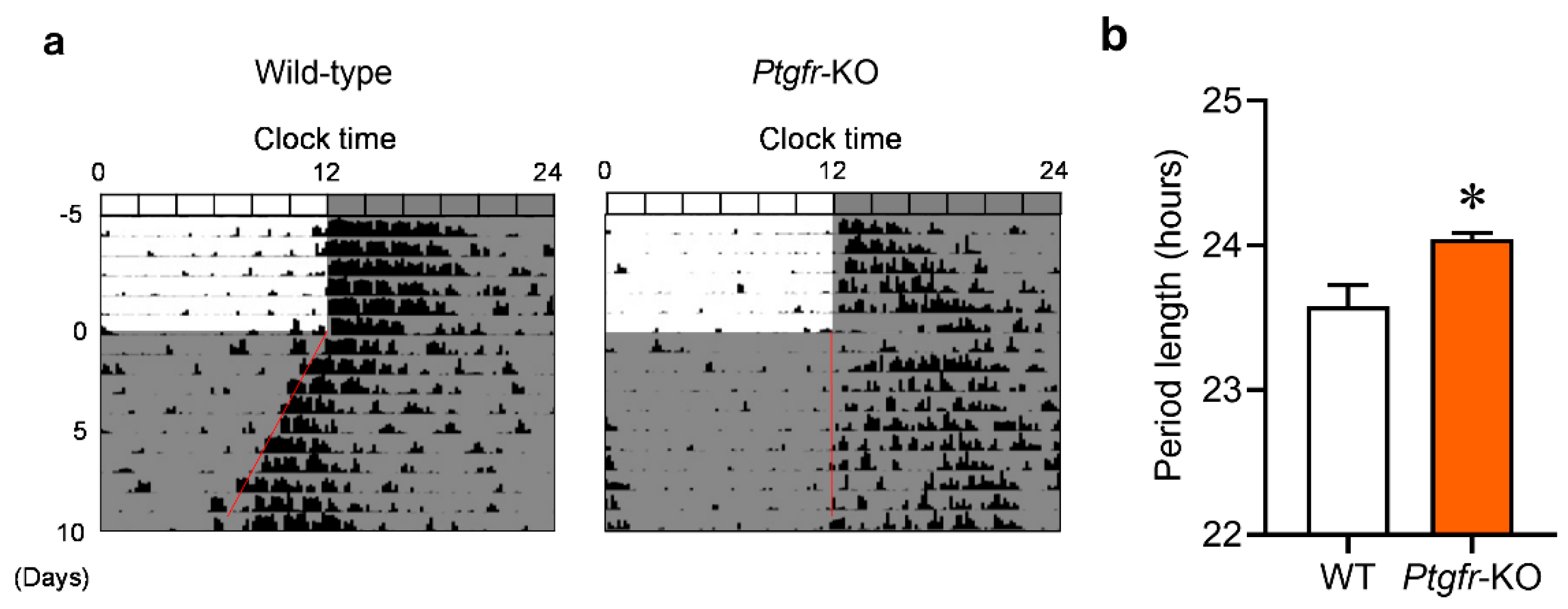
2.1. Ptgfr-KO Mice Exhibit Longer Behavioral Cycles under Constant Dark Conditions

We initially observed differences in the behavioral cycles, meaning the time length between the start of an activity and the start of the next activity, of wild-type and Ptgfr-KO mice under constant dark (DD) conditions. After acclimation to light–dark (LD) conditions for two weeks, each mouse was kept in DD conditions. Wild-type mice exhibited a behavioral cycle of 23.58 ± 0.293 h (Figure 1a), similar to those in previous reports [12]. In contrast, Ptgfr-KO mice exhibited a longer behavioral cycle (24.05 ± 0.085 h) than wild-type mice (Figure 1b). These results indicate that Ptgfr deficiency affects the periodicity of the biological circadian clock.

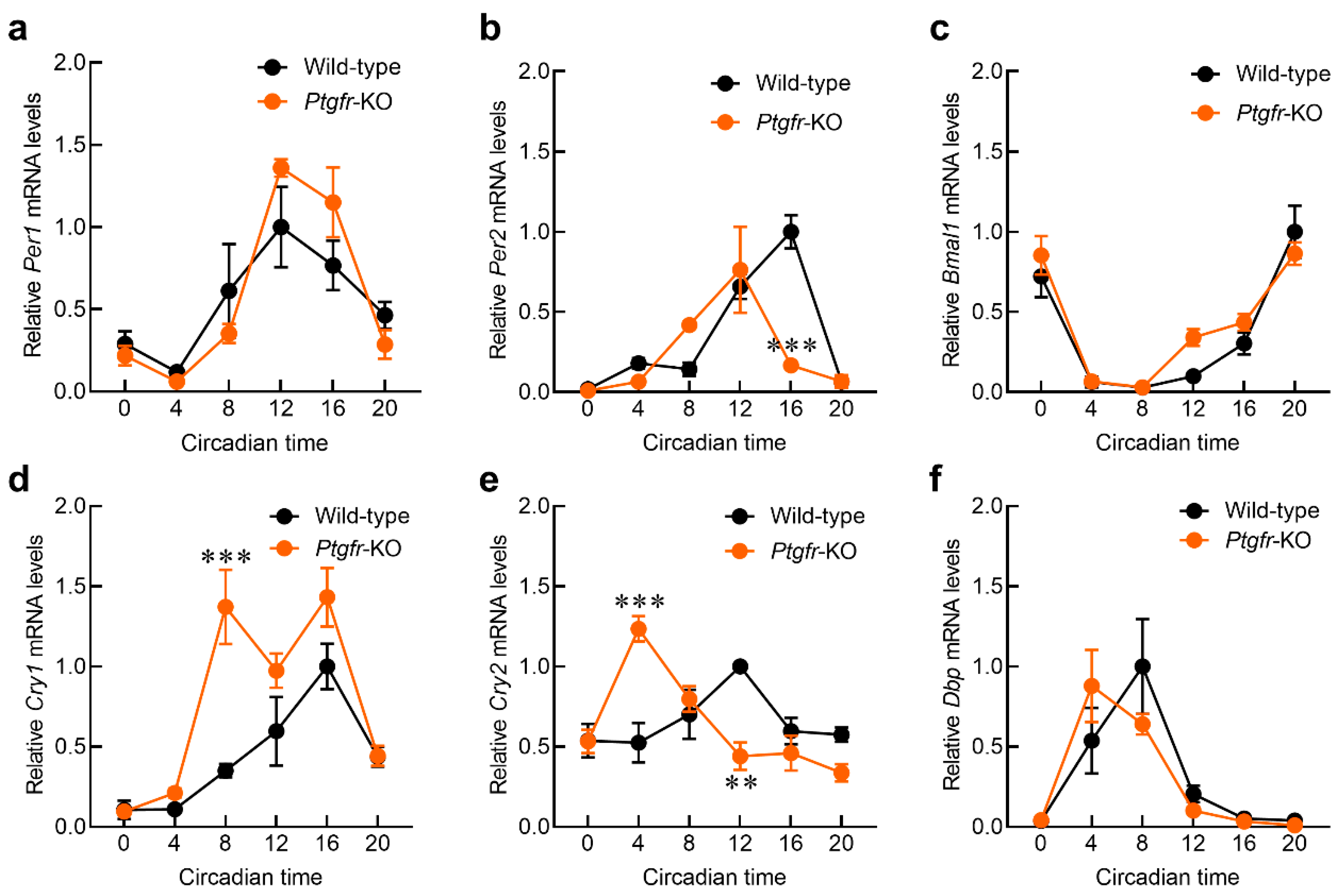
2.2. Time-Dependent Changes in Expression of Clock Genes in the Suprachiasmatic Nucleus (SCN) of Ptgfr-KO Mice

Changes in the amplitude of clock gene expression in the SCN affect the periodicity of behavior [20]. Because the locomotor activity cycle of Ptgfr-KO mice was longer than that of wild-type mice, we evaluated the expression rhythms of clock genes in the SCN of Ptgfr-KO mice. We measured the expression rhythms of Per1, Per2, Bmal1, and Cry1, which form the core of the circadian clock system, and whose own expression indicates circadian oscillations [21], using in situ hybridization. The results show that the mRNA expression of Per1, Per2, and Bmal1 was nearly equal in both wild-type and Ptgfr-KO mice (Figure 2a,b,d). In contrast, the peak time of Cry1 mRNA expression was regressed in Ptgfr-KO mice (Figure 2c; Supplementary Table S1: Cry1 in wild-type; acrophase = 8.99, Cry1 in Ptgfr-KO; acrophase = 13.60). In addition, the expression level at the peak time was also significantly downregulated. These results indicate that Ptgfr deficiency affects time-dependent Cry1 expression in SCN.

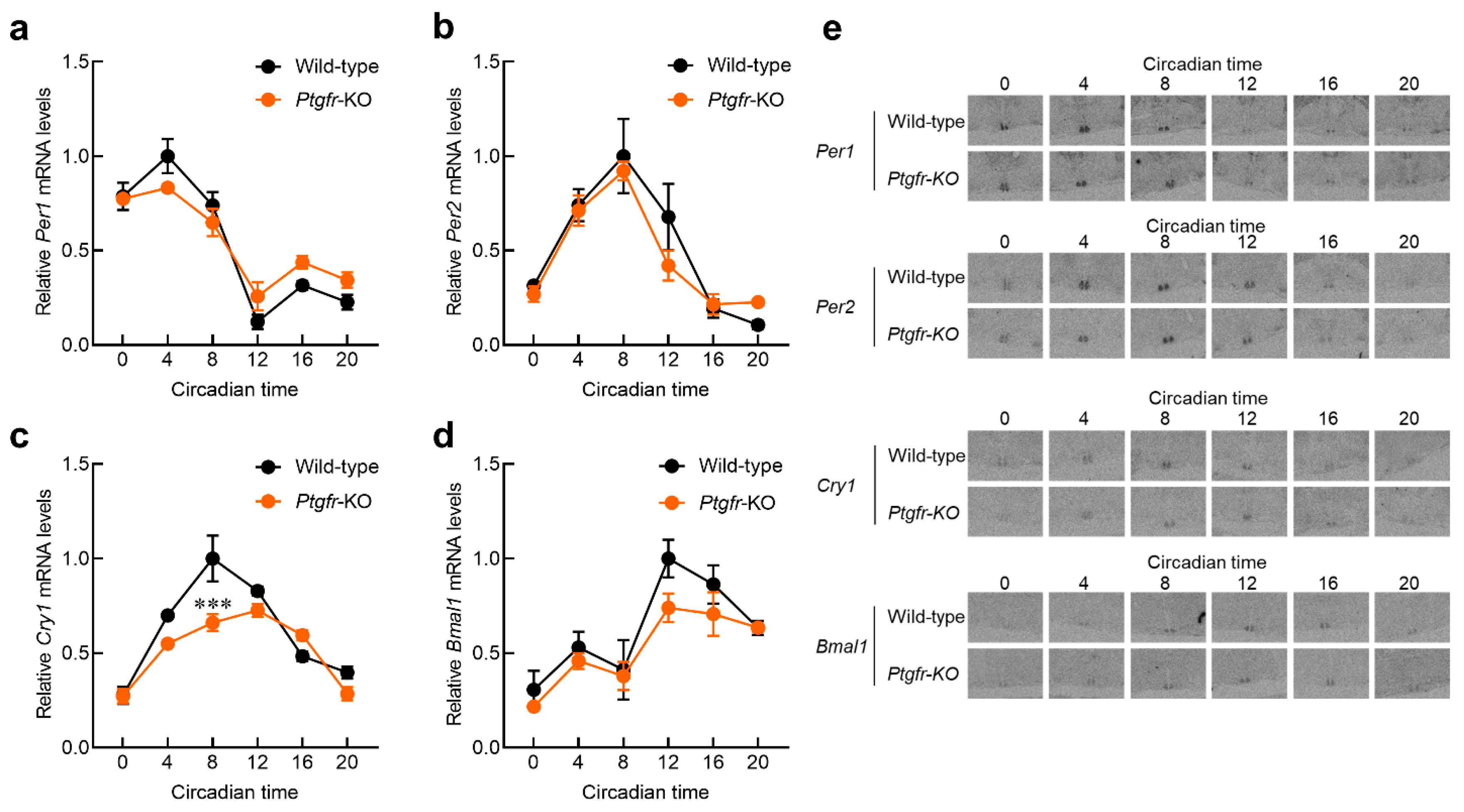
2.3. Time-Dependent Changes in Clock Gene Expression in the Liver of Ptgfr-KO Mice

The altered expression of clock genes in the SCN also alters their expression rhythms in peripheral organs, such as the liver and kidneys [10,11]. When changes are observed in the clock core–loop rhythms of peripheral organs, the expression of clock-regulated genes undergoes rhythmic changes [6,7]. Because changes in clock genes were observed in the SCN of Ptgfr-KO mice, we measured their expression levels in the liver. Total RNA was extracted from the livers of wild-type and Ptgfr-KO mice after 3 days under DD conditions and reverse-transcribed. The mRNA for clock genes was measured using a reverse transcription qualitative PCR (RT-qPCR) system. In wild-type mice, significant circadian rhythms were observed in the expression of each clock gene (Figure 3a–f). Ptgfr-KO mice also showed a significant circadian rhythm; however, the amplitude of Per2 mRNA expression was reduced in Ptgfr-KO mice compared to that in wild-type mice (Figure 3b; Supplementary Table S1). The expression amplitudes of Cry1 and Cry2 mRNAs in Ptgfr-KO mice were higher than those in wild-type mice (Figure 3d,e; Supplementary Table S1: Cry1 in wild-type; 0.424, Cry1 in Ptgfr-KO; 0.732, Cry2 in wild-type; 0.193, Cry2 in Ptgfr-KO; 0.424). In addition, the peak time of the Cry1/2 mRNA expression came faster. These results indicate that Ptgfr deficiency affects time-dependent clock gene expression in the peripheral organs.

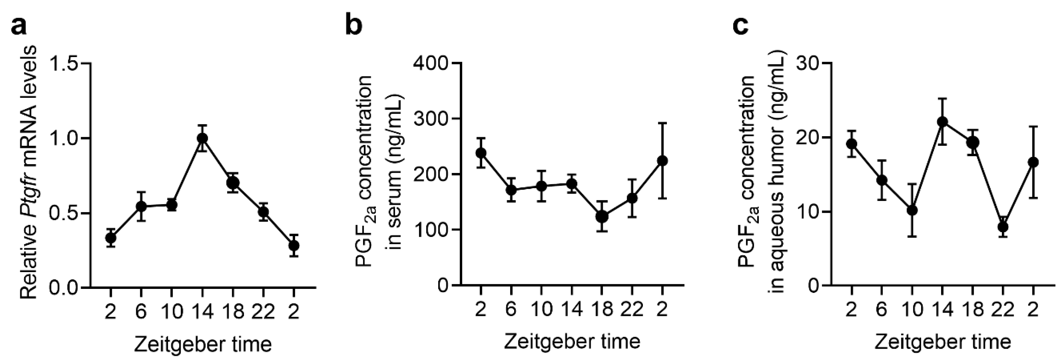
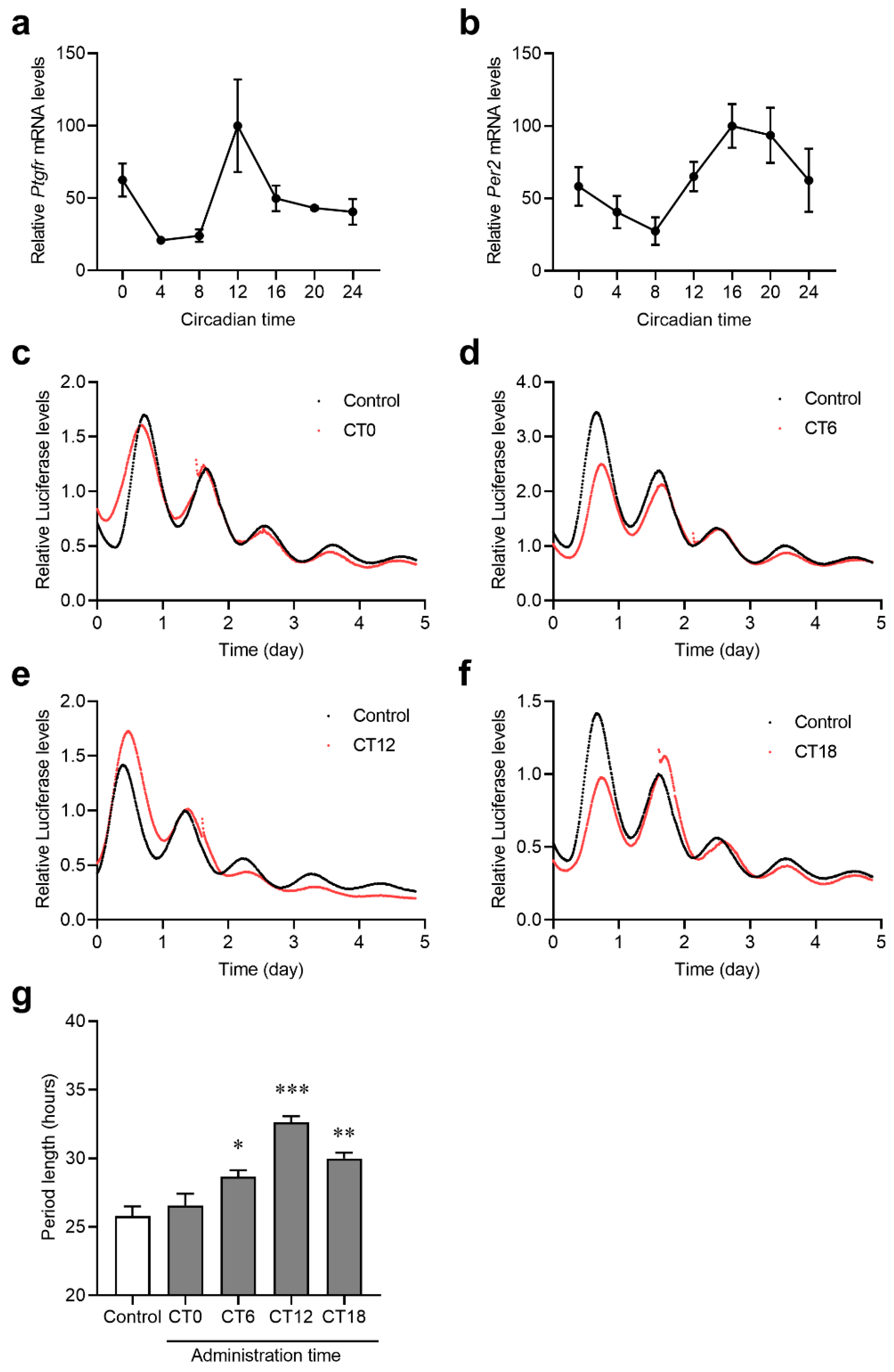
2.4. The Expression Rhythms of Ptgfr and the Secretion Rhythms of PGF in Mouse Retina

Although Ptgfr is expressed in organs throughout the body, it is highly expressed in the uterus and retina [17,22]. Circadian rhythms in prostaglandin E2 secretion in serum and prostaglandin D synthase expression in the brain have also been observed [16,23,24]. To clarify the activation rhythm of the Ptgfr cascade in the retina, we measured the expression rhythm of Ptgfr in the retinas of wild-type mice using an RT-qPCR system. The retinal Ptgfr mRNA expression showed a significant circadian rhythm, increasing during the dark period (Figure 4a). The secretory rhythm of PGF in mouse serum and aqueous humor was measured using a PGF ELISA kit. Serum PGF levels did not exhibit a significant circadian rhythm (Figure 4b). In contrast, the PGF concentrations in the aqueous humor exhibited the same phase rhythm as the expression rhythm of Ptgfr in the retinas of wild-type mice (Figure 4c). Based on these results, the PTGFR signal in the retina is considered high in the early dark period. Given the expression rhythm of Cry1 in the SCN of wild-type and Ptgfr-KO mice, PTGFR signaling in the early dark period may be involved in the Cry1 expression cycle.

2.5. Effects of PTGFR Agonists on Per2 Expression Rhythm in Per2::luc C6 Cells

The circadian rhythms of Ptgfr expression and PGF secretion were observed, suggesting that PGF-related signaling may have a time-dependent effect. To determine the effect of PGF signaling on the expression of clock genes, we evaluated the effect of PTGFR agonists in a model in which the clock genes were synchronized. Using Per2-fusion luciferase-expressing C6 rat glioma cells (Per2::luc C6 cells), we synchronized cell clock genes with dexamethasone, added PTGFR agonists at different times, and tested their effects on the periodicity of Per2::luc activity. After the Per2::luc C6 cells synchronized, the rhythms of Ptgfr and Per2 mRNA expression were measured.
A significant rhythm in the expression of Ptgfr and Per2 mRNA was observed in Per2::luc C6 cells (Figure 5a,b). Therefore, we added latanoprost, an PTGFR agonist, to CT0, 6, 12, and 18 and observed periodic changes in Per2::luc activity. A transient upregulation in Per2 expression was observed immediately after latanoprost administration at all time points (Figure 5c–f). The results show that adding latanoprost during periods of high Ptgfr and Per2 expression significantly prolonged the Per2::luc activity (Figure 5g). These results suggest that PTGFR signaling affects the expression rhythm of clock genes in a time-dependent manner.

3. Discussion

In this study, we investigated changes in the biological clocks of Ptgfr-KO mice. Ptgfr-KO mice showed a prolonged behavioral cycle of approximately 24 h. The measurement of the clock gene expression in the SCN and peripheral organs revealed changes in the expression levels and peak time of core clock genes. In particular, the expression level of Per2 at the peak time was attenuated in the livers of Ptgfr-KO mice compared to that in wild-type mice, whereas the peak-time expression of Cry1 and Cry2 mRNA in Ptgfr-KO mice was higher than that in wild-type mice. Increased Per2 mRNA expression and cycle prolongation were observed when PTGFR agonists were added. These results suggest that activating or inhibiting PTGFR may affect the expression cycle of clock genes.
Several types of prostaglandins affect clock gene expression in peripheral organs. The treatment of cells with prostaglandin E increases Per1 expression [16]. The administration of prostaglandin E alters the expression cycle of clock genes in mice [16]. The amplitude of clock gene expression is modified in prostaglandin D synthase KO mice [24]. However, administering prostaglandin E has no effect on the behavioral cycle [16]. Furthermore, in prostaglandin D synthase knockout mice, the biological clock cycle was unchanged from that of wild-type mice under DD conditions [24]. In Ptgfr-KO mice, the behavioral cycle was changed to 24 h under DD conditions (Figure 1). This result indicates that Ptgfr is the only prostanoid receptor involved in the behavioral cycle.
Although Ptgfr mRNA expression was not detected in the SCN, a regression in peak time and decreased expression at the peak time of Cry1 mRNA were observed within the SCN of Ptgfr-KO mice. Ptgfr is highly expressed in the retina of mammals [16]. Downstream PTGFR signaling, as well as that of photoreceptors, is mediated by PKC or PKG activation and Ca2+ signaling [18,25]. The activation of retinal Ca2+ signaling activates the optic nerve, causing increased CREB phosphorylation or c-Fos mRNA expression in the SCN [26,27]. Therefore, the activation of PTGFR signaling projects is likely to occur in the SCN via the optic nerve. In Ptgfr-KO mice, the periodicity of Cry1 mRNA expression in the SCN is maintained but its phase is regressed (Figure 2). Cry1-KO mice, in which the circadian cycle of Cry1 expression is completely lost, show a shortened behavioral cycle and hastening of behavioral phases [28]. As the rhythm of Cry1 mRNA expression is observed in Ptgfr-KO mice, it is unlikely that the same shortening of the behavioral cycle phase occurs in Ptgfr-KO mice as in Cry1-KO mice. The manipulation of the Cry1 expression cycle causes a regression in the circadian clock cycle [29]. Therefore, the regression in the Cry1 peak observed in Ptgfr-KO mice may be responsible for the regression in the behavioral cycle.
PTGFR not only activates Ca2+ signaling but also drives other physiological pathways in the livers of mice. The activation of Ptgfr in the liver accelerates glycogenesis via the CaMKIIg/p38/FOXO1 pathway [30]. Moreover, the hepatic accumulation of triglycerides is reduced with PTGFR activation. PTGFR signaling also inhibits adipose tissue differentiation by suppressing the PPARγ function [31]. The activation of these signals and pathways may affect the expression of respective clock genes by acting on their promoter regions. The transcriptional rhythm is created by the binding region of phosphorylated CREB, which is associated with Ca2+ signaling upstream of the Per2 gene [32]. The reduced expression amplitude of Per2, as shown in Figure 3b, may have been due to the loss of Ptgfr, which caused a reduction in Ca2+ signaling. The different effects of Ptgfr-KO on clock gene expression in the SCN and liver may be due to this peripheral-organ-specific function of PTGFR.
In this study, periodic changes in clock genes were evaluated in the SCN and liver. The liver clock mechanism is largely affected by changes in the SCN [33]. In addition, the liver is a relatively homogeneous peripheral organ, with most of its constituent cells being hepatic parenchymal cells [34]. Therefore, in many studies, the SCN is used as the central clock and the liver as the peripheral clock as the index for evaluation [5,35,36]. Because these results suggest that the changes in clock gene expression observed in the liver of Ptgfr-KO mice may also be observed in other organs in the periphery, further analysis of the relationship between PTGFR and the function of peripheral organs, including the liver, will help us understand the circadian clock mechanism in the periphery.
Latanoprost, a PTGFR agonist, is used to treat glaucoma. Although the mechanism is not clearly understood, latanoprost is thought to drive IP3/Ca2+ signaling in retinal cells and promote aqueous humor drainage [37]. Latanoprost is one of the PGF ligands that potentiates intracellular Ca2+ concentrations in in vitro experiments [38]. As shown in Figure 5, the transient increase in the transcriptional activity of Per2 after the addition of latanoprost is thought to be due to the influx of Ca2+. The Per2 cycle changes induced by the addition of latanoprost were time-dependent (Figure 5c–f). This is similar to the phase changes induced by light irradiation in mice [39]. Ca2+ signaling is also involved in light-irradiation-induced SCN activation and increases Per gene expression [11,32]. These findings suggest that the Ptgfr expression rhythm may be involved in Ca2+-signaling-induced time-dependent cyclic changes.
The central clock, which determines the overall internal clock of the body, is affected by light, physical exercise, and feeding. The peripheral clock is also driven by variations in the central clock and zeitgeber [40]. When the factors affecting the central and peripheral clocks differ from normal, the overall biological circadian clock system can modulate. The modulation of the biological clock can lead to obesity, diabetes, cardiovascular diseases, cancer, and other diseases [9,41,42,43]. Therefore, the genes that influence the biological clock and behavioral cycle have been investigated through studies focusing on several types of genes [8,44,45]. Molecules associated with the inflammatory response affect the circadian clock mechanism through various pathways [38]. Although a link between prostanoids, which are closely associated with inflammatory processes, and clock genes has been suggested, previous studies have not identified the prostaglandin receptors that initiate changes in the behavioral cycle [16,23]. The results regarding the behavioral cycle and clock gene analyses of Ptgfr-KO mice in this study indicate that PTGFR activation is involved in the biological circadian clock system. Various prostaglandin subtypes have different effects on clock genes. It may be possible to develop a medicine that regulates the biological clock by conducting a detailed analysis in the future.

4. Materials and Methods

4.1. Animal Experiments

Ptgfr-KO male mice (B6.129-Ptgfrtm1Sna, CARD ID: 2078) with C57BL/6J background and wild-type C57BL/6J male mice were maintained under a standard light–dark cycle (lights on at zeitgeber time [ZT] 0, off at ZT12) in a temperature (24 ± 1 °C)- and humidity (60 ± 5%)-controlled room with food and water ad libitum. The animals were acclimated to the LD cycle for two weeks before the experiments. There were no changes in water consumption or food intake and no significant differences in body weight between wild-type and Ptgfr-KO mice (Supplementary Figure S1). All experiments were conducted according to the protocol approved by the Internal Committee for Animal Experiments at Kyushu University (ID: A20-133-0). All animal experiments complied with the ARRIVE guidelines and were performed in accordance with the National Institutes of Health Guide for the Care and Use of Laboratory Animals (NIH Publication No. 8023; revised 1978).

4.2. Measurement of Locomotor Activity Rhythm

To measure locomotor activity, the cages were placed in an infrared ray area sensor (Neuroscience, Tokyo, Japan), and locomotor activity was measured under a 12 h LD cycle for 7 days and then under a DD cycle for 7 days. The period and onset of activity under the DD cycle were determined via Cosinor analysis using Clock Lab software (Version 2.36, Actimetrics, Evanston, IL, USA) by a chronobiology expert in a blinded manner.

4.3. In Situ Hybridization

Wild-type and Ptgfr-KO mice were euthanized via cervical dislocation following isoflurane anesthesia. At six time points every 4 h starting CT 2, the brain of each mouse was harvested and flash-frozen. Slices containing SCN were prepared from the frozen brains [46]. The expression levels of clock genes in the SCN were evaluated using in situ hybridization with reference to previous studies. Antisense 45-mer oligonucleotide probes (mBmal1:1755–1799 of AB015203; mPer1:3239–3283 of AB002108; mPer2:242–286 of AF035830; and mCry1:1742–1786 of AY034432) were labeled with [35S] dATP (New England Nuclear, Boston, MA, USA) using terminal deoxyribonucleotide transferase (GIBCO-BRL, Waltham, MA, USA).

4.4. Quantitative RT-PCR Analysis

RNA was extracted from mouse livers using RNAiso (Takara Bio, Osaka, Japan). Reverse transcription of RNA to cDNA was performed using a Rever Tra Ace quantitative real-time PCR kit (Toyobo, Osaka, Japan). For quantitative real-time PCR reactions, THUN-DERBIRD SYBR qPCR Mix (Toyobo) and LightCycler® 96 System (Roche, Basel, Switzerland) were used, and the mRNA expression levels of each gene were measured via the calibration curve method. The expression level of each gene was corrected in reference to the β-actin mRNA expression level. The PCR primer sequences are listed in Supplementary Table S2.

4.5. Quantification of PGF Using ELISA

Plasma and aqueous humor samples from wild-type mice were collected at seven time points (ZT2, 6, 10, 14, 18, 22, and ZT2 + 24 h). PGF concentrations in the collected samples were quantified using a PGF ELISA kit (ADI-900-069; Enzo Life Sciences, Farmingdale, NY, USA). After the antibody reaction, the PGF concentration was observed at a wavelength of 405 nm and calculated using the method described in the kit protocol.

4.6. Real-Time Monitoring of Circadian Bioluminescence

Per2-fusion luciferase-expressing C6 rat glioma cells (Per2::luc C6 cells) were maintained in Dulbecco’s Modified Eagle’s Medium (DMEM) supplemented with 10% fetal bovine serum (FBS) at 37 °C in a 5% CO2 humidified atmosphere. Per2::luc C6 cells were seeded in 35 mm culture dishes. After treatment with 100 μM DEX for 2 h, the medium was exchanged for DMEM with HEPES supplemented with 100 nM luciferin and 5% FBS. After wrapping the culture dishes with Parafilm, they were placed in a lumi-cycle and the intensity of Per2::luc was observed. After 48 h of DEX shock, latanoprost (0.005%, 50 μL) was added every 6 h (CT 0: 30 h, CT 6: 36 h, CT 12: 42 h, CT 18: 48 h). An equal volume of saline was administered to the control group. The amplitude of bioluminescence derived from Per2::luc was calculated 24 h after the addition of the reagent using Lumicycle analysis software (Version 2.04, Actimetrics), as previously reported [47].

4.7. Statistical Analysis

Statistical analyses were performed using the GraphPad Prism software (ver. 8; GraphPad Software, San Diego, CA, USA). The significance of the differences among the groups was analyzed using analysis of variance (ANOVA), followed by Tukey’s post hoc test. Differences between the groups were analyzed using two-way ANOVA, followed by Sidak’s post hoc test. Statistical significance was set at p < 0.05. Cosinor analyses were performed using Circadian Rhythm Laboratory Software accessed on 20 December 2023 (https://www.circadian.org/). p value, which were calculated from comparisons of the residuals before and after fitting, being less than 0.05 indicated the detection of a rhythm. Using this analysis, we also calculated the acrophase, the time of the peak value in the fitted cosine function. The values calculated via this analysis are shown in Supplementary Table S1. Although no statistical methods were used to predetermine sample sizes, our sample sizes were similar to those reported in previous studies [12,30,46]. The experiments were not randomized.

Supplementary Materials

The following supporting information can be downloaded at: https://www.mdpi.com/article/10.3390/ijms25031841/s1.

Author Contributions

Conceptualization, Y.T. and Y.Y.; Methodology, Y.T., Y.Y. and N.M.; Software, Y.T., Y.Y. and A.T.; Validation, Y.T., Y.Y., N.M., A.T., T.O., S.Y. (Sai Yasukochi), and S.K.; Formal analysis, Y.T., Y.Y., N.M., A.T., T.O., S.Y. (Sai Yasukochi), and S.K.; Investigation, Y.T., Y.Y., K.H., A.T., T.O., S.Y. (Sai Yasukochi), S.Y. (Shinobu Yasuo), A.I., T.Y., T.I., S.T., T.T., N.N., S.H., Y.S. and S.K.; Resources, Y.T., Y.Y., N.M. and S.O.; Data curation, T.I., S.T., T.T., N.N., S.H., Y.S. and S.K.; Writing—original draft preparation, Y.T. and Y.Y.; Writing—review and editing, Y.T., Y.Y., K.H., N.M. and S.O.; Visualization, Y.T.; Supervision, Y.Y., N.M. and S.O.; Project administration, Y.Y. and S.O.; Funding acquisition, N.M., S.K. and S.O. All authors have read and agreed to the published version of the manuscript.

Funding

This work was supported by the Japan Society for the Promotion of Science (JSPS) KAKENHI Grant-in-Aid for Scientific Research A (22H00442 to S.O.) and partially supported by a JSPS KAKENHI Grant-in-Aid for Challenging Exploratory Research (21K18249,17H06262 to S.O.). This research is supported by the Platform Project for Supporting Drug Discovery and Life Science Research (BINDS) from AMED (AMED under Grant Number JP22αm0101091).

Institutional Review Board Statement

Animals in this study were treated according to the guidelines stipulated by the Animal Care and Use Committee of Kyushu University. All experiments were conducted under the protocol approved by the Internal Committee for Animal Experiments at Kyushu University (protocol code, A30-166-0: approval date 30 March 2018).

Informed Consent Statement

Not applicable.

Data Availability Statement

Data from this study are available from the corresponding author upon reasonable request.

Acknowledgments

We are grateful for the technical support provided by the Research Support Center, Graduate School of Medical Sciences, Kyushu University.

Conflicts of Interest

Tatsuya Yoshihara was employed by “NMedical Co. Ouryokukai”, but no commercial or financial support was received from this firm in conducting this study. The remaining authors declare that the research was conducted in the absence of any commercial or financial relationships that could be construed as a potential conflict of interest.

References

  1. Langmesser, S.; Tallone, T.; Bordon, A.; Rusconi, S.; Albrecht, U. Interaction of circadian clock proteins PER2 and CRY with BMAL1 and CLOCK. BMC Mol. Biol. 2008, 9, 41. [Google Scholar] [CrossRef] [PubMed]
  2. Bass, J.; Takahashi, J.S. Circadian integration of metabolism and energetics. Science 2010, 330, 1349–1354. [Google Scholar] [CrossRef] [PubMed]
  3. Asher, G.; Schibler, U. Crosstalk between components of circadian and metabolic cycles in mammals. Cell Metab. 2011, 13, 125–137. [Google Scholar] [CrossRef]
  4. DeBruyne, J.P.; Weaver, D.R.; Reppert, S.M. CLOCK and NPAS2 have overlapping roles in the suprachiasmatic circadian clock. Nat. Neurosci. 2007, 10, 543–545. [Google Scholar] [CrossRef] [PubMed]
  5. Ueda, H.R.; Hayashi, S.; Chen, W.; Sano, M.; Machida, M.; Shigeyoshi, Y.; Iino, M.; Hashimoto, S. System-level identification of transcriptional circuits underlying mammalian circadian clocks. Nat. Genet. 2005, 37, 187–192. [Google Scholar] [CrossRef] [PubMed]
  6. Kwon, P.K.; Lee, K.H.; Kim, J.H.; Tae, S.; Ham, S.; Jeong, Y.H.; Kim, S.W.; Kang, B.; Kim, H.M.; Choi, J.H.; et al. hnRNP K Supports High-Amplitude D Site-Binding Protein mRNA (Dbp mRNA) Oscillation to Sustain Circadian Rhythms. Mol. Cell. Biol. 2020, 40, e00537-19. [Google Scholar] [CrossRef] [PubMed]
  7. Kanemitsu, T.; Tsurudome, Y.; Kusunose, N.; Oda, M.; Matsunaga, N.; Koyanagi, S.; Ohdo, S. Periodic variation in bile acids controls circadian changes in uric acid via regulation of xanthine oxidase by the orphan nuclear receptor PPARα. J. Biol. Chem. 2017, 292, 21397–21406. [Google Scholar] [CrossRef]
  8. Kim, W.; Shin, J.C.; Lee, K.H.; Kim, K.T. PTBP1 Positively Regulates the Translation of Circadian Clock Gene, Period1. Int. J. Mol. Sci. 2020, 21, 6921. [Google Scholar] [CrossRef]
  9. Mazzoccoli, G.; Colangelo, T.; Panza, A.; Rubino, R.; Tiberio, C.; Palumbo, O.; Carella, M.; Trombetta, D.; Gentile, A.; Tavano, F.; et al. Analysis of clock gene-miRNA correlation networks reveals candidate drivers in colorectal cancer. Oncotarget 2016, 7, 45444–45461. [Google Scholar] [CrossRef]
  10. Welsh, D.K.; Takahashi, J.S.; Kay, S.A. Suprachiasmatic nucleus: Cell autonomy and network properties. Annu. Rev. Physiol. 2010, 72, 551–577. [Google Scholar] [CrossRef]
  11. Ashton, A.; Foster, R.G.; Jagannath, A. Photic Entrainment of the Circadian System. Int. J. Mol. Sci. 2022, 23, 729. [Google Scholar] [CrossRef] [PubMed]
  12. Le Minh, N.; Damiola, F.; Tronche, F.; Schütz, G.; Schibler, U. Glucocorticoid hormones inhibit food-induced phase-shifting of peripheral circadian oscillators. EMBO J. 2001, 20, 7128–7136. [Google Scholar] [CrossRef]
  13. Entzian, P.; Linnemann, K.; Schlaak, M.; Zabel, P. Obstructive sleep apnea syndrome and circadian rhythms of hormones and cytokines. Am. J. Respir. Crit. Care Med. 1996, 153, 1080–1086. [Google Scholar] [CrossRef] [PubMed]
  14. Narumiya, S.; Sugimoto, Y.; Ushikubi, F. Prostanoid receptors: Structures, properties, and functions. Physiol. Rev. 1999, 79, 1193–1226. [Google Scholar] [CrossRef] [PubMed]
  15. Kato, K.; Murai, I.; Asai, S.; Takahashi, Y.; Nagata, T.; Komuro, S.; Mizuno, S.; Iwasaki, A.; Ishikawa, K.; Arakawa, Y. Circadian rhythm of melatonin and prostaglandin in modulation of stress-induced gastric mucosal lesions in rats. Aliment. Pharmacol. Ther. 2002, 16, 29–34. [Google Scholar] [CrossRef]
  16. Tsuchiya, Y.; Minami, I.; Kadotani, H.; Nishida, E. Resetting of peripheral circadian clock by prostaglandin E2. EMBO Rep. 2005, 6, 256–261. [Google Scholar] [CrossRef] [PubMed]
  17. Zhao, Y.; Lei, Y.; Ning, H.; Zhang, Y.; Chen, G.; Wang, C.; Wan, Q.; Guo, S.; Liu, Q.; Xie, R.; et al. PGF2α facilitates pathological retinal angiogenesis by modulating endothelial FOS-driven ELR+ CXC chemokine expression. EMBO Mol. Med. 2023, 15, e16373. [Google Scholar] [CrossRef]
  18. Xu, C.; You, X.; Liu, W.; Sun, Q.; Ding, X.; Huang, Y.; Ni, X. Prostaglandin F2α regulates the expression of uterine activation proteins via multiple signalling pathways. Reproduction 2015, 149, 139–146. [Google Scholar] [CrossRef]
  19. Sharif, N.A.; Kelly, C.R.; Crider, J.Y.; Williams, G.W.; Xu, S.X. Ocular hypotensive FP prostaglandin (PG) analogs: PG receptor subtype binding affinities and selectivities, and agonist potencies at FP and other PG receptors in cultured cells. J. Ocul. Pharmacol. Ther. 2003, 19, 501–515. [Google Scholar] [CrossRef]
  20. van der Vinne, V.; Swoap, S.J.; Vajtay, T.J.; Weaver, D.R. Desynchrony between brain and peripheral clocks caused by CK1δ/ε disruption in GABA neurons does not lead to adverse metabolic outcomes. Proc. Natl. Acad. Sci. USA 2018, 115, E2437–E2446. [Google Scholar] [CrossRef]
  21. Dunlap, J.C. Molecular bases for circadian clocks. Cell 1999, 96, 271–290. [Google Scholar] [CrossRef] [PubMed]
  22. Matsuo, T.; Cynader, M.S. Localisation of prostaglandin F2 alpha and E2 binding sites in the human eye. Br. J. Ophthalmol. 1992, 76, 210–213. [Google Scholar] [CrossRef] [PubMed]
  23. De Zavalía, N.; Fernandez, D.C.; Sande, P.H.; Keller Sarmiento, M.I.; Golombek, D.A.; Rosenstein, R.E.; Silberman, D.M. Circadian variations of prostaglandin E2 and F2 alpha release in the golden hamster retina. J. Neurochem. 2010, 112, 972–979. [Google Scholar] [CrossRef] [PubMed]
  24. Kawaguchi, C.; Shintani, N.; Hayata-Takano, A.; Hatanaka, M.; Kuromi, A.; Nakamura, R.; Yamano, Y.; Shintani, Y.; Nagai, K.; Tsuchiya, S.; et al. Lipocalin-type prostaglandin D synthase regulates light-induced phase advance of the central circadian rhythm in mice. Commun. Biol. 2020, 3, 557. [Google Scholar] [CrossRef] [PubMed]
  25. Yan, J.; Chen, Y.; Zhu, Y.; Paquet-Durand, F. Programmed Non-Apoptotic Cell Death in Hereditary Retinal Degeneration: Crosstalk between cGMP-Dependent Pathways and PARthanatos? Int. J. Mol. Sci. 2021, 22, 10567. [Google Scholar] [CrossRef] [PubMed]
  26. Warren, E.J.; Allen, C.N.; Brown, R.L.; Robinson, D.W. The light-activated signaling pathway in SCN-projecting rat retinal ganglion cells. Eur. J. Neurosci. 2006, 23, 2477–2487. [Google Scholar] [CrossRef] [PubMed]
  27. Masana, M.I.; Benloucif, S.; Dubocovich, M.L. Light-induced c-fos mRNA expression in the suprachiasmatic nucleus and the retina of C3H/HeN mice. Brain Res. Mol. Brain Res. 1996, 42, 193–201. [Google Scholar] [CrossRef]
  28. Vitaterna, M.H.; Selby, C.P.; Todo, T.; Niwa, H.; Thompson, C.; Fruechte, E.M.; Hitomi, K.; Thresher, R.J.; Ishikawa, T.; Miyazaki, J.; et al. Differential regulation of mammalian period genes and circadian rhythmicity by cryptochromes 1 and 2. Proc. Natl. Acad. Sci. USA 1999, 96, 12114–12119. [Google Scholar] [CrossRef]
  29. Ukai-Tadenuma, M.; Yamada, R.G.; Xu, H.; Ripperger, J.A.; Liu, A.C.; Ueda, H.R. Delay in feedback repression by cryptochrome 1 is required for circadian clock function. Cell 2011, 144, 268–281. [Google Scholar] [CrossRef]
  30. Wang, Y.; Yan, S.; Xiao, B.; Zuo, S.; Zhang, Q.; Chen, G.; Yu, Y.; Chen, D.; Liu, Q.; Liu, Y.; et al. Prostaglandin F2α Facilitates Hepatic Glucose Production through CaMKIIγ/p38/FOXO1 Signaling Pathway in Fasting and Obesity. Diabetes 2018, 67, 1748–1760. [Google Scholar] [CrossRef]
  31. Fujimori, K. Prostaglandins as PPARγ Modulators in Adipogenesis. PPAR Res. 2012, 2012, 527607. [Google Scholar] [CrossRef] [PubMed]
  32. Tischkau, S.A.; Mitchell, J.W.; Tyan, S.H.; Buchanan, G.F.; Gillette, M.U. Ca2+/cAMP response element-binding protein (CREB)-dependent activation of Per1 is required for light-induced signaling in the suprachiasmatic nucleus circadian clock. J. Biol. Chem. 2003, 278, 718–723. [Google Scholar] [CrossRef] [PubMed]
  33. Terazono, H.; Mutoh, T.; Yamaguchi, S.; Kobayashi, M.; Akiyama, M.; Udo, R.; Ohdo, S.; Okamura, H.; Shibata, S. Adrenergic regulation of clock gene expression in mouse liver. Proc. Natl. Acad. Sci. USA 2003, 100, 6795–6800. [Google Scholar] [CrossRef] [PubMed]
  34. Kholodenko, I.V.; Yarygin, K.N. Cellular Mechanisms of Liver Regeneration and Cell-Based Therapies of Liver Diseases. Biomed. Res. Int. 2017, 2017, 8910821. [Google Scholar] [CrossRef] [PubMed]
  35. Greco, C.M.; Koronowski, K.B.; Smith, J.G.; Shi, J.; Kunderfranco, P.; Carriero, R.; Chen, S.; Samad, M.; Welz, P.S.; Zinna, V.M.; et al. Integration of feeding behavior by the liver circadian clock reveals network dependency of metabolic rhythms. Sci. Adv. 2021, 7, eabi7828. [Google Scholar] [CrossRef] [PubMed]
  36. Asano, F.; Kim, S.J.; Fujiyama, T.; Miyoshi, C.; Hotta-Hirashima, N.; Asama, N.; Iwasaki, K.; Kakizaki, M.; Mizuno, S.; Mieda, M.; et al. SIK3-HDAC4 in the suprachiasmatic nucleus regulates the timing of arousal at the dark onset and circadian period in mice. Proc. Natl. Acad. Sci. USA 2023, 120, e2218209120. [Google Scholar] [CrossRef] [PubMed]
  37. Winkler, N.S.; Fautsch, M.P. Effects of prostaglandin analogues on aqueous humor outflow pathways. J. Ocul. Pharmacol. Ther. 2014, 30, 102–109. [Google Scholar] [CrossRef]
  38. Cuppoletti, J.; Malinowska, D.H.; Tewari, K.P.; Chakrabarti, J.; Ueno, R. Cellular and molecular effects of unoprostone as a BK channel activator. Biochim. Biophys. Acta 2007, 1768, 1083–1092. [Google Scholar] [CrossRef]
  39. Foster, R.G.; Hughes, S.; Peirson, S.N. Circadian Photoentrainment in Mice and Humans. Biology 2020, 9, 180. [Google Scholar] [CrossRef]
  40. Csoma, B.; Bikov, A. The Role of the Circadian Rhythm in Dyslipidaemia and Vascular Inflammation Leading to Atherosclerosis. Int. J. Mol. Sci. 2023, 24, 14145. [Google Scholar] [CrossRef]
  41. Škrlec, I.; Talapko, J.; Džijan, S.; Cesar, V.; Lazić, N.; Lepeduš, H. The Association between Circadian Clock Gene Polymorphisms and Metabolic Syndrome: A Systematic Review and Meta-Analysis. Biology 2021, 11, 20. [Google Scholar] [CrossRef] [PubMed]
  42. Early, J.O.; Menon, D.; Wyse, C.A.; Cervantes-Silva, M.P.; Zaslona, Z.; Carroll, R.G.; Palsson-McDermott, E.M.; Angiari, S.; Ryan, D.G.; Corcoran, S.E.; et al. Circadian clock protein BMAL1 regulates IL-1β in macrophages via NRF2. Proc. Natl. Acad. Sci. USA 2018, 115, E8460–E8468. [Google Scholar] [CrossRef] [PubMed]
  43. Estarlich, M.; Tolsa, C.; Trapero, I.; Buigues, C. Circadian Variations and Associated Factors in Patients with Ischaemic Heart Disease. Int. J. Environ. Res. Public Health 2022, 19, 15628. [Google Scholar] [CrossRef] [PubMed]
  44. Liu, Y.; Zhang, Y.; Li, T.; Han, J.; Wang, Y. The tight junction protein TJP1 regulates the feeding-modulated hepatic circadian clock. Nat. Commun. 2020, 11, 589. [Google Scholar] [CrossRef] [PubMed]
  45. Pignatelli, J.; de Sevilla, M.E.; Sperber, J.; Horrillo, D.; Medina-Gomez, G.; Aleman, I.T. Insulin-like Growth Factor I Couples Metabolism with Circadian Activity through Hypothalamic Orexin Neurons. Int. J. Mol. Sci. 2022, 23, 4679. [Google Scholar] [CrossRef] [PubMed]
  46. Watanabe, T.; Suzuki, T.; Ishikawa, A.; Yokota, Y.; Ueda, H.R.; Yamada, R.G.; Tei, H.; Imai, S.; Tomida, S.; Kobayashi, J.; et al. Genetic and Molecular Analysis of Wild-Derived Arrhythmic Mice. PLoS ONE 2009, 4, e4301. [Google Scholar] [CrossRef] [PubMed]
  47. Kusunose, N.; Matsunaga, N.; Kimoto, K.; Akamine, T.; Hamamura, K.; Koyanagi, S.; Ohdo, S.; Kubota, T. Mitomycin C modulates the circadian oscillation of clock gene period 2 expression through attenuating the glucocorticoid signaling in mouse fibroblasts. Biochem. Biophys. Res. Commun. 2015, 467, 157–163. [Google Scholar] [CrossRef]
Figure 1. Comparison of behavioral rhythms between wild-type and Ptgfr-KO mice under constant dark (DD) conditions. (a) Representative activity records from animals initially held in a 12:12 LD cycle and then transferred to DD conditions (left: wild-type mice, right: Ptgfr-KO mice). (b) Periodogram estimates of the period for wild-type and Ptgfr-KO mice. Each value represents the mean with standard error; (n = 4). * p < 0.05 significant difference from wild-type mice, t-test.
Ijms 25 01841 g001
Figure 2. Influence on the expression of clock genes in the SCN of wild-type and Ptgfr-KO mice. (ad) Temporal expression profiles of Per1 (a), Per2 (b), Cry1 (c), and Bmal1 (d) mRNA in the SCN of wild-type and Ptgfr-KO mice maintained under DD conditions. (e) Representative in situ hybridization images. *** p < 0.001; significantly different at the corresponding time points (two-way ANOVA with Sidak’s post hoc test; time: F5,10 = 35.38, p < 0.001; group: F1,2 = 37.72, p < 0.05; interaction: F5,10 = 6.968, p < 0.01). The results of the Cosinor analysis are shown in Supplementary Table S1.
Ijms 25 01841 g002
Figure 3. Time-dependent expression of the hepatic clock control gene in Ptgfr-KO mice. (af) Temporal expression roles of Per1 (a), Per2 (b), Bmal1 (c), Cry1 (d), Cry2 (e), and DBP (f) mRNA in the livers of wild-type and Ptgfr-KO mice maintained under DD conditions. Values are shown as means with standard error; (n = 3). ** p < 0.01, *** p < 0.001; significant difference from wild-type mice at the corresponding time points (two-way ANOVA with Sidak’s post hoc test). The results of the Cosinor analysis are shown in Supplementary Table S1.
Ijms 25 01841 g003
Figure 4. Diurnal variation in the retinal expression of Ptgfr mRNA and PGF concentration in the sera and aqueous humor of wild-type mice. (a) Temporal expression profiles of Ptgfr mRNA in wild-type mice. The mean peak values were set at 1.0. Values are shown as means with standard error (n = 4–6). Significant time-dependent variations were observed in Ptgfr mRNA levels in wild-type mice (p < 0.001, one-way ANOVA). (b,c) Diurnal variation in PGF concentrations in the serum (b) and the aqueous humor (c). Values are shown as means with standard error (n = 3–4). Significant time-dependent variations were observed in the PGF concentrations in the aqueous humor of mice (one-way ANOVA, serum: p = 0.227; aqueous humor: p < 0.05). The results of the Cosinor analysis are shown in Supplementary Table S1.
Ijms 25 01841 g004
Figure 5. Influence of latanoprost on Per2 gene transcriptional activity cycle in Per2::luc C6 cells. (a,b) Temporal expression profile of Ptgfr and Per2 mRNA in Per2::luc C6 cells after DEX treatment. CT0 represents 30 h after dexamethasone (DEX) addition. Each value represents the mean with standard error (n = 3–4). Significant time-dependent variations were observed in Ptgfr and Per2 mRNA expression (one-way ANOVA, Ptgfr mRNA: p < 0.05; Per2 mRNA: p < 0.05). (cf) Per2::luc activity variation with the treatment of latanoprost (red) or saline (black) on CT0 (c), 6 (d), 12 (e), and 18 (f). Latanoprost was added 1–2 days after DEX synchronization. Each value represents the mean with standard error (n = 4–5). (g) Per2::luc transcription period after latanoprost treatment. Each value represents the mean with standard error; (n = 4–5). * p < 0.05, ** p < 0.01, *** p < 0.001; significant difference from saline-treated samples (F4,19 = 20.47, p < 0.001; one-way ANOVA, Dunnet’s post hoc test). The results of the Cosinor analysis are shown in Supplementary Table S1.
Ijms 25 01841 g005
Disclaimer/Publisher’s Note: The statements, opinions and data contained in all publications are solely those of the individual author(s) and contributor(s) and not of MDPI and/or the editor(s). MDPI and/or the editor(s) disclaim responsibility for any injury to people or property resulting from any ideas, methods, instructions or products referred to in the content.

