Article

Salicylic Acid Derivatives as Antifungal Agents: Synthesis, In Vitro Evaluation, and Molecular Modeling

by
Ana Júlia de Morais Santos Oliveira
1,
Danielle da N. Alves
2,
Marcelo Cavalcante Duarte
3,
Ricardo Dias de Castro
2,
Yunierkis Perez-Castillo
4 and
Damião Pergentino de Sousa
1,*
1
Laboratory of Pharmaceutical Chemistry, Department of Pharmaceutical Sciences, Federal University of Paraíba, Cidade Universitária, João Pessoa 58051-900, PB, Brazil
2
Laboratory of Experimental Pharmacology and Cell Culture of the Health Sciences Center, Federal University of Paraíba, Cidade Universitária, João Pessoa 58051-900, PB, Brazil
3
Department of Pharmacy, Federal University of Sergipe, São Cristóvão 49100-000, SE, Brazil
4
Bio-Cheminformatics Research Group, Facultad de Ingeniería y Ciencias Aplicadas, Universidad de Las Américas, Quito 170516, Ecuador
*
Author to whom correspondence should be addressed.
Chemistry 2025, 7(5), 151; https://doi.org/10.3390/chemistry7050151
Submission received: 30 July 2025 / Revised: 3 September 2025 / Accepted: 10 September 2025 / Published: 17 September 2025

Abstract

A series of twenty-five salicylic acid derivatives was synthesized and structurally characterized by 1H and 13C-APT NMR and IR spectroscopic techniques, and HRMS analysis. The derivatives were subjected to biological evaluation against species of the genus Candida (C. albicans ATCC 90028, C. albicans CBS 5602, C. tropicalis CBS 94, and C. krusei CBS 573). In assays were used the broth microdilution method to determine the minimum inhibitory concentration (MIC) and verify the probable mechanism of action for antifungal activity. In the antifungal evaluation, compounds N-isobutyl-2-hidroxybenzamide (14), N-cyclohexyl-2-hydroxybenzamide (15), N-benzyl-2-hydroxybenzamide (16), N-4-methylbenzyl-2-hydroxybenzamide (17), N-4-methoxybenzyl-2-hydroxybenzamide (18), N-2,4-dimethoxybenzyl-2-hydroxybenzamide (19), N-4-fluorbenzyl-2-hiydroxybenzamide (22), and N-4-chlorobenzyl-2-hydroxybenzamide (23) were bioactive against at least one fungal strain. The compound with the best antifungal profile was N-cyclohexyl-2-hydroxybenzamide (15), which presented a MIC of 570.05 μM against most of the strains tested. The tests using ergosterol and sorbitol demonstrated that the compound does not act by altering cell wall functions or the plasmatic membrane in Candida species. The in silico analysis of 15 for antifungal activity in various biological targets suggested a probable multitarget mechanism. Therefore, the synthesis of salicylic acid derivatives resulted in compounds with a good antifungal profile.

1. Introduction

Over the years, an increase in yeast resistance to available antifungal agents has occurred, mainly in Candida species against azole therapy [1]. This resistance is resulting in major health problems, generating high mortality rates, and high costs to governments and healthcare sectors [2]. In immunocompromised patients may also be responsible for the progression of invasive systemic infections, septicemia, and high mortality rates. Currently, systemic Candida infections are the fourth leading cause of nosocomial bloodstream infections [3,4]. Of all invasive infections, 90% are caused by opportunistic Candida species that reside in healthy hosts, including Candida albicans, Candida glabrata, Candida tropicalis, Candida parapsilosis, and Candida krusei. When an individual becomes immunocompromised, these species can begin invasive infections that spread to internal organs, causing serious damage. Therefore, research into the development of new antifungal drugs is a priority [5].
Salicylic acid and some derivatives exhibit diverse pharmacological activities, including antifungal action (Figure 1) [6,7]. The phenolic hydroxyl and the carboxyl group are two functionalization sites of salicylic acid that enable the rapid synthesis of several derivatives with different chemical parameters, such as lipophilicity and stereochemistry, as well as novel chemical interactions with biological targets to enhance their biological action. Some of these compounds are pharmaceuticals used for various therapeutic purposes, such as analgesic and antipyretic activities [8,9]. The investigation of new therapeutic indications for approved drugs or bioactivities for their structural analogs is an interesting strategy due to the proven toxicological safety of these drugs. Therefore, in the present study, salicylic acid and its derivatives were prepared and evaluated against pathogenic Candida species. Analysis of the mechanism of action, as well as investigation of potential targets related to anti-Candida activity, were also performed.

2. Materials and Methods

2.1. Chemistry

2.1.1. Materials

Compound purification was performed using column chromatography, silica gel 60, ART 7734 MERCK with a Hexane/EtOAc solvent gradient, accompanied by thin-layer analytic chromatography on silica gel 60 F254, MERCK, exposed to ultraviolet irradiation of two wave lengths (254 and 366 nm) with a MINERALIGHT device, and using plates revealed by 5% H2SO4 solution in ethanol, plotted on heating plates. The structures of synthesized compounds were confirmed by FTIR, 1H NMR, 13C-APT NMR, and HRMS analysis. Melting points were also determined.

2.1.2. Preparation of Salicylic Acid Derivatives 16

In a 125 mL flask, 200 mg (1.45 mmol) of salicylic acid was dissolved in 40 mL of aliphatic alcohol. Subsequently, 0.4 mL of concentrated sulfuric acid (H2SO4) was added to the solution. The reaction was maintained under reflux with constant stirring for 24 h. The solvent was evaporated, and the residue was treated with water (10 mL), and then extracted with ethyl acetate (3 × 10 mL). The organic matter phase was pooled and treated with 5% sodium bicarbonate solution (10 mL) and distilled water (10 mL). The ethyl acetate phase was dried with anhydrous Na2SO4, and the solvent was evaporated. The residue was purified by column chromatography on silica gel using hexane as the mobile phase [10].

2.1.3. Preparation of Salicylic Acid Derivatives 713

In a 50 mL flask, 200 mg (1.45 mmol) of salicylic acid and aromatic alcohol (1.45 mmol) were dissolved with 4.82 mL of tetrahydrofuran (THF). The reaction was kept in an ice bath under stirring for about 30 min. Subsequently, diisopropyl azodicarboxylate (DIAD) (0.3 mL; 1.45 mmol), and triphenylphosphine (TPP) (0.38 g, 1.45 mmol) were added, with stirring maintained at room temperature for 72 h. The solvent was evaporated, and the residue was treated with water (10 mL) and then extracted with ethyl acetate (3 × 10 mL). The organic phase was combined and treated with a 1N hydrochloric acid solution (10 mL), followed by a 5% sodium bicarbonate solution (10 mL) and water (10 mL). The ethyl acetate phase was dried with anhydrous Na2SO4, and the solvent was evaporated. The residue was purified using silica gel column chromatography (hexane to Hexane/EtOAc, (9:1)) to obtain the compounds [11].

2.1.4. Preparation of Salicylic Acid Derivatives 1423

A mixture of salicylic acid (0.1 g, 0.72 mmol) and SOCl2 (4.8 mL, 66.45 mmol) was heated under reflux for 2 to 3 h. Excess SOCl2 was evaporated, being used without further purification. The amine (0.86 mmol) was dissolved in CH2Cl2 (4 mL) and then added to Et3N (1.08 mmol), the mixture was cooled to 0 °C, and the acid chloride was dissolved in CH2Cl2 (4 mL), and then added to the solution containing the amine, drop-wise, under magnetic stirring. The resulting mixture was left at room temperature for 2 h. The residue was purified using silica gel column chromatography (Hexane/EtOAc, 9:1 to 8:2) to obtain the compounds [12].

2.1.5. Preparation of Derivative 24

In a 50 mL flask, 100 mg (0.65 mmol) of methyl salicylate (1) was added to a 4 mL acetone solution containing K2CO3 (0.267 mg, 1.93 mmol) with 1-propanyl bromide (0.07 mL, 0.79 mmol). The reaction was maintained under reflux and magnetic stirring for 16 h. The solvent was evaporated, and the residue was added to a separatory funnel containing distilled water (10 mL), and extracted with dichloromethane (3 × 10 mL). The organic phase was treated with a 1 N NaOH solution (3 × 10 mL) and dried with anhydrous Na2SO4, filtered, and concentrated in a rotary evaporator. The residue was purified using silica gel column chromatography (Hexane/EtOAc, 97:3) [13].

2.1.6. Preparation of Derivative 25

In a 50 mL flask, 100 mg (0.56 mmol) of propyl salicylate (3) was added to a 10% aqueous sodium hydroxide solution (0.8 mL, 2 mmol) under magnetic stirring. Phenylacetyl chloride (0.07 mL, 0.56 mmol) was then added drop wise, maintaining room temperature and magnetic stirring for 2 h. The solvent was evaporated and the residue was added to a separatory funnel containing distilled water (10 mL) and extracted with dichloromethane (3 × 10 mL). The dichloromethane phase was treated with 5% sodium carbonate (Na2CO3), dried with Na2SO4, filtered, and then concentrated in a rotary evaporator. The product was purified using silica gel column chromatography (Hexane/EtOAc, 97:3) to obtain the compounds [14].
The compounds 15 [15], 6 [16], 7 [17], 9 [18], 10 [19], 12 [20], 14 [21,22], 1516 [23], 18 [24], 19 [25], 21 [22], 22 [21,26], 23 [24], 24 [27] are previously published. Its structural characterization data are in the Supplementary Materials.
4-Methylbenzyl 2-hydroxybenzoate (8). White liquid, yield 45.7% (160 mg, 0.66 mmol); Reaction time: 72 h; TLC (9:1 Hexane/EtOAc); Rf: 0.56; IR νmax (KBr, cm−1): 3152; 2966; 1674; 1614; 1485; 1299; 1248; 1211; 754; 1H NMR (500 MHz, CDCl3): δH 10.78 (s, 1H); 7.89 (ddd, J = 8.0, 1.8, 0.4 Hz, 1H); 7.47–7.43 (m, 1H); 7.35 (d, J = 8.0 Hz, 2H); 7.22 (d, J = 8.0 Hz, 2H); 6.99 (ddd, J = 8.4, 1.1, 0.4 Hz, 1H); 6.86 (ddd, J = 8.3, 7.2, 1.1 Hz, 1H); 5.35 (s, 2H); 2.38 (s, 3H); 13C-APT NMR (125 MHz, CDCl3): δC 170.17; 161.89; 138.64; 135.92; 132.48; 130.20; 129.55; 128.63; 119.32; 117.74; 112.67; 67.20; 21.42. HRMS (FT-ICR) m/z calculated C15H14O3 [M + H]+ = 243.1015; found 243.1013.
3-Methoxybenzyl 2-hydroxybenzoate (11). Yellow oil, yield 32.7% (122 mg; 0.47 mmol); Reaction time 72 h; TLC (9:1 Hexane/EtOAc); Rf: 0.62; IR νmax (KBr, cm−1): 3193, 2959, 1677, 1613, 1487, 1301, 1250, 1213, 758. 1H NMR (500 MHz, CDCl3): δH 10.73 (s, 1H); 7.89 (dd, J = 8.0; 1.7 Hz, 1H); 7.46 (ddd, J = 8.4; 7.3; 1.5 Hz, 1H); 7.32 (t, J = 7.9 Hz, 1H); 7.03 (dd, J = 7.6, 0.5 Hz, 1H); 6.99–6.98 (m, 2H); 6.92–6.86 (m, 2H); 5.36 (s, 2H); 3.83 (s, 3H); 13C-APT NMR (125 MHz, CDCl3): δC 169.83; 161.64; 159.73; 136.71; 135.70; 129.89; 129.66; 120.28; 119.07; 117.49; 113.77; 113.73; 112.28; 66.70; 55.18. HRMS (ESI) m/z calculated C15H14O4 [M + H]+ = 259.0964; found 259.0962.
4-Chlorobenzyl 2-hydroxybenzoate (13). Yellow amorphous solid, yield 45.5% (170 mg; 0.66 mmol); Reaction time 72 h; TLC (95:5 Hexane/EtOAc); M.P.: 61–62 °C; Rf: 0.80; IR νmax (KBr, cm−1): 3200, 2962, 1673, 1488, 1299, 1251, 1214, 1089, 754. 1H NMR (400 MHz, CDCl3): δH 10.69 (s, 1H); 7.86 (ddd, J = 8.0, 1.7, 0.2 Hz, 1H); 7.47 (ddd, J = 8.4, 7.3, 1.7 Hz, 1H); 7.38 (s, 4H); 6.99 (dd, J = 8.4, 0.8 Hz, 1H); 6.88 (ddd, J = 8.0, 7.2, 1.1 Hz, 1H); 5.35 (s, 2H). 13C-APT NMR (100 MHz, CDCl3): δC 169.67; 161.58; 135.79; 134.33; 133.59; 129.73; 129.52; 128.78; 119.07; 117.48; 112.02; 65.96. HRMS (ESI) m/z calculated C14H11ClO3 [M + H]+ = 263.0469; found 263.0467.
N-4-methylbenzyl-2-hydroxybenzamide (17). Colorless crystalline solid, yield 16.7% (29.3 mg; 0.12 mmol); Reaction time: 4 h; TLC (8:2 Hexane/EtOAc); Rf: 0.6; M.P.: 116–117 °C; IR νmax (KBr, cm−1): 3379; 2940; 1643; 1591; 1495; 1252; 750; 1H NMR (500 MHz, CDCl3): δH 7.44–7.39 (m, 1H); 7.37 (dd, J = 8.0, 1.3 Hz, 1H); 7.28 (dl, J = 6.7 Hz, 2H); 7.20 (dl, J = 7.9 Hz, 2H); 7.02 (dd, J = 8.3, 0.7 Hz, 1H); 6.87–6.82 (m, 1H); 6.63 (brs, 1H, NH); 4.61 (d, J = 5.6 Hz, 2H); 2.39 (s, 3H); 13C-APT NMR (125 MHz, CDCl3): δC 169.89; 161.73; 137.77; 134.50; 134.38; 129.68; 128.04; 125.47; 118.76; 118.75; 114.31; 43.79; 21.23; HRMS (FT-ICR) m/z calculated for C15H15NO2 [M + H]+ = 242.1175; found 242.1175.
N-3,4-dimethoxybenzyl-2-hydroxybenzamide (20). Yellow amorphous solid, yield 40.6% (84 mg, 0.29 mmol); Reaction time: 4 h; TLC (8:2 Hexane/EtOAc); Rf: 0.58; M.P.: 128–129 °C; IR νmax (KBr, cm−1): 3367; 2965; 1640; 1594; 1376; 1237; 756; 1H NMR (400 MHz, CDCl3): δH 7.39 (ddd, J = 8.4, 7.2, 1.6 Hz, 1H); 7.35 (dd, J = 8.0, 1.5 Hz, 1H); 7.01–6.98 (m, 1H); 6.89 (td, J = 8.0, 2.0 Hz, 2H); 6.85–6.80 (m, 2H); 6.60 (brs, 1H, NH); 4.56 (d, J = 5.6 Hz, 2H); 3,87 (s, 6H); 13C-APT NMR (100 MHz, CDCl3): δC 169.55; 161.42; 149.09; 148.58; 134.15; 129.68; 125.16; 120.16; 118.49; 118.48; 113.93; 111.10; 55.76 (3′,4′-MeO); 43.43; HRMS (FT-ICR) m/z calculated for C16H17NO4 [M + H]+ = 288.1230; found 288.1227.
Propyl 2-(2-phenylacetoxy)benzoate (25). Colorless oil, yield 27.5% (59 mg; 0.20 mmols); Reaction time: 2 h; TLC (8:2 Hexane/EtOAc); Rf: 0.76; IR νmax (KBr, cm−1): 3034, 2970, 1767, 1722, 1454, 1205, 1297, 1263, 745. 1H NMR (400 MHz, CDCl3): δH 8.02 (dd, J = 7.9, 1.6 Hz, 1H); 7.52 (ddd, J = 8.0, 7.5, 1.7 Hz, 1H); 7.44–7.35 (m, 4H); 7.33–7.28 (m, 2H); 7.05 (dd, J = 8.1, 0.9 Hz, 1H); 4.21 (t, J = 6.8 Hz, 2H); 3.96 (s, 2H); 1.75 (sex, J = 7.4 Hz, 2H); 1.01 (t, J = 7.4 Hz, 3H). 13C-APT NMR (100 MHz, CDCl3): δC 170.33; 164.65; 150.76; 133.74; 133.55; 131.76; 129.73; 128.72; 127.36; 126.13; 123.82; 123.72; 66.81; 41.19; 22.16; 10.57. HRMS (ESI) m/z calculated C18H18O4 [M + Na]+ = 321.1102; found 321.1109.

2.2. Antifungal Activity

The biological activity was carried out at the Laboratory of Experimental Pharmacology and Cell Culture (LAFECC), at the UFPB. Reference strains of Candida spp. from the American Type Culture Collection (ATCC, Rockville, MD, USA): Candida albicans ATCC 90028, from the Central Bureau vor Schimmelcultures (CBS): Candida albicans CBS 562, Candida krusei CBS94, and Candida tropicalis CBS 573. Nystatin, ketoconazole, DMSO (Dimethyl Sulfoxide), Tween 80%, and Ergosterol were obtained from Sigma-Aldrich® Chemical Co. (St. Louis, MO, USA). Sorbitol (anhydrous D-sorbitol) was purchased from INLAB® (São Paulo, Brazil).

2.2.1. Determination of Minimum Inhibitory Concentration (MIC) and Minimum Fungicide Concentration (CFM)

The MIC was determined using the microdilution technique described by the Clinical and Laboratory Standards Institute [28]. The yeast suspension was prepared in RPMI broth (Roswell Park Memorial Institute Medium) and adjusted with turbidity equivalent to 2.5 × 10³ CFU/mL, 530 nm, absorbance between 0.08 and 0.116. Serial dilutions of compounds were made in 96-well U-bottom microtiter plates containing RPMI, resulting in concentrations ranging from 1000 to 7.81 μg/mL. Nystatin and ketoconazole were used as controls and were tested at concentrations ranging, respectively, from 48 to 0.75 μg/mL and 16 to 0.125 μg/mL. These plates were incubated for 24 h at 35 °C, and the results were read by visually observing cell aggregates at the bottom of the wells. Controls for cell viability, sterility of the culture medium, and 5% DMSO solution, used to prepare the solutions of the compounds, were performed simultaneously with the assay. MIC was defined as the lowest concentration capable of inhibiting visible growth. To determine the CFM, 10 μL aliquots from the wells corresponding to CIM, CIM × 2, and CIM × 4 were subcultured on Sabouraud Dextrose Agar (KASVI1, kasv Imp and Dist de Prod/laboratories LTDA, Curitiba, Brazil). Then, the plates were incubated for 24 h at 35 °C, and the reading was performed by visually observing the fungal growth in the solid medium. CFM was defined as the lowest concentration capable of inhibiting visible growth by forming colonies in solid culture medium. The CFM/MIC ratio was calculated to determine whether the substance had fungistatic (CFM/MIC greater than or equal to 4) or fungicidal (CFM/MIC less than 4) activity [29]. The bioactivity of the compounds was determined from the MIC values and classified according to the following categories: (a) very strong bioactivity (MIC < 3.515 µg/mL); (b) strong bioactivity (MIC between 3.515 and 25 µg/mL); (c) moderate bioactivity (MIC between 26 and 100 µg/mL); (d) weak bioactivity (MIC from 101 to 500 µg/mL); (e) very weak bioactivity (MIC in the range of 501–2000 µg/mL) [30].

2.2.2. Investigation of the Mode of Action on the Fungal Cell Wall and Membrane

(1) Ergosterol Assay. The MIC in the presence of Ergosterol was defined as the lowest concentration of the substance capable of promoting the inhibition of visible microbial growth. The assay was also performed using the microdilution technique; however, in the presence of exogenous Ergosterol (Sigma-Aldrich, São Paulo, Brazil) at a concentration of 400 µg/mL., C. albicans strain ATCC 90028 was used, and the assay was conducted as described for MIC determination. Nystatin was used as a positive control [31].
(2) Sorbitol Assay. The assay was performed using the microdilution technique aiming at comparing the MIC values of compound 15 against C. albicans ATCC 90028 in the absence and presence of 0.8 μM Sorbitol. To conduct this experiment, the procedures described for determining the MIC were performed. After this step, the plates were incubated at 35 °C, and readings were taken 24 h after the incubation period. Caspofungin, at an initial concentration of 4 µg/mL, was used as a positive control. Sorbitol is an osmotic protector of the fungal cell wall and higher MIC values in media with the addition of this substance indicate a possible mode of action on targets that involve cell wall functions [32,33].
The assays were conducted in triplicate, and the result was expressed as the arithmetic mean of the minimum inhibitory concentration (MIC) and minimum fungicidal concentration (MFC) values obtained in the three assays.

2.3. Modeling Methods

The modeling methodology consisted of the sequential application of target fishing, molecular docking, and molecular dynamics (MD) simulations. Target fishing was used to identify a potential set of proteins that could interact with the studied compound. Next, molecular docking was applied to generate protein–ligand binding hypotheses. Finally, MD simulations were performed to evaluate the energetic stability of the docking complexes through the calculation of binding free energies. Unless otherwise noted, default parameters were used for all calculations.

2.3.1. Targets Selection

The potential biological targets of compound 15 in C. albicans were identified using the previously described homology-based computational target fishing methodology [34,35]. For this, molecular targets for compound 15 were predicted with the similarity ensemble approach web server [36]. Computational target fishing approaches rely mainly on the ligand-receptor interactions reported in databases such as chembl. These databases are known to contain information for interactions biased against H. sapiens proteins. Because of this, the potential targets identified by sea were used as queries in a blast search against the C. albicans proteome (tax id: 5476) reported in the reference proteins (refseq_protein) database. Any protein in C. albicans identical in at least 45% to any query protein and with its sequence covered in a minimum of 75% by the blast alignment was selected as a potential antifungal target of compound 15.

2.3.2. Molecular Docking

One initial 3D conformation was generated for compound 15, and all hydrogen atoms were added using OpenEye’s Omega 6.0.0 [37,38]. The 3D conformation of the compound was obtained with the Merck Molecular Force Field (MMFF94) as implemented in Omega using default parameters. Partial atomic charges of type am1bcc were added to the compound conformer with MolCharge 2.2.5 [39].
Among the studied proteins, only FBA1 has 3D coordinates deposited in the Protein Data Bank (PDB) database. For this receptor, the X-ray structure deposited with the code 7V6F in the PDB was selected for modeling studies. The rest of the proteins have no structure solved and for these homology models were generated with the SwissModel web server [40]. Different homology models were built for each target sequence and, among them, the one with the highest QMEAND is Co global score was selected for modeling studies.
Molecular docking of compound 15 to its potential targets was performed with the Gold 2025.1 software Chimera 1.19 and LigPlot+ 2.3 [41] through its Hermes graphical interface. Hydrogen atoms were added to the receptor and only functional relevant cofactors and metal ions were kept in the receptor for docking. The docking cavity of FBA1 was defined from the co-crystallized ligands. For the rest of the receptors, the binding pockets were defined from the ligands present in the homology models’ templates. Gold was run with the search efficiency parameter set to 200%. A total of 30 different docking solutions were generated. The GoldScore scoring function was selected for primary scoring and all predicted poses were processed with the ChemScore function. The top three scored poses of the ligand per target, according to ChemScore, were selected for additional assessments.

2.3.3. Molecular Dynamics (MD) Simulations and Estimation of the Binding Energies

MD analyses were applied with Amber 22 [42] following the previously described protocol [43,44]. The systems were subject to the same energy minimization, heating, equilibration, and production runs protocol. In brief, the force fields ff19SB and gaff2 were used to parametrize amino acids and the ligand, respectively. The parameters for cofactors were extracted from the Amber parameter database [45]. Zinc cation was treated via the Cationic Dummy Atom Approach [46]. On the other hand, the di-cation centers present in MAP2 and PPH3 were parameterized with the Metal Centre Parameter Builder (MCPB) utility of Amber 22 [47].
Each complex was enclosed in a truncated octahedron box that was solvated using OPC water molecules. Na+ and Cl ions were added at a concentration of 150 mM to neutralize excess charge according to the protocol described in [48]. Energy minimization was performed in two steps. The initial energy minimization was carried out using a protocol of 1000 steps, with the first 500 steps performed by the steepest descent method followed by 500 steps of conjugate gradient minimization. A non-bonded cutoff of 10 Å was used in both energy minimization steps. Positional restraints of 500 kcal·mol−1·Å−2 were imposed on the solute. Subsequently, a second minimization stage was performed consisting of 2500 steps, with the first 1000 using the steepest descent algorithm and the remaining steps with the conjugate gradient method. In this step no positional restraints were applied, allowing the entire system to relax.
The system was gradually heated from 0 to 300 K over 20 ps under periodic boundary conditions. Langevin dynamics was employed for temperature control with a collision frequency of 1.0 ps−1. A 2 fs integration step was used, with SHAKE applied to constrain bonds involving hydrogen atoms. Weak positional restraints of 10 kcal·mol−1·Å−2 were applied to everything except the solvent, allowing it to equilibrate around the restrained solute during this initial heating stage.
Equilibration of the system was performed for 100 ps under isothermal–isobaric (NPT) conditions at 300 K and 1 atm. Periodic boundary conditions were applied with a pressure relaxation time of 2.0 ps. Temperature was maintained at 300 K using Langevin dynamics with a collision frequency of 1.0 ps−1. A 2 fs integration step was employed, with all bonds involving hydrogen atoms constrained using SHAKE. No positional restraints were applied, allowing the entire complex to relax. Finally, the equilibrated system was used as input for five production runs, each one initialized with different random initial velocities and using the same parameters as for equilibration. Each production run lasted for 4 ns.
The binding free energies of 15 to its potential targets were estimated with the Molecular Mechanics Poisson–Boltzmann Surface Area (MM-PBSA) method as implemented in Amber 22 [49]. MM-PBSA calculations were performed from 100 MD snapshots evenly extracted from the 1 ns–4 ns time interval of the five production runs of each system. The ionic strength for calculating the binding energies has been established to 150 mM.

3. Results

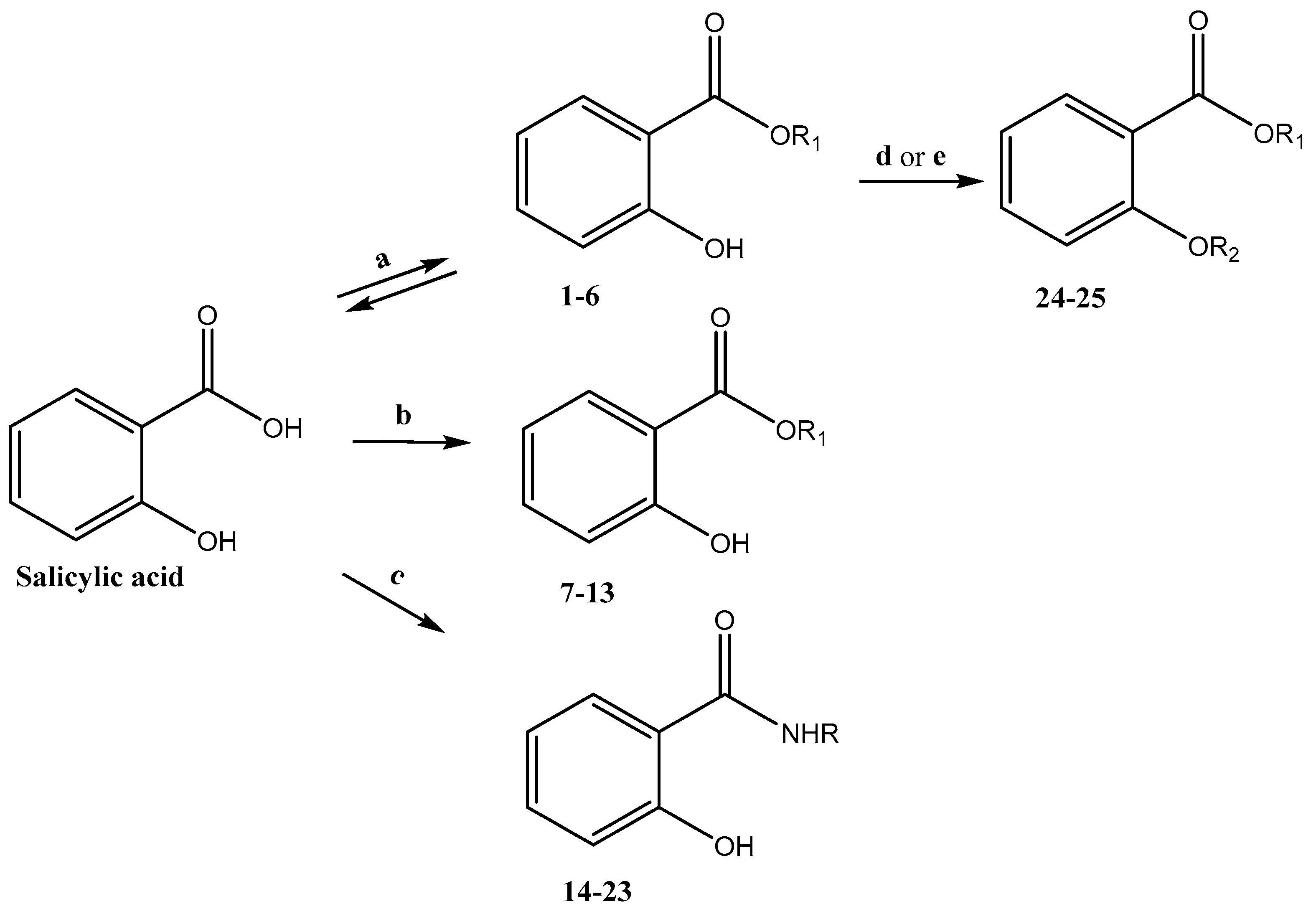
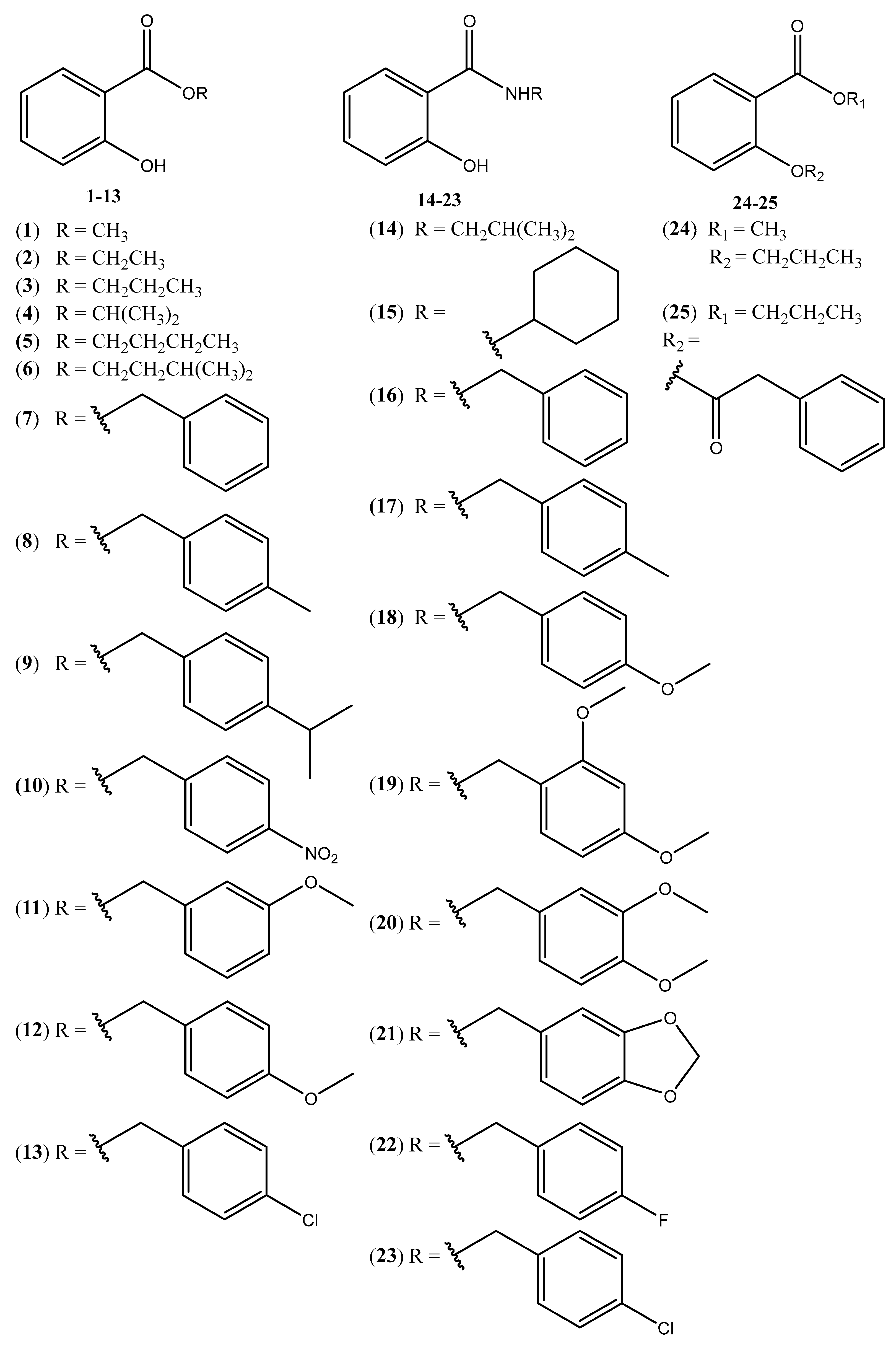
In planning the preparation of this collection of compounds, several chemical parameters were used, such as lipophilicity, stereochemistry, and electronic effects, aiming to identify new antifungal compounds. Thus, the chemical structures of twenty-five derivatives were selected and synthesized by functionalizing the two reaction sites of the starting material, the carboxylic group and the phenolic hydroxyl. The products were obtained using Fischer esterification reactions (16), Mitsunobu reaction (713), Schotten–Baumann reaction (1423, 25), and nucleophilic substitution reaction with alkyl halide (24), according to Scheme 1 and Figure 2.

3.1. Antifungal Activity

In the present study, twenty-five compounds were submitted to antifungal evaluations against Candida strains (Table 1a–d). One can visualize the results according to MFC/MIC ratios, MFC (minimum fungicidal concentration), and MIC (minimum inhibitory concentration). Salicylates did not show antifungal activity (Table 1a,b), however, several salicylamides were bioactive (Table 1c,d) indicating the importance of the amide functional group in activity against fungi.
Compound 15 presented the best antifungal profile (MIC of 570.05 μM) against C. albicans ATCC 90028, C. albicans CBS 5602, and C. krusei CBS 573. We investigated its mechanism of action against C. albicans ATCC 90028, via ergosterol and sorbitol assays to determine the likely action of the compound, whether on the plasma membrane or the cell wall, respectively (Table 2 and Table 3). Sterols participate in the constitution of all types of fungal cells; the principal sterol is ergosterol, which acts by modulating membrane fluidity, cell growth, and proliferation [50,51].

3.2. Molecular Modeling

Modeling studies were conducted to investigate the potential antifungal mechanism of action of compound 15 in C. albicans. This compound was selected for modeling because it exhibited the best bioactivity profile across the series of compounds. The modeling workflow, summarized in Figure 3, integrates computational target fishing, molecular docking, and the estimation of free energies of binding from a conformational ensemble obtained through MD simulations. Target fishing is first employed to identify a set of proteins in the fungus for structure-based modeling, in place of an unguided selection of proteins without a sound rationale. Molecular docking is then applied to generate receptor-ligand binding hypotheses. Subsequently, MD simulations were performed to produce a conformational ensemble for each complex, from which free energies of binding were estimated. Finally, the selection of the most probable targets of the compound in the fungus is performed from the MD-predicted free energies of binding.
The use of the MD-derived energies to select the most probable biological targets of the compound is motivated by to main aspects. The first is the higher accuracy in the description of inter-molecular interactions provided by MD simulations over molecular docking. Specifically, it has been shown that MM-PBSA calculations provide more reliable predictions for binding free energies than molecular docking [52,53]. The second aspect that was considered is that due to their simplicity, docking scoring functions can produce biased results when their results are compared across different targets [54,55].
The appropriate duration of MD simulations for estimating protein–ligand binding free energies with the MM-PBSA approach remains an open question in the literature. Several studies have shown that relatively short simulations (often below 5 ns) can be sufficient to obtain reliable estimates with this method [52,53]. In practice, the choice of simulation length is largely constrained by available computational resources, and no general consensus has been established on the minimum time required to sample conformational ensembles for MM-PBSA analyses. In our study, we adopted a multiple-replicate strategy rather than relying on a single long trajectory. Specifically, five independent production runs of 4 ns each were carried out per complex, with initial atomic velocities randomized for every replica. This scheme allowed for a more diverse sampling of conformational space while maintaining the total computational effort at a feasible level. From these trajectories, 100 snapshots were extracted and used to estimate the binding free energies.
The list of potential biological targets of the compound in C. albicans that were identified during the target fishing predictions is given in Table 4. The table includes the Accession number of each target sequence in the Uniprot database, the ID assigned to the proteins along the manuscript and a brief functional description for them. Among the 15 selected proteins, five of them are protein kinases and three are annotated as peptidyl-prolyl cis-trans isomerases. That is, most proteins are annotated with the two later functions.
Molecular docking calculations were processed as presented in the Methods section. For FBA1, two binding cavities were explored: the active site and an allosteric modulation site in the neighborhood of the first [56]. Likewise, three different binding regions previously identified in the homolog IDH1 of H. sapiens were explored for IDP1 and IDP2. These were the NADPH binding groove, the isocitrate binding region, and an allosteric modulator cavity located in the dimer interface [57,58].
The performance of the molecular docking protocol was assessed by re-docking the ligand co-crystallized with FBA1 in its allosteric site (PDB code 7V6G) using the parameters described in the Section 2. The top three docking solutions yielded RMSD values of 1.87 Å, 6.96 Å, and 1.06 Å relative to the experimental ligand conformation. These results indicate that, among the top three poses selected by our protocol for subsequent MD analyses, two deviate by less than 2 Å from the crystallographic structure.
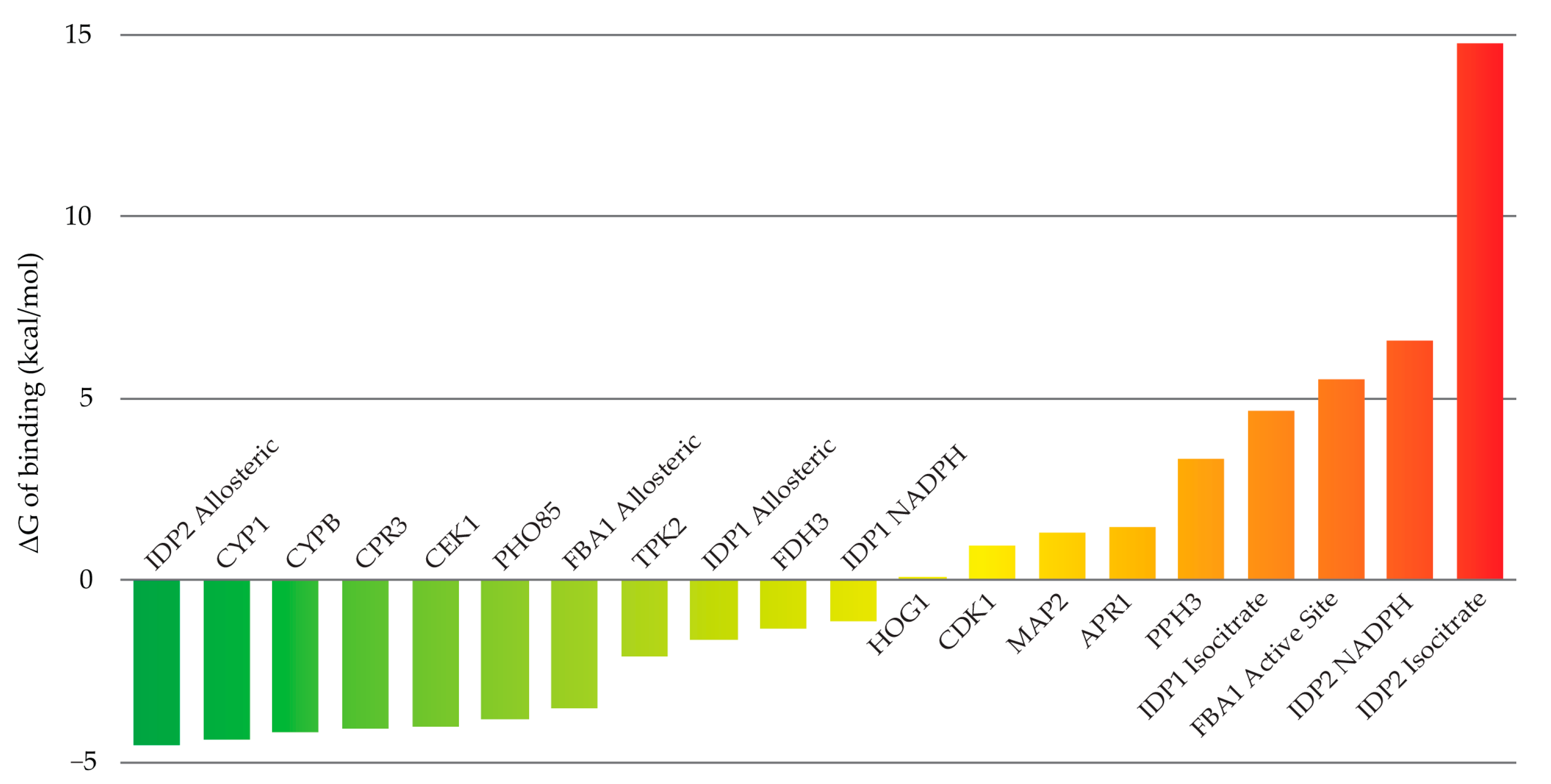
The results of the molecular docking simulations are in the Supplementary Materials (Table S1) and the PDB files of the docking—predicted complexes are given in file DockingResults.zip. Docking calculations lead to 60 compound 15 protein complexes for further analyses. As discussed above, molecular docking was used to generate ligand-receptor binding hypotheses whose energetic stability was next evaluated by MM-PBSA calculations performed from MD simulations snapshots. The applied modeling workflow led to a total of 1.2 µs MD simulation time. The calculated free energies of binding obtained for all complexes are provided as Supplementary Materials in Table S2 and are summarized in Figure 4.
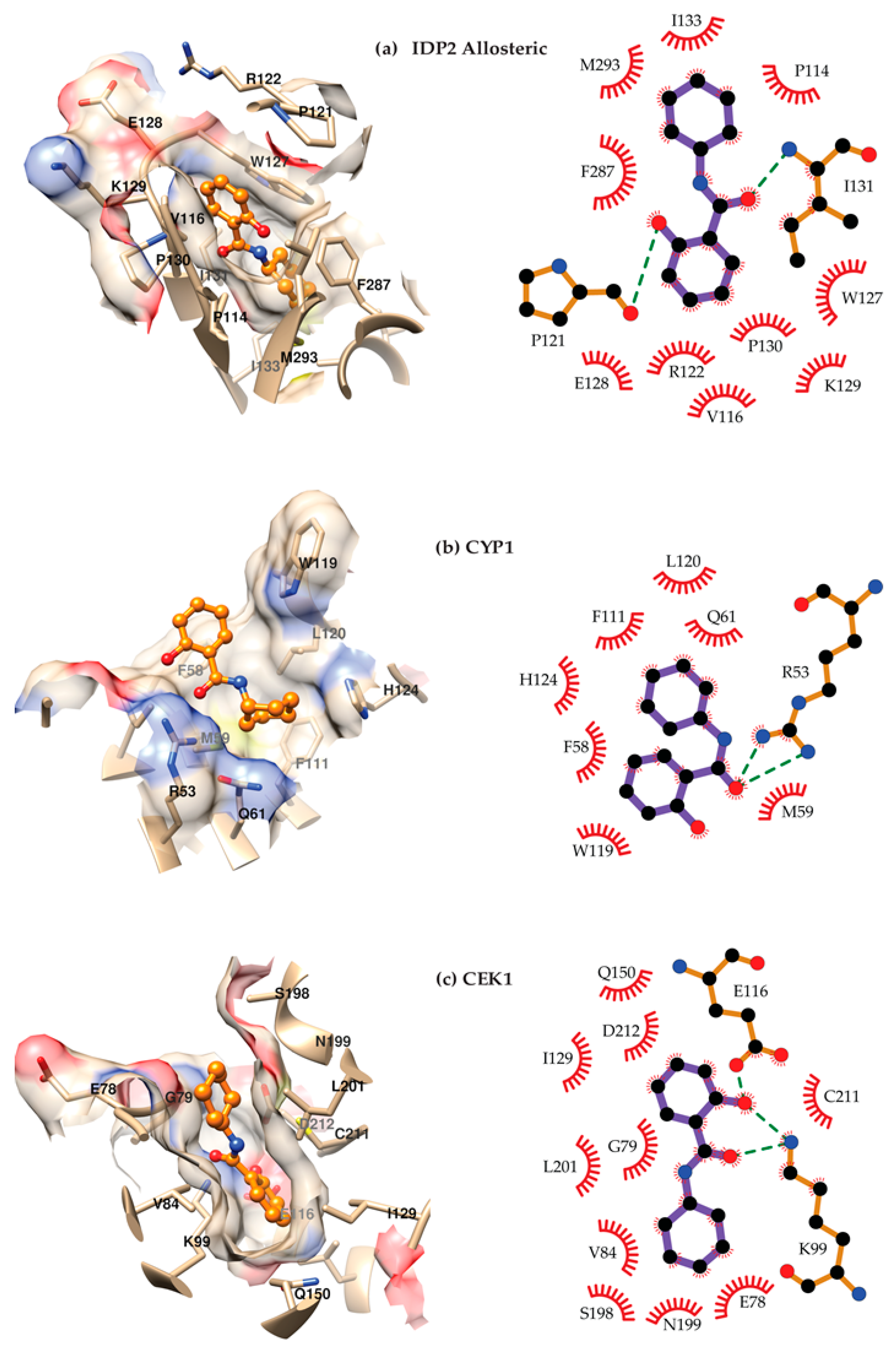
To obtain more insights into the potential inhibition of compound 15 against its three more probable target classes, the predicted binding modes to the allosteric site of IDP2, CYP1, and CEK1 were analyzed in detail. These binding conformations, as well as the predicted ligand-receptor interactions, are depicted in Figure 5. The ligand poses represented in the figure are the centroids of the largest clusters obtained from grouping the 100 MD snapshots used for MM-PBSA calculations. The figure was produced with UCSF Chimera [58] and LigPlot+ [59,60,61].
To further evaluate the stability of the predicted complexes of compound 15 with its most probable targets (IDP2, CYP1, and CEK1), the MD simulation time for these systems was extended up to 100 ns per replica. This resulted in an additional 480 ns of simulation per complex and a total of 1.44 µs of MD time. For these extended simulations, several stability metrics were examined, including the Root Mean Square Deviation (RMSD), Root Mean Square Fluctuation (RMSF), Solvent Accessible Surface Area (SASA), and Radius of Gyration (Rgyr). As shown in Supplementary Figures S25–S27, in all cases the ligand remained close to its docking-predicted pose, with RMSD values below 2 Å in all MD replicas for the three targets. The RMSD values of the protein backbone also indicated conformational stability, although in IDP2 and CEK1 the backbone deviated by up to ~4 Å from the starting structure.
To investigate whether protein flexibility could affect complex stability, the RMSF of each residue was analyzed (Supplementary Figures S28–S30). The results show that the largest fluctuations occur in regions distant from the ligand-binding cavity and therefore do not directly impact ligand binding. By contrast, residues within the binding site exhibited low flexibility in all three systems. Finally, the stability of SASA (Supplementary Figure S31) and Rgyr (Supplementary Figure S32) further support the conformational stability of the predicted complexes. In summary, these extended MD simulations provide consistent indicators of stability at longer timescales.

4. Discussion

4.1. 1H and 13C-APT NMR Spectroscopy Analysis

Analysis of 1H NMR spectra of salicylates and salicylamides showed the following signals: a chemical shift between δH 12.20–10.56 ppm, in the form of a broad singlet corresponding to hydrogen of the hydroxyl (OH) of the phenolic structure (sign present only in the ester derivatives); in the region between δH 7.90–6.80 ppm the four aromatic hydrogens common to all compounds. The amides also showed a chemical shift between δH 6.68–6.22 ppm, in the form of a singlet referring to nitrogen hydrogen (N-H). Analysis of 13C-APT NMR spectra showed seven common aromatic hydrogens. A signal between δC 170.71–164.65 ppm was attributed to the carbonyl carbon; a signal between δC 161.99–161.47 ppm was assigned to the carbon attached to the hydroxyl. The signs between δC 136.39–133.41 ppm indicate the carbon in the aromatic ring; the signal between δC 130.20–125.16 ppm is attributed to an ortho carbon of the aromatic ring, and the signal between δC 126.13–118.49 ppm and between δC 118.84–117.36 ppm attributed to two meta carbons, respectively. Furthermore, the signal between δC 123.72–111.96 ppm is assigned to the aromatic ring carbon attached to the carbonyl ester. Compound 18 showed according to 13C-APT NMR sp2 hybridization carbon atom at δ 118.64 ppm (methine carbon). For unpublished compounds 8, 11, 13, 17, 20, and 25, their spectra are in the Supplementary Materials. In the analyzes by infrared spectroscopy, the esters 113, 24 and 25 showed: signal above 3000 cm−1 (C-H sp2 stretching), paired ring stretching absorptions at 1600 and 1475 cm−1 (C=C), characteristic signs of ester-conjugated carbonyl in 1679–1666 cm−1 (C=O) and two bands in the range 1300 to 1000 cm−1 (C-O stretch). Regarding amides 1423, they showed absorption bands at about 3300 cm−1 to the N-H stretching of secondary amides; a strong band between 1680 and 1630 cm−1 attributed to the stretching of the amide carbonyl (C=O). The paired signals around 1600 and 1475 cm−1 refer to the stretching (C=C) of aromatic rings. Furthermore, the chemical structures of the unpublished compounds were confirmed by HRMS according to their molecular weights.

4.2. Antifungal Activity

Eight salicylamides presented inhibitory activity against at least one fungal strain and fungicidal action against all tested strains. However, none of the salicylates demonstrated antifungal bioactivity. In fact, some esters and amides contain similar substituents; however, only the amides exhibit antifungal activity. For example, derivatives with a four-carbon side chain (ester 5 and amide 14), a 4-methylbenzyl substituent (ester 8 and amide 17), a 4-methoxybenzyl substituent (ester 12 and amide 18), and a 4-chlorobenzyl substituent (ester 13 and amide 23). The presence of the ester function in these compounds resulted in inactivity against all strains. Of the salicylamide compounds, 14, 15, 17, 18, 22, and 23 were active against the Candida albicans strain ATCC 90028 at concentrations ranging from 485.83 to 4077.47 µM. Compounds 15 and 18 presented respective MICs of 570.05 and 485.83 μM. It was also observed that both cyclohexyl substituents and 4-methoxy-benzyl yielded similar antifungal potencies against most strains tested for both N-cyclohexyl-2-hydroxybenzamide (15) and N-4-methoxybenzyl-2-hydroxybenzamide (18). Compound 14 (isobutyl group) was bioactive against C. albicans strain ATCC 90028, with an MIC of 1293.73 µM. The compounds N-4-chlorobenzyl-2-hydroxybenzamide (23), and N-4-methylbenzyl-2-hydroxybenzamide (17) were bioactive against C. albicans ATCC 90028, presenting respective MICs of 1910.58 and 2072.20 μM, demonstrating a higher MIC value than the other compounds discussed. The compound N-4-fluorobenzyl-2-hydroxybenzamide (22) presented activity at the highest concentration tested against C. albicans ATCC 90028 (MIC = 4077.47 μM), as the presence of an electron withdrawing group such as fluorine in the para position of the aromatic ring increased the MIC. In tests using C. albicans CBS 5602, the bioactive compounds were 1418 and 22 at concentrations ranging from 485.83 to 2072.20 µM. Compounds 15, 16, and 18 were the most potent against C. albicans CBS 5602 presenting respective MICs of 570.05, 550.03, and 485.83 μM. Compound 15 (with a cyclohexyl group) was equipotent with compound 16 (with an aromatic ring). The presence of a methoxyl in the para position of the aromatic ring in compound 18 potentiated antifungal activity. Amides 17 and 22 were equipotent (MIC of 2072.20 and 2038.74 μM, respectively), while the isobutyl substituent in 14 increased antifungal activity, to yield an MIC of 1293.73 μM against C. albicans CBS 5602. In tests against C. tropicalis CBS 94, the bioactive compounds were 14, 15, 18, and 22 with concentrations ranging from 1140.10 to 2587.46 µM. Compound 15 was the most potent with an MIC of 1140.10 µM. Compounds 14, 18, and 22 presented respective MICs of 2587.46, 1943.33, and 2038.74 μM, and decreasing potency was observed. In tests using C. krusei CBS, the bioactive compounds were 1419, 22, and 23 with MIC values between 485.83 and 2072.20 µM. Compounds 15 and 18 presented the lowest MIC values (respectively, 570.05 and 485.83 μM) against C. krusei, with activity similar to tests against the Candida albicans ATCC 90028. It was observed that the presence of a six-membered cyclic alkyl side chain (15) or a 4-methoxy-benzyl substituent (18) potentiates antifungal activity. Compounds 14 and 16 presented similar activity against C. krusei CBS 573 (MIC = 1293.73 and 1100.06 µM, respectively). Compounds 17, 19, 22, and 23 presented similar activity with higher MIC values (2072.20, 1740.28, 2038.74, and 1910.58 µM, respectively). Corroborating results for compound 23 (MIC = 500 μg/mL) [34], presented similar results in antifungal evaluations of N-(4-halobenzyl) amides with a benzoic skeletal structure similar to that of N-4-chlorobenzyl-2-hydroxybenzamide, a compound submitted to evaluation against the C. krusei (ATCC 14243), which yielded an MIC of 250 µg/mL [44].
The salicylic acid, when combined with rifampicin and benzoic acid in the form of topical nanoemulsion, presented significant activity against C. albicans (when isolated and collected from a patient in a microbiology laboratory in Bangabandhu Sheikh Mujib Medical University) [62]. This demonstrated great antimicrobial potential in the incorporation of these compounds into antifungal formulations. Thus, the most potent salicylamides in the present study could be used in combination with other antifungal drugs in formulations for the development of new drugs against strains of Candida. In the present study, the reference drugs Nystatin and Ketoconazole were more potent than the tested derivatives. Despite the differences in antifungal potency, the synthetic derivatives were obtained via easy-to-perform, low-cost reactions.
The mechanism of action test for 15 consisted of adding ergosterol to a medium containing the fungus and the compound 15. There was no MIC variation, indicating that the compound did not act by either inhibiting ergosterol synthesis or directly binding to ergosterol. Azoles and polyenes (often used to treat fungal infections) act against ergosterol [63]. Sorbitol, an osmotic protector that acts by preventing changes in the fungal cell wall was added to a medium containing the fungus and compound 15, yet there was no increase in the MIC, indicating that the substance does not act by interfering with cell wall synthesis [64].

4.3. Molecular Docking

The results suggest that the antifungal mechanism of action of 15 could be related to a multi-target mechanism of action. This supposition is supported by the very similar binding energies obtained for the targets ranked in the first seven positions. Moreover, these top ranked proteins are related to four different functions: isocitrate dehydrogenase (IDP2), peptidyl-prolyl cis-trans isomerase (CYP1, CYPB, and CPR3), protein kinase (CEK1 and PHO85), and fructose-bisphosphate aldolase (FBA1). It is interesting to note that the three peptidyl-prolyl cis-trans isomerases rank consecutively in positions 2, 3, and 4. The later can be explained from the identical structures and sequences of these proteins in their ligand binding regions.
Compound 15 is predicted to form hydrogen bonds with the three receptors. These interactions take place with the backbones of P121 and I131 of IDP2, the side chain of R53 in CYP1, and the side chains of K99 and E116 of CEK1. In addition, the ligand orientates with its ortho-phenyl group to the entrance of the allosteric site of IDP2, interacting with V116, P121, R122, K129, and P130. The orientation of this substituent is favorable for the π-π stacking interaction with W127. On the other side, the cyclohexyl moiety occupies a hydrophobic region defined by P114, F287, M293, I131, and I133. In the predicted complex with CYP1, the rest of the ligand-receptor interactions occur with F58 and W119 for the ortho-phenyl substituent and with M59, Q61, F111, L120, and H124 for the cyclohexyl group. Finally, in the complex with CEK1, the later moiety is predicted to orient at the entrance of the cavity lined by E78, G79, V84, S198, N199, and L201. The 2-hydroxyphenyl group, in contrast, occupies the bottom of the active site of CEK1 and makes additional contacts with I129, Q150, C211, and D212. As previously discussed, the modeling results point to a probable multi-target antifungal mechanism of action of compound 15. Despite no experimental validation of the potential biological targets of this compound in C. albicans is performed, the modeling results can guide future experimentation in this direction. For example, proteins and cellular processes can be prioritized based on the modeling results. Additionally, the predicted binding modes of the compound to its potential targets can serve as the starting point for the optimization of the antifungal activity of compound 15. Molecular Docking data (Tables S1 and S2) are available in Supplementary Materials.

5. Conclusions

In the present study, twenty-five synthetic salicylates and salicylamides were structurally designed based on parameters such as lipophilicity, electron density, and stereochemistry, and evaluated against C. albicans ATCC 90028, C. albicans CBS 5602, C. krusei CBS 573, and C. tropicalis CBS 94. Among the 25 compounds prepared, six are new products. Compounds 1419, 22, and 23 were bioactive against at least one strain, with MIC values ranging from 485.83 to 4077.47 μM. Compound 15 presented the best antifungal activity against the tested Candida strains, suggesting that the presence of a six-membered cyclic alkyl group potentiates the antifungal activity. It was demonstrated that compound 15 does not act by causing alterations in the cell wall or plasma membrane functions in Candida species. Molecular modeling of compound 15 suggested a probable multi-targeted antifungal mechanism of action, which is of great importance for guiding future experiments using this chemical class as antifungal agents.

Supplementary Materials

The supporting information can be downloaded at: https://www.mdpi.com/article/10.3390/chemistry7050151/s1, Chemistry: FTIR, 1H NMR, 13C NMR, and HRMS spectra for compounds 8, 11, 13, 17, 20, and 25 (Figures S1–S24). RMSD for the predicted complexes of compound 15 with IDP2, CYP1, and CEK1 (Figures S25–S27). RMSF for the predicted complexes of compound 15 with IDP2, CYP1, and CEK1 (Figures S28–S30). SASA for the predicted complexes of compound 15 with IDP2, CYP1, and CEK1 (Figure S31). Radius of gyration for the predicted complexes of compound 15 with IDP2, CYP1, and CEK1 (Figure S32). Molecular Docking: Tables S1 and S2. PDB files for the docking predictions (File DockingResults.zip).

Author Contributions

A.J.d.M.S.O. conducted the synthesis of compounds and wrote the manuscript. Y.P.-C. performed the in silico tests, and D.d.N.A. and R.D.d.C. performed the biological tests. M.C.D. reviewed part of the data. D.P.d.S. edited the manuscript. All authors have read and agreed to the published version of the manuscript.

Funding

This research was funded by Coordination for the Improvement of Higher Education Personnel (CAPES)—Finance Code 001, and the National Council for Scientific and Technological Development (CNPq), grant number 312977/2023-9.

Institutional Review Board Statement

Not applicable.

Informed Consent Statement

Not applicable.

Data Availability Statement

All data generated or analyzed during this study are included in this article.

Conflicts of Interest

The authors declare no conflicts of interest.

References

  1. Bitew, A.; Abebaw, Y. Vulvovaginal Candidiasis: Species Distribution of Candida and Their Antifungal Susceptibility Pattern. BMC Women’s Health 2018, 18, 94. [Google Scholar] [CrossRef]
  2. Marak, M.B.; Dhanashree, B. Antifungal Susceptibility and Biofilm Production of Candida Spp. Isolated from Clinical Samples. Int. J. Microbiol. 2018, 2018, 7495218. [Google Scholar] [CrossRef]
  3. Medici, N.P.; Del Poeta, M. New Insights on the Development of Fungal Vaccines: From Immunity to Recent Challenges. Mem. Do Inst. Oswaldo Cruz 2015, 110, 966–973. [Google Scholar] [CrossRef]
  4. Nami, S.; Mohammadi, R.; Vakili, M.; Khezripour, K.; Mirzaei, H.; Morovati, H. Fungal Vaccines, Mechanism of Actions and Immunology: A Comprehensive Review. Biomed. Pharmacother. 2019, 109, 333–344. [Google Scholar] [CrossRef] [PubMed]
  5. Spampinato, C.; Leonardi, D. Candida Infections, Causes, Targets, and Resistance Mechanisms: Traditional and Alternative Antifungal Agents. Biomed. Res. Int. 2013, 2013, 204237. [Google Scholar] [CrossRef] [PubMed]
  6. Krátký, M.; Vinšová, J. Antifungal Activity of Salicylanilides and Their Esters with 4-(Trifluoromethyl)benzoic Acid. Molecules 2012, 17, 9426–9442. [Google Scholar] [CrossRef] [PubMed]
  7. Rainsford, K.D. Aspirin and Related Drugs; CRC Press: Boca Raton, FL, USA, 2004; ISBN 9781135732639. [Google Scholar]
  8. Sahoo, J.; Paidesetty, S.K. Antimicrobial, Analgesic, Antioxidant and in Silico Study of Synthesized Salicylic Acid Congeners and Their Structural Interpretation. Egypt. J. Basic Appl. Sci. 2015, 2, 268–280. [Google Scholar] [CrossRef]
  9. Djurendić, E.; Vujašković, S.D.; Sakač, M.; Ajduković, J.; Gaković, A.; Kojić, V.; Bogdanović, G.; Klisurić, O.; Gaši, K.P. Synthesis and Biological Evaluation of Some New 2-Oxazoline and Salicylic Acid Derivatives. Arkivoc 2010, 2011, 83–102. [Google Scholar] [CrossRef]
  10. Sanderson, J.T.; Clabault, H.; Patton, C.; Lassalle-Claux, G.; Jean-François, J.; Paré, A.F.; Hébert, M.J.G.; Surette, M.E.; Touaibia, M. Antiproliferative, Antiandrogenic and Cytotoxic Effects of Novel Caffeic Acid Derivatives in LNCaP Human Androgen-Dependent Prostate Cancer Cells. Bioorg. Med. Chem. 2013, 21, 7182–7193. [Google Scholar] [CrossRef]
  11. Chapado, L.; Linares-Palomino, P.J.; Salido, S.; Altarejos, J.; Rosado, J.A.; Salido, G.M. Synthesis and Evaluation of the Platelet Antiaggregant Properties of Phenolic Antioxidants Structurally Related to Rosmarinic Acid. Bioorg. Chem. 2009, 38, 108–114. [Google Scholar] [CrossRef] [PubMed]
  12. Ji, X.-Y.; Wang, H.-Q.; Hao, L.-H.; He, W.-Y.; Gao, R.-M.; Li, Y.-P.; Li, Y.-H.; Jiang, J.-D.; Li, Z.-R. Synthesis and Antiviral Activity of N-Phenylbenzamide Derivatives, a Novel Class of Enterovirus 71 Inhibitors. Molecules 2013, 18, 3630–3640. [Google Scholar] [CrossRef] [PubMed]
  13. Coolen, H.K.A.C.; Meeuwis, J.A.M.; van Leeuwen, P.W.M.N.; Nolte, R.J.M. Substrate Selective Catalysis by Rhodium Metallohosts. J. Am. Chem. Soc. 1995, 117, 11906–11913. [Google Scholar] [CrossRef][Green Version]
  14. Sadeghian, H.; Seyedi, S.M.; Saberi, M.R.; Arghiani, Z.; Riazi, M. Design and Synthesis of Eugenol Derivatives, as Potent 15-Lipoxygenase Inhibitors. Bioorg. Med. Chem. 2008, 16, 890–901. [Google Scholar] [CrossRef]
  15. Masetto, E.; Lazaroto, A.; Oleinik, G.; Lima, F.; Gallina, A.; Soares, L. Salicilatos Como Inibidores do Processo Oxidativo Mediado por Cobre e Ferro no Biodiesel B100. Quim. Nova 2022, 45. [Google Scholar] [CrossRef]
  16. Zhao, Q.; Chu, J.; Jiang, T.; Yin, H. Synthesis of Isoamyl Salicylate Using a Novel Mesoporous Titania Superacid as a Catalyst. Korean J. Chem. Eng. 2008, 25, 1008–1013. [Google Scholar] [CrossRef]
  17. Yamada, K.; Yoshida, S.; Fujita, H.; Kitamura, M.; Kunishima, M. O-Benzylation of Carboxylic Acids Using 2,4,6-Tris(Benzyloxy)-1,3,5-Triazine (TriBOT) under Acidic or Thermal Conditions. Eur. J. Org. Chem. 2015, 2015, 7997–8002. [Google Scholar] [CrossRef]
  18. Villa, C.; Baldassari, S.; Gambaro, R.; Mariani, E.; Loupy, A. Eco-Friendly Methodologies for the Synthesis of Some Aromatic Esters, Well-Known Cosmetic Ingredients. Int. J. Cosmet. Sci. 2005, 27, 11–16. [Google Scholar] [CrossRef] [PubMed]
  19. Dev, D.; Palakurthy, N.B.; Thalluri, K.; Chandra, J.; Mandal, B. Ethyl 2-Cyano-2-(2-Nitrobenzenesulfonyloxyimino) Acetate (O-NosylOXY): A Recyclable Coupling Reagent for Racemization-Free Synthesis of Peptide, Amide, Hydroxamate, and Ester. J. Org. Chem. 2014, 79, 5420–5431. [Google Scholar] [CrossRef]
  20. Bamoharram, F.F.; Heravi, M.M.; Roshani, M.; Jahangir, M.; Gharib, A. Preyssler Catalyst, [NaP5W30O110]14−: A Green, Efficient and Reusable Catalyst for Esterification of Salicylic Acid with Aliphatic and Benzylic Alcohols. Appl. Catal. A Gen. 2006, 302, 42–47. [Google Scholar] [CrossRef]
  21. Carcelli, M.; Rogolino, D.; Bacchi, A.; Rispoli, G.; Fisicaro, E.; Compari, C.; Sechi, M.; Stevaert, A.; Naesens, L. Metal-chelating 2-hydroxyphenyl amide pharmacophore for inhibition of influenza virus endonuclease. Mol Pharm. 2014, 11, 304–316. [Google Scholar] [CrossRef]
  22. Gao, B.; Chen, K.; Bi, X.; Wang, J. Intramolecular Functionalization of c (Sp3) H Bonds Adjacent to an Amide Nitrogen Atom: Metal-Free Synthesis of 2-Hydroxy-Benzoxazinone Derivatives. Tetrahedron 2017, 73, 7005–7010. [Google Scholar] [CrossRef]
  23. Madasamy, K.; Balakrishnan, M.H.; Korivi, R.; Mannathan, S. Trifluoroacetic Acid-Mediated Denitrogenative Ortho-Hydroxylation of 1,2,3-Benzotriazin-4(3H)-Ones: A Metal-Free Approach. J. Org. Chem. 2022, 87, 8752–8756. [Google Scholar] [CrossRef]
  24. Waisser, K.; Peřina, M.; Klimešová, V.; Kaustová, J. On the Relationship between the Structure and Antimycobacterial Activity of Substituted N-Benzylsalicylamides. Coll. Czech. Chem. Commun. 2003, 68, 1275–1294. [Google Scholar] [CrossRef]
  25. Grintsevich, S.; Sapegin, A.; Reutskaya, E.; Peintner, S.; Erdélyi, M.; Krasavin, M. An Alternative Approach to the Hydrated Imidazoline Ring Expansion (HIRE) of Diarene-Fused [1.4] Oxazepines. Eur. J. Org. Chem. 2020, 2020, 5664–5676. [Google Scholar] [CrossRef]
  26. Zhang, H.; Wang, J.; Ji, Z.; Sun, X.; Tian, Q.; Wei, S.; Ji, Z. Discovery, SAR, and Putative Mode of Action of N-Benzyl-2-Methoxybenzamides as Potential Bleaching Herbicides. Pest Manag. Sci. 2021, 77, 2804–2811. [Google Scholar] [CrossRef] [PubMed]
  27. Ahmed, M.N.; Ahmad, K.; Yasin, K.A.; Farooq, T.; Khan, B.A.; Roy, S.K. Ligand-Free Cu(Ii)-Catalyzed Aerobic Etherification of Aryl Halides with Silanes: An Experimental and Theoretical Approach. New J. Chem. 2019, 43, 11316–11333. [Google Scholar] [CrossRef]
  28. Rex, J.H.; CLSI. Reference Method for Broth Dilution Antifungal Susceptibility Testing of Yeasts: Approved Standard, 3rd ed.; CLSI: Wayne, PA, USA, 2008; ISBN 9781562386665. [Google Scholar]
  29. Siddiqui, Z.N.; Farooq, F.; Musthafa, T.N.M.; Ahmad, A.; Khan, A.U. Synthesis, Characterization and Antimicrobial Evaluation of Novel Halopyrazole Derivatives. J. Saudi Chem. Soc. 2013, 17, 237–243. [Google Scholar] [CrossRef]
  30. Alves, D.d.N.; Ferreira, A.R.; Duarte, A.B.S.; Melo, A.K.V.; de Sousa, D.P.; Castro, R.D. Breakpoints for the Classification of Anti-Candida Compounds in Antifungal Screening. Biomed. Res. Int. 2021, 2021, e6653311. [Google Scholar] [CrossRef]
  31. Lima, I.O.; Pereira, F.O.; Oliveira, W.A.; Lima, E.O.; Menezes, E.A.; Cunha, F.A.; Diniz, M.F.F.M. Antifungal Activity and Mode of Action of Carvacrol against Candida albicans Strains. J. Essent. Oil Res. 2013, 25, 138–142. [Google Scholar] [CrossRef]
  32. Escalante, A.; Gattuso, M.; Pérez, P.; Zacchino, S. Evidence for the Mechanism of Action of the Antifungal Phytolaccoside B Isolated from Phytolacca tetramera Hauman. J. Nat. Prod. 2008, 71, 1720–1725. [Google Scholar] [CrossRef]
  33. Freires, I.A.; Murata, R.M.; Furletti, V.F.; Sartoratto, A.; Alencar, S.M.; Figueira, G.M.; Rodrigues, J.A.O.; Duarte, M.C.T.; Rosalen, P.L. Coriandrum sativum L. (Coriander) Essential Oil: Antifungal Activity and Mode of Action on Candida Spp., and Molecular Targets Affected in Human Whole-Genome Expression. PLoS ONE 2014, 9, e99086. [Google Scholar] [CrossRef]
  34. Ferreira, A.R.; Alves, D.N.; Castro, R.D.; Perez-Castillo, Y.; Sousa, D.P. Synthesis of Coumarin and Homoisoflavonoid Derivatives and Analogs: The Search for New Antifungal Agents. Pharmaceuticals 2022, 15, 712. [Google Scholar] [CrossRef] [PubMed]
  35. Lopes, S.P.; Yepes, L.M.; Pérez-Castillo, Y.; Robledo, S.M.; Sousa, D.P. Alkyl and Aryl Derivatives Based on P-Coumaric Acid Modification and Inhibitory Action against Leishmania braziliensis and Plasmodium falciparum. Molecules 2020, 25, 3178. [Google Scholar] [CrossRef] [PubMed]
  36. Keiser, M.J.; Roth, B.L.; Armbruster, B.N.; Ernsberger, P.; Irwin, J.J.; Shoichet, B.K. Relating Protein Pharmacology by Ligand Chemistry. Nat. Biotechnol. 2007, 25, 197–206. [Google Scholar] [CrossRef]
  37. Hawkins, P.C.D.; Skillman, A.G.; Warren, G.L.; Ellingson, B.A.; Stahl, M.T. Molecular Modeling Software|OpenEye Scientific. Available online: https://www.eyesopen.com/omega (accessed on 3 June 2025).
  38. Hawkins, P.C.D.; Skillman, A.G.; Warren, G.L.; Ellingson, B.M.; Stahl, M.T. Conformer Generation with OMEGA: Algorithm and Validation Using High Quality Structures from the Protein Databank and Cambridge Structural Database. J. Chem. Inf. Model. 2010, 50, 572–584. [Google Scholar] [CrossRef] [PubMed]
  39. Santa Fe, N.M. Molecular Modeling Software|OpenEye Scientific. Available online: https://www.eyesopen.com/quacpac (accessed on 27 May 2025).
  40. Waterhouse, A.; Bertoni, M.; Bienert, S.; Studer, G.; Tauriello, G.; Gumienny, R.; Heer, F.T.; Beer, T.A.; Rempfer, C.; Bordoli, L.; et al. SWISS-MODEL: Homology Modelling of Protein Structures and Complexes. Nucleic Acids Res. 2018, 46, W296–W303. [Google Scholar] [CrossRef]
  41. Jones, G.; Willett, P.; Glen, R.C.; Leach, A.R.; Taylor, R. Development and Validation of a Genetic Algorithm for Flexible Docking 1 1Edited by F. E. Cohen. J. Mol. Biol. 1997, 267, 727–748. [Google Scholar] [CrossRef]
  42. Case, D.A.; Aktulga, H.M.; Belfon, K.; Ben-Shalom, I.; Berryman, J.T.; Brozell, S.R.; Cerutti, D.S.; Cheatham, T.E.; Cisneros, G.A.; Cruzeiro, V.W.D.; et al. Amber 2022; University of California: San Francisco, CA, USA, 2022. [Google Scholar]
  43. Castro, M.; Perez-Castillo, Y.; Silva, V.R.; Santos, L.S.; Botelho, M.; Bezerra, D.P.; Castro, R.D.; Sousa, D.P. Cytotoxic and Antifungal Amides Derived from Ferulic Acid: Molecular Docking and Mechanism of Action. Biomed. Res. Int. 2021, 2021, 3598000. [Google Scholar] [CrossRef]
  44. Perez-Castillo, Y.; Montes, R.C.; Silva, C.R.; Neto, J.B.A.; Dias, C.S.; Duarte, A.B.S.; Nobre, H.V.; Sousa, D.P. Antifungal Activity of N-(4-Halobenzyl) Amides against Candida Spp. And Molecular Modeling Studies. Int. J. Mol. Sci. 2021, 23, 419. [Google Scholar] [CrossRef]
  45. AMBER Parameter Database (Bryce Group: Computational Biophysics and Drug Design—University of Manchester). Available online: http://amber.manchester.ac.uk/ (accessed on 7 September 2022).
  46. Pang, Y.P.; Xu, K.; Yazal, J.E.; Prendergast, F.G. Successful Molecular Dynamics Simulation of the Zinc-Bound Farnesyltransferase Using the Cationic Dummy Atom Approach. Protein Sci. 2000, 9, 1857–1865. [Google Scholar] [CrossRef]
  47. Li, P.; Merz, K.M. MCPB.py: A Python Based Metal Center Parameter Builder. J. Chem. Inf. Model. 2016, 56, 599–604. [Google Scholar] [CrossRef] [PubMed]
  48. Machado, M.R.; Pantano, S. Split the Charge Difference in Two! A Rule of Thumb for Adding Proper Amounts of Ions in MD Simulations. J. Chem. Theory Comput. 2020, 16, 1367–1372. [Google Scholar] [CrossRef]
  49. Miller, B.R.; McGee, T.D.; Swails, J.M.; Homeyer, N.; Gohlke, H.; Roitberg, A.E. MMPBSA.py: An Efficient Program for End-State Free Energy Calculations. J. Chem. Theory Comput. 2012, 8, 3314–3321. [Google Scholar] [CrossRef]
  50. Czub, J.; Baginski, M. Comparative Molecular Dynamics Study of Lipid Membranes Containing Cholesterol and Ergosterol. Biophys. J. 2006, 90, 2368–2382. [Google Scholar] [CrossRef]
  51. Vandeputte, P.; Ferrari, S.; Coste, A.T. Antifungal Resistance and New Strategies to Control Fungal Infections. Int. J. Microbiol. 2012, 2012, 713687. [Google Scholar] [CrossRef]
  52. Poli, G.; Granchi, C.; Rizzolio, F.; Tuccinardi, T. Application of MM-PBSA Methods in Virtual Screening. Molecules 2020, 25, 1971. [Google Scholar] [CrossRef]
  53. Wang, E.; Sun, H.; Wang, J.; Wang, Z.; Liu, H.; Zhang, J.Z.H.; Hou, T. End-Point Binding Free Energy Calculation with MM/PBSA and MM/GBSA: Strategies and Applications in Drug Design. Chem. Rev. 2019, 119, 9478–9508. [Google Scholar] [CrossRef]
  54. Lapillo, M.; Tuccinardi, T.; Martinelli, A.; Macchia, M.; Giordano, A.; Poli, G. Extensive Reliability Evaluation of Docking-Based Target-Fishing Strategies. Int. J. Mol. Sci. 2019, 20, 1023. [Google Scholar] [CrossRef] [PubMed]
  55. Luo, Q.; Zhao, L.; Hu, J.; Jin, H.; Liu, Z.; Zhang, L. The Scoring Bias in Reverse Docking and the Score Normalization Strategy to Improve Success Rate of Target Fishing. PLoS ONE 2017, 12, e0171433. [Google Scholar] [CrossRef]
  56. Wen, W.; Cao, H.; Huang, Y.; Tu, J.; Wan, C.; Wan, J.; Han, X.; Chen, H.; Liu, J.; Rao, L.; et al. Structure-Guided Discovery of the Novel Covalent Allosteric Site and Covalent Inhibitors of Fructose-1,6-Bisphosphate Aldolase to Overcome the Azole Resistance of Candidiasis. J. Med. Chem. 2022, 65, 2656–2674. [Google Scholar] [CrossRef] [PubMed]
  57. Jakob, C.G.; Upadhyay, A.K.; Donner, P.L.; Nicholl, E.; Addo, S.N.; Qiu, W.; Ling, C.; Gopalakrishnan, S.M.; Torrent, M.; Cepa, S.P.; et al. Novel Modes of Inhibition of Wild-Type Isocitrate Dehydrogenase 1 (IDH1): Direct Covalent Modification of His315. J. Med. Chem. 2018, 61, 6647–6657. [Google Scholar] [CrossRef] [PubMed]
  58. Duarte, A.B.S.; Perez-Castillo, Y.; Andrade, P.N.; Castro, R.D.; Sousa, D.P. 3,5-Dinitrobenzoate and 3,5-Dinitrobenzamide Derivatives: Mechanistic, Antifungal, and in Silico Studies. J. Chem. 2022, 2022, e2336175. [Google Scholar] [CrossRef]
  59. Pettersen, E.F.; Goddard, T.D.; Huang, C.C.; Couch, G.S.; Greenblatt, D.M.; Meng, E.C.; Ferrin, T.E. UCSF Chimera--a Visualization System for Exploratory Research and Analysis. J. Comput. Chem. 2004, 25, 1605–1612. [Google Scholar] [CrossRef] [PubMed]
  60. Laskowski, R.A.; Swindells, M.B. LigPlot+: Multiple Ligand–Protein Interaction Diagrams for Drug Discovery. J. Chem. Inf. Model. 2011, 51, 2778–2786. [Google Scholar] [CrossRef]
  61. Silva, R.H.N.; Machado, T.Q.; Fonseca, A.C.C.; Tejera, E.; Perez-Castillo, Y.; Robbs, B.K.; Sousa, D.P. Molecular Modeling and in Vitro Evaluation of Piplartine Analogs against Oral Squamous Cell Carcinoma. Molecules 2023, 28, 1675. [Google Scholar] [CrossRef]
  62. Begum, K.; Shahid, M.S.; Jalil, R.U.J. Topical Nanoemulsion of Rifampicin with Benzoic Acid and Salicylic Acid: Activity against Staphylococcus aureus, Stap. epidermis and Candida albicans. Bangladesh Pharm. J. 2019, 22, 1–6. [Google Scholar] [CrossRef]
  63. Tavares, W. Manual de Antibióticos E Quimioterápicos Antiinfecciosos, 3rd ed.; Atheneu: São Paulo, Brazil, 2021. [Google Scholar]
  64. Leite, M.C.A.; Bezerra, A.P.B.; Sousa, J.P.; Lima, E.O. Investigating the Antifungal Activity and Mechanism(S) of Geraniol against Candida albicans Strains. Med. Mycol. 2014, 53, 275–284. [Google Scholar] [CrossRef]
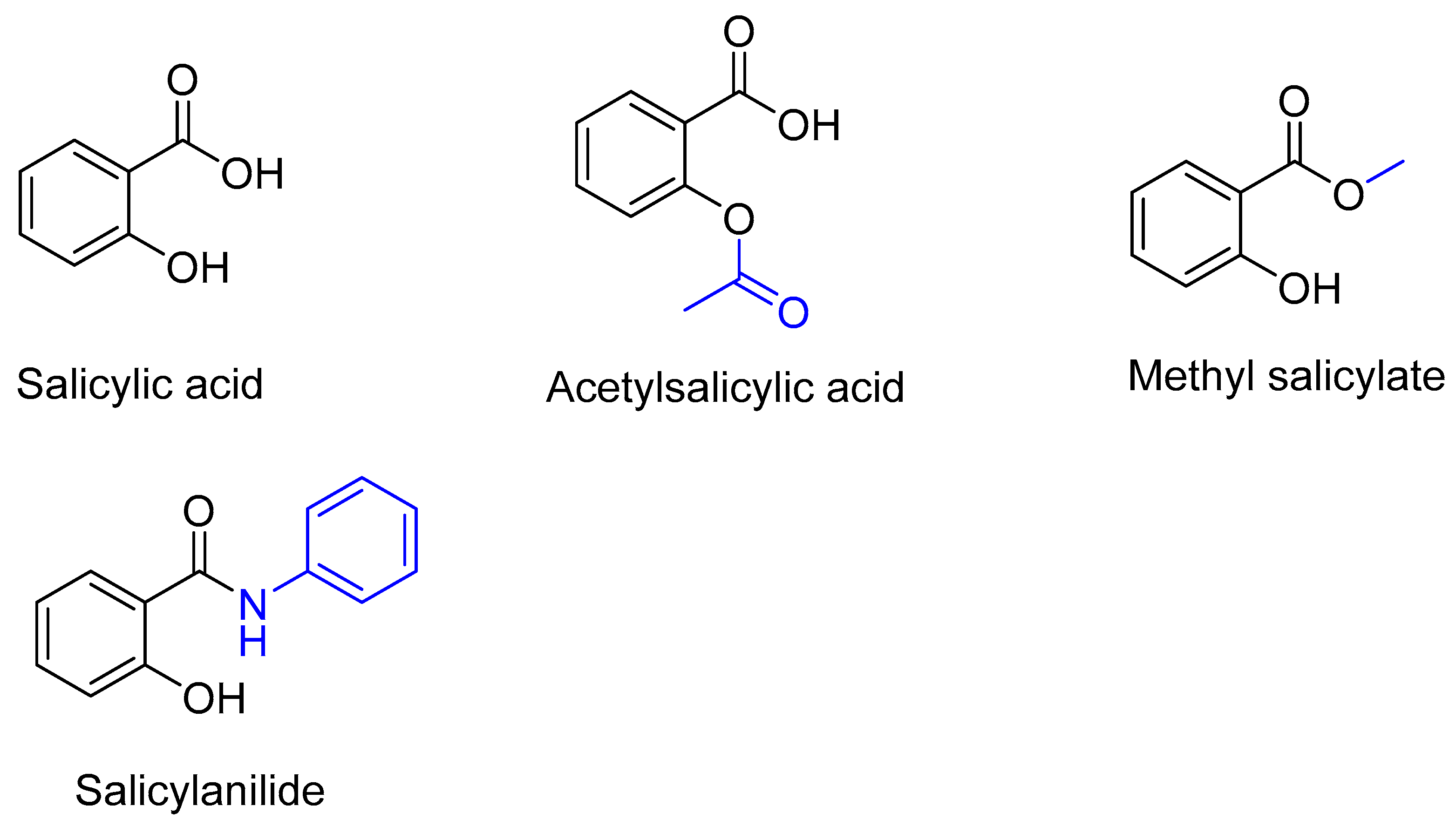
Figure 1. Salicylic acid and derivatives with antifungal activity.
Chemistry 07 00151 g001
Scheme 1. Preparation of compounds 125. (a) R1OH, H2SO4 (cat.), reflux; (b) R1OH, diisopropyl azodicarboxylate (DIAD), TPP, THF, 0 °C to r.t.; (c) I. SOCl2; reflux; II. RNH2, Et3N, CH2Cl2, 0 °C to r.t. (d) 1, acetone, K2CO3, C3H7Br, reflux; (e) 3, NaOH (10%), C8H7ClO to r.t.
Chemistry 07 00151 sch001
Figure 2. Chemical structures of the compounds 125.
Chemistry 07 00151 g002
Figure 3. Workflow of the modeling studies.
Chemistry 07 00151 g003
Figure 4. Predicted free energies of binding of compound 15 to its potential biological targets.
Chemistry 07 00151 g004
Figure 5. Predicted binding modes of compound 15 to the allosteric site of IDP2 (a), CYP1 (b), and CEK1 (c). The ligand is represented as orange balls and sticks in the complexes’ pictures, while atoms are colored as: carbon tan, nitrogen blue, oxygen red and sulfur yellow. All atoms are represented in the interaction diagrams only for residues forming hydrogen bonds with the compound. The labeled residues are those interacting with the compound in at least 40% of the snapshots employed for MM-PBSA calculations.
Chemistry 07 00151 g005
Table 1. (ac). Minimum inhibitory concentration (MIC) and minimum fungicidal concentration (MFC) of compounds against Candida spp. MIC and MFC values are expressed in μg/mL and μM. MFC values are expressed in μM. (d). Minimum inhibitory concentration (MIC) and minimum fungicidal concentration (MFC) of compounds against Candida spp. expressed in μg/mL and/or μM.
(a)
CompoundsC. albicans  ATCC 90028C. albicans  CBS 5602
MIC
(μg/mL)
MIC
(μM)
MFCMFC/
MIC
MIC
(μg/mL)
MIC
(μM)
MFCMFC/
MIC
SA--------
1--------
2--------
3--------
4--------
5--------
6--------
7--------
8--------
9--------
10--------
11--------
12--------
13--------
Nystatin1.51.621.6211.51.621.621
Ketoconazole0.50.9400.940111.881.881
DMSO--------
(b)
CompoundsC. tropicalis CBS 94C. krusei CBS 573
MIC
(μg/mL)
MIC
(μM)
MIC
(μg/mL)
MIC
(μM)
MIC
(μg/mL)
MIC
(μM)
MIC
(μg/mL)
MIC
(μM)
SA--------
1--------
2--------
3--------
4--------
5--------
6--------
7--------
8--------
9--------
10--------
11--------
12--------
13--------
Nystatin1.51.621.51.621.51.621.51.62
Ketoconazole47.5347.5347.5347.53
DMSO--------
(c)
CompoundsC. albicans
ATCC 90028
C. albicans
CBS 5602
MIC
(μg/mL)
MIC
(μM)
MFCMFC/
MIC
MIC
(μg/mL)
MIC
(μM)
MFCMFC/
MIC
142501293.731293.7312501293.731293.731
15125570.05570.051125570.05570.051
16----125550.03550.031
175002072.202072.2015002072.202072.201
18125485.83485.831125485.83485.831
19--------
20--------
21--------
2210004077.474077.4715002038.742038.741
235001910.581910.581----
24--------
25--------
Nystatin1.51.621.6211.51.621.621
Ketoconazole0.50.9400.940111.881.881
DMSO--------
(d)
CompoundsC. tropicalis  CBS 94C. krusei  CBS 573
MIC
(μg/mL)
MIC
(μM)
MFCMFC
/MIC
MIC
(μg/mL)
MIC
(μM)
MFCMFC
/MIC
145002587.462587.4612501293.731293.731
152501140.101140.101125570.05570.051
16----2501100.061100.061
17----5002072.202072.201
185001943.331943.331125485.83485.831
19----5001740.281740.281
20--------
21--------
225002038.742038.7415002038.742038.741
23----5001910.581910.581
24--------
25--------
Nystatin1.51.621.6211.51.621.621
Ketoconazole47.537.5310.50.9400.9401
DMSO--------
Table 2. The effect of exogenous ergosterol (1.008 mM) on the MIC of 15 and nystatin.
C. albicans ATCC 90028
15Nistatin
Concentration (μM)Without ErgosterolWith ErgosterolConcentration (μM)Without ErgosterolWith Ergosterol
912052
456026+
228013+
11406.5+
5703.2+
290++1.6+
140++0.8++
70++0.4++
Note: +, fungal growth; −, no fungal growth.
Table 3. MIC values of 15 and caspofungin in the absence and presence of sorbitol (0.8 M) against strains of C. albicans ATCC 90028. Values are expressed in μM.
C. albicans ATCC 90028
15Caspofungin
Concentration (μM)Without SorbitolWith SorbitolConcentration (μM)Without SorbitolWith Sorbitol
91203.6
45601.8
22800.9+
11400.45+
5700.228+
290++0.114++
140++0.056++
70++0.028++
Note: +, fungal growth; −, no fungal growth.
Table 4. Potential biological targets of compound 15 in C. albicans.
UniProt AccessionIDDescription
A0A1D8PHU1TPK2cAMP-dependent protein kinase
P43063CDK1Cyclin-dependent kinase 1
A0A1D8PDA6PHO85Cyclin-dependent serine/threonine-protein kinase
Q5A1D3CEK1Extracellular signal-regulated kinase 1
Q9URB4FBA1Fructose-bisphosphate aldolase
A0A1D8PS79IDP2Isocitrate dehydrogenase
A0A1D8PHH7IDP1Isocitrate dehydrogenase
Q59LF9MAP2Methionine aminopeptidase 2
Q5ALM6CPR3Peptidyl-prolyl cis-trans isomerase
P22011CYP1Peptidyl-prolyl cis-trans isomerase
A0A8H6F4I1CYPBPeptidyl-prolyl cis-trans isomerase
Q59U59APR1Proteinase A
A0A1D8PU61FDH3S-(hydroxymethyl)glutathione dehydrogenase
A0A1D8PSJ8PPH3Serine/threonine-protein phosphatase
Q92207HOG1Mitogen-activated protein kinase HOG1
Disclaimer/Publisher’s Note: The statements, opinions and data contained in all publications are solely those of the individual author(s) and contributor(s) and not of MDPI and/or the editor(s). MDPI and/or the editor(s) disclaim responsibility for any injury to people or property resulting from any ideas, methods, instructions or products referred to in the content.