Article

Synthesis and Characterization of a New Hydrogen-Bond-Stabilized 1,10-Phenanthroline–Phenol Schiff Base: Integrated Spectroscopic, Electrochemical, Theoretical Studies, and Antimicrobial Evaluation

by
Alexander Carreño
1,2,*,
Evys Ancede-Gallardo
1,
Ana G. Suárez
1,3,
Marjorie Cepeda-Plaza
2,
Mario Duque-Noreña
2,
Roxana Arce
2,4,
Manuel Gacitúa
5,6,7,
Roberto Lavín
5,6,7,
Osvaldo Inostroza
8,
Fernando Gil
8,9,
Ignacio Fuentes
10,11,12 and
Juan A. Fuentes
10,12,*
1
Laboratory of Organometallic Synthesis (CANS), Departamento de Ciencias Químicas, Facultad de Ciencias Exactas, Universidad Andres Bello, República 330, Santiago 8370186, Chile
2
Departamento de Ciencias Químicas, Facultad de Ciencias Exactas, Universidad Andres Bello, Av. República 275, Santiago 8370146, Chile
3
Doctorado en Físicoquímica Molecular, Facultad de Ciencias Exactas, Universidad Andres Bello, Av. República 275, Santiago 8370146, Chile
4
Millennium Institute on Green Ammonia as Energy Vector (MIGA), Av. Vicuña Mackenna 4860, Macul, Santiago 7820436, Chile
5
Facultad de Ingeniería y Ciencias, Universidad Diego Portales, Ejercito 441, Santiago 8370191, Chile
6
Center for the Development of Nanoscience and Nanotechnology, CEDENNA, Avenida Alameda Libertador Bernardo O’Higgins 3363, Santiago 9170124, Chile
7
Centro de Investigación en Nanomateriales y Medio Ambiente, CINAMA, Universidad Diego Portales, Av. Ejército Libertador 441, Santiago 8370191, Chile
8
Microbiota-Host Interactions & Clostridia Research Group, Center for Biomedical Research and Innovation (CIIB), Universidad de los Andes, Santiago 7620001, Chile
9
School of Medicine, Faculty of Medicine, Universidad de los Andes, Santiago 7620001, Chile
10
Laboratorio de Genética y Patogénesis Bacteriana, Facultad de Ciencias de la Vida, Universidad Andres Bello, Santiago 8370186, Chile
11
Doctorado en Biotecnología, Facultad de Ciencias de la Vida, Universidad Andres Bello, Santiago 8370186, Chile
12
Centro de Investigación de Resiliencia a Pandemias, Facultad de Ciencias de la Vida e Instituto de Salud Pública, Universidad Andres Bello, Santiago 8370186, Chile
*
Authors to whom correspondence should be addressed.
Chemistry 2025, 7(4), 135; https://doi.org/10.3390/chemistry7040135
Submission received: 30 May 2025 / Revised: 8 August 2025 / Accepted: 12 August 2025 / Published: 21 August 2025

Abstract

A new Schiff base, (E)-2-(((1,10-phenanthrolin-5-yl)imino)methyl)-4,6-di-tert-butylphenol (Fen-IHB), was designed to incorporate an intramolecular hydrogen bond (IHB) between the phenolic OH and the azomethine nitrogen with the goal of modulating its physicochemical and biological properties. Fen-IHB was synthesized by condensation of 5-amino-1,10-phenanthroline with 3,5-di-tert-butyl-2-hydroxybenzaldehyde and exhaustively characterized by HR-ESI-MS, FTIR, 1D/2D NMR (1H, 13C, DEPT-45, HH-COSY, CH-COSY, D2O exchange), and UV–Vis spectroscopy. Cyclic voltammetry in anhydrous CH3CN revealed a single irreversible cathodic peak at −1.43 V (vs. Ag/Ag+), which is consistent with the intramolecular reductive coupling of the azomethine moiety. Density functional theory (DFT) calculations, including MEP mapping, Fukui functions, dual descriptor analysis, and Fukui potentials with dual descriptor potential, identified the exocyclic azomethine carbon as the principal nucleophilic site and the phenolic ring (hydroxyl oxygen and adjacent carbons) as the main electrophilic region. Noncovalent interaction (NCI) analysis further confirmed the strength and geometry of the intramolecular hydrogen bond (IHB). In vitro antimicrobial assays indicated that Fen-IHB was inactive against Gram-negative facultative anaerobes (Salmonella enterica serovar Typhimurium and Typhi, Escherichia coli) and strictly anaerobic Gram-positive species (Clostridioides difficile, Roseburia inulinivorans, Blautia coccoides), as any growth inhibition was indistinguishable from the DMSO control. Conversely, Fen-IHB displayed measurable activity against Gram-positive aerobes and aerotolerant anaerobes, including Bacillus subtilis, Streptococcus pyogenes, Enterococcus faecalis, Staphylococcus aureus, and Staphylococcus haemolyticus. Overall, these comprehensive characterization results confirm the distinctive chemical and electronic properties of Fen-IHB, underlining the crucial role of the intramolecular hydrogen bond and electronic descriptors in defining its reactivity profile and selective biological activity.

1. Introduction

Schiff bases are structurally diverse organic compounds containing an imine (–C=N–) group, typically formed by the condensation of primary amines with aldehydes or ketones [1]. The azomethine linkage acts as a versatile reactive center, facilitating diverse chemical transformations relevant to catalysis, materials science, and medicinal chemistry. Schiff bases exhibit structural flexibility due to their ability to incorporate various alkyl and aryl groups, allowing fine-tuning of their electronic and steric properties. Additionally, derivatives with hydrazide groups display amide-iminol tautomerism, contributing further electronic complexity and functional diversity [2].
In catalysis, Schiff bases function effectively as ligands in metal-mediated oxidation and asymmetric reactions by stabilizing intermediates and regulating electron distribution [3,4,5,6]. In biomedical applications, their antimicrobial, antifungal, and anticancer properties highlight their promise as candidates for therapeutic development [3,7].
The functional properties of Schiff bases are strongly influenced by the type and position of substituents on their aromatic rings, which affect electronic distribution, hydrogen bonding, redox behavior, and molecular geometry. Strategically designed intramolecular hydrogen bonds (IHB) offer a valuable approach to control conformation and responsiveness to external stimuli [8,9,10]. IHBs can significantly influence the photophysical behavior of Schiff bases by enhancing emission, conformational rigidity, and chemical stability. In certain systems, hydrogen bonding acts as a dynamic switch, modulating polarity and proton transfer, which are features valuable for optoelectronic and photoresponsive applications. Moreover, IHBs contribute to improved biological activity and stability, reinforcing the therapeutic potential of these compounds [8,11].
In this context, 1,10-phenanthroline stands out as a valuable scaffold due to its planar structure, aromaticity, and ability to engage in π–π interactions and redox tuning [12,13]. Its incorporation into Schiff base systems, particularly those with phenolic units capable of forming IHBs, expands their electronic and bioactive potential, making phenol-imine derivatives with π-conjugated frameworks promising candidates for further exploration [14,15].
The present study reports the design, synthesis, and characterization of a novel phenol-based Schiff base, (E)-2-(((1,10-phenanthrolin-5-yl)imino)methyl)-4,6-di-tert-butylphenol, herein referred to as Fen-IHB (Figure 1). This compound was designed to promote a stable intramolecular hydrogen bond between the phenolic –OH group and the azomethine nitrogen, thereby establishing a quasi-six-membered ring that enforces molecular planarity and facilitates internal electronic polarization. This interaction promotes molecular planarity and internal electronic polarization, leading to modified redox behavior, optical properties, and reactivity [16].
To investigate these properties, Fen-IHB was synthesized and subjected to extensive structural and spectroscopic characterization, including high-resolution mass spectrometry (HRMS), FTIR, UV–Vis spectroscopy, and 1H/13C NMR (with DEPT-45, HH-COSY, CH-COSY, and D2O exchange). Electrochemical properties were probed via cyclic voltammetry, and electronic structure and reactivity descriptors were further explored through quantum chemical calculations using density functional theory (DFT) as implemented in Gaussian 16, which employed the long-range corrected ωB97X-D functional in combination with the 6-31+G(d) basis set, including geometry optimization, time-dependent density functional theory (TD-DFT), molecular electrostatic potential (MEP), Fukui functions, dual descriptor (DD) mapping, dual descriptor potential (DDP), and noncovalent interaction (NCI) analysis. Finally, to evaluate its bioactivity, Fen-IHB was tested against a panel of aerobic and anaerobic microorganisms.

2. Results and Discussion

2.1. Synthesis and Structural Features of Fen-IHB

The synthesis of (E)-2-(((1,10-phenanthrolin-5-yl)imino)methyl)-4,6-di-tert-butylphenol (Fen-IHB) was accomplished through a straightforward condensation reaction between equimolar amounts of 5-amino-1,10-phenanthroline and 3,5-di-tert-butyl-2-hydroxybenzaldehyde, following a slightly modified protocol based on previously reported methodologies [17]. The chemical structure of Fen-IHB is shown in Figure 1. This compound belongs to the class of ortho-hydroxyaryl Schiff bases, characterized by the presence of a hydroxyl group in the ortho-position relative to the imine moiety, which promotes the formation of a stable intramolecular hydrogen bond (IHB) [18,19]. The existence of phenol-imine to keto-amine tautomerism (O–H···N ⇌ O···H–N) has been proposed, with its occurrence strongly dependent on the formation of intramolecular hydrogen bonds [18,20]. These tautomeric forms have been previously reported in salicylaldimine-type Schiff base derivatives and are known to be influenced by solvent polarity [20,21,22].
Prior studies indicate that these structural arrangements favor the enol-imine tautomer and the thermodynamically stable E-configuration, as supported by experimental data. Our computational analysis using Gaussian 16 (ωB97X-D/6-31+G(d)) further confirms that the E-configuration of Fen-IHB is the most energetically favorable (see Section 2.5). Deviations between experimental and theoretical vibrational modes are attributed to phase differences, with gas-phase calculations lacking the intermolecular interactions present in condensed phases [18,23]. Deviations observed between experimental and theoretical vibrational modes in FTIR analyses are attributed to phase differences, as gas-phase calculations lack the intermolecular interactions present in condensed phases [24,25]. These aspects will be discussed in more detail below.
The molecular architecture of Fen-IHB integrates two nitrogen donor atoms from the 1,10-phenanthroline ring, offering potential coordination sites for metal ions. The inclusion of sterically demanding tert-butyl substituents at the 4- and 6-positions of the phenolic ring imparts hydrophobic character and introduces steric hindrance, which may influence both the solubility profile and the interaction with biomolecular or coordination environments. These features collectively suggest potential applications of Fen-IHB in materials science and supramolecular chemistry [26,27,28,29].
Due to the limited solubility of 5-amino-1,10-phenanthroline in methanol, the reaction was initiated by dissolving this reagent in methanol with the addition of a few drops of dichloromethane to facilitate solubilization. The subsequent addition of the aldehyde yielded a clear, yellow solution. Upon standing at room temperature, a yellow precipitate formed, which was filtered, washed, and recrystallized from hot methanol. Slow cooling of the filtrate resulted in the isolation of Fen-IHB as a crystalline solid with a yield exceeding 70%, highlighting the efficiency of the synthetic route. The final product displayed moderate solubility in dimethyl sulfoxide (DMSO), acetonitrile, and methanol; meanwhile, the solubility of Fen-IHB was low in dichloromethane at room temperature. The physicochemical parameters, including molecular weight, yield, and solid-state color, are summarized in Table S1. The compound exhibited a melting point in the range of 114–116 °C prior to decomposition.

2.2. High-Resolution Mass Spectrometry, FTIR, UV–Vis Spectroscopy, and NMR Analysis

The molecular identity of Fen-IHB was confirmed by high-resolution electrospray ionization mass spectrometry (HR-ESI-MS) performed in positive ionization mode. A single, well-defined molecular ion peak was observed at m/z 412.2390, corresponding to the [M + H]+ species (Figure S1). This result is in complete agreement with the expected monoprotonated form of the synthesized Schiff base, supporting the successful formation of the target compound.
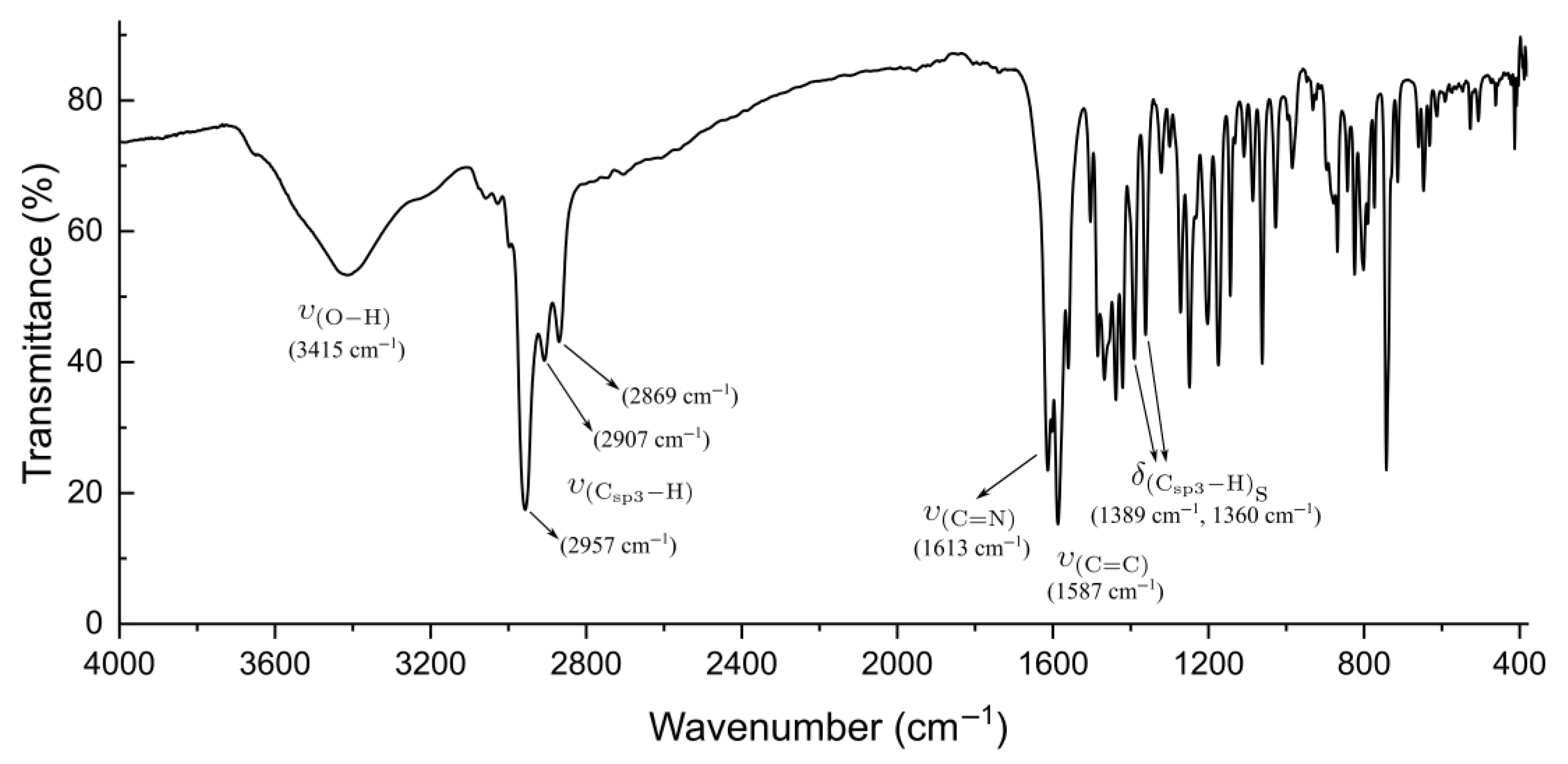
FTIR spectroscopy (KBr pellet, Figure 2) provided additional structural evidence supporting the formation of intramolecular hydrogen bonding in Fen-IHB. As expected for salicyl-derived Schiff bases, the strength and presence of O–H···N intramolecular interactions can be inferred from diagnostic shifts in the stretching frequencies of both the phenolic O–H and the azomethine C=N bonds. In particular, the observed red shift of the O–H stretch and the characteristic position of the C=N band are consistent with literature reports for related systems bearing strong intramolecular hydrogen bonds [30,31].
A broad O–H stretching band at 3415 cm−1, significantly red-shifted from the typical free phenolic O–H (~3600 cm−1), indicates strong intramolecular hydrogen bonding (IHB) with the azomethine nitrogen, which is consistent with literature reports [8,17,32,33]. Additional key bands include aliphatic C–H stretches at 2957, 2907, and 2869 cm−1, aromatic C=C at 1587 cm−1, and a sharp C=N stretch at 1613 cm−1, which is characteristic of Schiff bases with IHB motifs. Methyl bending vibrations appeared at 1389 and 1360 cm [8,17,32,33]. Additional key bands include aliphatic C–H stretches at 2957, 2907, and 2869 cm−1, aromatic C=C at 1587 cm−1, and a sharp C=N stretch at 1613 cm−1, which is characteristic of Schiff bases with IHB motifs. Methyl bending vibrations appeared at 1389 and 1360 cm−1 [18,34].
These vibrational features align with quantum chemical predictions (Section 2.4), confirming the presence of all functional groups and highlighting the structural and electronic role of the IHB. Overall, the FTIR data support the successful synthesis and structural integrity of Fen-IHB.
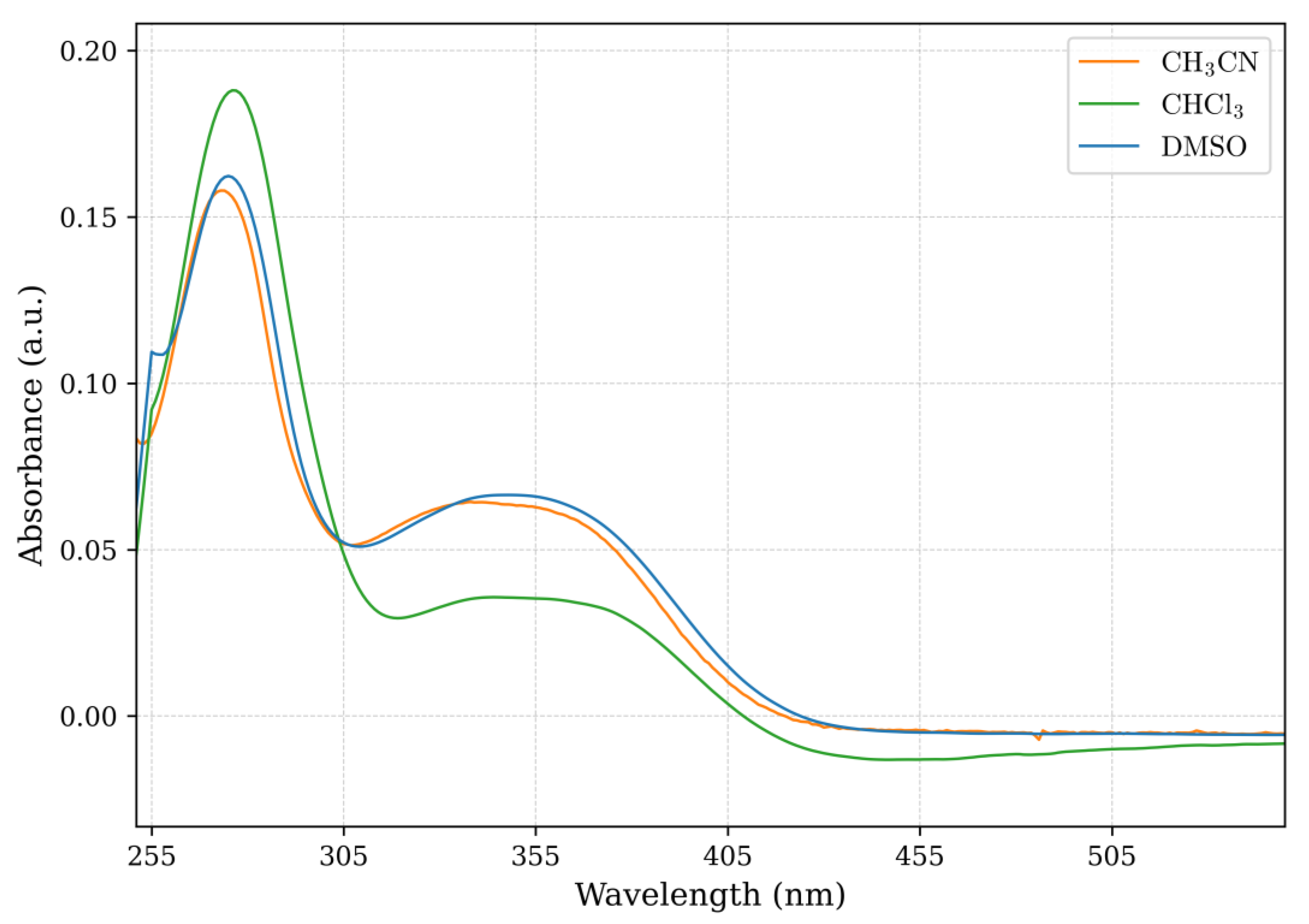
To evaluate the optical properties of Fen-IHB, UV–Vis absorption spectra were recorded in aerated solutions using three solvents of differing polarity: chloroform (ε = 4.81), acetonitrile (ε = 37.5), and dimethyl sulfoxide (DMSO; ε = 46.7), at room temperature. Figure 3 presents the UV–Vis absorption spectra of Fen-IHB in the three solvents analyzed. In all cases, two well-defined absorption bands are observed. The high-energy band, centered around 273 nm in acetonitrile, is attributed to a localized π → π* transition associated with the aromatic and conjugated segments of the molecule [35,36,37]. The lower-energy band, appearing near 340 nm, corresponds to a mixed π → π* and n → π* transition involving the azomethine moiety and the extended conjugated system [38,39,40]. Table 1 summarizes the main spectroscopic features of Fen-IHB in each solvent. Notably, the low-energy band exhibits consistently lower molar absorptivity across all solvents, suggesting a more delocalized electronic transition modulated by intramolecular hydrogen bonding (IHB) [41]. In DMSO, a polar aprotic solvent capable of strong dipole–dipole interactions, a modest red shift is observed relative to less polar media. This bathochromic shift likely reflects stabilization of the excited state through solvent–solute interactions that influence the electronic distribution within the conjugated framework [42,43].
To further complement these experimental assignments, time-dependent density functional theory (TD-DFT) calculations were performed in DMSO as solvent (Section 2.5). A HOMO → LUMO transition was identified for the absorption band near 345 nm. Notably, the lowest-energy transition, which exhibits the weakest molar absorptivity, aligns with the region of the molecule involved in intramolecular hydrogen bonding. This suggests that the IHB contributes to the stabilization of the ground state and the reorganization of electron density during excitation, thereby influencing the optical signature of the compound. An extended discussion on the results from TD-DFT calculations and the assignment of bands is provided in Section 2.5. The UV–Vis spectra of Fen-IHB are consistent with those reported for structurally related Schiff bases incorporating 1,10-phenanthroline moieties [44,45]. These comparisons support our spectroscopic assignments and underscore the structural role of the phenanthroline fragment in modulating the optical properties.
To further elucidate the structural features of Fen-IHB, 1H NMR spectroscopy (400 MHz, DMSO-d6, 25 °C) was performed, confirming the expected chemical structure (see Supplementary Figure S2 for arbitrary proton numbers and Figures S3–S5 for complete spectral data). A broad singlet at δ 13.60 ppm was assigned to the phenolic –OH proton, a chemical shift characteristic of strong intramolecular O–H···N hydrogen bonding, as verified by D2O exchange. This finding is consistent with previous reports on salicylic acid-derived Schiff bases bearing similar intramolecular interactions. The 1H NMR spectra in both deuterated DMSO and acetonitrile indicate that the compound predominantly adopts the phenol-imine tautomeric form in solution (Figures S6 and S7). This tautomeric preference is supported by the FTIR spectrum, which displays a red-shifted O–H stretching vibration at 3415 cm−1 (Figure 2), further corroborating the presence of a stable intramolecular hydrogen bond. These observations align with those reported for related Schiff base systems [18].
To validate that the phenol–imine (enol–imine) form represents the most thermodynamically stable tautomer, we performed a comparative stabilization energy analysis of the enol and keto–imine forms using DFT methods, as detailed in Section 2.5.
Continuing the characterization in deuterated DMSO, the azomethine (–CH=N–) proton appears as a sharp singlet at δ 9.20 ppm, a value consistent with classical chemical shifts reported for Schiff base systems. The aromatic region (δ 7.5–9.2 ppm) exhibits well-resolved multiplets corresponding to the protons of the 1,10-phenanthroline and substituted phenol rings. The observed chemical shifts in this region reflect the electronic effects of π-conjugation and the deshielding influence exerted by the adjacent imine functionality [46].
The 1H NMR spectrum of Fen-IHB revealed additional aromatic signals at δ 7.93, 7.92, and 7.79 ppm, consistent with the diastereotopic proton environments of the 1,10-phenanthroline moiety. Signals at δ 7.63 and 7.50 ppm were assigned to aromatic protons of the substituted phenol ring, with assignments confirmed via 2D HH-COSY analysis (Figure S8). In the aliphatic region, two singlets at δ 1.48 and 1.24 ppm were observed, corresponding to the methyl groups of the tert-butyl substituents at the 4- and 6-positions of the phenol ring.
The 13C NMR spectrum (100 MHz, DMSO-d6, 25 °C; Figure S9) exhibited well-resolved signals consistent with the 26 carbon atoms present in the molecule, accounting for chemical equivalence and signal overlap. The azomethine carbon was clearly identified at δ 168.06 ppm, while the phenolic quaternary carbon appeared at δ 157.03 ppm, both values aligning with those reported for structurally related Schiff bases [46,47]. Aromatic carbon signals were distributed between δ 110 and 151 ppm, with partial overlap noted at δ 128.66 and 124.42 ppm. These assignments were supported by DEPT-45 and CH-COSY spectra (Figures S10 and S11).
Signals corresponding to the methyl carbons of the tert-butyl groups were found at δ 29.67 and 31.52 ppm, while quaternary and bridging carbons of the aliphatic framework were observed between δ 34 and 41 ppm. In total, ten tertiary and nine quaternary carbon environments were resolved, in agreement with the molecular symmetry and substitution pattern of Fen-IHB. A complete summary of chemical shifts and NMR correlations is provided in Table S3.
As noted previously, salicylidene Schiff base compounds with aromatic substituents flanking opposite sides of the C=N bond typically adopt the thermodynamically favored E-configuration. In agreement with this, the Schiff bases derived from related salicylidene are in the E-configuration about the C=N double bond and are found to exist in this form, as revealed by X-ray crystallographic analysis. This configuration corresponds to the spatial arrangement in which the substituents on the carbon and nitrogen atoms of the azomethine group are positioned on opposite sides of the double bond [48,49,50].
In the case of Fen-IHB, this configuration was supported by comprehensive spectroscopic evidence, including 1H and 13C NMR, DEPT-45, HH-COSY, CH-COSY, FTIR, UV–Vis, and high-resolution mass spectrometry. Collectively, these data confirm the proposed molecular structure and indicate a high degree of chemical purity. Furthermore, they support the conclusion that the tautomeric equilibrium strongly favors the phenol–imine form, which is an observation that is further explored in Section 2.5 through DFT-based computational analysis.

2.3. Cyclic Voltammetry

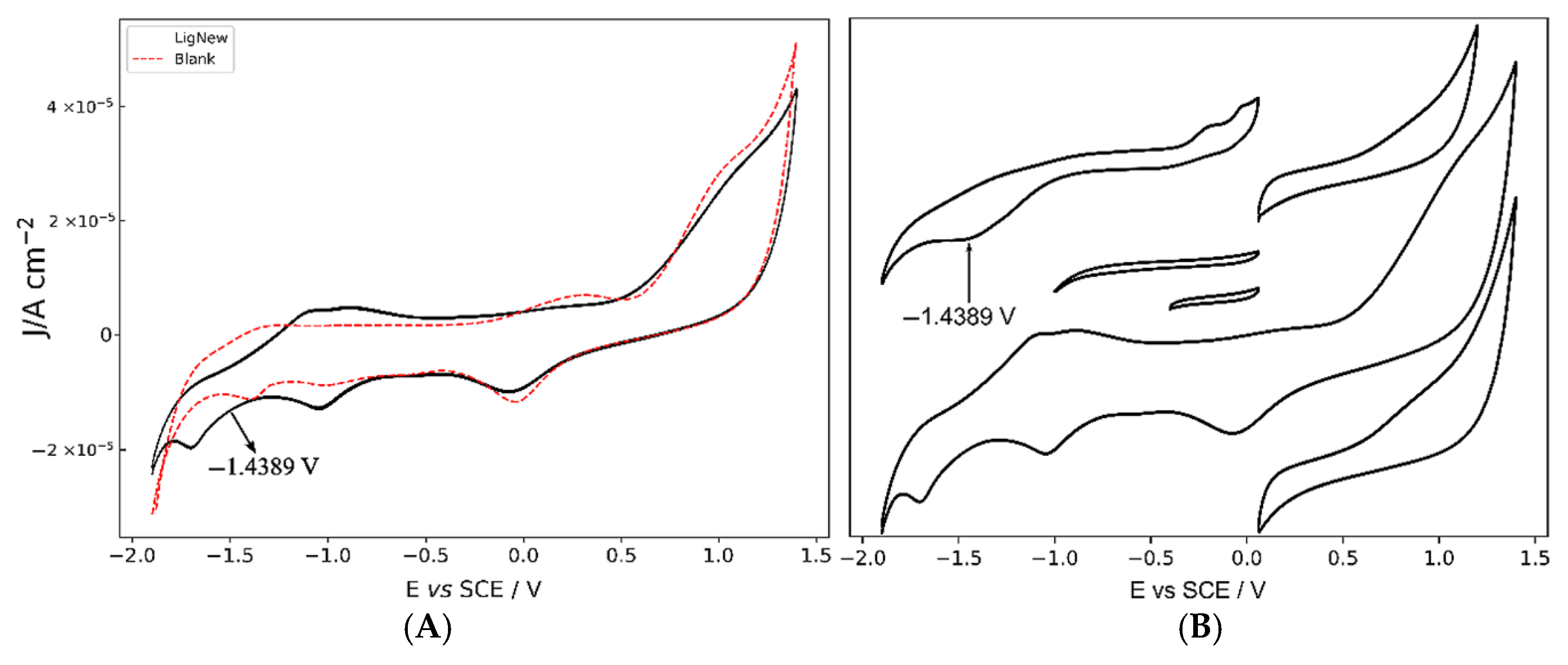
The redox properties of Fen-IHB were probed by cyclic voltammetry in anhydrous acetonitrile containing 0.10 M tetrabutylammonium hexafluorophosphate (TBAPF6) as the supporting electrolyte, using a platinum working electrode and an argon atmosphere to exclude oxygen (Figure 4). A blank voltammogram of the supporting electrolyte alone exhibited only capacitive currents and no faradaic features. In contrast, the voltammogram of Fen-IHB showed (1) no oxidative waves within the solvent window, which is consistent with the absence of easily oxidizable –NH2 groups and in agreement with related non-metallic Schiff bases [51], and (2) a single irreversible cathodic peak at −1.43 V (vs. Ag/AgCl), which we assign to the reductive coupling of the azomethine (–C=N–) unit.
To rule out contributions from solvent or electrolyte reduction, a working-window study was carried out by varying the lower potential limit (Figure 4B). The irreversible wave at −1.43 V (vs. Ag/AgCl) persisted across all windows, confirming its origin in Fen-IHB rather than in acetonitrile or TBAPF6. Typically, these irreversible processes are attributed to an intramolecular reductive coupling of the azomethine group, involving a self-protonation reaction, as previously described for other related Schiff bases [52]. This irreversible reduction wave suggests a chemical transformation following electron uptake, likely influenced by the presence of the intramolecular hydrogen bond.
Frontier molecular orbital (FMO) analysis from DFT calculations (see below) supports this assignment: the highest occupied molecular orbital (HOMO) is predominantly localized on the phenolic ring, explaining the lack of oxidation, whereas the lowest unoccupied molecular orbital (LUMO) is centered on the azomethine linkage, identifying it as the site of electron uptake. The pronounced intramolecular hydrogen bond likely stabilizes the protonated intermediate generated during reduction, shifting the process into an irreversible regime.
Together with spectroscopic (HRMS, 1D and 2D NMR, FTIR, UV–Vis) and computational data (see below), these electrochemical results not only confirm the structural integrity of Fen-IHB but also demonstrate its distinct redox activity, which may inform future design of organic materials with tailored electron-transfer properties.

2.4. Quantum Chemistry: Geometry Optimizations, TD-DFT, Reactivity Descriptors, and Noncovalent Interaction (NCI) Index

We were unable to obtain a suitable crystal of Fen-IHB for X-ray diffraction analysis. However, based on X-ray structures of Schiff base derivatives of related salicylidene that exhibit an intramolecular hydrogen bond [53], we performed DFT calculations for geometry optimization and subsequent computational analyses.
In this study, the geometry of the Fen-IHB molecule was optimized using density functional theory (DFT) as implemented in Gaussian 16. The optimization employed the long-range corrected ωB97X-D functional in combination with the 6-31+G(d) basis set and the polarizable continuum model (PCM) to simulate DMSO as the solvent [54,55]. Subsequent single-point energy and vibrational frequency calculations were performed at the same level of theory to confirm the stability of the optimized geometry and to support spectroscopic interpretations. Given the lack of suitable single crystals for X-ray diffraction, we employed DFT-based computational analysis to gain insight into the molecular structure. This approach has been widely validated for reproducing structural features (including tautomeric preferences, planarity, and intramolecular interactions) in systems where crystallographic data are unavailable [56,57].
To support our experimental FTIR assignments and obtain detailed insight into the vibrational characteristics of Fen-IHB, we performed quantum chemical frequency calculations at the ωB97X-D/6-31+G(d) level using the PCM solvation model for DMSO [58,59]. The geometry optimization confirmed that the E-isomer—stabilized by a quasi-six-membered intramolecular O–H···N hydrogen bond—corresponds to the global energy minimum, which is in agreement with similar Schiff bases [50,53]. Key bond lengths in the optimized structure, including C=N at 1.2865 Å, C–O at 1.3445 Å, and O–H at 0.9921 Å, are in close agreement with values reported for structurally related salicylidene Schiff bases [53,60,61] (see Figure S12 for atom labeling and Table S4). The calculated dihedral angle between the aromatic planes is approximately –43.11°, which is consistent with the literature-reported geometries for analogous Schiff bases that exhibit E-configuration and intramolecular hydrogen bonding [53]. In agreement with this, Schiff bases derived from salicyl are in the E-configuration around the C=N double bond and are found to exist predominantly in this form, as revealed by X-ray crystallographic analysis [53,62].
These structural parameters further corroborate the presence of a strong intramolecular O–H···N hydrogen bond and support the predominance of the enol–imine tautomeric form in solution. As documented in previous studies, salicylidene-based Schiff bases commonly adopt E-configured conformers stabilized by a six-membered intramolecular hydrogen-bonded ring. This structural motif significantly enhances thermodynamic stability by promoting planarity and extended conjugation within the molecular framework [30,53,63].
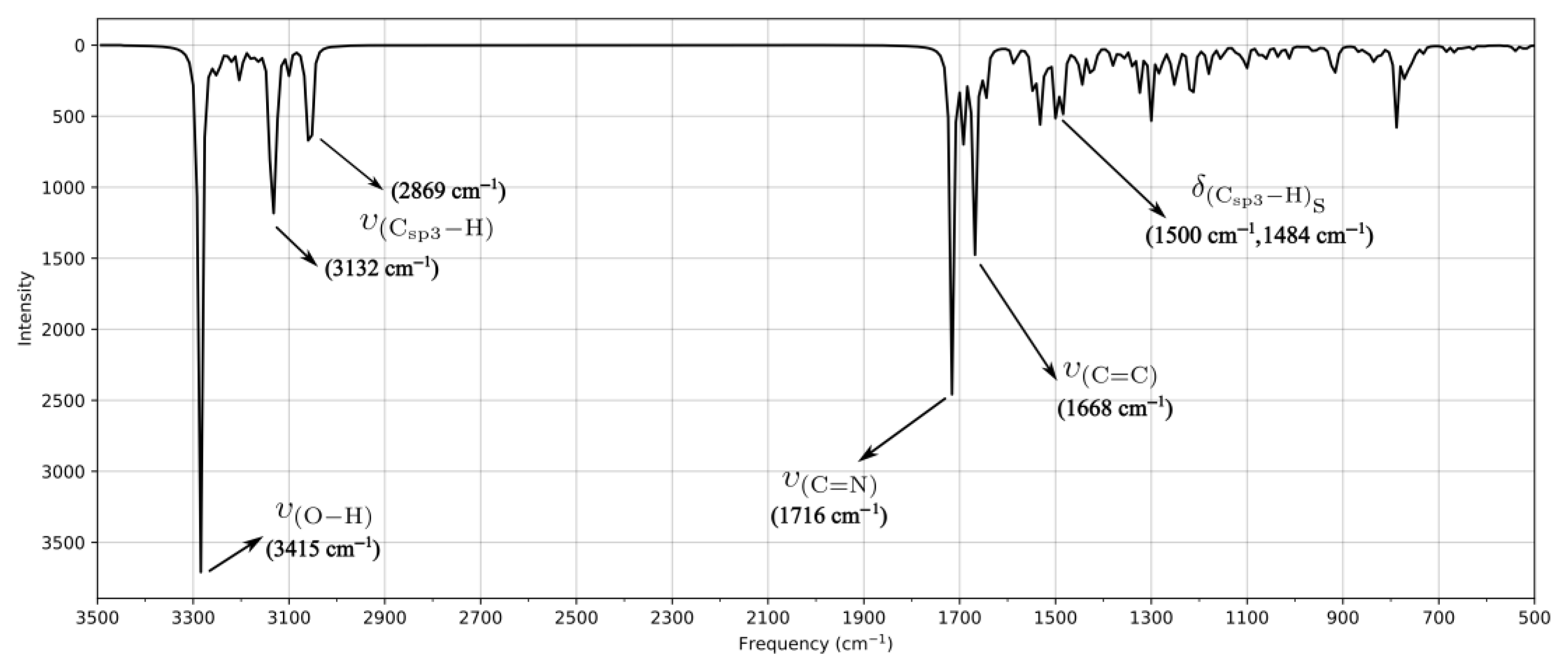
The DFT-computed FTIR spectrum of Fen-IHB (Figure 5) describes most of the main bands displayed in the experimental spectrum, allowing a reliable assignment of the vibrational modes involved in the observed spectral features. However, some differences are observed. Although deviations are generally below 50 cm−1, some bands display discrepancies around 100 cm−1. These differences could be attributed to the differences in phase conditions of each spectra. While the experimental data were obtained in the solid phase, the theoretical calculations were performed in the gas phase, where intermolecular interactions are absent. Importantly, the O–H stretching mode, calculated at 3415 cm−1, matches exactly with the band observed at 3415 cm−1. This agreement supports the presence of a strong intramolecular hydrogen bond and is consistent with the FTIR data (see Figure 2) and with previously reported salicylidene Schiff bases [22,30,64,65]. The calculated hydrogen-bond geometry of Fen-IHB was also in agreement with similar compounds, confirming the structural integrity of the model [65,66]. The IR-calculated bands for the tert-butyl substituents appear at 3132 and 2869 cm−1, in good agreement with the experimental absorptions between 2957 and 2869 cm−1. The azomethine C=N stretch is predicted at 1716 cm−1, which is deviated approximately 100 cm−1 from the observed peak at 1613 cm−1, while the aromatic C=C vibration is calculated at 1668 cm−1, which is consistent with the band at 1587 cm−1 in the FTIR spectrum. Therefore, although there are differences, the comparison between the computed and experimental FTIR spectra facilitates the assignment of the main vibrational modes, confirming the key bands associated with the Fen-IHB structure. In addition, the DFT-calculated results align well with those reported by other authors using the same level of theory, further supporting the proposed vibrational mode assignments and validating the reliability of our approach [67,68,69].
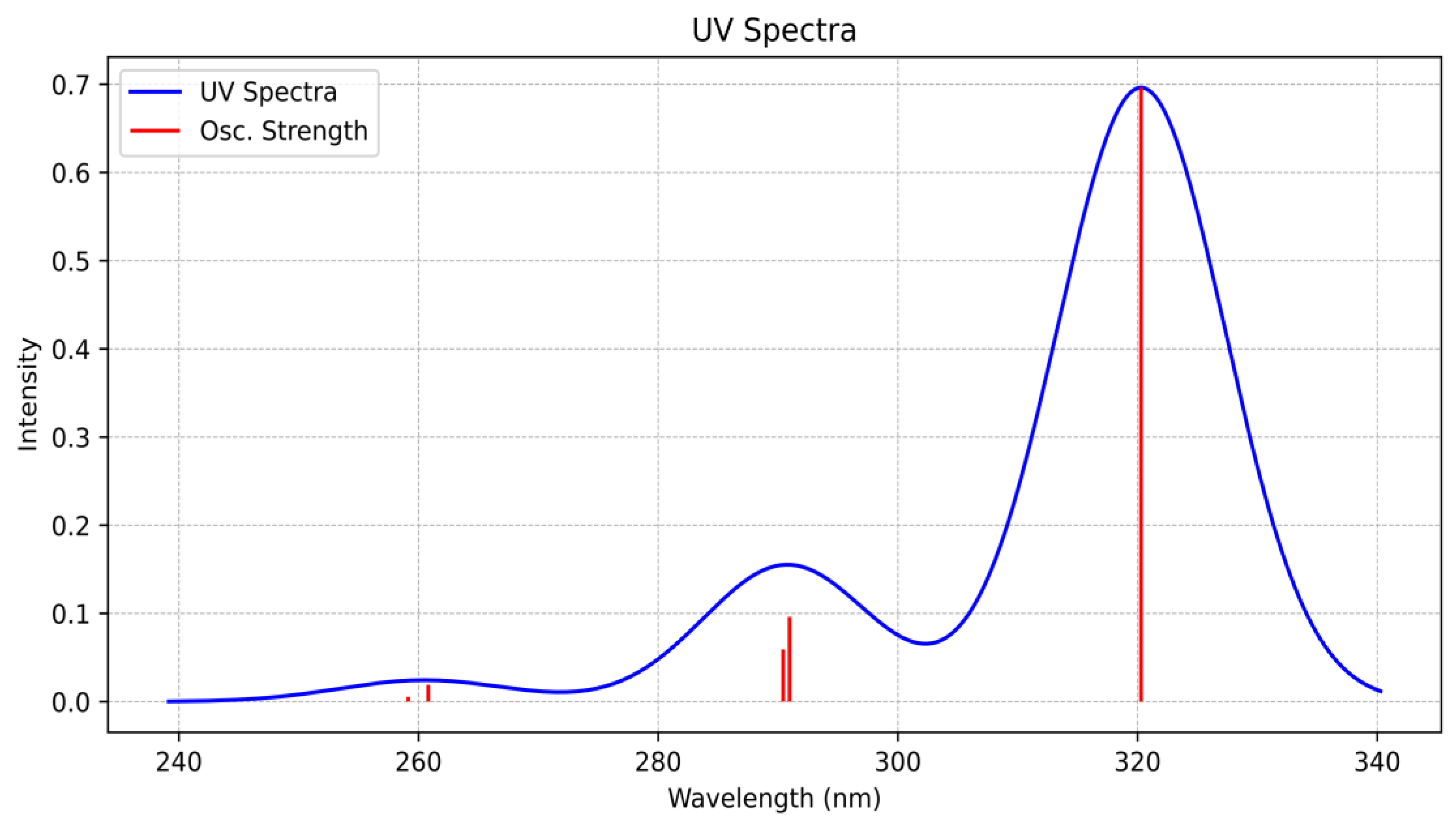
To rationalize the UV–Vis spectral features of Fen-IHB, the electronic absorption spectrum was simulated based on vertical excitation energies and oscillator strengths obtained from time-dependent density functional theory (TD-DFT) calculations performed with Gaussian 16, using the ωB97X-D functional and the 6-31+G(d) basis set [70,71]. Solvent effects were considered using the PCM model with DMSO as the implicit solvent. The simulated UV–Vis spectrum (Figure 6) closely matches the experimental data, enabling an unambiguous assignment of the electronic origins of the observed bands. While experimental results are obtained in solution where the bands are influenced by solvent interactions, the theoretical calculations are often performed in the gas phase, where such interactions are not accounted for. This distinction highlights the importance of including solvation models to improve the accuracy of computational predictions [72,73].
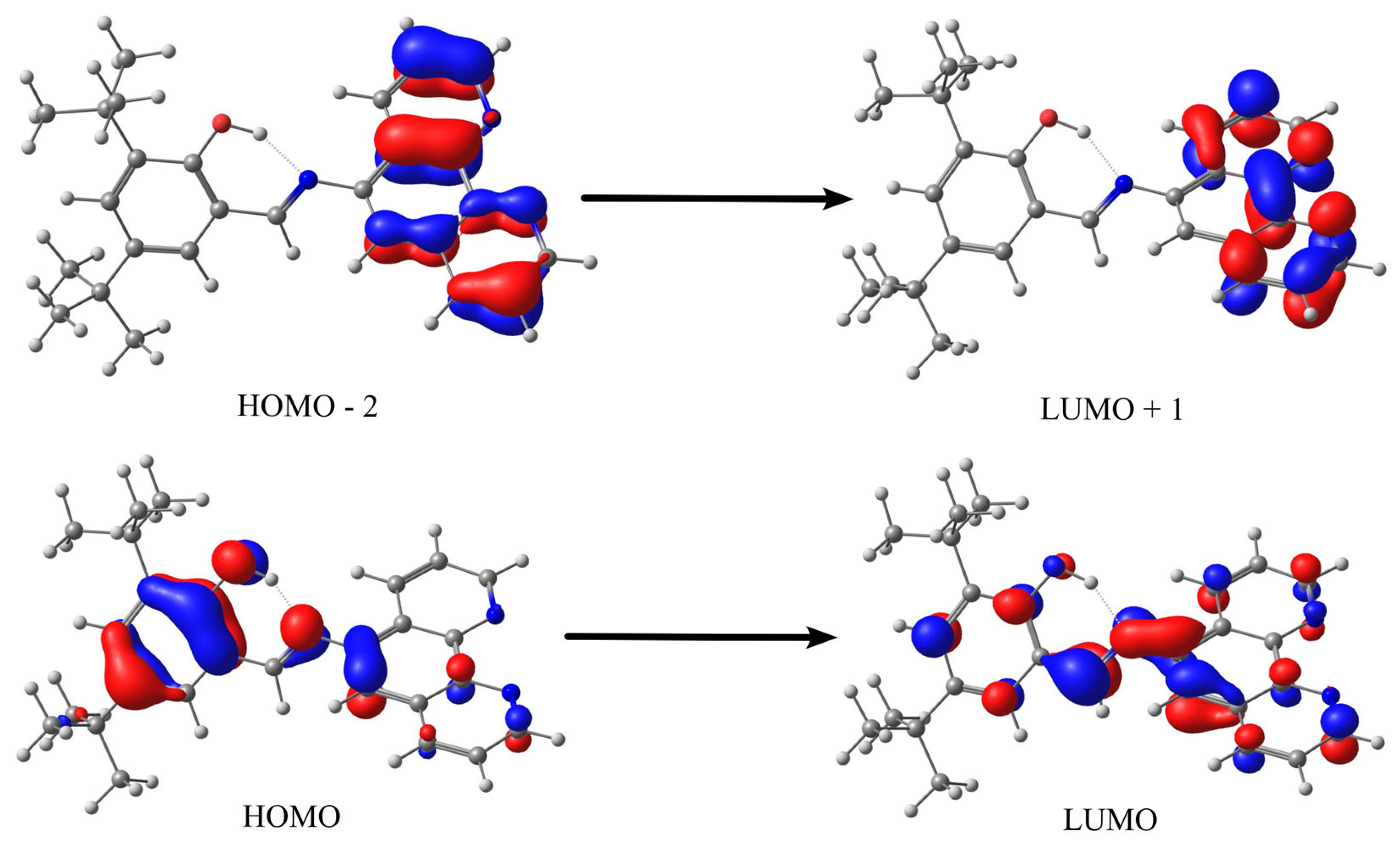
The high-energy band at 291 nm is dominated by a π → π* transition arising from the HOMO–2 → LUMO+1 excitation (oscillator strength f = 0.1). Orbital isosurface plots reveal that this transition is localized on the 1,10-phenanthroline moiety, reflecting the rigid aromatic framework. The lowest-energy band at 320.3 nm (f = 0.7) arises from the HOMO → LUMO excitation (Figure 7). In this transition, the HOMO is predominantly localized on the phenolic –OH and adjacent aromatic carbon involved in the intramolecular hydrogen bond, whereas the LUMO extends over the azomethine unit and 1,10-phenanthroline ring. This HOMO → LUMO excitation thus exhibits combined π → π* and n → π* characters, a signature of the extended conjugation and IHB stabilization [74,75]. The calculated wavelengths, oscillator strengths, and dominant orbital contributions are compiled in Table S6. This isosurface plots analysis supports the cyclic voltammetry assignments (see above).
To complement the experimental 1H NMR data obtained in deuterated DMSO and acetonitrile, which indicate that the enol–imine tautomer is predominant in solution, we performed a computational analysis to evaluate the relative stability of the enol and keto–imine forms. The calculations were conducted using Gaussian 16, employing the long-range corrected ωB97X-D functional with the 6-31+G(d) basis set, and incorporating DMSO as the solvent via the PCM model [76,77] (see Figure S13).
The computed energy difference between the two isomers was 13.27 kJ/mol, indicating that the phenol–imine tautomer is significantly more stable under the experimental conditions. This result strongly supports our NMR observations and aligns with previous reports on salicylidene Schiff bases, where the enol–imine form is generally favored in polar aprotic solvents due to intramolecular hydrogen bonding and electronic delocalization.

2.4.1. Comparative Analysis of DFT-Based Reactivity Descriptors

To understand the local reactivity in Fen-IHB, a hierarchical approach based on conceptual DFT was used, incorporating complementary descriptors [78,79,80,81,82,83]. To address this limitation and provide a more chemically meaningful view, Fukui functions were calculated using the finite-difference method, revealing local electron density changes upon electron addition or removal [84]. The f + Fukui function identifies regions where electron density increases upon the addition of an electron, corresponding to sites with enhanced nucleophilic susceptibility. Conversely, the f function maps electron depletion upon electron removal, indicating regions of electrophilic susceptibility. In Fen-IHB, the f + surface highlights the exocyclic carbon of the azomethine group, along with the ortho and para positions of the phenolic ring (relative to the imine carbon), as the most reactive nucleophilic centers. Meanwhile, the f + surface analysis suggests that the phenolic ring and azomethine group may exhibit electrophilic behavior due to easier electron density removal. However, this interpretation requires further validation, as it does not fully align with other reactivity descriptors. Detailed parameters are provided in Table S7. To synthesize the complementary insights from the f + and f functions, we evaluated the dual descriptor ( Δ f = f + f ) [85,86]. This unified descriptor generates a single reactivity surface, where positive values ( Δ f > 0 ) indicate nucleophilic sites and negative values ( Δ f < 0 ) correspond to electrophilic regions. The surface confirmed the azomethine carbon and phenolic ring as key nucleophilic sites. Unexpectedly, the phenolic oxygen appeared electrophilic, which is likely due to intramolecular hydrogen bonding reducing its electron-donating ability. Although phenanthroline nitrogens seem electron-rich in MEP maps, their low values suggest limited reactivity. Full isosurface parameters are listed in Table S7.
We computed the Fukui potentials ( f p + and f p ), which integrate both charge redistribution and electrostatic effects to depict how a test charge interacts with induced density changes [87]. The f p + potential confirmed the azomethine carbon and phenolic ring as preferred nucleophilic sites, while f p identified the phenolic oxygen as the dominant electrophilic center. In contrast, the phenanthroline region, though electrostatically negative and theoretically f p -active, exhibited low Fukui potential values, indicating weak practical reactivity. The discrepancy between MEP and Fukui-based descriptors underscores that electron density alone does not fully determine chemical reactivity. Instead, reactivity is better described by how a system responds to perturbations, which is more accurately captured by orbital- and energy-based descriptors (see Table S8 for threshold values). Finally, the dual descriptor potential [88] ( Δ f p = f p + f p ) identifies the azomethine carbon as the main nucleophilic site and the phenolic group as the primary electrophilic region. Despite initial indications from MEP, the phenanthroline ring showed low reactivity in orbital and potential-based analyses. This highlights the value of an integrated approach: MEP offers quick visualization, Fukui functions reveal orbital trends, and Fukui potentials validate predictions energetically. Together, these tools enable accurate identification of reactive centers, avoiding the limitations of relying on a single descriptor. Isosurface criteria are detailed in Table S8.

2.4.2. Noncovalent Interaction (NCI) Index

To further elucidate the role of noncovalent interactions in stabilizing the Fen-IHB scaffold, we employed an RDG-based NCI analysis to complement our spectroscopic and electrochemical data. In this approach, electron density and its gradient were computed at the same level of DFT used for geometry optimizations, and RDG isosurfaces were plotted at 0.6 a.u. to visualize interaction regions [89]. In the resulting NCI map (Figure S14), three distinct interaction motifs emerge. First, the prominent blue torus between the phenolic O–H and the imine nitrogen marks the intramolecular hydrogen bond, corroborating the red-shifted ν(O–H) band in the FTIR spectrum (Figure 2) and the downfield 1H NMR resonance at 13.60 ppm (Figure S3). Second, extensive green patches encircling the tert-butyl substituents reveal weak van der Waals contacts that reinforce the hydrophobic pocket around the aromatic core, in agreement with the MEP and dual-descriptor potential maps that highlighted these regions as electronically neutral yet sterically significant. Third, localized red regions at the phenanthroline periphery indicate steric repulsion (“cage effect”) within the fused ring system [90,91], which is consistent with the slight distortions observed in the optimized geometry.
Collectively, this NCI analysis confirms that the robust intramolecular hydrogen bond and complementary dispersive interactions jointly enforce the planarity and electronic polarization of Fen-IHB. These insights align with our cyclic voltammetry and TD-DFT findings by demonstrating how noncovalent forces shape both the ground-state conformation and the molecule’s reactivity profile.

2.5. Biocidal Activity of Fen-IHB

To assess the biological relevance of Fen-IHB, we determined its antimicrobial activity against a panel of clinically relevant Gram-negative and Gram-positive bacteria representing diverse envelope architectures and oxygen requirements. Measuring minimum inhibitory concentrations (MICs) is essential for establishing the correlation between predicted chemical reactivity and biological effectiveness, thereby providing a direct experimental validation of our computational analyses. MIC values were obtained using standard broth microdilution assays performed in Luria–Bertani (LB) medium. Bacterial inocula, standardized to 0.5 McFarland in PBS and further diluted 1:1000 in LB, were incubated for 24 h at 37 °C with increasing concentrations of Fen-IHB. Dimethyl sulfoxide (DMSO), the compound solvent, served as a negative control. MIC was defined as the lowest concentration at which no visible bacterial growth was observed compared to the DMSO control.
The antimicrobial activity of Fen-IHB is summarized in Table 2. Fen-IHB showed no detectable inhibitory effect against the Gram-negative facultative anaerobic bacteria tested (Salmonella enterica serovar Typhimurium ATCC 14028s and Typhi STH2370, and clinical isolate Escherichia coli). Similarly, no inhibitory activity was detected against strictly anaerobic Gram-positive bacteria (Clostridioides difficile R20291, Roseburia inulinivorans, and Blautia coccoides) at the highest concentrations tested.
In contrast, Fen-IHB demonstrated detectable inhibitory activity exclusively against selected Gram-positive aerobic or aerotolerant anaerobic bacteria. The lowest MIC observed was against Streptococcus pyogenes (3.2 ± 0.0 µM), indicating substantial susceptibility to the compound. Moderate inhibitory activity was also observed against Staphylococcus aureus (12.6 ± 0.0 µM) and other strict aerobes or aerotolerant anaerobic Gram-positive bacteria (Table 2).
Overall, Fen-IHB exhibited pronounced selectivity for Gram-positive bacteria, with MIC values in the low-to-mid micromolar range against B. subtilis, S. pyogenes, E. faecalis, S. aureus, and S. haemolyticus, while showing no detectable inhibition of Gram-negative facultative anaerobes (e.g., Salmonella spp., E. coli) or strictly anaerobic Gram-positive species under the tested conditions. Although we have not yet performed dedicated mechanistic assays, integration of our DFT-derived reactivity descriptors with literature precedents provides a coherent framework to rationalize this selectivity.
First, the two bulky tert-butyl substituents increase the overall lipophilicity of Fen-IHB, promoting partitioning into lipid bilayers. In Gram-positive bacteria, whose single membrane is almost directly exposed to the environment, this enhanced membrane affinity can lead to fluidization or transient pore formation, as commonly observed for other lipophilic Schiff bases [92]. Our MEP maps and Fukui analyses reveal a hydrophobic phenolic surface contiguous with the azomethine linkage, consistent with a propensity for membrane insertion.
Second, conceptual DFT descriptors highlight a pronounced intramolecular O–H···N hydrogen bond that rigidifies the molecule into a near-planar conformation. The dual descriptor and Fukui potential surfaces identify the exocyclic azomethine carbon as the principal nucleophilic site and the phenolic OH region as the primary electrophilic domain—features that optimize orbital overlap and reduce conformational entropy penalties during target binding [93]. Such rigidity may not only facilitate deeper membrane penetration but also orient reactive centers toward specific macromolecular targets, such as nucleic acids or enzymes [94].
Third, the azomethine (C=N) moiety functions as a chemically reactive center capable of engaging in hydrogen bonding and π-stacking interactions. This functional group may contribute to binding at enzymatic sites, particularly through interactions with catalytic or structural residues. Previous studies have highlighted the critical role of the azomethine linkage in mediating both membrane-related and intracellular antimicrobial mechanisms [92]. In particular, it is possible that the electrophilic regions of Fen-IHB could form non-covalent or even reversible covalent interactions with the active-site cysteine of sortase A, a transpeptidase essential for anchoring surface proteins to the peptidoglycan layer in Gram-positive pathogens [95,96]. By interfering with sortase-mediated protein anchoring or directly inhibiting peptidoglycan transglycosylases, Fen-IHB may impair cell-wall assembly.
Fourth, electrostatic complementarity may further modulate the interaction of Fen-IHB with biological targets. Molecular electrostatic potential (MEP) analysis reveals a pronounced negative potential around the phenolic oxygen and imine nitrogen. These regions may engage in favorable electrostatic or hydrogen-bonding interactions with positively charged residues on bacterial enzymes, such as sortases or peptidoglycan biosynthetic enzymes, or with D-alanylated teichoic acids in the Gram-positive cell wall, potentially interfering with cell wall integrity or protein function [97,98].
Finally, the lack of activity against Gram-negative bacteria such as Salmonella and E. coli is consistent with an entry-restricted mechanism. The outer membrane of Gram-negative bacteria, rich in lipopolysaccharides (LPS), presents a formidable barrier to hydrophobic and bulky molecules. In contrast, the relatively more permeable cell envelope of Gram-positive organisms allows easier access to intracellular targets [99]. Consequently, the selective Gram-positive antimicrobial activity of Fen-IHB can be rationalized by a combination of lipophilic membrane interaction, conformational rigidity, electronic features of the azomethine functionality, and electrostatic complementarity with bacterial targets. These preliminary hypotheses provide a foundation for future mechanistic investigations, including membrane-disruption assays, enzyme inhibition studies, and intracellular localization experiments. Accordingly, detailed mechanistic studies to elucidate Fen-IHB’s mode of action can be pursued in future investigations.

3. Experimental

3.1. Materials and Methods

All chemicals and solvents were obtained from Sigma-Aldrich (St. Louis, MO, USA) and used as received without further purification. Infrared spectra (FTIR) were recorded in KBr pellets on a Bruker Vertex80V spectrophotometer (Bruker Corp., Billerica, MA, USA), Universidad Diego Portales. Nuclear magnetic resonance (NMR) experiments, including 1H, 13C, DEPT-45, and 2D HH- and CH-COSY, were performed on a Bruker AVANCE400 instrument (400MHz for 1H; 100MHz for 13C; Bruker Corp.) at 25 °C, with samples dissolved in DMSO-d6. Chemical shifts (δ) are reported in ppm relative to residual solvent peaks (Pontificia Universidad Católica de Chile). Melting points were determined in open capillaries on a Stuart Scientific SMP3 apparatus (UK) and are uncorrected.
For UV–Vis spectroscopy, stock solutions of Fen-IHB (1.25 × 10⁻³ M) were prepared in methanol and serially diluted to the desired concentrations. Absorption spectra were recorded at ambient conditions on an Agilent 8454 diode-array spectrophotometer (Agilent Technologies, Santa Clara, CA, USA) using 1 cm quartz cuvettes. Measurements were conducted in aerated solutions of chloroform, methanol, and DMSO. Molar absorptivities (ε) were calculated from the slope of absorbance versus concentration plots according to the Beer–Lambert law. HRMS was performed using a Bruker Daltonik GmbH (40 Manning Road, Billerica, MA, USA) Fondequip EQM170172, at Pontificia Universidad Católica de Chile.
Electrochemical characterization was conducted using cyclic voltammetry in anhydrous acetonitrile (CH3CN), with 0.10 M tetrabutylammonium hexafluorophosphate (TBAPF6) as the supporting electrolyte. To further evaluate the electrochemical behavior, a working-window study was performed by applying different potential ranges. This allowed us to examine the dependence of each signal on others and assess their reversible nature. All experimental procedures were carried out in accordance with previously established methodologies [100]. FenIHB (1.0 × 10⁻³ M) solutions were degassed by purging with high-purity argon for 10 min, and an argon blanket was maintained throughout each experiment. A non-annealed polycrystalline platinum disc (2 mm diameter) served as the working electrode; a platinum gauze separated by sintered glass acted as the counter electrode; and an Ag/AgCl reference electrode (in tetramethylammonium chloride) was used, calibrated against a saturated calomel electrode (SCE) at room temperature. Voltammograms were collected at 200 mV s-1 using a CHI 900 B bipotentiostat (CH Instruments, Austin, TX, USA) and controlled via CHI 9.12 software (Universidad Diego Portales, Santiago, Chile).

3.2. Synthesis of (E)-2-(((1,10-Phenanthrolin-5-yl)imino)methyl)-4,6-di-tert-butylphenol (Fen-IHB)

Fen-IHB was synthesized via a direct reaction between 5-amine-1,10-phenanthroline and 3,5-di-tert-butyl-2-hydroxybenzaldehyde in a 1:1 molar ratio, using 20 mL of methanol as the solvent, following a previously described method [60,61]. Initially, 5-amine-1,10-phenanthroline was dissolved in methanol with a few drops of 2-chloroethanol, and the mixture was stirred at room temperature until complete dissolution occurred. Subsequently, 3,5-di-tert-butyl-2-hydroxybenzaldehyde was added to the solution. The reaction mixture was stirred for an additional 72 h at room temperature without the need for heating or an inert atmosphere. The resulting precipitate was filtered and washed with a 50:50 v/v mixture of ethanol and diethyl ether, then dried under vacuum. Recrystallized from methanol, washed with acetone, and dried, the product yielded a yellow powder with a yield exceeding 70% of the desired product.
Decomposition point: 116 °C desc. FTIR (KBr, cm−1): 3415 νOH, 1613 νC=N, 1587 νC=C. UV–Vis: (chloroform, room temperature) λ (nm)(ε) = 245 (25.54 × 10³ mol−1 dm3 cm−1), 276 (33.77 × 10³ mol−1 dm3 cm−1), 345 (14.00 ×10³ mol−1 dm3 cm−1); (acetonitrile, room temperature) λ (nm) (ε)= 224 (35.87 × 10³ mol−1 dm3 cm−1), 276 (33.46 ×10³ mol−1 dm3 cm−1), 338 (14.15 ×10³ mol−1 dm3 cm−1); (DMSO, room temperature) λ (nm)(ε) = 276 (33.36 ×10³ mol−1 dm3 cm−1), 352 (14.03 ×10³ mol−1 dm3 cm−1).
1H NMR (400 MHz, CD3CN): δ 13.54 (s, 1H), 9.17 (s, 1H), 9.07 (s, 1H), 8.99 (s, 1H), 8.71 (d, J = 8.3 Hz, 1H), 8.37 (d, J = 8.3 Hz, 1H), 7.81–7.75 (m, 1H), 7.71 (t, J = 3.8 Hz, 1H), 7.67 (s, 1H), 7.59 (s, 1H), 7.52 (s, 1H), 1.51 (s, 9H), 1.36 (s, 9H).
1HNMR (400 MHz, DMSO-d6, ppm): δ = 13.60 (s, 1H, –OH), 9.20 (s, 1H, H8), 9.19 (m, 1H, H5), 9.08 (d, J = 4.2 Hz, 1H, H1), 8.64 (d, J= 8.2 Hz, 1H, H7), 8.48 (d, J= 8.1 Hz, 1H, H3), 7.92 (s, 1H, H4), 7.91–7.87 (m, 1H, H6), 7.79 (dt, J= 12.5, 6.3 Hz, 1H, H2), 7.63 (s, 1H, H9), 7.50 (s, 1H, H10).
13C-NMR (100 MHz, DMSO-d6, ppm): δ = 168.41 (azomethine carbon), 158.31, 152.68, 151.31, 150.33, 146.26, 141.30, 136.62, 132.26, 131.96, 128.95, 124.20, 119.24, 113.89, 35.27, 34.67, 31.98, 29.94. DEPT 45 (100 MHz, DMSO-d6, ppm): δ = 168.45 (azomethine carbon), 151.07, 149.91, 136.80, 131.75, 128.66, 124.42, 114.07, 31.98, 29.94. HRMS (ESI, m/z): calcd for C27H29N3O [M+ H]+ 412.2311; found 412.2390.

3.3. Computational Methods

All quantum chemical calculations were carried out using the Gaussian 16 software package, Revision B.01 [101]. The molecular structure of the Fen-IHB compound was fully optimized at the long-range corrected hybrid functional level ωB97X-D, which incorporates empirical dispersion corrections [102], which is in conjunction with the 6-31+G(d) basis set. This level of theory has been widely validated for its accuracy in describing noncovalent interactions and has shown excellent performance in studies of electronic reactivity within the framework of conceptual DFT (CDFT). Implicit solvation effects were incorporated using the polarizable continuum model (PCM) [103,104] with dimethyl sulfoxide (DMSO) as the solvent, implemented via the self-consistent reaction field (SCRF) formalism in Gaussian. To confirm that the optimized structure corresponds to a minimum on the potential energy surface, vibrational frequency analysis was performed at the same level of theory. The absence of imaginary frequencies verified the stability of the geometry for further reactivity analyses.
Following geometry optimization, the wavefunction files necessary for reactivity analysis were generated. Electron density distributions and molecular electrostatic potential (MEP) surfaces were obtained using the cubegen utility in the Gaussian 16 software suite, producing cube files suitable for visualization and post-processing.
The analysis of local reactivity indices was performed using the Multiwfn program [105]. Specifically, the Conceptual DFT (CDFT) module [106] was employed to compute Fukui functions f + and f , the dual descriptor (DD), as well as the Fukui potentials f p + , f p , and the dual descriptor potential (DDP). These descriptors were derived using the finite difference approach (FDA), based on the electron density distributions of the neutral, anionic (N+1), and cationic (N−1) states. The three-dimensional visualizations of the molecular descriptors were rendered using the Visual Molecular Dynamics (VMD) software version 1.9.4a57 [107].

3.4. Antimicrobial Activity

The in vitro antimicrobial potency of Fen-IHB was assessed by determining minimum inhibitory concentrations (MICs) against a panel of aerobic and anaerobic bacteria. MICs for aerobic and facultative anaerobic strains were determined by the broth microdilution method, following the CLSI guidelines [108,109,110]. Briefly, stock solutions of Fen-IHB were prepared in DMSO and serially diluted two-fold in appropriate growth media. Gram-negative facultative anaerobes (i.e., Salmonella enterica subsp. enterica serovar Typhimurium ATCC 14028s and a clinical Escherichia coli isolate from Hospital Clínico de la Universidad de Chile [111], and Salmonella enterica subsp. enterica serovar Typhi STH2370 [110,111,112] enterica serovar Typhi STH2370 [109,110,111,112] were tested alongside Gram-positive aerobes: the sporulating Bacillus subtilis [17] and non-sporulating clinical isolates of Streptococcus pyogenes and Enterococcus faecalis [17]. Each well of a 96-well plate received a Fen-IHB dilution and bacterial suspension (0.5 McFarland standard, ~105 CFU/mL). Plates were incubated at 37 °C for 24 h, and the MIC was defined as the lowest concentration that prevented visible growth compared to the DMSO control [17].
Anaerobic susceptibility testing was performed for Clostridioides difficile R20291, Roseburia inulinivorans DSM 841, and Blautia coccoides DSM 935. Strains were cultured on brain heart infusion agar supplemented with cysteine (BHIc) [113] under strict anaerobic conditions (BACTRON EZ chamber, Sheldon Manufacturing, Cornelius, OR, USA) at 37 °C for 48 h. MICs were determined by the CLSI-recommended agar dilution method for anaerobes: bacterial suspensions equivalent to a 0.5 McFarland standard in BHIS (~104–105 CFU per spot) were applied to agar plates containing two-fold serial dilutions of Fen-IHB. Plates were incubated anaerobically at 37 °C for 24–48 h, and the MIC was recorded as the lowest concentration with no detectable growth compared to the drug-free control.

4. Conclusions

In conclusion, we successfully synthesized and characterized Fen-IHB, a novel phenolic Schiff base derived from 1,10-phenanthroline, specifically designed to feature a robust intramolecular hydrogen bond (IHB) and phenol-imine tautomeric forms. The structural elucidation, confirmed through comprehensive spectroscopic techniques (HRMS, FTIR, UV–Vis, and NMR), revealed the presence and stability of this key interaction, significantly influencing its physicochemical behavior.
The electrochemical studies highlighted an irreversible reductive process localized at the azomethine group, attributed to intramolecular reductive coupling facilitated by the strong intramolecular hydrogen bond. Quantum chemical calculations further corroborated these findings by identifying the electronic transitions, reactive sites, and noncovalent interaction landscapes. Specifically, the DFT-based descriptors, including molecular electrostatic potential (MEP), Fukui functions, Fukui potentials, and the dual descriptor potential (DDP), consistently indicated the azomethine carbon as a nucleophilic center and the phenolic ring, particularly the hydroxyl oxygen and adjacent aromatic positions, as electrophilic hotspots.
Antimicrobial assays demonstrated selective biocidal activity of Fen-IHB against certain Gram-positive, strictly aerobic, or aerotolerant anaerobic bacteria, while no detectable inhibitory effect was observed against Gram-negative or anaerobic Gram-positive bacteria under the tested conditions.
Overall, these comprehensive characterization results confirm the distinctive chemical and electronic properties of Fen-IHB, underlining the crucial role of the intramolecular hydrogen bond and electronic descriptors in defining its reactivity profile and selective biological activity.

Supplementary Materials

The following supporting information can be downloaded at: https://www.mdpi.com/article/10.3390/chemistry7040135/s1, Figure S1. High-resolution electrospray ionization mass spectrum of Fen–IHB. Figure S2. Structural diagram of Fen-IHB showing the arbitrary proton numbering used for NMR assignments. Each label (H1–H10 and the tert-butyl methyl groups) corresponds to the signals discussed in the 1H and 13C NMR, COSY, and HSQC/HMBC spectra, facilitating unambiguous correlation of resonances with molecular positions. Figure S3. 1HNMR spectrum of Fen-IHB recorded at 400 MHz in DMSO-d6 at 25 °C. The spectrum shows characteristic signals corresponding to aromatic, imine, phenolic –OH, and tert-butyl protons. The broad resonance at 13.60 ppm is attributed to the phenolic hydroxyl proton involved in intramolecular hydrogen bonding (IHB). Signal assignments are consistent with the proposed molecular structure and supported by D2O exchange and HH-COSY experiments. Figure S4. 1H NMR spectrum of Fen-IHB (400 MHz, DMSO-d6, 25 °C) after D2O exchange. The disappearance of the broad singlet at 13.60 ppm confirms the assignment of this resonance to the phenolic –OH proton involved in intramolecular hydrogen bonding. Solvent residual peaks at 2.50 ppm (DMSO) and 3.29 ppm (H2O) are indicated. Figure S5. Expanded aromatic region of the 1H NMR spectrum of Fen-IHB recorded at 400 MHz in DMSO-d6 at 25 °C. Figure S6. 1HNMR spectrum of Fen-IHB recorded at 400 MHz in deuterated acetonitrile at 25 °C. The spectrum shows characteristic signals corresponding to aromatic, imine, phenolic –OH, and tert-butyl protons. The broad resonance at 13.60 ppm is attributed to the phenolic hydroxyl proton involved in intramolecular hydrogen bonding (IHB). Figure S7. Expanded aromatic region of the 1H NMR spectrum of Fen-IHB recorded at 400 MHz in deuterated ACN at 25 °C. Figure S8. 2D 1H–1H HH-COSY spectrum of Fen-IHB recorded at 400 MHz in DMSO-d6 at 25 °C. Figure S9. 13C NMR spectrum of Fen-IHB recorded at 100 MHz in deuterated DMSO (DMSO-d6) at 25 °C. The spectrum exhibits 17 resolved carbon resonances corresponding to aromatic, imine, phenolic, and aliphatic environments, including the characteristic signal of the azomethine carbon at 168.06 ppm and the phenolic quaternary carbon at 157.03 ppm. Overlapping signals were detected at 128.66 ppm and 124.42 ppm, consistent with the presence of magnetically similar aromatic carbons. Assignments were confirmed by DEPT-45 and 2D 1H-13C CH-COSY correlation experiments. Figure S10. DEPT-45 13C NMR spectrum of Fen-IHB recorded at 100 MHz in DMSO-d6 at 25 °C. Positive-phase signals correspond to CH, CH2, and CH3 carbons, enabling differentiation of tertiary and quaternary centers. Figure S11. 2D 1H–13C CH-COSY spectrum of Fen-IHB recorded at 400 MHz (1H) and 100 MHz (13C) in DMSO-d6 at 25 °C. Cross-peaks correlate directly bonded proton–carbon pairs, facilitating unambiguous assignment of overlapping aromatic and aliphatic signals. Figure S12. DFT-optimized E-conformation of Fen-IHB (ωB97X-D/6-31+G(d) level with the PCM–DMSO) with atom numbering scheme used for all spectroscopic assignments and computational analyses. The labels correspond to the carbon and hydrogen positions referenced in NMR, FTIR, and vibrational mode discussions. Figure S13. Representation of the phenol-imine and keto-amine tautomers of Fen-IHB. The phenol-imine tautomer, stabilized by bifurcated N⋯H hydrogen bonds, is energetically favored. Density Functional Theory (DFT) calculations using ωB97X-D/6-31+G(d) level show that it is 13.27 kJ/mol more stable than the keto-amine tautomer, which features O⋯H hydrogen bonding. Figure S14. Non-covalent interaction (NCI) analysis of Fen-IHB. Top: Three-dimensional RDG isosurface (ρ = 0.6 a.u.) overlaid on the optimized structure, colored by sign(λ2)·ρ to highlight attractive hydrogen bonds (blue), van der Waals contacts (green), and steric repulsion (red). Bottom: RDG vs. Sign (λ2)·ρ scatter plot for the five lowest-energy minima (kcal·mol−1), with peaks corresponding to the key interaction regions shown above. Color scale: −0.02 < sign (λ2)·ρ< 0.02 a.u. All NCI calculations were performed at the Reduced Density Gradient (RDG) level. Table S1. Characteristic constants of Fen-IHB Schiff bases. Table S2. Summarized comparison of the 1H NMR spectra of Fen-IHB in deuterated DMSO and acetonitrile. Table S3. Summarized protons associated with their respective carbon atoms in the aromatic zone for Fen-IHB NMR analysis. Table S4. Optimized geometry parameters for Fen-IHB in the ground state. Table S5. Calculated frequencies (cm−1) for Fen-IHB. Table S6. Calculated wavelengths (nm), oscillator strengths (f), and corresponding transition of Fen-IHB in DMSO. Table S7. Isosurfaces highlighting electrostatic polarity (MEP) and local reactivity trends ( f + , f ). Isovalues were selected to maximize visual clarity and emphasize chemically significant regions. Table S8. Unified and energy-weighted descriptors ( Δ f , f P + , f P , Δ f P ) revealing nucleophilic and electrophilic domains. Isovalues were chosen for optimal spatial resolution and interpretability. References [114,115,116,117,118,119,120] are cited in the Supplementary Materials.

Author Contributions

Conceptualization, A.C. and J.A.F.; methodology, A.C., M.G. and J.A.F.; software, M.C.-P. and M.D.-N.; validation, R.A., R.L., M.C.-P., A.C. and J.A.F.; formal analysis, A.C., E.A.-G. and J.A.F.; investigation, A.C., M.G., M.D.-N., F.G. and J.A.F.; resources, A.C., M.G., R.L., M.D.-N., F.G. and J.A.F.; data curation, A.G.S., E.A.-G., M.D.-N., O.I. and I.F.; writing—original draft preparation, A.C. and J.A.F.; writing—review and editing, A.C., R.A., M.G., M.C.-P. and J.A.F.; visualization, A.C. and J.A.F.; supervision, A.C. and J.A.F.; project administration, A.C.; funding acquisition, A.C. and J.A.F. All authors have read and agreed to the published version of the manuscript.

Funding

This work was supported by FONDECYT Grant 1230917 (ANID). J.A. Fuentes also acknowledges support from FONDECYT Grant 1220584 (ANID). The authors thank Dr. Dayán Páez-Hernández (Departamento de Ciencias Químicas, Facultad de Ciencias Exactas, UNAB) for access to computational and instrumental facilities; Andrea Ovalle, Belen Gómez, and Vania Artigas for assistance with UV–Vis measurements, recrystallization methods, and melting point data; Pedro Marchant for support with antimicrobial assays; and Juan Manuel Ortega Martínez (Fondecyt 1230917) for help with English editing.

Conflicts of Interest

The authors declare no conflict of interest.

References

  1. Mushtaq, I.; Ahmad, M.; Saleem, M.; Ahmed, A. Pharmaceutical significance of Schiff bases: An overview. F J. Pharm. Sci. 2024, 10, 16. [Google Scholar] [CrossRef]
  2. Raczuk, E.; Dmochowska, B.; Samaszko-Fiertek, J.; Madaj, J. Different Schiff Bases-Structure, Importance and Classification. Molecules 2022, 27, 787. [Google Scholar] [CrossRef]
  3. De, S.; Jain, A.; Barman, P. Recent Advances in the Catalytic Applications of Chiral Schiff-Base Ligands and Metal Complexes in Asymmetric Organic Transformations. ChemistrySelect 2022, 7, e202104334. [Google Scholar] [CrossRef]
  4. Al Zoubi, W.; Ko, Y.G. Schiff base complexes and their versatile applications as catalysts in oxidation of organic compounds: Part I. Appl. Organomet. Chem. 2016, 31, e3574. [Google Scholar] [CrossRef]
  5. Senthil Kumar, R.; Archana, S.; Archana, T.; Divya, R.; Praveen, S.; Shridharshini, K. Synthetic approaches of medicinally important Schiff bases: An updated Review. World J. Adv. Res. Rev. 2022, 16, 838–852. [Google Scholar] [CrossRef]
  6. Soliman, A.I.A.; Sayed, M.; Elshanawany, M.M.; Younis, O.; Ahmed, M.; Kamal El-Dean, A.M.; Abdel-Wahab, A.A.; Wachtveitl, J.; Braun, M.; Fatehi, P.; et al. Base-Free Synthesis and Photophysical Properties of New Schiff Bases Containing Indole Moiety. ACS Omega 2022, 7, 10178–10186. [Google Scholar] [CrossRef]
  7. Iacopetta, D.; Ceramella, J.; Catalano, A.; Saturnino, C.; Bonomo, M.G.; Franchini, C.; Sinicropi, M.S. Schiff Bases: Interesting Scaffolds with Promising Antitumoral Properties. Appl. Sci. 2021, 11, 1877. [Google Scholar] [CrossRef]
  8. Krupka, K.M.; Pochec, M.; Panek, J.J.; Jezierska, A. Comprehensive Empirical Model of Substitution-Influence on Hydrogen Bonding in Aromatic Schiff Bases. Int. J. Mol. Sci. 2022, 23, 12439. [Google Scholar] [CrossRef]
  9. Irfan, A.; Al-Sehemi, A.G.; Kalam, A. Tuning the Electronic and Charge Transport Properties of Schiff Base Compounds by Electron Donor and/or Acceptor Groups. Materials 2022, 15, 8590. [Google Scholar] [CrossRef]
  10. Sarkar, C.G. Theoretical Explorations of Structural, Electronic, and Spectroscopic Properties of a Series of Ruthenium Schiff-Base Complexes. J. Sci. Res. 2023, 15, 489–508. [Google Scholar] [CrossRef]
  11. Jezierska-Mazzarello, A.; Panek, J.J.; Vuilleumier, R.; Koll, A.; Ciccotti, G. Direct observation of the substitution effects on the hydrogen bridge dynamics in selected Schiff bases--a comparative molecular dynamics study. J. Chem. Phys. 2011, 134, 034308. [Google Scholar] [CrossRef] [PubMed]
  12. Reddy, P.R.; Rajeshwar, S.; Satyanarayana, B. Synthesis, characterization of new copper (ii) Schiff base and 1,10 phenanthroline complexes and study of their bioproperties. J. Photochem. Photobiol. B 2016, 160, 217–224. [Google Scholar] [CrossRef] [PubMed]
  13. Accorsi, G.; Listorti, A.; Yoosaf, K.; Armaroli, N. ChemInform Abstract: 1,10-Phenanthrolines: Versatile Building Blocks for Luminescent Molecules, Materials and Metal Complexes. ChemInform 2009, 38, 1690–1700. [Google Scholar] [CrossRef]
  14. Ashrafuzzaman, M.; Camellia, F.K.; Hossain, M.D.; Shovon, M.G.M.; Rahman, H.; Hossain, M.T.; Rahman, M.H.; Kudrat-E-Zahan, M.; Haque, M.M. Mixed Ligand Complexes of Ni2+, Cu2+ and Zn2+ Ions Containing N-(4-methoxybenzylidene)isonicotinohydrazone Schiff Base and 1,10-Phenanthroline: Synthesis, Characterization, Antimicrobial and Antioxidant Properties. Asian J. Chem. Sci. 2023, 13, 46–57. [Google Scholar] [CrossRef]
  15. Sogukomerogullari, H.G.; Başaran, E.; Sarıoğlu, A.O.; Köse, A.; Akkoç, S. Synthesis, Characterization, Photoluminescence Properties and Antiproliferative Activity of New Pd(II), Ni(II) and Cu(II) Mixed Complexes Bearing Schiff Base Ligand and 1,10-phenanthroline. ChemistrySelect 2023, 8, e202301014. [Google Scholar] [CrossRef]
  16. Chang, C.; Shieh, T.L.; Floss, H.G. Model studies of pyridoxal Schiff’s bases. Coplanarity and intramolecular hydrogen bonding. J. Med. Chem. 1977, 20, 176–178. [Google Scholar] [CrossRef]
  17. Carreno, A.; Morales-Guevara, R.; Cepeda-Plaza, M.; Paez-Hernandez, D.; Preite, M.; Polanco, R.; Barrera, B.; Fuentes, I.; Marchant, P.; Fuentes, J.A. Synthesis, Physicochemical Characterization, and Antimicrobial Evaluation of Halogen-Substituted Non-Metal Pyridine Schiff Bases. Molecules 2024, 29, 4726. [Google Scholar] [CrossRef]
  18. Sidir, I.; Gulseven Sidir, Y.; Gobi, S.; Berber, H.; Fausto, R. Structural Relevance of Intramolecular H-Bonding in Ortho-Hydroxyaryl Schiff Bases: The Case of 3-(5-bromo-2-hydroxybenzylideneamino) Phenol. Molecules 2021, 26, 2814. [Google Scholar] [CrossRef]
  19. Szady-Chelmieniecka, A.; Kolodziej, B.; Morawiak, M.; Kamienski, B.; Schilf, W. Spectroscopic studies of the intramolecular hydrogen bonding in o-hydroxy Schiff bases, derived from diaminomaleonitrile, and their deprotonation reaction products. Spectrochim. Acta A Mol. Biomol. Spectrosc. 2018, 189, 330–341. [Google Scholar] [CrossRef]
  20. Yıldız, M.; Kılıç, Z.; Hökelek, T. Intramolecular hydrogen bonding and tautomerism in Schiff bases. Part I. Structure of 1,8-di[N-2-oxyphenyl-salicylidene]-3,6-dioxaoctane. J. Mol. Struct. 1998, 441, 1–10. [Google Scholar] [CrossRef]
  21. Flores-Leonar, M.; Esturau-Escofet, N.; Méndez-Stivalet, J.M.; Marín-Becerra, A.; Amador-Bedolla, C. Factors determining tautomeric equilibria in Schiff bases. J. Mol. Struct. 2011, 1006, 600–605. [Google Scholar] [CrossRef]
  22. Schilf, W.; Kamienski, B.; Szady-Chelmieniecka, A.; Kolodziej, B.; Grech, E.; Zarzeczanska, D.; Wcislo, A.; Ossowski, T. Structure investigation of intramolecular hydrogen bond in some substituted salicylaldehydes and 4-aminoantipyrine derivatives in solution and in the solid state. Spectrochim. Acta A Mol. Biomol. Spectrosc. 2013, 109, 47–54. [Google Scholar] [CrossRef]
  23. Sıdır, İ.; Sıdır, Y.G.; Berber, H.; Fausto, R. Effects of Enol-imine/Keto-amine tautomerism and conformational changes on the electronic spectra of a novel 1,2,4-triazole ortho-hydroxyaryl Schiff base in different solvents. J. Mol. Struct. 2023, 1292, 136191. [Google Scholar] [CrossRef]
  24. Kujawski, J.; Drabinska, B.; Dettlaff, K.; Skotnicki, M.; Olszewska, A.; Ratajczak, T.; Napierala, M.; Chmielewski, M.K.; Kasprzak, M.; Kujawski, R.; et al. Structural and Spectroscopic Properties of Magnolol and Honokiol-Experimental and Theoretical Studies. Int. J. Mol. Sci. 2025, 26, 6085. [Google Scholar] [CrossRef] [PubMed]
  25. Ditler, E.; Luber, S. Vibrational spectroscopy by means of first-principles molecular dynamics simulations. WIREs Comput. Mol. Sci. 2022, 12, e1605. [Google Scholar] [CrossRef]
  26. Shimakoshi, H.; Hisaeda, Y. Synthesis of Cyclic and Acyclic Schiff-base Compounds and Development of Unique Dicobalt Complexes with Supramolecular Functions. J. Synth. Org. Chem. Jpn. 2012, 70, 60–70. [Google Scholar] [CrossRef]
  27. Dong, Y.W.; Fan, R.Q.; Chen, W.; Zhang, H.J.; Song, Y.; Du, X.; Wang, P.; Wei, L.G.; Yang, Y.L. Different conjugated system Zn(ii) Schiff base complexes: Supramolecular structure, luminescent properties, and applications in the PMMA-doped hybrid materials. Dalton Trans. 2017, 46, 1266–1276. [Google Scholar] [CrossRef]
  28. Tesfaye, D.; Linert, W.; Gebrezgiabher, M.; Bayeh, Y.; Elemo, F.; Sani, T.; Kalarikkal, N.; Thomas, M. Iron(II) Mediated Supramolecular Architectures with Schiff Bases and Their Spin-Crossover Properties. Molecules 2023, 28, 1012. [Google Scholar] [CrossRef]
  29. Jin, Y.; Zhu, Y.; Zhang, W. Development of organic porous materials through Schiff-base chemistry. CrystEngComm 2013, 15, 1484–1499. [Google Scholar] [CrossRef]
  30. Hansen, P.E.; Spanget-Larsen, J. NMR and IR Investigations of Strong Intramolecular Hydrogen Bonds. Molecules 2017, 22, 552. [Google Scholar] [CrossRef]
  31. Szady-Chełmieniecka, A.; Rozwadowski, Z.; Dziembowska, T.; Grech, E.; Wojciechowski, G.; Brzezinski, B. Intramolecular hydrogen bonds in 3-diethylaminomethyl-5-R-salicylic aldehydes. J. Mol. Struct. 2000, 520, 39–43. [Google Scholar] [CrossRef]
  32. Yadav, A.K.; Singh, N.; Silwal, M.; Adhikari, A.; Yadav, P.N. Synthesis, characterization, anticancer activity, molecular docking and DFT calculation of 3-acetylcoumarin thiosemicarbazones and Schiff’s bases. Results Chem. 2024, 11, 101794. [Google Scholar] [CrossRef]
  33. Yousif, E.; Hasan, A.; El-Hiti, G.A. Spectroscopic, Physical and Topography of Photochemical Process of PVC Films in the Presence of Schiff Base Metal Complexes. Polymers 2016, 8, 204. [Google Scholar] [CrossRef] [PubMed]
  34. Filarowski, A.; Koll, A.; Karpfen, A.; Wolschann, P. Intramolecular hydrogen bond in molecular and proton-transfer forms of Schiff bases. Chem. Phys. 2004, 297, 323–332. [Google Scholar] [CrossRef]
  35. Carreno, A.; Paez-Hernandez, D.; Cantero-Lopez, P.; Zuniga, C.; Nevermann, J.; Ramirez-Osorio, A.; Gacitua, M.; Oyarzun, P.; Saez-Cortez, F.; Polanco, R.; et al. Structural Characterization, DFT Calculation, NCI, Scan-Rate Analysis and Antifungal Activity against Botrytis cinerea of (E)-2-{[(2-Aminopyridin-2-yl)imino]-methyl}-4,6-di-tert-butylphenol (Pyridine Schiff Base). Molecules 2020, 25, 2741. [Google Scholar] [CrossRef] [PubMed]
  36. Li, H.Q.; Yang, S.H.; Li, Y.; Ye, W.X.; Liao, Z.Y.; Lu, J.Q.; Wang, Z.Y. Schiff Base Compounds Derived from 5-Methyl Salicylaldehyde as Turn-On Fluorescent Probes for Al(3+) Detection: Experimental and DFT Calculations. Molecules 2025, 30, 1128. [Google Scholar] [CrossRef]
  37. Reddy, K.R.; Raghu, A.V.; Jeong, H.M. Siddaramaiah, Synthesis and Characterization of Pyridine-Based Polyurethanes. Des. Monomers Polym. 2012, 12, 109–118. [Google Scholar] [CrossRef]
  38. Whiteoak, C.J.; Salassa, G.; Kleij, A.W. Recent advances with pi-conjugated salen systems. Chem. Soc. Rev. 2012, 41, 622–631. [Google Scholar] [CrossRef]
  39. Al-Hakimi, A.N.; Alresheedi, T.M.; Albarrak, R.A.; Albadri, A.E.A.E.; Abd El-Hady, M.M.; Saeed, S.E.-S. Synthesis and Characterization of a Fluorinated Schiff Base from Benzimidazole and Its Metal Complexes for Antimicrobial and UV-Protective Cotton Fabrics. Coatings 2025, 15, 380. [Google Scholar] [CrossRef]
  40. Caviglia, M.; Li, Z.; Santini, C.; Del Gobbo, J.; Cimarelli, C.; Du, M.; Dolmella, A.; Pellei, M. New Cu(II), Cu(I) and Ag(I) Complexes of Phenoxy-Ketimine Schiff Base Ligands: Synthesis, Structures and Antibacterial Activity. Molecules 2025, 30, 1893. [Google Scholar] [CrossRef]
  41. Houjou, H.; Shingai, H.; Yagi, K.; Yoshikawa, I.; Araki, K. Mutual interference between intramolecular proton transfer sites through the adjoining pi-conjugated system in Schiff bases of double-headed, fused salicylaldehydes. J. Org. Chem. 2013, 78, 9021–9031. [Google Scholar] [CrossRef]
  42. Sigalov, M.V.; Shainyan, B.A.; Chipanina, N.N.; Oznobikhina, L.P. Molecular Structure, Intramolecular Hydrogen Bonding, Solvent-Induced Isomerization, and Tautomerism in Azolylmethylidene Derivatives of 2-Indanone. Eur. J. Org. Chem. 2017, 2017, 1353–1364. [Google Scholar] [CrossRef]
  43. Mabesoone, M.F.J.; Palmans, A.R.A.; Meijer, E.W. Solute-Solvent Interactions in Modern Physical Organic Chemistry: Supramolecular Polymers as a Muse. J. Am. Chem. Soc. 2020, 142, 19781–19798. [Google Scholar] [CrossRef] [PubMed]
  44. Ashok, U.P.; Kollur, S.P.; Anil, N.; Arun, B.P.; Jadhav, S.N.; Sarsamkar, S.; Helavi, V.B.; Srinivasan, A.; Kaulage, S.; Veerapur, R.; et al. Preparation, Spectroscopic Characterization, Theoretical Investigations, and In Vitro Anticancer Activity of Cd(II), Ni(II), Zn(II), and Cu(II) Complexes of 4(3H)-Quinazolinone-Derived Schiff Base. Molecules 2020, 25, 24. [Google Scholar] [CrossRef] [PubMed]
  45. Bal, M.; Köse, A. Schiff bases containing 1,2,3-triazole group and phenanthroline: Synthesis, characterization, and investigation of DNA binding properties. J. Photochem. Photobiol. A 2024, 448, 115320. [Google Scholar] [CrossRef]
  46. Issa, Y.M.; Hassib, H.B.; Abdelaal, H.E. 1H NMR, 13C NMR and mass spectral studies of some Schiff bases derived from 3-amino-1,2,4-triazole. Spectrochim. Acta A Mol. Biomol. Spectrosc. 2009, 74, 902–910. [Google Scholar] [CrossRef]
  47. Gurusamy, S.; Sankarganesh, M.; Revathi, N.; Asha, R.N.; Mathavan, A. Synthesis and structural investigation of o-Vanillin scaffold Schiff base metal complexes: Biomolecular interaction and molecular docking studies. J. Mol. Liq. 2023, 369, 120941. [Google Scholar] [CrossRef]
  48. Fabbrizzi, L. Beauty in Chemistry: Making Artistic Molecules with Schiff Bases. J. Org. Chem. 2020, 85, 12212–12226. [Google Scholar] [CrossRef]
  49. Arumugam, A.; Shanmugam, R.; Munusamy, S.; Muhammad, S.; Algarni, H.; Sekar, M. Study of the Crystal Architecture, Optoelectronic Characteristics, and Nonlinear Optical Properties of 4-Amino Antipyrine Schiff Bases. ACS Omega 2023, 8, 15168–15180. [Google Scholar] [CrossRef]
  50. Akitsu, T.; Nakane, D.; Miroslaw, B. Viewpoints Concerning Crystal Structure from Recent Reports on Schiff Base Compounds and Their Metal Complexes. Symmetry 2024, 16, 1525. [Google Scholar] [CrossRef]
  51. Ahmed, A.S.; Mohamed, M.B.I.; Bedair, M.A.; El-Zomrawy, A.A.; Bakr, M.F. A new Schiff base-fabricated pencil lead electrode for the efficient detection of copper, lead, and cadmium ions in aqueous media. RSC Adv. 2023, 13, 15651–15666. [Google Scholar] [CrossRef]
  52. Aytac, S.; Gundogdu, O.; Bingol, Z.; Gulcin, I. Synthesis of Schiff Bases Containing Phenol Rings and Investigation of Their Antioxidant Capacity, Anticholinesterase, Butyrylcholinesterase, and Carbonic Anhydrase Inhibition Properties. Pharmaceutics 2023, 15, 779. [Google Scholar] [CrossRef]
  53. Chen, K.Y.; Tsai, H.Y. Synthesis, X-ray structure, spectroscopic properties and DFT studies of a novel Schiff base. Int. J. Mol. Sci. 2014, 15, 18706–18724. [Google Scholar] [CrossRef] [PubMed]
  54. Da Silva, H.C.; Hernandes, I.S.; De Almeida, W.B. Modeling Solvent Effects in Quantum Chemical Calculation of Relative Energies and NMR Chemical Shifts for Azithromycin. J. Phys. Chem. A 2025, 129, 2200–2216. [Google Scholar] [CrossRef] [PubMed]
  55. Malloum, A.; Conradie, J. Accurate binding energies of ammonia clusters and benchmarking of hybrid DFT functionals. Comput. Theor. Chem. 2021, 1200, 113236. [Google Scholar] [CrossRef]
  56. Hughes, C.E.; Reddy, G.N.M.; Masiero, S.; Brown, S.P.; Williams, P.A.; Harris, K.D.M. Determination of a complex crystal structure in the absence of single crystals: Analysis of powder X-ray diffraction data, guided by solid-state NMR and periodic DFT calculations, reveals a new 2’-deoxyguanosine structural motif. Chem. Sci. 2017, 8, 3971–3979. [Google Scholar] [CrossRef]
  57. Barbas, R.; Portell, A.; Prohens, R.; Frontera, A. DFT–Assisted Structure Determination from Powder X-ray Diffraction Data of a New Zonisamide/ϵ-Caprolactam Cocrystal. Crystals 2022, 12, 1020. [Google Scholar] [CrossRef]
  58. Mancinelli, M.; Franzini, R.; Renzetti, A.; Marotta, E.; Villani, C.; Mazzanti, A. Determination of the absolute configuration of conformationally flexible molecules by simulation of chiro-optical spectra: A case study. RSC Adv. 2019, 9, 18165–18175. [Google Scholar] [CrossRef]
  59. Rad, A.S.; Ardjmand, M.; Esfahani, M.R.; Khodashenas, B. DFT calculations towards the geometry optimization, electronic structure, infrared spectroscopy and UV-vis analyses of Favipiravir adsorption on the first-row transition metals doped fullerenes; a new strategy for COVID-19 therapy. Spectrochim. Acta A Mol. Biomol. Spectrosc. 2021, 247, 119082. [Google Scholar] [CrossRef]
  60. Carreno, A.; Ladeira, S.; Castel, A.; Vega, A.; Chavez, I. (E)-2-{[(2-Amino-pyridin-3-yl)imino]-meth-yl}-4,6-di-tert-butyl-phenol. Acta Crystallogr. Sect. E Struct. Rep. Online 2012, 68 Pt 8, o2507–o2508. [Google Scholar] [CrossRef]
  61. Gacitúa, M.; Carreño, A.; Morales-Guevara, R.; Páez-Hernández, D.; Martínez-Araya, J.I.; Araya, E.; Preite, M.; Otero, C.; Rivera-Zaldívar, M.M.; Silva, A.; et al. Physicochemical and Theoretical Characterization of a New Small Non-Metal Schiff Base with a Differential Antimicrobial Effect against Gram-Positive Bacteria. Int. J. Mol. Sci. 2022, 23, 2553. [Google Scholar] [CrossRef]
  62. Raza, M.A.; Dege, N.; DoĞAn, O.E.; AĞAr, T.; Sumrra, S.H. Synthesis of two new Schiff bases; crystal structure, Hirshfeld surface analysis, density functional theory and molecular docking. J. Mol. Struct. 2021, 1226, 129330. [Google Scholar] [CrossRef]
  63. Thakor, P.M.; Patel, J.D.; Patel, R.J.; Chaki, S.H.; Khimani, A.J.; Vaidya, Y.H.; Chauhan, A.P.; Dholakia, A.B.; Patel, V.C.; Patel, A.J.; et al. Exploring New Schiff Bases: Synthesis, Characterization, and Multifaceted Analysis for Biomedical Applications. ACS Omega 2024, 9, 35431–35448. [Google Scholar] [CrossRef] [PubMed]
  64. Panova, E.V.; Safin, D.A. Novel cyclohexyl-containing N-salicylidene aniline Schiff bases: Synthesis, characterization, DFT studies, photophysical properties and potency against SARS-CoV-2 proteins. J. Mol. Struct. 2025, 1323, 140443. [Google Scholar] [CrossRef]
  65. de Toledo, T.A.; Pizani, P.S.; da Silva, L.E.; Teixeira, A.M.R.; Freire, P.T.C. Spectroscopy studies on Schiff base N,N′-bis(salicylidene)-1,2-phenylenediamine by NMR, infrared, Raman and DFT calculations. J. Mol. Struct. 2015, 1097, 106–111. [Google Scholar] [CrossRef]
  66. Sánchez-Pacheco, A.D.; Hernández-Vergara, M.; Jaime-Adán, E.; Hernández-Ortega, S.; Valdés-Martínez, J. Schiff bases as possible hydrogen bond donors and acceptors. J. Mol. Struct. 2021, 1234, 130136. [Google Scholar] [CrossRef]
  67. Shaner, S.E.; Stone, K.L. Determination of Stretching Frequencies by Isotopic Substitution Using Infrared Spectroscopy: An Upper-Level Undergraduate Experiment for an In-Person or Online Laboratory. J. Chem. Educ. 2023, 100, 2347–2352. [Google Scholar] [CrossRef]
  68. Henschel, H.; Andersson, A.T.; Jespers, W.; Mehdi Ghahremanpour, M.; van der Spoel, D. Theoretical Infrared Spectra: Quantitative Similarity Measures and Force Fields. J. Chem. Theory Comput. 2020, 16, 3307–3315. [Google Scholar] [CrossRef]
  69. Giechaskiel, B.; Clairotte, M. Fourier Transform Infrared (FTIR) Spectroscopy for Measurements of Vehicle Exhaust Emissions: A Review. Appl. Sci. 2021, 11, 7416. [Google Scholar] [CrossRef]
  70. Tirri, B.; Mazzone, G.; Ottochian, A.; Gomar, J.; Raucci, U.; Adamo, C.; Ciofini, I. A combined Monte Carlo/DFT approach to simulate UV-vis spectra of molecules and aggregates: Merocyanine dyes as a case study. J. Comput. Chem. 2021, 42, 1054–1063. [Google Scholar] [CrossRef]
  71. Safi, Z.S.; Wazzan, N.; Aqel, H. Calculation of vertical and adiabatic ionization potentials for some benzaldehydes using hybrid DFT, multilevel G3B3 and MP2 methods. Chem. Phys. Lett. 2022, 791, 139349. [Google Scholar] [CrossRef]
  72. Skyner, R.E.; McDonagh, J.L.; Groom, C.R.; van Mourik, T.; Mitchell, J.B. A review of methods for the calculation of solution free energies and the modelling of systems in solution. Phys. Chem. Chem. Phys. 2015, 17, 6174–6191. [Google Scholar] [CrossRef] [PubMed]
  73. Tomasi, J.; Cammi, R.; Mennucci, B.; Cappelli, C.; Corni, S. Molecular properties in solution described with a continuum solvation model. Phys. Chem. Chem. Phys. 2002, 4, 5697–5712. [Google Scholar] [CrossRef]
  74. Yusof, E.N.M.; Latif, M.A.M.; Tahir, M.I.M.; Sakoff, J.A.; Simone, M.I.; Page, A.J.; Veerakumarasivam, A.; Tiekink, E.R.T.; Ravoof, T. o-Vanillin Derived Schiff Bases and Their Organotin(IV) Compounds: Synthesis, Structural Characterisation, In-Silico Studies and Cytotoxicity. Int. J. Mol. Sci. 2019, 20, 854. [Google Scholar] [CrossRef]
  75. Pis-Diez, R.; Echeverría, G.A.; Piro, O.E.; Jios, J.L.; Parajón-Costa, B.S. A structural, spectroscopic and theoretical study of an o-vanillin Schiff base derivative involved in enol-imine and keto-amine tautomerism. New J. Chem. 2016, 40, 2730–2740. [Google Scholar] [CrossRef]
  76. Gray, M.; Bowling, P.E.; Herbert, J.M. Comment on “Benchmarking Basis Sets for Density Functional Theory Thermochemistry Calculations: Why Unpolarized Basis Sets and the Polarized 6-311G Family Should Be Avoided”. J. Phys. Chem. A 2024, 128, 7739–7745. [Google Scholar] [CrossRef]
  77. Loco, D.; Chataigner, I.; Piquemal, J.P.; Spezia, R. Efficient and Accurate Description of Diels-Alder Reactions Using Density Functional Theory. Chemphyschem 2022, 23, e202200349. [Google Scholar] [CrossRef]
  78. Politzer, P.; Murray, J.S. The fundamental nature and role of the electrostatic potential in atoms and molecules. Theor. Chem. Acc. 2002, 108, 134–142. [Google Scholar] [CrossRef]
  79. Murray, J.S.; Politzer, P. The electrostatic potential: An overview. WIREs Comput. Mol. Sci. 2011, 1, 153–163. [Google Scholar] [CrossRef]
  80. Lakshminarayanan, S.; Jeyasingh, V.; Murugesan, K.; Selvapalam, N.; Dass, G. Molecular electrostatic potential (MEP) surface analysis of chemo sensors: An extra supporting hand for strength, selectivity & non-traditional interactions. J. Photochem. Photobiol. 2021, 6, 100022. [Google Scholar] [CrossRef]
  81. Martinez-Araya, J.I. An intermediate level of approximation for computing the dual descriptor. J. Mol. Model. 2013, 19, 2811–2820. [Google Scholar] [CrossRef]
  82. Martinez-Araya, J.I. The Dual Descriptor Reveals the Janus-Faced Behaviour of Diiodine. Front. Chem. 2022, 10, 869110. [Google Scholar] [CrossRef] [PubMed]
  83. Martinez-Araya, J.I. Explaining reaction mechanisms using the dual descriptor: A complementary tool to the molecular electrostatic potential. J. Mol. Model. 2013, 19, 2715–2722. [Google Scholar] [CrossRef] [PubMed]
  84. Balawender, R.; Komorowski, L. Atomic Fukui function indices and local softness ab initio. J. Chem. Phys. 1998, 109, 5203–5211. [Google Scholar] [CrossRef]
  85. Morell, C.; Grand, A.; Toro-Labbe, A. New dual descriptor for chemical reactivity. J. Phys. Chem. A 2005, 109, 205–212. [Google Scholar] [CrossRef]
  86. Morell, C.; Grand, A.; Toro-Labbé, A. Theoretical support for using the Δf(r) descriptor. Chem. Phys. Lett. 2006, 425, 342–346. [Google Scholar] [CrossRef]
  87. Cardenas, C.; Tiznado, W.; Ayers, P.W.; Fuentealba, P. The Fukui potential and the capacity of charge and the global hardness of atoms. J. Phys. Chem. A 2011, 115, 2325–2331. [Google Scholar] [CrossRef]
  88. Martínez-Araya, J.I. The dual descriptor potential. J. Math. Chem. 2024, 62, 1094–1112. [Google Scholar] [CrossRef]
  89. Wu, P.; Chaudret, R.; Hu, X.; Yang, W. Noncovalent Interaction Analysis in Fluctuating Environments. J. Chem. Theory Comput. 2013, 9, 2226–2234. [Google Scholar] [CrossRef]
  90. Jablonski, M. Determining Repulsion in Cyclophane Cages. Molecules 2022, 27, 3969. [Google Scholar] [CrossRef]
  91. Tu, D.; Li, J.; Sun, F.; Yan, H.; Poater, J.; Sola, M. Cage(-)...Cage(-) Interaction: Boron Cluster-Based Noncovalent Bond and Its Applications in Solid-State Materials. JACS Au 2021, 1, 2047–2057. [Google Scholar] [CrossRef] [PubMed]
  92. da Silva, C.M.; da Silva, D.L.; Modolo, L.V.; Alves, R.B.; de Resende, M.A.; Martins, C.V.B.; de Fátima, Â. Schiff bases: A short review of their antimicrobial activities. J. Adv. Res. 2011, 2, 1–8. [Google Scholar] [CrossRef]
  93. Parr, R.G.; Yang, W. Density functional approach to the frontier-electron theory of chemical reactivity. J. Am. Chem. Soc. 2002, 106, 4049–4050. [Google Scholar] [CrossRef]
  94. Ceramella, J.; Iacopetta, D.; Catalano, A.; Cirillo, F.; Lappano, R.; Sinicropi, M.S. A Review on the Antimicrobial Activity of Schiff Bases: Data Collection and Recent Studies. Antibiotics 2022, 11, 191. [Google Scholar] [CrossRef]
  95. Lei, S.; Hu, Y.; Yuan, C.; Sun, R.; Wang, J.; Zhang, Y.; Zhang, Y.; Lu, D.; Fu, L.; Jiang, F. Discovery of Sortase A covalent inhibitors with benzofuranene cyanide structures as potential antibacterial agents against Staphylococcus aureus. Eur. J. Med. Chem. 2022, 229, 114032. [Google Scholar] [CrossRef]
  96. Spirig, T.; Weiner, E.M.; Clubb, R.T. Sortase enzymes in Gram-positive bacteria. Mol. Microbiol. 2011, 82, 1044–1059. [Google Scholar] [CrossRef]
  97. Zhou, H.X.; Pang, X. Electrostatic Interactions in Protein Structure, Folding, Binding, and Condensation. Chem. Rev. 2018, 118, 1691–1741. [Google Scholar] [CrossRef]
  98. Saar-Dover, R.; Bitler, A.; Nezer, R.; Shmuel-Galia, L.; Firon, A.; Shimoni, E.; Trieu-Cuot, P.; Shai, Y. D-alanylation of lipoteichoic acids confers resistance to cationic peptides in group B streptococcus by increasing the cell wall density. PLoS Pathog. 2012, 8, e1002891. [Google Scholar] [CrossRef]
  99. Alias, Y.; Bhangwar, S.; Ali, A.; Sherino, B.; Bahron, H.; Sarwar, A. A review of current trends of antibacterial Schiff base complexes: Lower and higher transition metal complexes. Malays. J. Microbiol. 2023, 19, 333–347. [Google Scholar] [CrossRef]
  100. Rojas-Poblete, M.; Carreño, A.; Gacitúa, M.; Páez-Hernández, D.; Rabanal-León, W.A.; Arratia-Pérez, R. Electrochemical behaviors and relativistic DFT calculations to understand the terminal ligand influence on the [Re6(μ3-Q)8X6]4− clusters. New J. Chem. 2018, 42, 5471–5478. [Google Scholar] [CrossRef]
  101. Frisch, M.J.; Trucks, G.W.; Schlegel, H.B.; Scuseria, G.E.; Robb, M.A.; Cheeseman, J.R.; Scalmani, G.; Barone, V.; Petersson, G.A.; Nakatsuji, H.L.X.; et al. Gaussian 16, Revision C.01; Gaussian, Inc.: Wallingford, CT, USA, 2016. [Google Scholar]
  102. Chai, J.D.; Head-Gordon, M. Long-range corrected hybrid density functionals with damped atom-atom dispersion corrections. Phys. Chem. Chem. Phys. 2008, 10, 6615–6620. [Google Scholar] [CrossRef] [PubMed]
  103. Chai, J.D.; Head-Gordon, M. Systematic optimization of long-range corrected hybrid density functionals. J. Chem. Phys. 2008, 128, 084106. [Google Scholar] [CrossRef] [PubMed]
  104. Tomasi, J.; Mennucci, B.; Cammi, R. Quantum mechanical continuum solvation models. Chem. Rev. 2005, 105, 2999–3093. [Google Scholar] [CrossRef] [PubMed]
  105. Lu, T.; Chen, F. Multiwfn: A multifunctional wavefunction analyzer. J. Comput. Chem. 2012, 33, 580–592. [Google Scholar] [CrossRef]
  106. Lu, T.; Chen, Q. Realization of Conceptual Density Functional Theory and Information-Theoretic Approach in Multiwfn Program. In Conceptual Density Functional Theory: Towards a New Chemical Reactivity Theory; Liu, S., Ed.; Wiley: Hoboken, NJ, USA, 2022; Volume 2. [Google Scholar]
  107. Humphrey, W.; Dalke, A.; Schulten, K. VMD: Visual molecular dynamics. J. Mol. Graph. 1996, 14, 33–38. [Google Scholar] [CrossRef]
  108. Cuenca-Estrella, M.; Moore, C.B.; Barchiesi, F.; Bille, J.; Chryssanthou, E.; Denning, D.W.; Donnelly, J.P.; Dromer, F.; Dupont, B.; Rex, J.H.; et al. Multicenter evaluation of the reproducibility of the proposed antifungal susceptibility testing method for fermentative yeasts of the Antifungal Susceptibility Testing Subcommittee of the European Committee on Antimicrobial Susceptibility Testing (AFST-EUCAST). Clin. Microbiol. Infect. 2003, 9, 467–474. [Google Scholar] [CrossRef]
  109. Marchant, P.; Carreno, A.; Vivanco, E.; Silva, A.; Nevermann, J.; Otero, C.; Araya, E.; Gil, F.; Calderon, I.L.; Fuentes, J.A. “One for All”: Functional Transfer of OMV-Mediated Polymyxin B Resistance From Salmonella enterica sv. Typhi DtolR and DdegS to Susceptible Bacteria. Front. Microbiol. 2021, 12, 672467. [Google Scholar] [CrossRef]
  110. Marchant, P.; Vivanco, E.; Silva, A.; Nevermann, J.; Fuentes, I.; Barrera, B.; Otero, C.; Calderon, I.L.; Gil, F.; Fuentes, J.A. b-lactam-induced OMV release promotes polymyxin tolerance in Salmonella enterica sv. Typhi. Front. Microbiol. 2024, 15, 1389663. [Google Scholar] [CrossRef]
  111. Parra, F.; Carreño, A.; Ancede-Gallardo, E.; Majluf, D.; Soto, J.A.; Sepúlveda, R.V.; Aguayo, D.; Otero, M.C.; Calderón, I.L.; Gil, F.; et al. Benzimidazole-Derived B2 as a Fluorescent Probe for Bacterial Outer Membrane Vesicle (OMV) Labeling: Integrating DFT, Molecular Dynamics, Flow Cytometry, and Confocal Microscopy. Int. J. Mol. Sci. 2025, 26, 4682. [Google Scholar] [CrossRef]
  112. Fuentes, I.; Parra, F.; Rojas, D.; Silva, A.; Nevermann, J.; Otero, M.C.; Gil, F.; Calderon, I.L.; Fuentes, J.A. Hypervesiculation Meets Sec-Targeting: Enhancing Heterologous Protein Loading in Salmonella Typhi Outer Membrane Vesicles for Delivery and Immune Response. Int. J. Mol. Sci. 2025, 26, 4223. [Google Scholar] [CrossRef]
  113. Zhang, C.; Ma, K.; Nie, K.; Deng, M.; Luo, W.; Wu, X.; Huang, Y.; Wang, X. Assessment of the safety and probiotic properties of Roseburia intestinalis: A potential “Next Generation Probiotic”. Front. Microbiol. 2022, 13, 973046. [Google Scholar] [CrossRef] [PubMed]
  114. Buckingham, A.D.; Schaefer, T.; Schneider, W.G. Solvent Effects in Nuclear Magnetic Resonance Spectra. J. Chem. Phys. 1960, 32, 1227–1233. [Google Scholar] [CrossRef]
  115. Masnabadi, N.; Thalji, M.R.; Alhasan, H.S.; Mahmoodi, Z.; Soldatov, A.V.; Ali, G.A.M. Structural, Electronic, Reactivity, and Conformational Features of 2,5,5-Trimethyl-1,3,2-diheterophosphinane-2-sulfide, and Its Derivatives: DFT, MEP, and NBO Calculations. Molecules 2022, 27, 4011. [Google Scholar] [CrossRef] [PubMed]
  116. Gadre, S.R.; Suresh, C.H.; Mohan, N. Electrostatic Potential Topology for Probing Molecular Structure, Bonding and Reactivity. Molecules 2021, 26, 3289. [Google Scholar] [CrossRef]
  117. Liu, S.; Pedersen, L.G. Estimation of molecular acidity via electrostatic potential at the nucleus and valence natural atomic orbitals. J. Phys. Chem. A 2009, 113, 3648–3655. [Google Scholar] [CrossRef]
  118. Zamora, P.P.; Bieger, K.; Cuchillo, A.; Tello, A.; Muena, J.P. Theoretical determination of a reaction intermediate: Fukui function analysis, dual reactivity descriptor and activation energy. J. Mol. Struct. 2021, 1227, 129369. [Google Scholar] [CrossRef]
  119. Kenouche, S.; Sandoval-Yañez, C.; Martínez-Araya, J.I. The antioxidant capacity of myricetin. A molecular electrostatic potential analysis based on DFT calculations. Chem. Phys. Lett. 2022, 801, 139708. [Google Scholar] [CrossRef]
  120. Martínez-Araya, J.I. Why is the dual descriptor a more accurate local reactivity descriptor than Fukui functions? J. Math. Chem. 2014, 53, 451–465. [Google Scholar] [CrossRef]
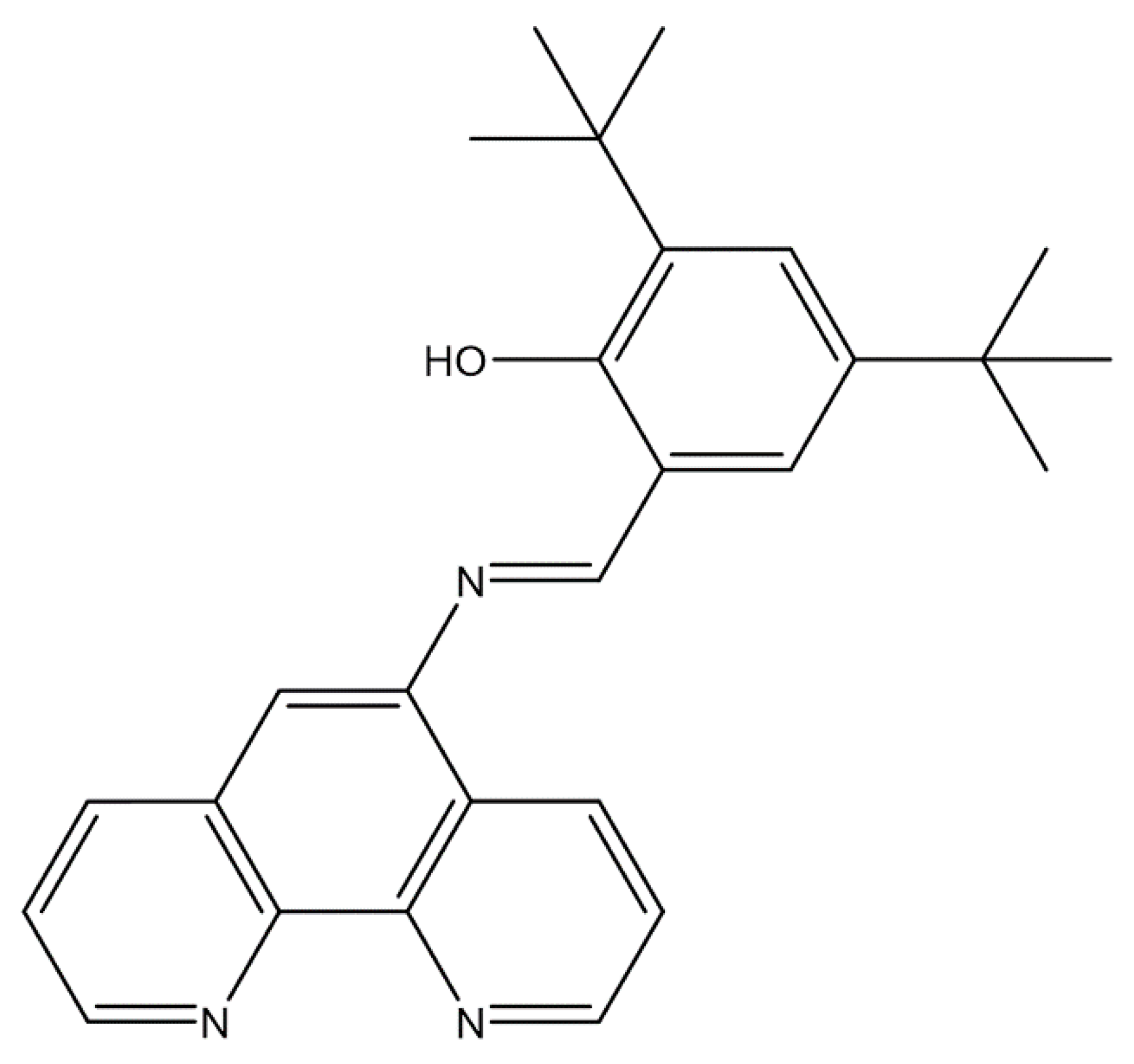
Figure 1. Chemical structure of (E)-2-(((1,10-phenanthrolin-5-yl)imino)methyl)-4,6-di-tert-butylphenol (Fen-IHB).
Chemistry 07 00135 g001
Figure 2. FTIR spectrum of Fen-IHB recorded in KBr under ultra-high vacuum conditions. The spectrum displays characteristic vibrational bands corresponding to the O–H stretching [ν(O–H)], aliphatic C–H stretching [ν(C–H)], azomethine C=N stretching [ν(C=N)], aromatic C=C stretching [ν(C=C)], and symmetric deformation of methyl groups [δ(C–H)]. The ν(O–H) band indicates strong intramolecular hydrogen bonding within the molecule.
Chemistry 07 00135 g002
Figure 3. UV–Vis spectra of Fen-IHB were recorded in aerated chloroform (green), acetonitrile (orange), and DMSO (blue) at room temperature.
Chemistry 07 00135 g003
Figure 4. (A) Cyclic voltammograms of Fen-IHB (— solid line) and blank supporting electrolyte (--- dashed line) recorded at a platinum working electrode under argon. Interface: Pt| 10−3 mol L−1 of compound + 10−1 mol L−1 of TBAPF6 in anhydrous CH3CN under an argon atmosphere. Scan rate: 200 mV s−1. (B) Working-window study for Fen-IHB demonstrating the persistence of the irreversible reduction peak at −1.43 V vs. Ag/AgCl across varying lower potential limits.
Chemistry 07 00135 g004
Figure 5. DFT-computed FTIR spectrum of Fen-IHB obtained at the ωB97X-D/6-31+G(d) level with a PCM model for DMSO (IR peak half-width at half height of 4). Labeled peaks correspond to the O–H stretching vibration involved in the intramolecular hydrogen bond (3415 cm−1), tert-butyl C–H asymmetric and symmetric stretches (3132 and 2869 cm−1), azomethine C=N stretch (1716 cm−1), and aromatic C=C stretch (1668 cm−1). Full frequency values and normal-mode descriptions are listed in Table S5.
Chemistry 07 00135 g005
Figure 6. UV–Vis absorption spectrum of Fen-IHB in DMSO (solid line) with peak width of 7 and the TD-DFT computed oscillator strength profile (red lines). The calculated spectrum was obtained at the wB97XD/6-31+G(d) level with the PCM solvation model.
Chemistry 07 00135 g006
Figure 7. Frontier molecular orbitals involved in the electronic transition of Fen-IHB in DMSO (see Table S6). Isosurface plots (with contour value of 0.05787) show the HOMO localized on the phenolic –OH and adjacent aromatic carbon (highlighting the intramolecular hydrogen bond region) and the LUMO delocalized over the azomethine linkage and the 1,10-phenanthroline ring. This HOMO → LUMO excitation accounts for the 375.8 nm band, exhibiting combined π→π* and n→π* characters.
Chemistry 07 00135 g007
Table 1. Experimental maximum wavelengths (λ) and molar extinction coefficients (ε) of Fen-IHB.
Schiff BaseSolventλ abs (nm)ε (L/mol × cm)Assignment
Fen-IHBChloroform27633,774.41π → π*
34514,009.92n → π*, π → π*
Acetonitrile27333,469.59π → π*
34014,155.42n → π*, π → π*
DMSO275333,618.74π → π*
34514,035.21n → π*, π → π*
Table 2. Minimal inhibitory concentration (MIC, μM) of Fen-IHB against selected bacterial strains.
Gram ClassificationOxygen RequirementSpeciesMIC (µM)
NegativeFacultative anaerobeSalmonella enterica subsp. Enterica sv Typhimurium ATCC 14028sNo effect **
NegativeFacultative anaerobeSalmonella enterica subsp. Enterica sv Typhi STH2370No effect
NegativeFacultative anaerobeEscherichia coliNo effect
PositiveStrict aerobeBacillus subtilis18.9 ± 3.6 *
PositiveAerotolerant anaerobeStreptococcus pyogenes3.2 ± 0.0 *
PositiveAerotolerant anaerobeEnterococcus faecalis22.1 ± 3.1 *
PositiveAerotolerant anaerobeStaphylococcus aureus12.6 ± 0.0 *
PositiveAerotolerant anaerobeStaphylococcus haemolyticus18.9 ± 3.6 *
PositiveStrict anaerobeClostridioides difficile R20291No effect
PositiveStrict anaerobeRoseburiainulinivoransNo effect
PositiveStrict anaerobeBlautia coccoidesNo effect
* Statistically significant difference compared to DMSO control (Mann–Whitney U test, one-sided, p < 0.05). ** “No effect” indicates no detectable growth inhibition distinguishable from the vehicle control (DMSO) alone.
Disclaimer/Publisher’s Note: The statements, opinions and data contained in all publications are solely those of the individual author(s) and contributor(s) and not of MDPI and/or the editor(s). MDPI and/or the editor(s) disclaim responsibility for any injury to people or property resulting from any ideas, methods, instructions or products referred to in the content.