Article

Characterization and Computational Insights into the Potential Biological Activity of 4-Hydroxyphenyl 8-Chlorooctanoate Purified from Endophytic Fusarium solani

1
Center of Biotechnology and Microbiology, University of Peshawar, Peshawar 25120, Pakistan
2
Department of Health and Biological Sciences, Abasyn University, Peshawar 25000, Pakistan
*
Authors to whom correspondence should be addressed.
Chemistry 2025, 7(4), 130; https://doi.org/10.3390/chemistry7040130 (registering DOI)
Submission received: 26 June 2025 / Revised: 26 July 2025 / Accepted: 8 August 2025 / Published: 14 August 2025

Abstract

Endophytes are important sources of bioactive secondary metabolites with therapeutic and agricultural relevance. This study reports the isolation and characterization of bioactive compounds from endophytic Fusarium solani associated with Solanum surattense. The fungal strain, selected after preliminary screening for its antimicrobial potential, was identified through morphological and molecular methods. A pure compound, 4-hydroxyphenyl 8-chlorooctanoate with a molecular mass of 270, was obtained and structurally characterized using GC–MS, FTIR, and NMR spectroscopy. Its anti-microbial potential was evaluated through molecular docking against key bacterial (Staphylococcus aureus) and fungal (Aspergillus fumigatus) targets, showing notable binding affinities with ClpP protease (−7.1 kcal/mol) and 14α-demethylase (−7.4 kcal/mol), respectively. Molecular dynamics simulations further confirmed the stability of the 5FRB-compound complex, with lower RMSD and RMSF values indicating strong structural integrity. Supporting analyses (B-factor and radius of gyration) confirmed the compactness and rigidity of the complex. These findings highlight the potential of 4-hydroxyphenyl 8-chlorooctanoate as a promising antimicrobial agent and provide a strong basis for further in vitro and in vivo validation of the purified compound as an antimicrobial candidate.

1. Introduction

Antibiotics play a crucial role in the treatment of infectious diseases; however, their overuse and the adaptive nature of pathogenic microbes have led to the emergence of multidrug-resistant (MDR) strains, particularly those capable of forming biofilms [1]. This escalating resistance necessitates efforts to restrict the spread of MDR pathogens and, importantly, to discover novel antimicrobial compounds [2]. Filamentous fungi, known for their immense and relatively untapped diversity of secondary metabolites, are increasingly recognized as promising candidates for novel antibiotic discovery [3]. Natural products derived from fungi possess remarkable industrial and therapeutic applications due to the biochemical diversity and structural complexity of fungal secondary metabolites [4]. Among these, endophytic fungi, i.e., those that live symbiotically within plant tissues without causing apparent harm, are especially significant. These endophytes are known to synthesize a broad range of bioactive compounds with applications in agriculture (as bio-pesticides and herbicides), industry (in food production), environmental management (bioremediation), and medicine (as antibiotics and other therapeutics) [5,6]. S. surattense, a medicinal plant traditionally used for various ailments, including liver disorders, urinary tract infections, and rheumatism, harbours diverse endophytic fungi capable of producing pharmaceutically relevant secondary metabolites. In South Asian countries, particularly Pakistan, hot aqueous extracts of S. surattense fruits are used to manage diabetes. The stem, fruits, and flowers are known to alleviate burning sensations in the feet, while its seeds are traditionally used to treat asthma and cough. Furthermore, seed preparations are employed for throat infections and inflammation, and root pastes are applied in the treatment of hernia. Seeds and fruits are also used as remedies for gum swelling, tooth pain, and cavities [7]. Phytochemical investigations have identified various classes of natural compounds from S. surattense, including alkaloids, phlobatannins, saponins, tannins, flavonoids, terpenoids, steroids, glycosides, quinones, and phenols. These compounds have been extensively studied for their bioactivity and commercial potential. Consequently, S. surattense remains a popular medicinal plant in India and Pakistan, widely used as a home remedy for conditions such as bronchitis, fever, asthma, and parasitic infections [8].
Endophytic fungi associated with S. surattense are considered valuable microbial resources capable of producing secondary metabolites with key therapeutic properties [9,10]. F. solani, in particular, is a potent endophyte inhabiting the fruits and stems of S. surattense. It is known to biosynthesize a variety of bioactive compounds, including azaanthraquinones (e.g., javancin), sterols (e.g., cerevesterol), and quinone metabolites (e.g., fusarubin and 3-O-methylfusarubin) [11]. The integration of fungal natural product research with bioinformatics tools presents a powerful strategy for identifying and evaluating new therapeutics, especially against drug-resistant pathogens. Computational methods, such as molecular docking and molecular dynamics simulations, can be instrumental in predicting the therapeutic potential of secondary metabolites [12].
In the present study, a bioactive compound was isolated from F. solani, an endophytic fungus associated with S. surattense. The compound was evaluated for its antimicrobial potential through in vitro screening against selected bacterial strains. Additionally, in silico analyses, including molecular docking and molecular dynamics simulations, were performed to investigate the compound’s binding affinity and stability against multiple biological targets: bacterial ClpP protease (S. aureus, PDB ID: 3V5I), fungal 14α-demethylase (A. fumigatus, PDB ID: 5FRB), cyclin-dependent kinase 9 (CDK9, PDB ID: 3LQ5), and HIV capsid protein (PDB ID: 3H4E). The main aim of this investigation was to isolate and structurally characterize a secondary metabolite from F. solani and to comprehensively evaluate its antimicrobial potential through experimental and computational approaches, thereby identifying its possible role as a lead compound for future therapeutic development.

2. Methodology

2.1. Preliminary Bioactivity Screening and Identification of Endophytic Fungi Isolated from S. surattense

S. surattense (Burm.f.), a widely distributed medicinal plant, was collected from various regions of Central Kurram, Khyber Pakhtunkhwa. Healthy plant material, including stems and fruits, was gathered using clean, sterile polyethylene bags to prevent contamination. The plant species was taxonomically authenticated at the Department of Botany, University of Peshawar, and a voucher specimen was deposited in the Mycology Research Laboratory, Centre of Biotechnology and Microbiology (COBAM), University of Peshawar, under the accession number SS-MRLCOBAM-2023-07. To isolate bioactive endophytic fungi, the collected stems and fruits were surface-sterilized using a sequential treatment of 0.1% mercuric chloride (HgCl2) solution and 95% ethanol. The sterilized parts were then cut into small segments, approximately 2 cm in size, and aseptically transferred onto Petri plates containing autoclaved Potato Dextrose Agar (PDA). The plates were incubated at 28 °C for 10 to 14 days. Emerging fungal colonies were subcultured to obtain pure isolates. For morphological identification, the isolates were grown using the slide culture technique and examined under a compound microscope equipped with 10×, 40×, and 100× objectives. Key microscopic features such as the structure of fruiting bodies, hyphal characteristics, spore morphology, and septation were observed. Colony morphology, including pigmentation and texture, was also recorded. Molecular identification of the fungal isolate was performed via ribotyping of the 18S rRNA gene region. Genomic DNA was amplified using the universal ITS primers: ITS1 (5′-TCCGTAGGTGAACCTGCGG-3′) and ITS4 (5′-TCCTCCGCTTATTGATATG-3′). PCR amplification followed by sequencing confirmed the taxonomic identity of the fungus. Preliminary screening of the fungal extract for antimicrobial activity was carried out against two bacterial strains: Escherichia coli and S. aureus. The isolated endophytic fungal strains were initially screened for antimicrobial activity against S. aureus and E. coli. For this purpose, the fungi were cultivated on Potato Dextrose Agar (PDA) plates, and 1 cm agar plugs (also referred to as mycelial plugs) containing actively growing mycelium were carefully excised in the form of uniform cubes, approximately the size of a standard cover slip. These plugs were then aseptically placed onto nutrient agar plates pre-inoculated with the respective bacterial strains to create uniform bacterial lawns. After incubation under appropriate conditions, zones of inhibition were observed around the fungal plugs, indicating the production of bioactive metabolites with antibacterial properties [13,14].

2.2. Biosynthesis of Fungal Natural Products Using Czapek Broth Medium

The isolated endophytic fungus, identified as F. solani, was cultured in liquid media to facilitate the biosynthesis of secondary metabolites. The Czapek Yeast Broth (CYB) medium was prepared and autoclaved at 121 °C under 15 psi pressure for 30 min to ensure sterility. The medium formulation included 1% peptone, 1% glucose, 0.05% KCl, 0.05% MgSO4·7H2O, and 0.001% FeSO4·7H2O, with the pH adjusted to 7.3 ± 0.2 prior to sterilization. To enhance the metabolic output, the medium was further enriched with 3% starch and an additional 2% glucose supplementation. The sterilized broth was inoculated with an actively growing fungal culture under aseptic conditions. The inoculated flasks were then incubated in a rotary shaking incubator at 150 rpm for 12 to 14 days, maintained at a controlled temperature of 26–28 °C to optimize fungal growth and metabolite production [15].

2.3. Extraction of the Crude Metabolite-Enriched Fraction

Following the completion of the incubation period, each culture flask was treated with 0.25 mL of 40% hydrochloric acid (HCl) to facilitate the breakdown of media components and promote phase separation. An equal volume of ethyl acetate was then added to each flask to extract the bioactive secondary metabolites. The flasks were agitated vigorously for 30 min to ensure efficient extraction. Subsequently, the fungal biomass (mycelia) was removed by filtration using a sterile cheesecloth. The resulting liquid phase was transferred into a separating funnel to isolate the ethyl acetate layer. The upper organic phase was washed with 2 M brine solution to remove residual polar impurities. To remove traces of moisture, the organic extract was dehydrated using anhydrous sodium sulfate (Na2SO4). Finally, the purified organic layer was concentrated using a rotary evaporator set at 45 °C, yielding a semi-solid crude extract enriched in fungal secondary metabolites [16].

2.4. Purification of Fungal Metabolites Using Column and Thin Layer Chromatography

The concentrated crude extract was initially dissolved in methanol, and a slurry was prepared by thoroughly mixing the methanolic extract with silica gel (60 mesh size) using a mortar and pestle. The chromatography column was pre-washed with 75% ethanol and allowed to dry completely before being set vertically for packing. To prepare the stationary phase, silica gel was mixed with n-hexane in a beaker and carefully poured into the column. Once the silica gel settled and packed uniformly, forming a stable bed, the crude slurry was gently loaded onto the column using a funnel. A small cotton plug was placed above the loaded sample to prevent disturbance during solvent flow. Elution was initiated with 100% n-hexane, and the polarity of the mobile phase was gradually increased by stepwise addition of ethyl acetate in increasing proportions. Eluted fractions corresponding to each solvent ratio were collected in separate vials. To monitor the separation and identify fractions with similar components, preparative thin-layer chromatography (TLC) was performed on each collected fraction. TLC was performed using pre-coated silica gel plates (Merck, Darmstadt, Germany). The mobile phase consisted of the solvent system (EtoAc: n-hexane, 90:10), and the development was carried out in a saturated TLC chamber at room temperature. After development, the plates were air-dried and visualized under 254 nm and 365 nm UV light. To improve visibility, the plates were also exposed to iodine vapor in a sealed chamber [17].

2.5. Sample Preparation and Analysis Using GC–MS

For GC–MS analysis, the purified compound was analyzed by dissolving 2 mg of the pure compound in 1 mL of methanol. From this stock solution, 1 μL was injected into the instrument using a 10 μL micro-syringe. The injection was performed in a splitless mode to ensure optimal sensitivity. The analysis was carried out on a GC–MS system (Agilent Technologies, Inc., Santa Clara, California, USA; Model: 5977B) equipped with a DB-1 capillary column (25 m × 0.25 mm i.d., 0.25 μm film thickness). The carrier gas was high-purity helium, maintained at a constant flow rate of 8.81 psi. The column oven temperature was programmed to rise from 50 °C to 300 °C during the total run time of 36 min to ensure efficient separation and analysis. During the run, the sample was vaporized in the injector and ionized within the mass spectrometer. The resulting fragments were analyzed based on their mass-to-charge (m/z) ratios. The obtained mass spectra were compared to reference spectra available in the NIST mass spectral library for compound identification. Chromatographic peaks corresponding to individual components were recorded and displayed as total ion chromatograms (TICs) [18,19].

2.6. Fourier Transform Infrared (FTIR) Spectroscopic Analysis

Fourier Transform Infrared (FTIR) analysis was conducted to identify the functional groups present in the purified compound. A small amount of the sample was finely ground and mixed with spectroscopic-grade potassium bromide (KBr) to form a translucent pellet. The spectral data were acquired using an Agilent Cary 630 FTIR spectrometer (Agilent Technologies, Inc., Santa Clara, California, USA) equipped with an Attenuated Total Reflectance (ATR) mode. Measurements were recorded in the range of 4000 to 400 cm−1, using 32 sample scans and 32 background scans at a resolution of 8 cm−1. The Happ–Genzel apodization function was used during data acquisition. The system was purged with dry nitrogen to prevent atmospheric interference from water vapor and CO2. Functional group assignments were made by comparing the observed peaks with standard reference values [20].

2.7. Structure Elucidation of the Compound Using Nuclear Magnetic Resonance (NMR) Spectroscopy

NMR analyses were performed to elucidate the molecular structure of the purified compound. Approximately 5 mg of the sample was dissolved in 0.65 mL of deuterated dimethyl sulfoxide (DMSO-d6), and the solution was transferred into a 5 mm NMR tube. All spectra were acquired on Varian 400 and 500 MHz NMR spectrometers operating at 500 MHz for 1H, 125 MHz for 13C, and 67.8 MHz for 17O nuclei. One-dimensional 1H and 13C NMR spectra were recorded using standard pulse sequences. Chemical shifts (δ) are reported in parts per million (ppm) relative to tetramethylsilane (TMS) as an internal standard. In addition to 1D spectra, two-dimensional heteronuclear single quantum coherence (HSQC) experiments were carried out to establish direct 1H and 13C correlations, allowing unambiguous assignment of protonated carbon signals. Furthermore, 17O NMR spectroscopy was performed to investigate the oxygen environment within the molecule, providing additional confirmation of functional groups involving oxygen atoms [21].

2.8. ADMET Analysis of the Compound-270

The adsorption, distribution, metabolism, excretion, and toxicity (ADMET) analysis of the compound was performed using the SwissADME online web server [22]. This analysis was important to shed light on the compound’s pharmacokinetic behavior during experimental in vitro and in vivo analysis [23]. The toxicity of the compound was predicted by the pkCSM server [24].

2.9. In Silico Analysis of the Compound-270

For molecular docking analysis of compounds with different protein targets, the PyRx v8.0 software was used [25]. Before molecular docking analysis, the target proteins were retrieved from PDB using their specific ID. For example, 3V5I [26], 3LQ5 [27], 3H4E [28], and 5FRB were used herein and were representative PDB IDs of potential, attractive, and high-resolution biological targets. 3V5I represents a potential target protein (bacterial virulence regulator caseinolytic protease P) for drugs against pathogenic S. aureus. Similarly, 5FRB is a 14-alpha demethylase from the pathogenic fungus A. fumigatus. The 3LQ5 is a cell division protein kinase 9 and is used as an anti-cancer target. The 3H4E is a capsid protein HIV virus. USCF Chimera v1.17 [29] was used to fetch and prepare the structure of the targeted proteins. Energy minimization of the proteins was carried out using steepest descent and conjugate gradient algorithms in UCSF Chimera v1.17. In the process, the FF19SB force field was applied [30]. The protein structures were uploaded one by one to the PyRx v0.8. The compound-270 was energy minimized using the MM2 force field and loaded to AutoDock Vina v4.1 through the PyRx v0.8 interface. After loading the proteins and ligand, the docking analysis was run by using default values. The number of iterations set for the compound was 10. The results of the docking analysis were ranked by the lowest value of binding energy in kcal/mol. The complexes of various targets with compound 270 were analyzed using Bio Discovery Studio [31], followed by molecular dynamics simulation analysis [32].

2.10. Molecular Dynamics Simulation

Molecular dynamics simulation was performed using the AMBER software c2022 to evaluate the structural stability and dynamic behavior of the complexes [33]. The ff19SB force field was used for the protein, while the General AMBER force field 2 was applied for compound parameterization [34]. The partial atomic charges for the selected compound were assigned using the AM1-BCC method using the Antechamber module [35]. Furthermore, the complexes were solvated in an octahedral box of TIP3P water molecules, keeping a 12 Å buffer between the docked complex and the box boundary. The counter ions (Na+ or Cl) were further added to neutralize the system. Next, the energy minimization was carried out in two stages: in the first phase, minimizing water molecules and ions while restraining the solute 5000 steps, followed by minimizing the entire system without restraints using 5000 steps. After the minimization of the complex, the system was slowly heated from 0 K to 300 kelvin over 100 ps under constant volume (NVT) conditions, using Langevin dynamics for temperature control [36]. Next, a 500 ps equilibration stage at the constant pressure NPT ensemble with positional restraints on the complex. Finally, a 100 ns production MD simulation was run under NPT conditions at 1 atm and 300 K with a time step of 2 fs. The SHAKE algorithm was employed to restrain all covalent bonds that involve hydrogen atoms [37]. In the last CPPTRAJ, commands were used for trajectory analysis, which was carried to investigate different simulation trajectory parameters such as root mean square deviation (RMSD), root mean square fluctuation (RMSF), beta factor (β-factor), and radius of gyration (Rg) [38,39], which were examined to understand the structural dynamics and stability of the docked complex during the whole simulation time [40].

3. Results

3.1. Culturing and Identification of F. solani Based on Morphological and Microscopic Characteristics

The isolated fungal colony initially exhibited a pure white appearance, which gradually developed into a grayish-white pigmentation with a cottony, fluffy texture as growth progressed. A characteristic concentric radial pattern was observed on PDA medium as shown in Figure 1A. For microscopic examination, a coverslip preparation was made using the slide culture technique and stained with lactophenol cotton blue. Microscopic analysis under 10× magnification confirmed the presence of fungal growth, while further observations under 40× and 100× magnifications revealed distinct morphological features. The hyphae were septate, and macroconidia appeared spindle-shaped with slightly curved ends, which are diagnostic characteristics of F. solani as shown in Figure 1C.

3.2. Molecular Identification

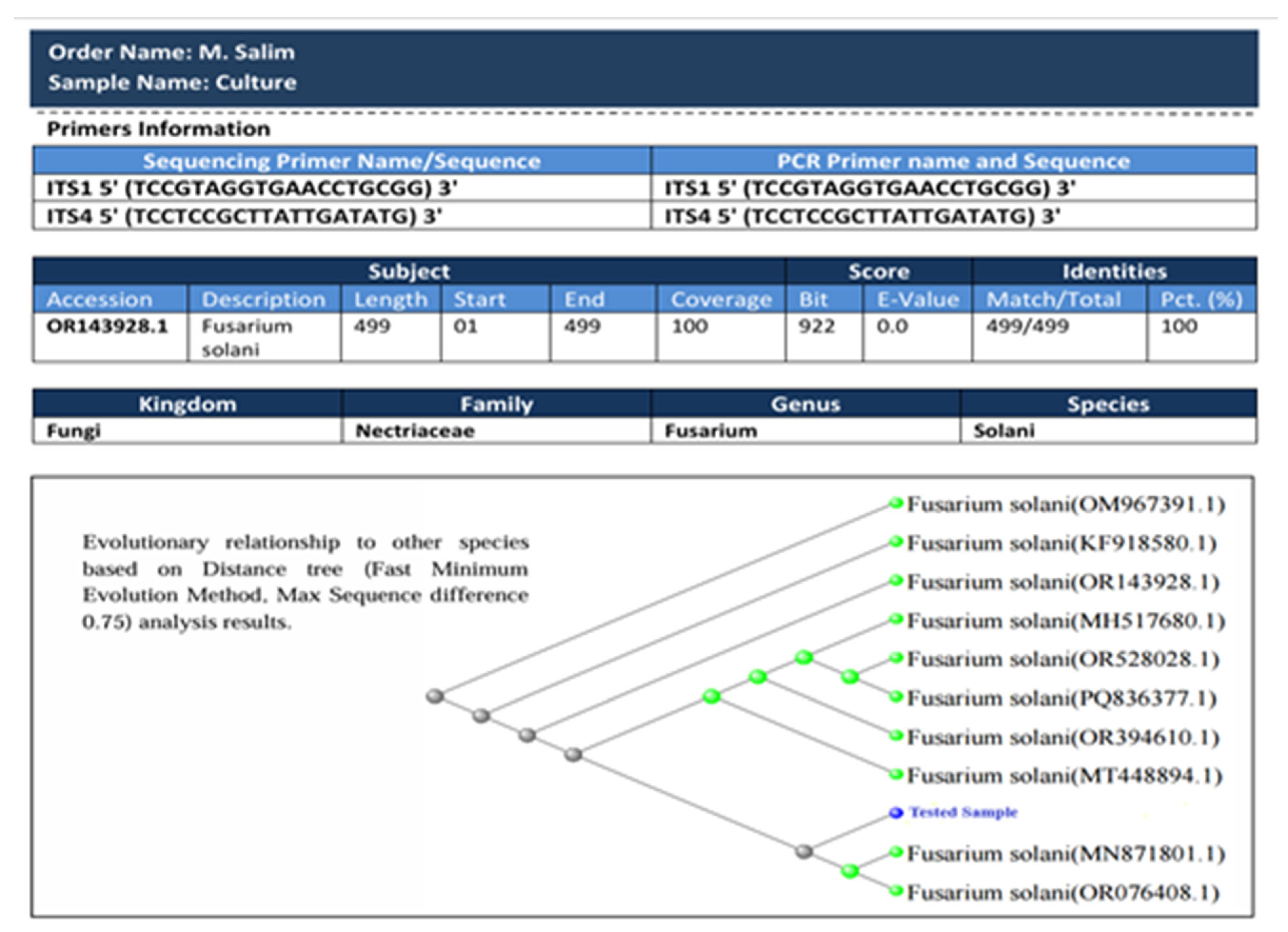
Molecularly, the fungus showed 100% evolutionary resemblance with the F. solani isolate AMWb. The phylogenetic tree is presented in Figure 2. As can be seen in the tree, two evolutionary clades are reported where the test sample falls into the small clade of F. solani MN871801 and F. solani OR076408. The tested isolate F. solani sequence chromatogram and FASTA sequence are provided as File S1 and File S2. Similarly, the ribotyping findings are given in File S3.

3.3. Preliminary Bioactivity Testing of the Fungal Isolates

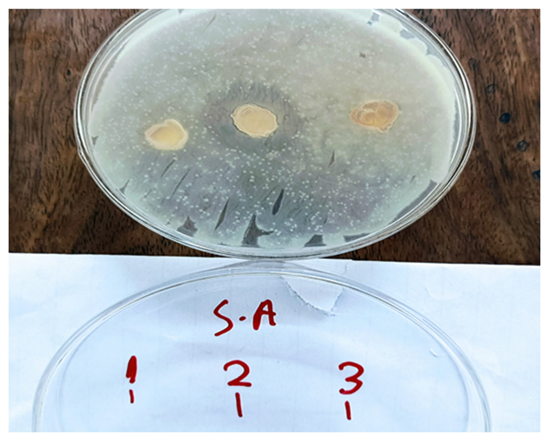
The isolated endophytic fungal strains were initially screened for antimicrobial activity against S. aureus and E. coli. This preliminary screening facilitated the selection of the most bioactive strain for further processing and metabolite production. Among the three fungal isolates obtained from S. surattense, isolate no. 2, identified as F. solani, exhibited the strongest inhibitory activity, particularly against S. aureus, with a prominent inhibition zone of 19 mm, as shown in Figure 3. The other isolates, i.e., isolate no. 1, showed some bioactivity, but the zone of inhibition was not clear, while isolate no. 3 formed no prominent zone of inhibition as shown in Figure 3.

3.4. Purification of the Compound Using Chromatographic Techniques

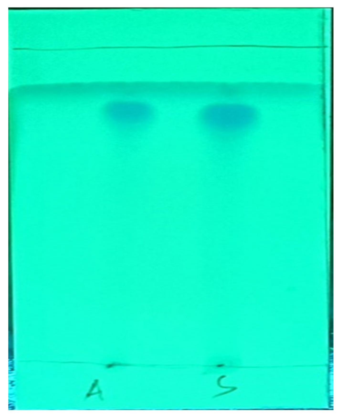
To purify the bioactive compound, the fungal crude extract was processed using silica gel column chromatography. We applied a gradient elution system starting with n-hexane and gradually increased the proportion of ethyl acetate. This allowed us to separate the extract into several fractions based on polarity. One of the key bioactive fractions was isolated using an ethyl acetate to n-hexane ratio of 85:15 (v/v), which yielded approximately 1.5 mg of compound from 5 L of culture extract. For further purification and confirmation of compound homogeneity, preparative thin-layer chromatography (TLC) was performed using pre-coated silica gel plates (Merck). The mobile phase consisted of the solvent system (EtoAc: n-hexane, 90:10), and the development was carried out in a saturated TLC chamber at room temperature. After development, the plates were air-dried and visualized under 254 nm and 365 nm UV light. To improve visibility, the plates were also exposed to iodine vapor in a sealed chamber, allowing for the clear identification of compound bands. Distinct spots representing the same compound were observed from two separately concentrated vials, labeled as “A” and “S”. These bands are clearly visible in the TLC as shown in Figure 4.

3.5. GC–MS Analysis of the Pure Compound

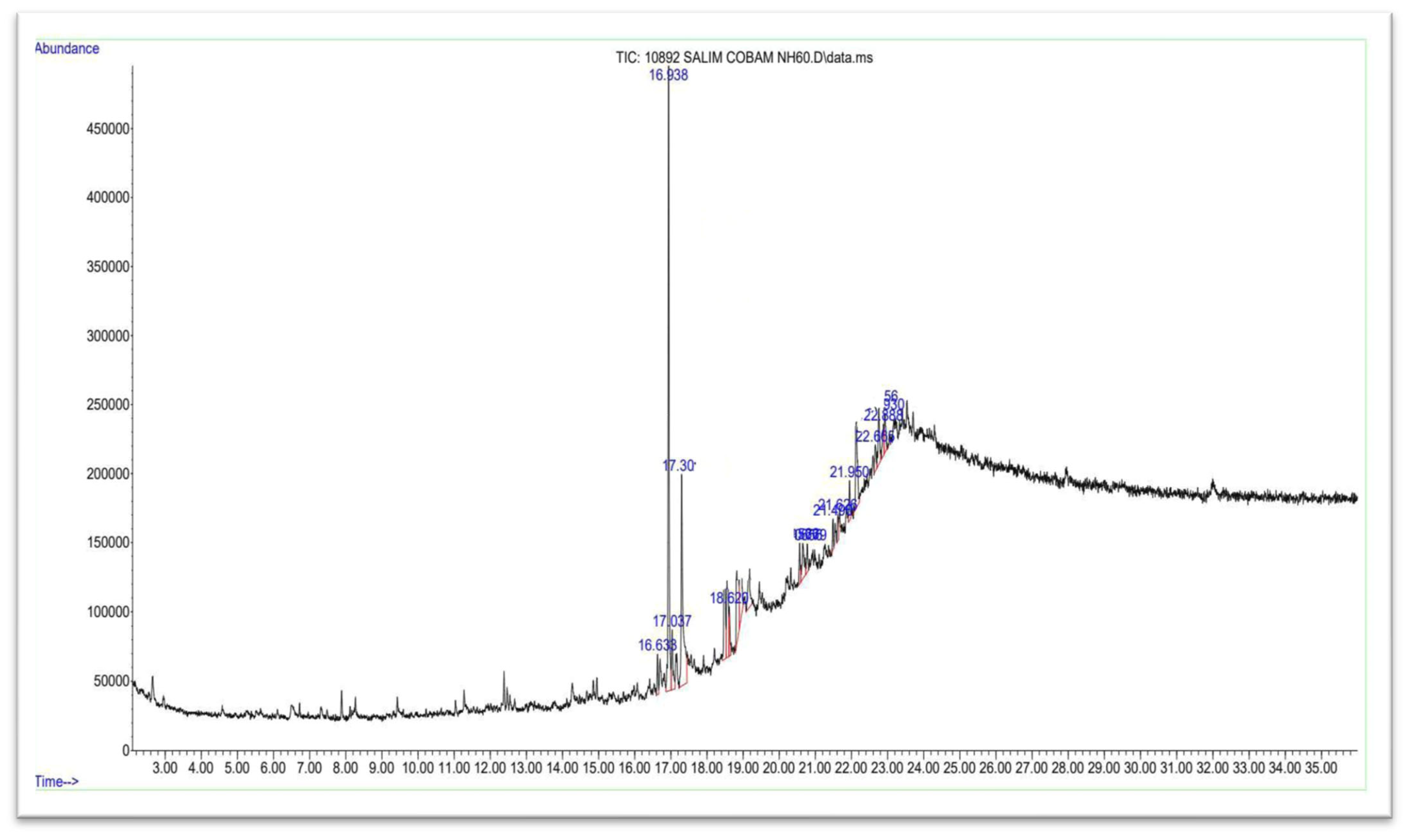
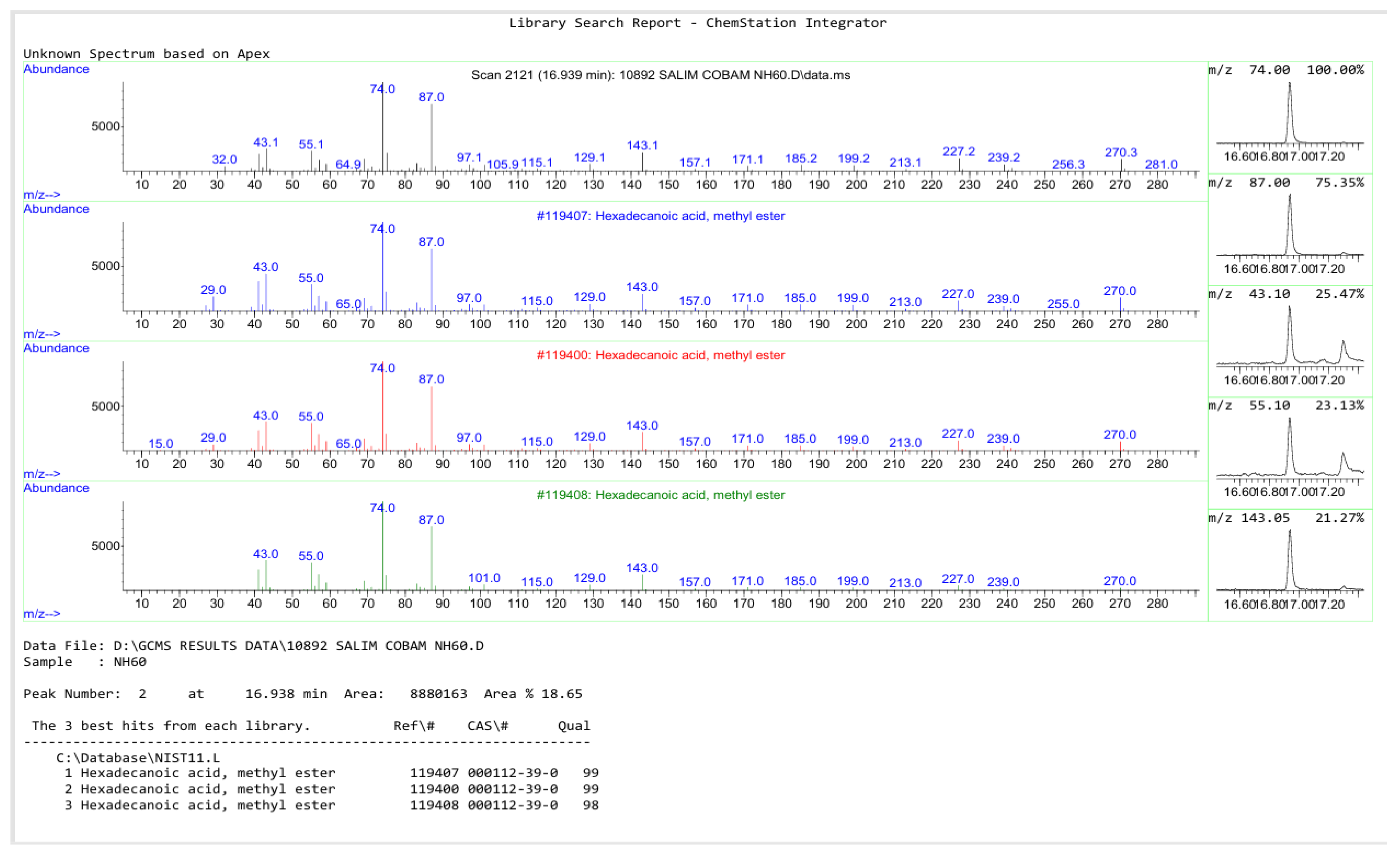
GC–MS analysis of the purified compound was performed using an Agilent Technologies GC–MS system equipped with a DB-1 capillary column (30 m length, 0.25 mm inner diameter, and 0.25 µm film thickness). Approximately 2 mg of the purified compound was dissolved in 1 mL of molecular-grade methanol, and 1 µL of the solution was injected into the GC–MS system in the splitless mode. The total ion chromatogram (TIC) revealed a prominent peak at a retention time (RT) of 16.938 min, as shown in Figure 5, indicative of a major constituent. The mass spectrum corresponding to this peak displayed a molecular ion peak at m/z 270, consistent with hexadecanoic acid, methyl ester (methyl palmitate). A detailed comparison with the NIST11.L spectral library confirmed the identity of the compound with a high match quality (98–99% similarity), cross-validated across three database entries (CAS No. 000112-39-8). The fragmentation pattern exhibited characteristic peaks at m/z 74, 87, 143, 171, 199, and 227, as presented in Figure 6, which align with typical fragmentation pathways of saturated fatty acid methyl esters. Based on the intensity and spectral similarity, methyl palmitate was conclusively identified as the predominant component in the purified sample. Methyl palmitate is well-known for its antimicrobial, anti-inflammatory, and cytotoxic properties, which highlight the compound’s potential pharmacological importance. For additional confirmation, FTIR analysis of the compound was performed.

3.6. FTIR Analysis

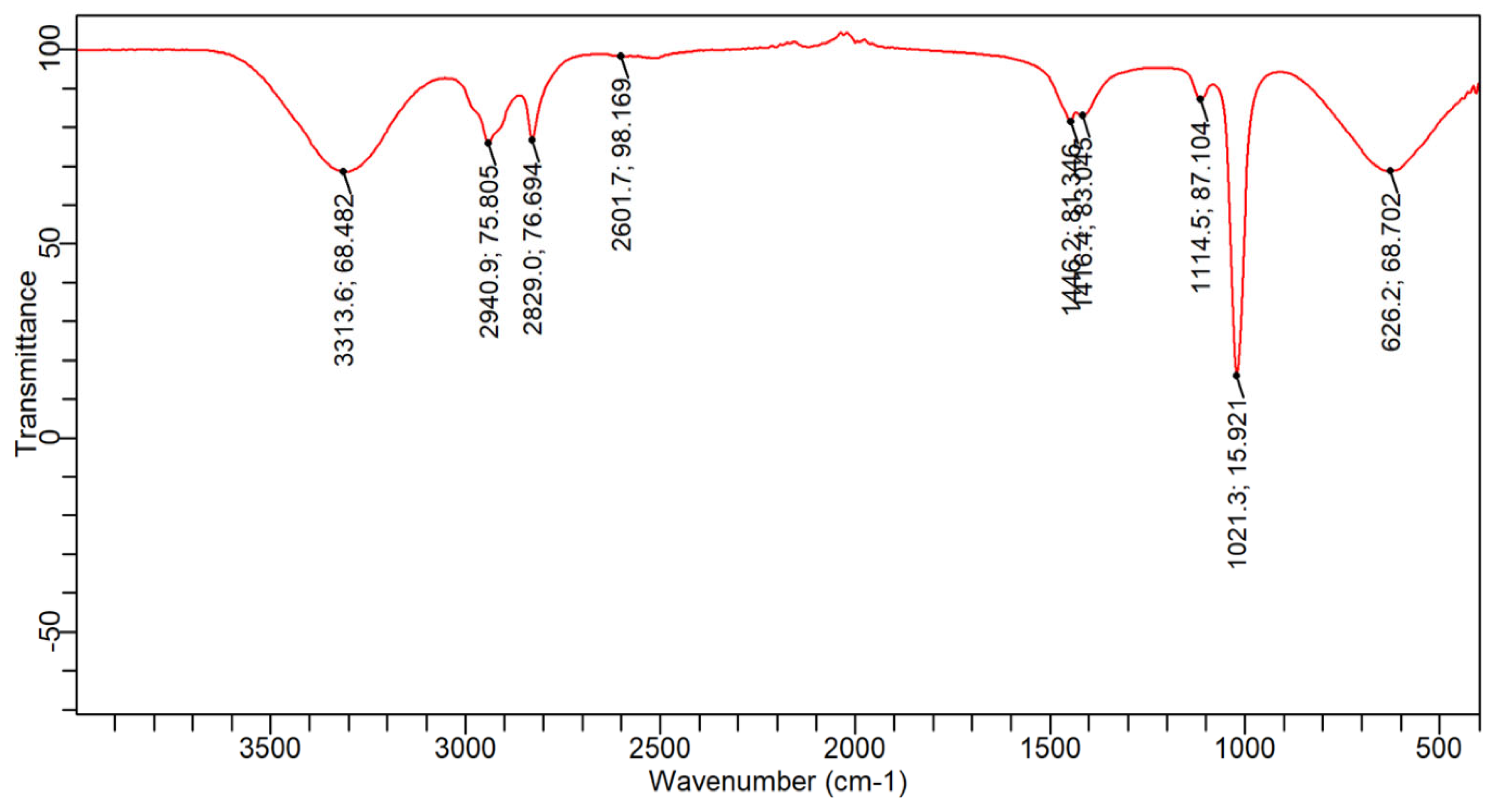
To further characterize the compound, FTIR spectroscopy was used to investigate the functional groups present. As presented in Figure 7, the spectrum showed a broad absorption band at 3313 cm−1, which is typically attributed to O–H or N–H stretching vibrations, indicating the presence of hydroxyl or amine groups. Absorption bands at 2940 cm−1 and 2829 cm−1 were assigned to C–H stretching in aliphatic chains. An additional peak at 2601 cm−1 may represent overtone or combination bands related to carboxylic acid O–H bending. In the fingerprint region, sharp peaks at 1446 cm−1 and 1416 cm−1 suggest C–H bending vibrations or possibly aromatic ring breathing. Peaks at 1114 cm−1 and 1021 cm−1 indicate C–N stretching, consistent with amine functionalities. A band observed at 626 cm−1 may reflect out-of-plane C–H bending, often seen in substituted aromatic compounds. Interestingly, the FTIR profile of the isolated compound did not entirely match that of methyl palmitate. The presence of strong O–H and N–H stretching signals and the absence of a distinct ester C=O peak around 1740 cm−1 suggest that the isolated compound may not be a simple fatty acid methyl ester. Instead, it may possess additional functional groups such as hydroxyl or amide linkages, indicating a structurally distinct, possibly novel, fungal metabolite. This discrepancy highlights the need for further structural confirmation using complementary techniques like NMR.

3.7. NMR-Based Structural Elucidation of 4-Hydroxyphenyl 8-Chlorooctanoate

The chemical structure of the purified compound, identified as 4-hydroxyphenyl 8-chlorooctanoate, was thoroughly characterized using one-dimensional (1H and 13C) and two-dimensional (HSQC, 17O) NMR spectroscopy in DMSO-d6. The 1H NMR spectrum (500 MHz, DMSO-d6) exhibited characteristic signals that confirmed the proposed structure. A singlet appearing distinctly at δ 8.82 ppm indicated the presence of the phenolic hydroxyl proton. Aromatic protons were evident as multiplets resonating between δ 6.99 and 6.81 ppm, consistent with the para-substituted phenyl moiety. A clearly defined doublet at δ 3.58 ppm corresponded to methylene protons adjacent to the ester linkage (–O–CH2–CO–), affirming the ester connectivity within the molecule. The aliphatic methylene protons exhibited multiple signals within the δ 2.82–1.31 ppm range, representative of a flexible alkyl chain. The signals at δ 1.78 and δ 1.56 ppm were indicative of methylene groups positioned near the terminal chloro substituent, highlighting chlorine’s electronic influence.
Further structural insights were provided by the 13C NMR spectrum (125 MHz, DMSO-d6). A distinct carbonyl resonance at δ 173.38 ppm confirmed the ester functionality, while aromatic carbon resonances at δ 154.81, 144.37, 123.22, and 117.15 ppm precisely matched a para-substituted phenolic system. The methylene carbon directly bonded to the ester oxygen appeared at δ 45.61 ppm, with additional aliphatic carbon resonances distributed clearly between δ 34.52 and δ 27.32 ppm, characteristic of the extended alkyl chain structure. HSQC analysis reinforced these assignments by clearly correlating proton resonances with their directly bonded carbons, facilitating accurate and unambiguous spectral interpretation. Moreover, 17O NMR spectroscopy revealed resonances confirming the ester carbonyl oxygen and phenolic hydroxyl oxygen atoms, adding definitive structural verification. Together, the NMR analyses supported by GC–MS and FTIR data confirm the structure of the isolated compound as 4-hydroxyphenyl 8-chlorooctanoate, a chlorinated phenolic ester derivative with considerable biological potential derived from the fungal isolate F. solani. The full NMR spectra of the compound in given in File S4. The structure of the compound is shown in Figure 8.

3.8. ADMET Analysis of the Compound-270

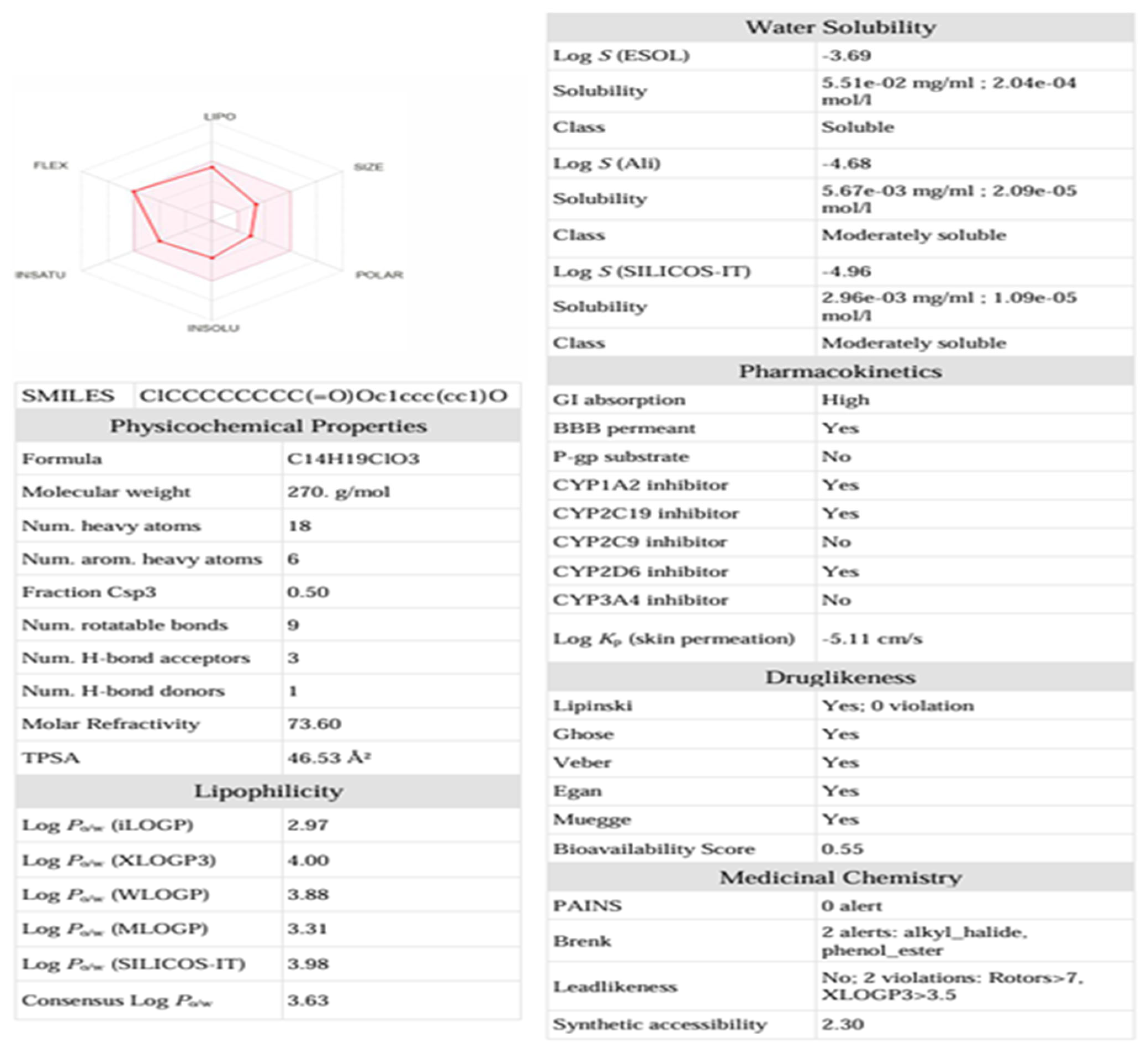
ADME analysis of compound-270 (4-hydroxyphenyl 8-chlorooctanoate) shows good drug-like properties with high GI absorption. It follows all major drug-like rules with no pain alerts and shows a moderate level of solubility. The compound 270 has acceptable lipophilicity and is easy to synthesize (synthetic accessibility 2.30), which makes it a promising drug candidate. The complete information about the ADME analysis of the compound can be seen in Figure 9. Furthermore, the compound was found to be non-toxic and non-mutagenic.

3.9. Molecular Docking Studies

Molecular docking analysis is primarily used to study the binding interactions between a ligand and receptor. In the current study, the said approach was utilized to analyze the binding affinity of 4-hydroxyphenyl 8-chlorooctanoate with different microbial and cancer drug targets, including fungal protein (14-alpha demethylase), bacterial protein (calpP-Protease), HIV (capsid protein), and anti-cancer (cyclin-dependent kinase 9). The docking process predicted different binding energies for the compound against each target. During visualization using BIOVIA Discovery Studio, it was confirmed that the ligand binds to the targeted receptors through interactions including pi-alkyl, Hydrogen Bonding, and van der Waals forces.

3.10. Fungal Protein and Compound-270 Complex

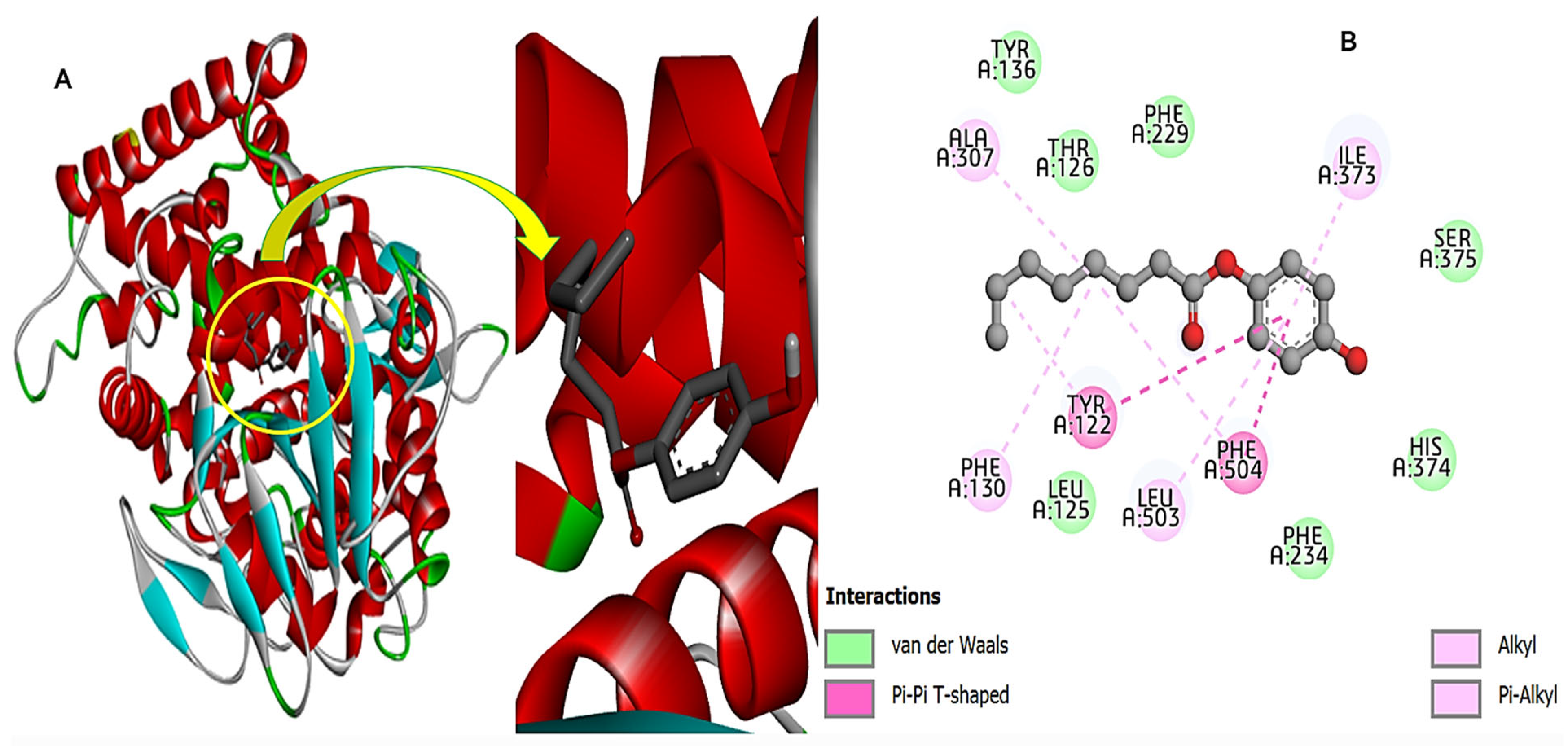
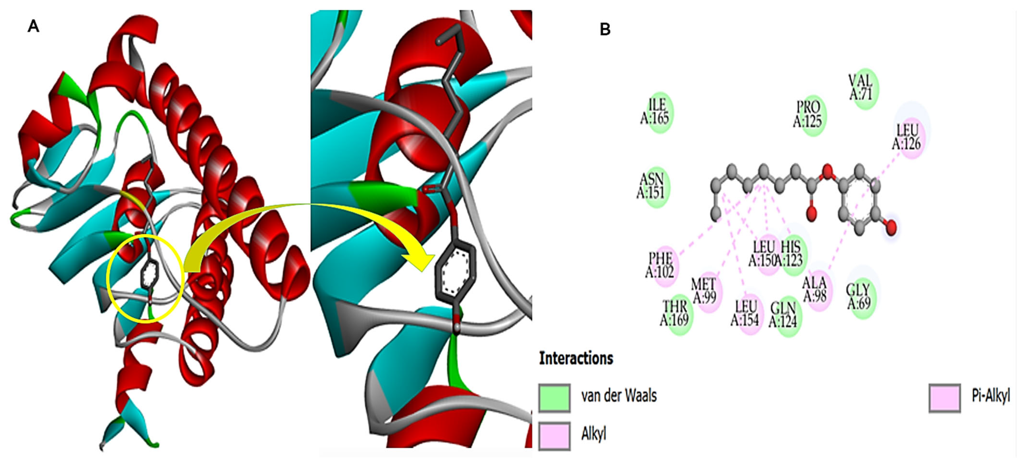
Molecular docking analysis of 4-hydroxyphenyl 8-chlorooctanoate with 14-alpha demethylase revealed a binding energy of −7.4 kcal/mol. Visualization of the complex reported that there is a stable ligand binding to the active site of 14-alpha demethylase. The ligand achieved a stable binding conformation and has deep access to the binding pocket. The study also revealed that the above ligand showed interactions with different amino acids through various bonding, including Alkyl, Pi-Alkyl, van der Waals, and conventional Pi-Pi T-shaped structures, as shown in Figure 10.

3.11. Bacterial Protein and Compound-270 Complex

Molecular docking analysis of 4-hydroxyphenyl 8-chlorooctanoate with CalpP-Protease of S. aureus unveiled a binding energy of −7.1 kcal/mol. Visualization of the complex indicated that the compound adjusted along the length of the CalpP-Protease active cavity. The study found that the ligand showed a rich interaction network and is engaged by pi-alkyl, van der Waals, and alkyl as shown in Figure 11.

3.12. HIV Protein and Compound-270 Complex

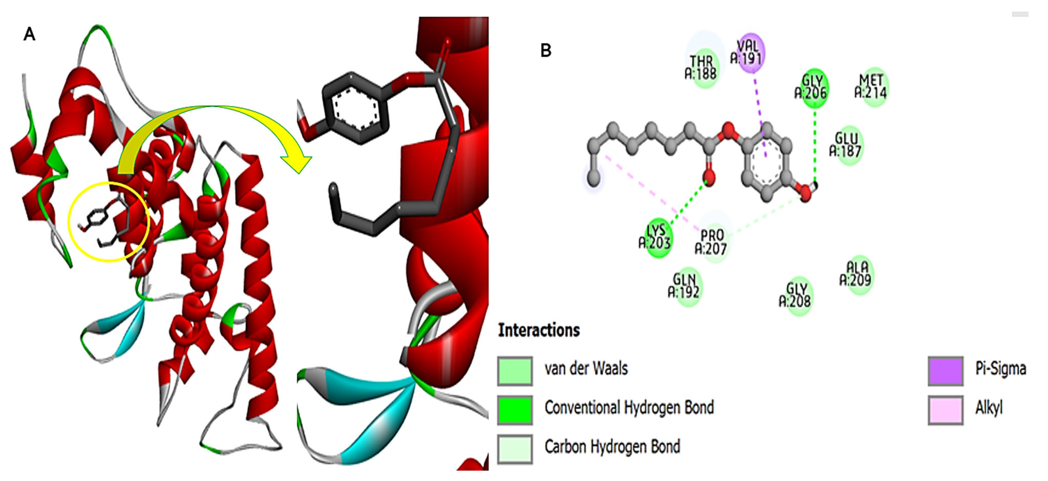
The 4-hydroxyphenyl 8-chlorooctanoate with HIV capsid protein has a binding energy score of −4.9 kcal/mol. The majority of the compound’s interactions with the receptor were found due to the terminal benzene ring; in particular, the oxygen atoms showed strong short-distance hydrogen bonding with Gly206 and Lys203. The rest of the compound structure was engaged in hydrophobic bonds, especially with Glu187, Met214, Thr188, Pro207, Ala209, Gly208, Pro207, and Gln192. The intermolecular interactions involve Pi-sigma, covalent bond, van der Waals, Pi-Alkyl, and H-bonding, as shown in Figure 12.

3.13. Cdk-9 Protein and the Compound-270 Complex

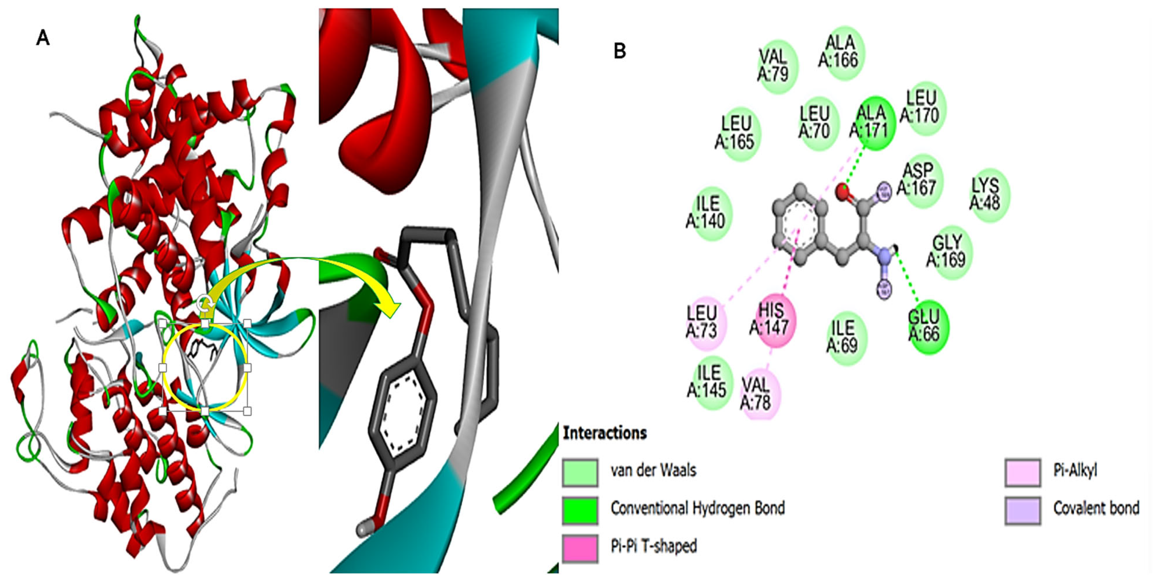
The 4-hydroxyphenyl 8-chlorooctanoate docks deep inside the binding pocket of the Cdk-9 protein. The docking analysis revealed a binding energy of −6.7 kcal/mol for the intermolecular complex of Cdk-9 and compound-270. The compound was reported to have deep binding at the active cavity. The compound aligned itself vertically in the pocket and is placed as such to stop access of the natural ligand competitively. The compound was observed to form mostly hydrophobic interactions, including van der Waals, pi-alkyl, and pi-pi T-shape bonding, as shown in Figure 13. The hydrogen bonds were formed between the compounds and residues Ala171 and Glu66. The hydrogen bonds were reported for the highly electronegative atoms, such as oxygen and nitrogen bonds to the hydrogen atom.

3.14. Molecular Dynamics Simulation

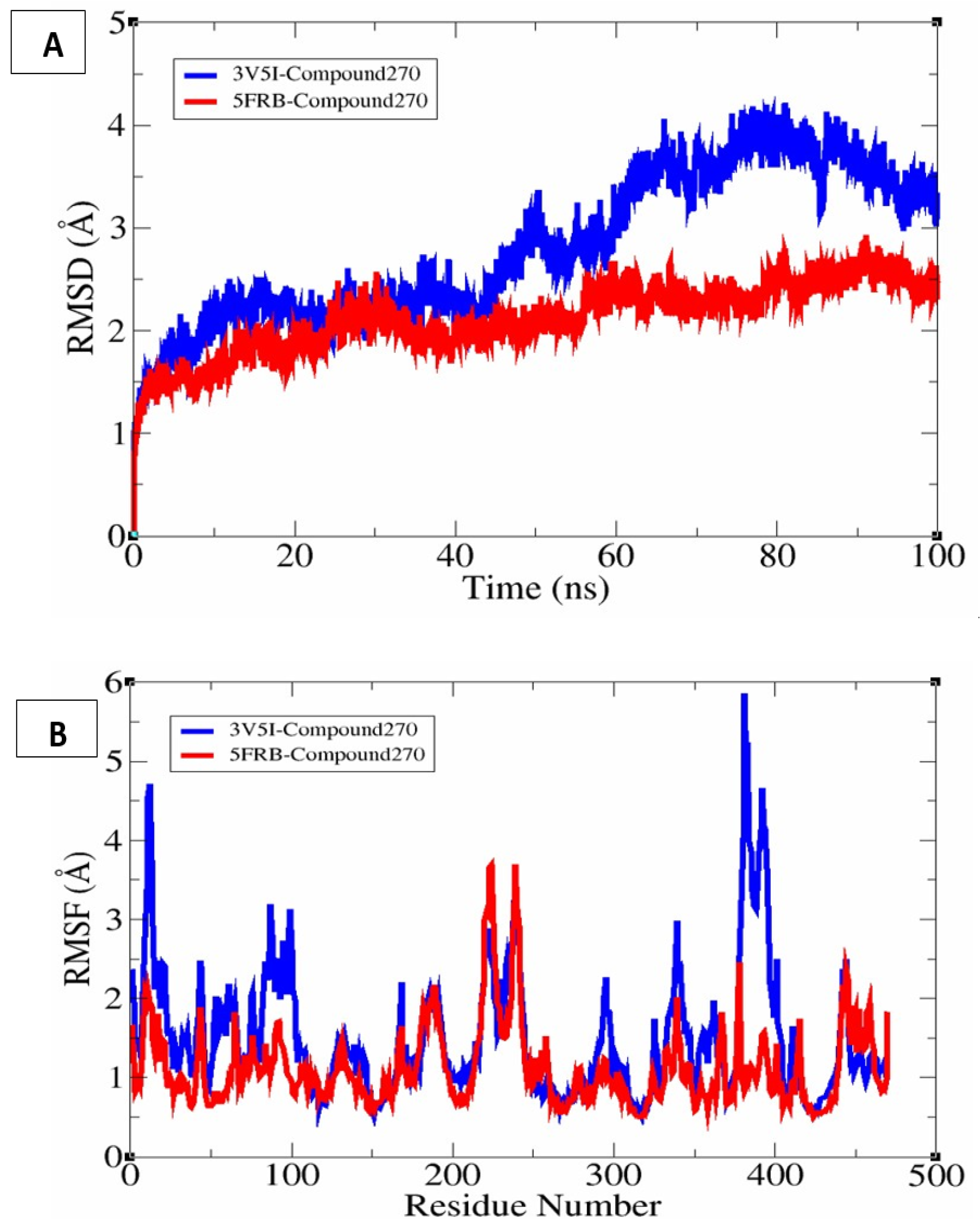
In molecular dynamics simulation, only two complexes (3V5I-Compound270 and 5FRB-Compound270) were selected for dynamics studies due to their higher intermolecular affinity. The complexes 3V5I-Compound270 (blue) and 5FRB-Compound270 (red) showed an initial increase in RMSD, indicating structural adjustment compared to the initial conformation when the sudden dynamics conditions were applied. The blue system (3V5I-Compound270) exhibits a higher RMSD value, suggesting greater structural deviation and flexibility over time. The red system (5FRB-Compound270) showed a lower RMSD value, indicating that it is more stable compared to the blue system. The RMSD plot is presented in Figure 14A. The y-axis represents the RMSD in angstroms (Å), while the x-axis represents time in nanoseconds (ns). The Mean RMSD of the 3V5I-Compound270 was 2.80917 (Å).
In Figure 14B, the y-axis represents RMSF (Å), while the x-axis corresponds to residue numbers. RMSF measures the flexibility of individual residues during the molecular dynamics simulation. Both systems exhibit similar fluctuation patterns, with some regions showing higher flexibility. Certain regions (e.g., around residue 400) showed pronounced fluctuations in the blue system, indicating increased flexibility. The red system (5FRB-Compound270) has generally lower RMSF values, suggesting more rigid and stable residues compared to the blue system. For the 5FRB-Compound270, a 5.8569 (Å) mean value was calculated.
The 5FRB-Compound270 system (red) appears to be more stable as it has lower RMSD and RMSF values. The 3V5I-Compound270 system (blue) shows higher deviations and fluctuations, indicating more structural flexibility or potential instability. These results suggest that the 5FRB-Compound270 may be a more stable candidate for further investigations, whereas 3V5I-Compound270 may exhibit more conformational changes over the simulation time.

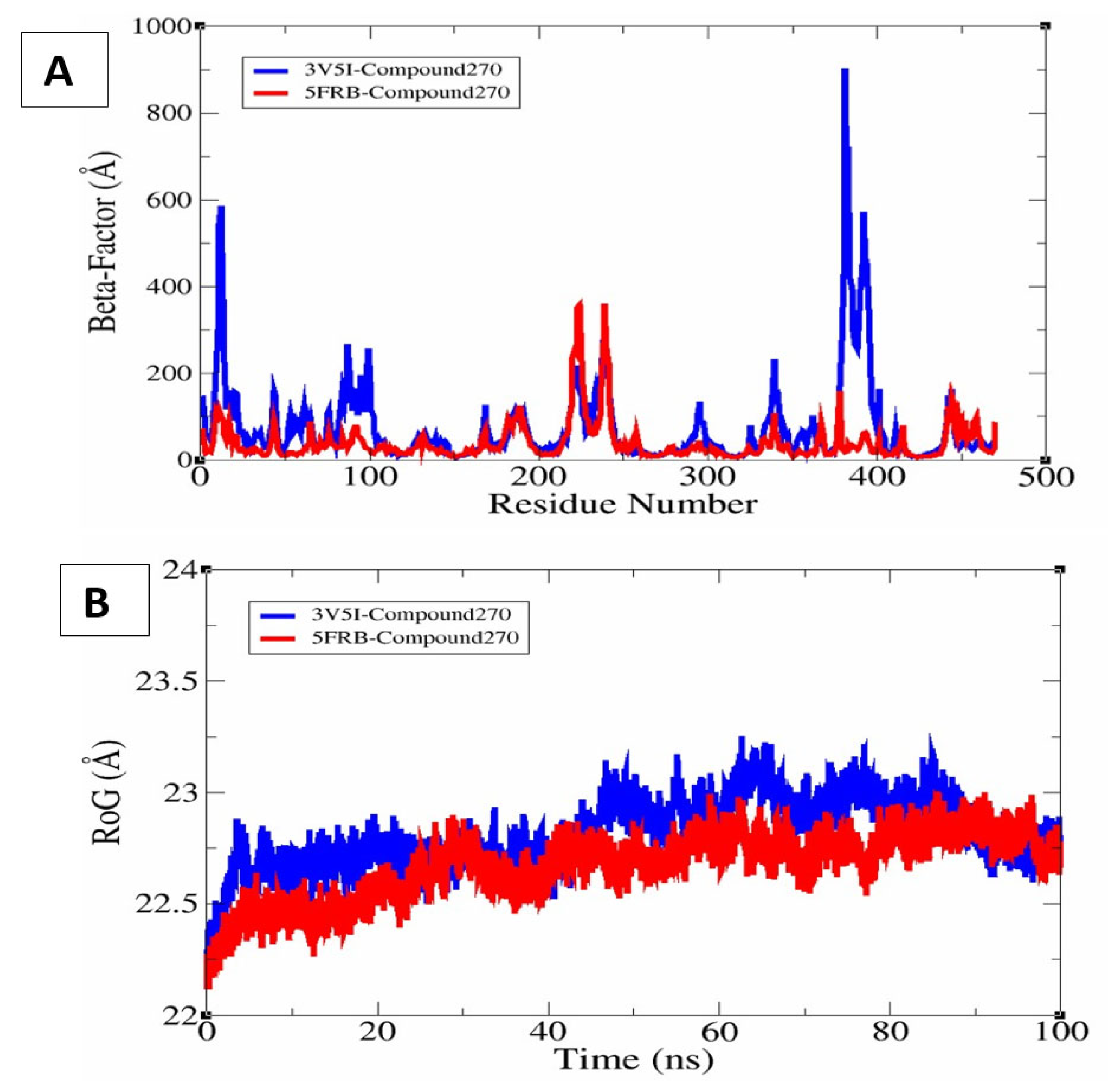
3.15. β-Factor Analysis and Rg

The β-factor reflects the flexibility of each residue, similar to RMSF but often derived from averaged atomic displacement over time [41]. The 3V5I-Compound270 system (blue) exhibits higher β-factor values, indicating more flexibility in these regions. The 5FRB-Compound270 system (red) shows relatively lower fluctuations, suggesting a more rigid and stable structure. Residues around 400 in 3V5I-Compound270 display significant fluctuations, which may indicate a flexible loop or unstructured region. The beta factor plot is shown in Figure 15A. The Rg quantifies the distribution of atomic mass around the centre of mass, providing insights into the compactness of the protein. Both systems show a stable Rg profile over time, suggesting that neither undergoes significant unfolding. 3V5I-Compound270 (blue) has slightly higher Rg values compared to 5FRB-Compound270 (red), indicating that it may adopt a slightly more expanded conformation. The 5FRB-Compound270 system (red) maintains a more compact structure throughout the simulation, suggesting greater structural stability. These findings suggest that 5FRB-Compound270 is structurally more stable, whereas 3V5I-Compound270 may have more dynamic regions that could influence binding or function, as the Rg plot is in Figure 15B.

4. Discussion

Endophytic fungi have emerged as prolific producers of bioactive secondary metabolites, and F. solani isolated from S. surattense is no exception. In this study, compound-270 (4-hydroxyphenyl 8-chlorooctanoate) was isolated and demonstrated promising antimicrobial properties. Molecular docking analysis against a panel of biological targets revealed that compound-270 exhibited the strongest binding affinity with fungal 14 alpha-demethylase (5FRB) at −7.4 kcal/mol, indicating potent antifungal activity. This was followed by favorable interactions with bacterial ClpP protease (−7.1 kcal/mol), cancer-associated CDK9 (−6.7 kcal/mol), and HIV capsid protein (−4.9 kcal/mol), highlighting the compound’s broad-spectrum bioactivity.
To further validate these interactions, molecular dynamics (MDs) simulations were performed on the 5FRB-Compound270 and 3V5I-Compound270 complexes. The 5FRB-Compound270 complex exhibited lower RMSD and RMSF values throughout the simulation, reflecting greater structural stability and minimal residue flexibility. In contrast, 3V5I-Compound270 showed higher fluctuations, particularly around residue ~400, indicating greater conformational dynamics and reduced complex stability.
Beta-factor analysis confirmed these findings, with 5FRB-Compound270 maintaining a more rigid structure and 3V5I-Compound270 displaying higher flexibility in several regions. Radius of Gyration (RoG) analysis further revealed that 5FRB-Compound270 retained a more compact conformation over time, while 3V5I-Compound270 adopted a slightly expanded structure, suggesting reduced binding stability. Collectively, these analyses demonstrate that compound-270 forms a more stable and effective complex with fungal 14-alpha-demethylase (5FRB), aligning with its strong docking score and suggesting a potential antifungal agent. Additionally, the compound’s favorable ADME profile supports its potential for further development as a bioactive lead compound.
The isolated fungal metabolite, structurally identified as 4-hydroxyphenyl 8-chlorooctanoate, demonstrated significant antimicrobial potential as predicted by our in silico molecular docking and MD simulation studies against selected bacterial and fungal targets. Structurally, the presence of a chlorinated alkyl chain coupled to a phenolic ester group strongly suggests a functional basis for its observed bioactivity. Chlorinated compounds are well-documented for their antimicrobial effectiveness due to their enhanced membrane permeability and interaction capabilities with microbial enzymes and cell wall structures [42,43,44]. Notably, the phenolic component (4-hydroxyphenyl moiety) of this compound is analogous to known phenolic antimicrobial agents such as parabens, thymol, and carvacrol, which have established efficacy due to their hydroxyl groups, conferring potent microbial membrane disruption capabilities. The hydroxyl functionality significantly contributes to their activity by increasing their lipophilicity, thereby facilitating enhanced penetration into microbial cells. Similarly, the phenolic moiety in 4-hydroxyphenyl 8-chlorooctanoate likely provides comparable antimicrobial interactions.
Furthermore, the ester linkage within the compound structure resembles that of certain naturally derived antimicrobials, such as esterified phenolic lipids found in various plant extracts, which demonstrate robust inhibitory effects against bacterial and fungal pathogens by disrupting essential cellular processes [45,46,47]. The presence of a long-chain aliphatic group further potentiates bioactivity by promoting membrane interactions, as observed in other long-chain antimicrobial esters [48,49,50].
Although the structure of 4-hydroxyphenyl 8-chlorooctanoate may seem uncommon at first, it actually fits within the natural biosynthetic abilities of fungi, especially endophytic species. Many fungi are known to produce complex secondary metabolites that include aromatic rings, ester bonds, and sometimes even halogen atoms like chlorine. In fact, phenolic esters and chlorinated fatty acid derivatives have been reported from several fungal genera, including Fusarium, Aspergillus, and Penicillium [51]. The formation of compounds through the esterification of long-chain fatty acids with phenolic groups is a common biosynthetic pathway in fungi [52]. Additionally, halogenation, especially chlorination, is a well-documented feature in fungal metabolites and often contributes to their biological activity [53]. The structure of our compound, which combines a phenolic group with a chlorinated fatty acid via an ester bond, closely resembles known natural products from fungi.
The functional groups identified, namely the phenolic hydroxyl, ester carbonyl, and terminal chloroalkyl chain, are well documented in the literature for their contribution to antimicrobial activity. Collectively, the presence of the phenolic hydroxyl group (which can disrupt microbial membranes), the ester linkage (which may enhance membrane permeability), and the chlorinated alkyl chain (which can increase lipophilicity and target binding affinity) provides a strong structural basis for the binding interactions predicted in our molecular docking and molecular dynamics (MDs) simulation studies. Therefore, supported by both structural similarities to known antimicrobial agents and in silico evidence, 4-hydroxyphenyl 8-chlorooctanoate appears to be a promising candidate for further exploration as a potential antimicrobial therapeutic option.

5. Conclusions

In the present study, a bioactive compound named 4-hydroxyphenyl 8-chlorooctanoate was isolated from endophytic F. solani and structurally characterized using FTIR, GC–MS, and NMR techniques. The presence of phenolic, ester, and chlorinated alkyl moieties supported its potential antimicrobial nature, further validated by molecular docking and simulation studies. The compound showed favorable binding affinities with both bacterial (ClpP protease) and fungal (14-alpha demethylase) targets and demonstrated stable protein–ligand interactions in MD simulations. These results suggest that 4-hydroxyphenyl 8-chlorooctanoate is a promising candidate for further antimicrobial evaluation and drug development.

Supplementary Materials

The following supporting information can be downloaded at https://www.mdpi.com/article/10.3390/chemistry7040130/s1: File S1: Sequence chromatogram of isolated endophytic F. solani; File S2: FASTA sequence file of the 18SRNA sequence of rDNA amplified from isolated endophytic F. solani; File S3: Ribotyping output file of sequenced endophytic F. solani; File S4: Full NMR spectrum of the compound.

Author Contributions

Conceptualization, S.A. and S.U.K.; Data curation, S.A. and S.U.K.; Formal analysis, M.S., S.A. and S.U.K.; Investigation, M.S., S.A. and S.U.K.; Methodology, M.S., S.A. and S.U.K.; Project administration, S.A. and S.U.K.; Resources, M.S.; Software, M.S. and S.A.; Supervision, S.A.; Validation, S.A.; Writing—original draft, M.S.; Writing—review and editing, S.A. and S.U.K. All authors have read and agreed to the published version of the manuscript.

Funding

This research received no external funding.

Data Availability Statement

The data generated in the manuscript is either presented in the main manuscript or provided as supplementary. Additionally, the authors can provide the raw data upon request to the corresponding authors.

Conflicts of Interest

The authors declare no conflicts of interest.

References

  1. Azeem, K.; Fatima, S.; Ali, A.; Ubaid, A.; Husain, F.M.; Abid, M. Biochemistry of bacterial biofilm: Insights into antibiotic resistance mechanisms and therapeutic intervention. Life 2025, 15, 49. [Google Scholar] [CrossRef]
  2. Niño-Vega, G.A.; Ortiz-Ramírez, J.A.; López-Romero, E. Novel Antibacterial Approaches and Therapeutic Strategies. Antibiotics 2025, 14, 404. [Google Scholar] [CrossRef]
  3. Correia, J.; Borges, A.; Simões, M.; Simões, L.C. Beyond Penicillin: The potential of filamentous fungi for drug discovery in the age of antibiotic resistance. Antibiotics 2023, 12, 1250. [Google Scholar] [CrossRef]
  4. Gunatilaka, A.A.L. Natural products from plant-associated microorganisms: Distribution, structural diversity, bioactivity, and implications of their occurrence. J. Nat. Prod. 2006, 69, 509–526. [Google Scholar] [CrossRef] [PubMed]
  5. De Groot, A.S.; Moise, L.; Terry, F.; Gutierrez, A.H.; Hindocha, P.; Richard, G.; Hoft, D.F.; Ross, T.M.; Noe, A.R.; Takahashi, Y. Better epitope discovery, precision immune engineering, and accelerated vaccine design using immunoinformatics tools. Front. Immunol. 2020, 11, 442. [Google Scholar] [CrossRef] [PubMed]
  6. Baker, P.; Tiroumalechetty, A.; Mohan, R. Fungal enzymes for bioremediation of xenobiotic compounds. In Recent Advancement in White Biotechnology Through Fungi. Volume 3: Perspective for Sustainable Environments; Springer: Cham, Switzerland, 2019; pp. 463–489. [Google Scholar]
  7. Tekuri, S.K.; Pasupuleti, S.K.; Konidala, K.K.; Amuru, S.R.; Bassaiahgari, P.; Pabbaraju, N. Phytochemical and pharmacological activities of Solanum surattense Burm. f.–A review. J. Appl. Pharm. Sci. 2019, 9, 126–136. [Google Scholar] [CrossRef]
  8. Narayanan, M.; Jayashree, T.; Kandasamy, S.; Natarajan, D.; Liu, G.; Elesawy, B.H.; Elfasakhany, A.; Pugazhendhi, A. An in vitro investigation of the antidermatophytic, antioxidant, and nephroprotective activity of Solanum surattense. Process Biochem. 2021, 109, 178–185. [Google Scholar] [CrossRef]
  9. Parvez, M.K.; Al-Dosari, M.S.; Arbab, A.H.; Alam, P.; Alsaid, M.S.; Khan, A.A. Hepatoprotective effect of Solanum surattense leaf extract against chemical-induced oxidative and apoptotic injury in rats. BMC Complement. Altern. Med. 2019, 19, 154. [Google Scholar] [CrossRef]
  10. Sashikumar, J.M.; Remya, M.; Janardhanan, K. Antimicrobial activity of ethno medicinal plants of Nilgiri biosphere reserve and Western Ghats. Asian J. Microbiol. Biotechnol. Environ. Sci. 2003, 5, 183–185. [Google Scholar]
  11. Khan, N.; Afroz, F.; Begum, M.N.; Rony, S.R.; Sharmin, S.; Moni, F.; Hasan, C.M.; Shaha, K.; Sohrab, M.H. Endophytic Fusarium solani: A rich source of cytotoxic and antimicrobial napthaquinone and aza-anthraquinone derivatives. Toxicol. Rep. 2018, 5, 970–976. [Google Scholar] [CrossRef]
  12. Gupta, C.M.; Tripathi, K.; Tiwari, S.; Rathore, Y.; Nema, S.; Dhanvijay, A.G. Current trends of clinicomycological profile of dermatophytosis in Central India. IOSR-JDMS 2014, 13, 23–26. [Google Scholar]
  13. James, J.E.; Santhanam, J.; Zakaria, L.; Mamat Rusli, N.; Abu Bakar, M.; Suetrong, S.; Sakayaroj, J.; Abdul Razak, M.F.; Lamping, E.; Cannon, R.D. Morphology, phenotype, and molecular identification of clinical and environmental Fusarium solani species complex isolates from Malaysia. J. Fungi 2022, 8, 845. [Google Scholar] [CrossRef]
  14. Ortlieb, N.; Klenk, E.; Kulik, A.; Niedermeyer, T.H.J. Development of an agar-plug cultivation system for bioactivity assays of actinomycete strain collections. PLoS ONE 2021, 16, e0258934. [Google Scholar] [CrossRef]
  15. Tan, W.-N.; Nagarajan, K.; Lim, V.; Azizi, J.; Khaw, K.-Y.; Tong, W.-Y.; Leong, C.-R.; Chear, N.J.-Y. Metabolomics analysis and antioxidant potential of endophytic Diaporthe fraxini ED2 grown in different culture media. J. Fungi 2022, 8, 519. [Google Scholar] [CrossRef]
  16. Khattak, S.U.; Ahmad, M.; Ahmad, J.; Ikram, S.; Ahmad, S.; Alshabrmi, F.M.; Alatawi, E.A. Purification of Potential Antimicrobial Metabolites from Endophytic Fusarium oxysporum Isolated from Myrtus communis. Appl. Biochem. Biotechnol. 2024, 196, 8940–8964. [Google Scholar] [CrossRef]
  17. Felisbino, J.K.R.P.; Vieira, B.S.; de Oliveira, A.; da Silva, N.A.; Martins, C.H.G.; Santiago, M.B.; Munoz, R.A.A.; Cunha, L.C.S.; Sousa, R.M.F. Identification of substances produced by Cercospora brachiata in absence of light and evaluation of antibacterial activity. J. Fungi 2021, 7, 680. [Google Scholar] [CrossRef] [PubMed]
  18. Adams, R.P. Identification of Essential Oil Components by Gas Chromatography/Mass Spectrometry, 5th ed.; Texensis Publishing: Gruver, TX, USA, 2017; pp. 46–52. [Google Scholar]
  19. Devi, N.N.; Singh, M.S. GC-MS Analysis of metabolites from endophytic fungus Colletotrichum gloeosporioides isolated from Phlogacanthus thyrsiflorus Nees. Int. J. Pharm. Sci. 2013, 23, 392–395. [Google Scholar]
  20. Coates, J. Interpretation of infrared spectra, a practical approach. Encycl. Anal. Chem. 2000, 12, 10815–10837. [Google Scholar]
  21. Elyashberg, M. Identification and structure elucidation by NMR spectroscopy. TrAC Trends Anal. Chem. 2015, 69, 88–97. [Google Scholar] [CrossRef]
  22. Ghosh, S.; Chetia, D.; Gogoi, N.; Rudrapal, M. Design, molecular docking, drug-likeness, and molecular dynamics studies of 1,2,4-trioxane derivatives as novel plasmodium falciparum falcipain-2 (Fp-2) inhibitors. Biotechnologia 2021, 102, 257–275. [Google Scholar] [CrossRef]
  23. Jia, C.-Y.; Li, J.-Y.; Hao, G.-F.; Yang, G.-F. A drug-likeness toolbox facilitates ADMET study in drug discovery. Drug Discov. Today 2020, 25, 248–258. [Google Scholar] [CrossRef]
  24. El Aissouq, A.; Bouachrine, M.; Bouayyadi, L.; Ouammou, A.; Khalil, F. Structure-based virtual screening of novel natural products as chalcone derivatives against SARS-CoV-2 Mpro. J. Biomol. Struct. Dyn. 2023, 41, 13235–13249. [Google Scholar] [CrossRef]
  25. Dallakyan, S.; Olson, A.J. Small-molecule library screening by docking with PyRx. In Chemical Biology; Springer: Berlin/Heidelberg, Germany, 2015; pp. 243–250. [Google Scholar]
  26. Gersch, M.; List, A.; Groll, M.; Sieber, S.A. Insights into structural network responsible for oligomerization and activity of bacterial virulence regulator caseinolytic protease P (ClpP) protein. J. Biol. Chem. 2012, 287, 9484–9494. [Google Scholar] [CrossRef]
  27. Bettayeb, K.; Baunbæk, D.; Delehouze, C.; Loaëc, N.; Hole, A.J.; Baumli, S.; Endicott, J.A.; Douc-Rasy, S.; Bénard, J.; Oumata, N. CDK inhibitors roscovitine and CR8 trigger Mcl-1 down-regulation and apoptotic cell death in neuroblastoma cells. Genes Cancer 2010, 1, 369–380. [Google Scholar] [CrossRef] [PubMed]
  28. Pornillos, O.; Ganser-Pornillos, B.K.; Kelly, B.N.; Hua, Y.; Whitby, F.G.; Stout, C.D.; Sundquist, W.I.; Hill, C.P.; Yeager, M. X-ray structures of the hexameric building block of the HIV capsid. Cell 2009, 137, 1282–1292. [Google Scholar] [CrossRef] [PubMed]
  29. Pettersen, E.F.; Goddard, T.D.; Huang, C.C.; Meng, E.C.; Couch, G.S.; Croll, T.I.; Morris, J.H.; Ferrin, T.E. UCSF ChimeraX: Structure visualization for researchers, educators, and developers. Protein Sci. 2021, 30, 70–82. [Google Scholar] [CrossRef]
  30. Raguette, L.E.; Cuomo, A.E.; Belfon, K.A.A.; Tian, C.; Hazoglou, V.; Witek, G.; Telehany, S.M.; Wu, Q.; Simmerling, C. phosaa14SB and phosaa19SB: Updated Amber force field parameters for phosphorylated amino acids. J. Chem. Theory Comput. 2024, 20, 7199–7209. [Google Scholar] [CrossRef]
  31. BIOVIA, D.S. BIOVIA Discovery Studio 2019. Available online: https://discover.3ds.com/discovery-studio-visualizer-download (accessed on 7 August 2025).
  32. Bouribab, A.; Errougui, A.; Chtita, S. CADD Methods for Developing Novel Compounds Synthesized to Inhibit Tyrosine Kinase Receptors. Curr. Top. Med. Chem. 2025, 25, 1141–1164. [Google Scholar] [CrossRef]
  33. Ismail, S.; Ahmad, S.; Azam, S.S. Immunoinformatics characterization of SARS-CoV-2 spike glycoprotein for prioritization of epitope based multivalent peptide vaccine. J. Mol. Liq. 2020, 314, 113612. [Google Scholar] [CrossRef] [PubMed]
  34. Love, O.; Pacheco Lima, M.C.; Clark, C.; Cornillie, S.; Roalstad, S.; Cheatham III, T.E. Evaluating the accuracy of the AMBER protein force fields in modeling dihydrofolate reductase structures: Misbalance in the conformational arrangements of the flexible loop domains. J. Biomol. Struct. Dyn. 2023, 41, 5946–5960. [Google Scholar] [CrossRef]
  35. Naz, A.; Yousaf, H.; Zaman, N.; Rauff, B.; Obaid, A.; Awan, F.M. Comprehensive immunoinformatics and structural biology based design for novel peptide vaccines against Epstein-Barr virus. Gene Rep. 2025, 38, 102137. [Google Scholar] [CrossRef]
  36. Siddiqui, B.; Yadav, C.S.; Akil, M.; Faiyyaz, M.; Khan, A.R.; Ahmad, N.; Hassan, F.; Azad, M.I.; Owais, M.; Nasibullah, M. Artificial intelligence in computer-aided drug design (cadd) tools for the finding of potent biologically active small molecules: Traditional to modern approach. Comb. Chem. High Throughput Screen. 2025; online ahead of print. [Google Scholar]
  37. Kräutler, V.; Van Gunsteren, W.F.; Hünenberger, P.H. A fast SHAKE algorithm to solve distance constraint equations for small molecules in molecular dynamics simulations. J. Comput. Chem. 2001, 22, 501–508. [Google Scholar] [CrossRef]
  38. Miandad, K.; Ullah, A.; Bashir, K.; Khan, S.; Abideen, S.A.; Shaker, B.; Alharbi, M.; Alshammari, A.; Ali, M.; Haleem, A. Virtual Screening of Artemisia annua Phytochemicals as Potential Inhibitors of SARS-CoV-2 Main Protease Enzyme. Molecules 2022, 27, 8103. [Google Scholar] [CrossRef] [PubMed]
  39. Al-Harbi, A.I.; Ullah, A.; Almanaa, T.N.; Gul, F.; Khan, S.; Waheed, Y.; Ul Haq, M.; Muhammad, R.; Khurram, M.; Ullah, A. A chemoinformatic-biophysics based approach to identify novel anti-virulent compounds against Pseudomonas aeruginosa disulfide-bond protein A1. J. Biomol. Struct. Dyn. 2023, 1–10. [Google Scholar] [CrossRef]
  40. Roe, D.R.; Cheatham III, T.E. PTRAJ and CPPTRAJ: Software for processing and analysis of molecular dynamics trajectory data. J. Chem. Theory Comput. 2013, 9, 3084–3095. [Google Scholar] [CrossRef] [PubMed]
  41. Bornot, A.; Etchebest, C.; De Brevern, A.G. Predicting protein flexibility through the prediction of local structures. Proteins Struct. Funct. Bioinform. 2011, 79, 839–852. [Google Scholar] [CrossRef] [PubMed]
  42. Peredo-Lovillo, A.; Romero-Luna, H.E.; Juárez-Trujillo, N.; Jiménez-Fernández, M. Antimicrobial efficiency of chlorine dioxide and its potential use as anti-SARS-CoV-2 agent: Mechanisms of action and interactions with gut microbiota. J. Appl. Microbiol. 2023, 134, lxad133. [Google Scholar] [CrossRef]
  43. Maillard, J.-Y.; Pascoe, M. Disinfectants and antiseptics: Mechanisms of action and resistance. Nat. Rev. Microbiol. 2024, 22, 4–17. [Google Scholar] [CrossRef]
  44. Boecker, D.; Zhang, Z.; Breves, R.; Herth, F.; Kramer, A.; Bulitta, C. Antimicrobial efficacy, mode of action and in vivo use of hypochlorous acid (HOCl) for prevention or therapeutic support of infections. GMS Hyg. Infect. Control 2023, 18, Doc07. [Google Scholar]
  45. López-Arellanes, M.E.; López-Pacheco, L.D.; Elizondo-Luevano, J.H.; González-Meza, G.M. Algae and Cyanobacteria Fatty Acids and Bioactive Metabolites: Natural Antifungal Alternative Against Fusarium sp. Microorganisms 2025, 13, 439. [Google Scholar] [CrossRef]
  46. Ham, H.N.; Mention, M.M.; Godon, B.; Brunissen, F.; Thomas, M.; Allais, F.; Lopez, M.; Imatoukene, N. Evaluation of the antimicrobial and antioxidant properties of synthetic phenolipids. Process Biochem. 2024, 141, 190–198. [Google Scholar] [CrossRef]
  47. Durmus, N.; Gulsunoglu-Konuskan, Z.; Kilic-Akyilmaz, M. Recovery, Bioactivity, and Utilization of Bioactive Phenolic Compounds in Citrus Peel. Food Sci. Nutr. 2024, 12, 9974–9997. [Google Scholar] [CrossRef]
  48. Cheng, C.; Liu, P.; Zhao, P.; Du, G.; Wang, S.; Liu, H.; Cao, X.; Zhao, Q.; Wang, X. Developing novel oenological tannins from 44 plants sources by assessing astringency and color in model wine. J. Sci. Food Agric. 2023, 103, 1499–1513. [Google Scholar] [CrossRef]
  49. Vavina, A.V.; Seitkalieva, M.M.; Strukova, E.N.; Ananikov, V.P. Fatty acid-derived ionic liquids as soft and sustainable antimicrobial agents. J. Mol. Liq. 2024, 410, 125483. [Google Scholar] [CrossRef]
  50. Paduszynska, M.A.; Neubauer, D.; Kamysz, W.; Kamysz, E. Anticandidal Activity of Lipopeptides Containing an LL-37-Derived Peptide Fragment KR12. Molecules 2025, 30, 1598. [Google Scholar] [CrossRef]
  51. Anwer, S.S.; Sdiq, K.H.; Muhammad, K.R.; Aladdin, L.M. Phenolic compound and fatty acid properties of some microalgae species isolated from Erbil City. Brazilian J. Biol. 2022, 82, e256927. [Google Scholar] [CrossRef] [PubMed]
  52. Balik, M.; Sułkowska-Ziaja, K.J.; Ziaja, M.; Muszyńska, B. Phenolic acids–occurrence and significance in the world of higher fungi. Med. Int. Rev. 2020, 29, 72–81. [Google Scholar]
  53. Gribble, G.W. Naturally occuring organohalogen compounds—A comprehensive survery. In Progress in the Chemistry of Organic Natural Products; Springer: Berlin/Heidelberg, Germany, 1996; pp. 1–423. [Google Scholar]
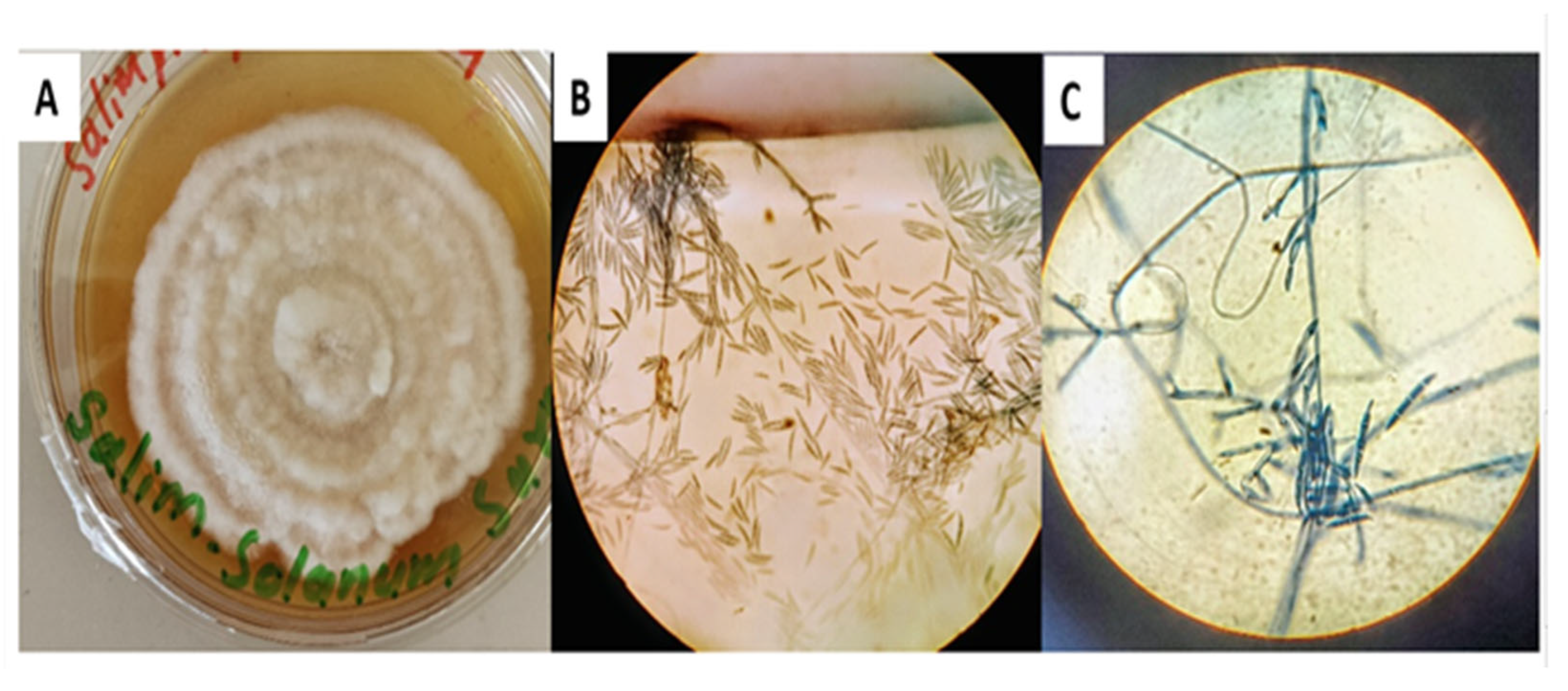
Figure 1. Morphological and microscopic characterization of F. solani: (A) Colony morphology on PDA medium showing concentric growth with cottony, fluffy texture; (B) Microscopic view showing septate hyphae and curved spindle-shaped macroconidia stained with lactophenol cotton blue (40×); (C) High magnification image (100×) highlighting conidial structures characteristic of F. solani.
Chemistry 07 00130 g001
Figure 2. Phylogenetic tree based on ITS rDNA sequence analysis showing the relationship of the tested endophytic fungal isolate (highlighted as “Tested Sample”) with other reference strains of F. solani retrieved from GenBank. The tree was constructed using the neighbor-joining method, indicating close evolutionary relatedness between the tested strain and previously reported F. solani sequences.
Chemistry 07 00130 g002
Figure 3. Antibacterial activity screening of fungal isolates against S. aureus using an agar plug diffusion assay. Three fungal isolates were tested (positions 1, 2, and 3), with isolate 2 (F. solani) showing the largest inhibition zone, indicating stronger antibacterial activity.
Chemistry 07 00130 g003
Figure 4. Visualization of the purified compound using TLC under UV light. Two concentrated vials (A and S) were applied to the plate, showing distinct fluorescent bands corresponding to the isolated compound.
Chemistry 07 00130 g004
Figure 5. GC–MS chromatogram of the purified compound isolated from F. solani, showing a major peak at a retention time of 16.938 min. The strong signal intensity at this retention time indicates the successful separation and high abundance of the target compound. The total ion chromatogram (TIC) reflects the purity and dominant presence of the compound in the analyzed sample.
Chemistry 07 00130 g005
Figure 6. Mass spectral fragmentation pattern and library search results of the purified compound corresponding to the retention time of 16.938 min. The molecular ion peak at m/z 270, along with prominent fragment ions at m/z 74, 87, 43, and 55, aids in the identification of the compound. The NIST library search matched the spectrum to hexadecanoic acid, a methyl ester (CAS# 119407, 119408), with high similarity scores (99%, 99%, and 98%).
Chemistry 07 00130 g006
Figure 7. FTIR spectrum of the purified compound showing characteristic peaks corresponding to the respective functional groups present within the molecule.
Chemistry 07 00130 g007
Figure 8. Elucidated chemical structure of 4-hydroxyphenyl 8-chlorooctanoate isolated from F. solani. The structure features a para-substituted phenolic ring connected via an ester linkage to an eight-carbon aliphatic chain terminating with a chlorine atom.
Chemistry 07 00130 g008
Figure 9. ADME analysis of the compound-270 with its physicochemical properties, solubility in water, pharmacokinetics, drug likeness, and its medicinal chemistry. The drug-like redar is also given, where red line shows the permitted range for a drug-ike molecule.
Chemistry 07 00130 g009
Figure 10. Figure (A) represents the docked complex of ligand 4-hydroxyphenyl 8-chlorooctanoate and its target receptor (14-alpha demethylase) of A. fumigatus and (B) shows different bonding and different interactions with various amino acid residues.
Chemistry 07 00130 g010
Figure 11. Figure (A) represents the docked complex of ligand 4-hydroxyphenyl 8-chlorooctanoate and its target receptor (CalpP-Protease) of S. aureus and (B) shows different bonding and different interactions with various amino acid residues.
Chemistry 07 00130 g011
Figure 12. Figure (A) represents the docked complex of ligand 4-hydroxyphenyl 8-chlorooctanoate and its target receptor (capsid protein) of HIV. (B) Shows different bonding and different interactions with various amino acid residues.
Chemistry 07 00130 g012
Figure 13. Figure (A) represents the docked complex of ligand 4-hydroxyphenyl 8-chlorooctanoate and its target receptor (Cdk-9) of cancer cells. (B) Shows different bonding and different interactions with various amino acid residues.
Chemistry 07 00130 g013
Figure 14. Molecular dynamics simulation analysis of 3V5I-Compound270 (blue) and 5FRB-Compound270 (red). (A) RMSD and (B) RMSF.
Chemistry 07 00130 g014
Figure 15. (A) represents the β-factor plot, while (B) represents the Rg plot.
Chemistry 07 00130 g015
Disclaimer/Publisher’s Note: The statements, opinions and data contained in all publications are solely those of the individual author(s) and contributor(s) and not of MDPI and/or the editor(s). MDPI and/or the editor(s) disclaim responsibility for any injury to people or property resulting from any ideas, methods, instructions or products referred to in the content.