Review

Revolutionizing Cancer Vaccine: The Power of Advanced Nanotechnology

by
Saranya Udayakumar
1,†,
Shangavy Pandiarajan
1,†,
Devadass Jessy Mercy
1,
Jayaprakash Suresh
1,
Jashwanth Raj Jagadeesh kumar
1,
Agnishwar Girigoswami
1,* and
Koyeli Girigoswami
2,*
1
Medical Bionanotechnology, Faculty of Allied Health Sciences, Chettinad Hospital and Research Institute, Chettinad Academy of Research and Education, Chettinad Health City, Kelambakkam, Chennai 603103, India
2
Centre for Global Health Research, Saveetha Medical College, Saveetha Institute of Medical and Technical Sciences, Thandalam, Chennai 602101, India
*
Authors to whom correspondence should be addressed.
These authors contributed equally to this work.
Chemistry 2025, 7(3), 97; https://doi.org/10.3390/chemistry7030097
Submission received: 15 March 2025 / Revised: 8 May 2025 / Accepted: 10 June 2025 / Published: 13 June 2025
(This article belongs to the Section Medicinal Chemistry)

Abstract

:
Developing an effective vaccine that is safer is the main focus in the field of cancer immunotherapy. Among other therapeutic approaches, cancer nanovaccination is formulated to deliver tumor adjuvant or antigen to the antigen-presenting cells (APCs) to prevent cancer relapse and metastasis. It has shown excellent efficacy in inhibiting cancer growth. Herein, we discussed various forms of nanovaccines, including lipid-based nanovaccines, metal-based nanovaccines, carbon nanotube-based nanovaccines, PLGA-based nanovaccines, exosome-based nanovaccines, dendritic cell-based nanovaccines, and self-adjuvant nanovaccines in cancer immunotherapy, including their therapeutic effect. We expect that the investigated content will provide a valuable reference for future research and the development of nanovaccines for cancer treatment.

Graphical Abstract

1. Introduction

A vaccine is one of the most momentous discoveries in medicine. It fights against pathogens by activating our body’s immune system and helps our body to distinguish pathogens. To develop immunity, these vaccines mimic the pathogens to educate the immune system. Various studies confirm that vaccines have effectively prevented diseases caused by bacteria, viruses, and other pathogens [1]. Chickenpox, mumps, typhoid, and pertussis are some diseases completely eradicated by vaccines [2]. The cytotoxic T lymphocyte-based multi-epitope vaccine has also been designed to fight the most deadly pandemic, SARS-CoV-2 [3]. Life-threatening diseases like cancer are considered to be a heavy burden for human life, although much effort has been made to find a cure. Surgery, radiotherapy, chemotherapy, and immunotherapy are the most common clinical strategies to treat cancer. Among these, cancer immunotherapy involving nanoparticles is seen as a revolutionary strategy that focuses on modulating the immune system instead of cancer growth [4]. Many natural compounds and their nanoformulations have shown promising results in combating cancer cells [5,6,7] and other diseases [8,9], although targeting the cancer stem cells and killing them can become a potential treatment modality.
On the other hand, immunocytochemical markers have also been used to differentiate between primary and secondary neoplasms in hepatic masses [10]. Thus, an amalgamation of immunology and nanotechnology can lead to improved diagnosis and therapy. Getting knowledge about the cancer cell death pathway can result in an effective vaccine production plan. Nanotechnology is a promising field in nanomedicine with its application in cancer imaging [11], targeted drug delivery [12], selective killing of cancer cells [13], wound healing [14,15], antimicrobials [16,17], biosensing [18], and cancer therapy [7]. Compared to conventional methods, nanotechnology has numerous prospects for enhancing the therapeutic efficiency of cancer vaccines. It can combine the adjuvant and antigen components to optimize immune stimulation, making it one of the significant benefits. Various applications have been used to load antigens and adjuvants into specific nanoparticles. (i) encapsulating the antigen inside the nanoparticles and (ii) attaching the antigens or adjuvants to the nanoparticle’s surface. Adjuvants are systemically dangerous to the body and produce side effects like fever, tiredness, diarrhea, and nausea. Nanoparticulate administration shields the adjuvant from degradation and helps safeguard the body from side effects [19].
In recent times, nanoparticles have also been used to design new cancer vaccines. These vaccines are aimed at triggering a strong immune response to fight and prevent cancer. Based on the tumor-specific antigens (TSAs), three types of vaccines have been developed: cell-based vaccines, protein/peptide vaccines, and genetic vaccines (including mRNA vaccines) [20]. Cell-based vaccines primarily target all tumor cells, including allogeneic and autologous tumor cells. Meanwhile, peptide vaccines are created using the amino acid sequence of a specific epitope found in the pathogen’s antigenic gene. A selected complete protein contains both protective and harmful sequences, and these peptide vaccines can avoid immunopathological proinflammatory regions and prevent mutational escape by using extensively conserved recessive epitopes. Nanoparticles modified with sugars (mannose) and bone-building molecules (bisphosphonates) can target antigen-presenting cells (APCs) and boost DNA vaccines. Liposome vaccines containing specific fats and sugars can target dendritic cells (DCs), a key player in the immune system, and activate innate and adaptive immunity [21]. Researchers are also exploring nanoparticles that can encapsulate antigens and help DCs mature and present them more effectively. Genetic vaccines are developed to target specific tumor-affected proteins, and they provide immunity for an extended period compared to other types of vaccines. Scientists are developing lipid nanoparticles to deliver messenger RNA (mRNA), the genetic material that the cells use to make proteins. These nanoparticles can protect the mRNA and deliver it to the right immune cells. Another approach uses a specific kala peptide (KALA) to target DCs and deliver mRNA vaccines, improving immune cell activation. These are a few examples, and the document highlights the promise of nanotechnology to create more effective cancer vaccines [22,23].

2. Cancer Immunotherapy

Cancer immunotherapy has received a lot of attention due to the significant clinical application of immune checkpoint blockade and chimeric antigen receptor T cell therapeutics. Recently, nano-delivery systems in vaccination have provided a highly adaptable platform for developing methods to enhance anti-tumor activity. This approach improved protection and immunogenicity and achieved targeting capabilities with minimal toxicity. In addition, combination therapy with other treatments, carrier design, targeting site, and cargo release techniques are some of the approaches that have been explored to improve the anticancer efficacy of nanovaccines. Apart from conventional vaccines consisting of adjuvants and antigens, nanoparticle-based chemotherapy, radiation, and other treatments can trigger the release of tumor antigens in the target tissue, thereby imitating anti-tumor immune responses. This type of vaccine-like nanomedicine not only eradicates primary tumors but also helps get rid of metastatic tumors and stops tumor recurrence [24]. These typical immunotherapies can be implemented in several ways, including immune checkpoint blockage, tumor therapeutic vaccines, and adoptive cell transfer therapy. These vaccinations have the potential to reduce local immunosuppression and stimulate immune response to the targeted site of diseases. They can be substantially effective with the use of nanomaterials. Antigens, nano adjuvants, and nanocarriers like liposomes and polylactide-co-glycolide (PLGA) are the main components of cancer nanovaccines. Tumor-associated antigens (TAA) can be expressed by multiple types of cancer cells and by healthy normal cells. Genetic processes like uncontrolled gene expression in malignant cells produce neoantigens. Biogenic nanocarriers like outer membrane vesicles and exosomes were demonstrated to have excellent immunotherapeutic efficacy. Schneider et al., 2018 [25] established that tumor antigens with virus-like particles stimulated an effective immune response to treat cancer. Moreover, antigens conjugated or encapsulated with nanomaterials have improved antigen-specific cytotoxic T lymphocyte proliferation [25]. Geall et al. developed an mRNA-encapsulated liposomal vaccine, proving that synthetic mRNA-encoding antigens are safer than plasmid DNA and significantly improve immunogenicity [26].
The fields of biology, medicine, and even engineering are paying more attention to virus-like particles, or VLPs. They were initially employed to investigate the intricate structure of viruses, but they soon discovered a new purpose in the creation of vaccines, initially to protect against viruses and later as adaptable platforms for creating various vaccines [27]. They have more recently been modified to function as tiny delivery systems that deliver medications straight to the body’s desired cells [28]. Without any infectious genetic material, VLPs are composed of viral proteins that spontaneously come together to form shapes that mimic those of actual viruses. Similar to real viruses, they are highly efficient at activating the immune system, particularly by stimulating dendritic cells, which are essential for initiating immune responses. They are, therefore, excellent candidates for creating vaccines that are safe, efficient, and reasonably priced to prevent cancer and infectious diseases. Scientists make viral proteins in non-human cells, purify them, and then let them self-assemble into empty virus shells to produce VLPs. The immune system responds strongly to these because they closely resemble the original virus, especially by activating dendritic cells. In the lymph nodes, these cells subsequently notify and stimulate helper and killer T cells, with CD8+ T cells being essential for locating and eliminating infected cells [29].
Particles with repeating, regularly spaced patterns on their surface are the ones that B cells react to the best. According to research, 12–16 of these repeating units, known as epitopes, can significantly activate B cells when they are positioned 5–10 nanometers apart. Many RNA viruses naturally have this configuration, called an “immunons,” which is made up of 180 copies of the coat protein spaced about 5 nanometers apart on a shell that is 30 nm wide [28]. The immune system is effectively stimulated by these virus-like structures. They are readily identified by natural IgM antibodies and firmly cluster B-cell receptors, inducing a potent immune response. This identification stimulates the formation of germinal centers, helps deposit antigens on follicular dendritic cells, activates the classical complement pathway, and aids in the development of durable plasma cells that generate antibodies. VLPs can be modified to carry full-length antigens and even RNA, and they mimic these virus structures. They are, therefore, effective instruments for triggering B-cells and producing enduring immunity. Certain VLPs, particularly those derived from RNA viruses, can also contain unique immune-boosting sequences, such as CpG DNA or bacterial RNA, which activate B cell toll-like receptors (TLR7/8 or TLR9) [30]. This increases plasma cell formation, improves the quality of the antibody response, and further increases the production of particular antibody types, such as IgG2a and IgA. Bacterial RNA seems to be the most effective at boosting B-cell activity.
In an effort to lower the incidence of virus-associated cancer, VLPs have been used as preventive vaccines against a number of oncogenic viruses. Only VLP-based vaccines against the human papillomavirus (HPV) and hepatitis B virus (HBV) have been approved by regulators for use in humans thus far; candidates against other carcinogenic viruses are still in the preclinical stage of development [31]. The first virus for which a VLP-based vaccine was created was HBV, a hepatotropic virus associated with hepatocellular carcinoma and chronic liver disease. The initial formulations made from infected plasma were eventually replaced by recombinant yeast-derived VLPs expressing the HBV surface antigen (HBsAg), which improved immunogenicity and safety. Recent innovations include Sci-B-Vac, which is produced in CHO cells with multiple antigens and glycosylation to enhance immune response, and Heplisav-B, which adds a CpG adjuvant for enhanced immunostimulation. Hyperglycosylated VLPs and novel expression systems like HEK293F and Pichia pastoris are being used in ongoing efforts to maximize immunogenicity and safety. The most frequent cause of genital viral infections is non-enveloped DNA viruses called human papillomaviruses (HPVs). The majority of HPV infections go away on their own in a matter of months to two years, but persistent infections with high-risk genotypes can cause cancers of the cervix, anus, vulva, vagina, penis, and oropharynx. Early in the 1990s, VLP-based HPV vaccines were created by expressing outer capsid proteins, specifically the L1 protein. As a result, two FDA-approved vaccines were created: Gardasil, which is currently known as Gardasil 9 and covers nine HPV types, and Cervarix, which targets HPV types 16 and 18. Adjuvants such as monophosphoryl lipid A (MPL) and aluminum salts are used in both vaccines to increase immunogenicity. High production costs make it difficult for them to be widely used in environments with limited resources despite their effectiveness. In order to increase protection across HPV genotypes, more recent approaches concentrate on the conserved L2 protein. L2 is integrated into chimeric platforms like L1-L2 VLPs or engineered constructs like Trx-L2-OVX313 because it cannot form VLPs on its own. Strong immunogenicity and cross-protection have been shown for these designs. Notably, preclinical models have demonstrated that a bacteriophage-based VLP vaccine (16L2-MS2) is more effective than current vaccines [32].
A promising strategy for targeted tumor treatment is provided by vaccines that strengthen the immune system’s defenses against antigens unique to cancer. Tumor-associated antigens (TAAs) can be presented effectively by VLPs using two primary methods: (1) functionalizing the VLP surface to bind TAAs, such as proteins, peptides, glycans, or haptens, for the best immune cell presentation and (2) altering the structural protein coding sequence of the VLP to produce a chimeric protein that presents the TAA. One recombinant protein is expressed and self-assembled into particles that exhibit the TAA in the second strategy, whereas VLPs, peptides, and linkers are synthesized independently and joined via cross-linking in the first strategy.
Although the conventional vaccines have served society for a long time period, the limitations are less bioavailability, targeted delivery, and extended circulation time. Nanotechnology is crucial in advancing vaccine development because it improves how vaccines are delivered, how well they work, and how stable they remain over time. This technology plays a key role in creating different types of vaccines, such as mRNA vaccines (like those from Pfizer-BioNTech and Moderna that use lipid nanoparticles), DNA-based vaccines, and subunit vaccines, which use specific components of a virus. Additionally, nanotechnology opens the door for vaccines to be given through the nose or skin, offering less invasive and more practical options—especially helpful during large-scale vaccination efforts [33].

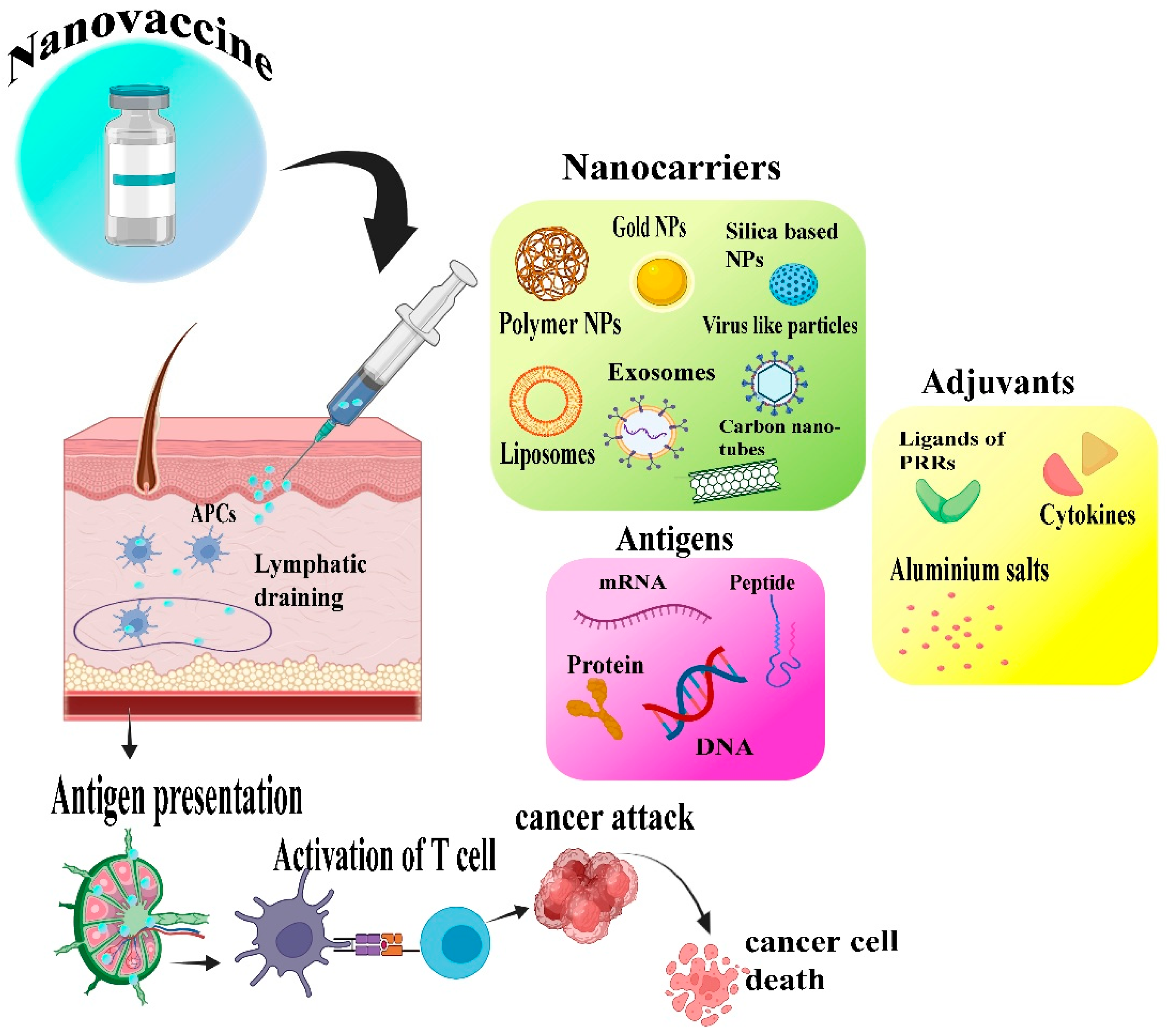
3. Nanotechnology’s Role in the Development of Cancer Vaccine

Nanotechnology has unfurled its application in various biomedical fields, such as the development of anticancer agents [7,34], antiamyloid agents [13], nanocarriers for delivering genes [35], antimicrobial agents [17], etc. Naturally, our body has a defense mechanism against abnormal cell development; it can detect and eliminate them to a certain extent. Numerous studies have been conducted on immunosurveillance. Immunotherapy’s purpose is to facilitate tumor remission by altering the scales in favor of immunosurveillance. Existing techniques, such as adoptive T cell transfer and immune checkpoint inhibition, show promise but are limited and may cause side effects. In such conditions, nanoparticles emerge as a game changer with a powerful immune response for effective tumor eradication [36]. These small particles serve as effective delivery vehicles, transporting antigens (foreign molecules) and adjuvants (immune-boosting chemicals) straight to specific immune cells, particularly DCs. Nanoparticles have the potential to greatly improve vaccination efficacy by improving antigen delivery to DCs and stimulating cytotoxic T lymphocytes (CTLs), which are immune cells that kill cancer cells. Figure 1 shows the different roles of nanoparticles in cancer vaccine development.
In nanotechnology, vaccines are designed by involving micro-adjuvants and antigens in order to extend immune activation. This improves systemic immune response against tumor cells. These developments in the design of the vaccine assist in the targeting of antigens through lymph nodes and APC, which influence the immune response in cells. In cancer vaccines, the utilization of nanocarriers for targeting antigens and delivering micro adjuvants could relieve lymph node constriction. The progress in the cancer vaccine has been magnificently improved by strengthening the immune reactions by fine-tuning the vaccine with natural or synthetic nanomaterials. By lowering the adverse effects, these nanomaterials demonstrate how important T-cell activation and antigen kinetics are for creating successful cancer vaccines. Vaccines such as Vx-001 and GV1001 demonstrated extended immunogenicity and improved survival rates in patients with stage 5 melanoma and small lung cell carcinoma [37].
Endosomal escape is a fundamental element of nanoparticles’ antigen delivery (Figure 2). Endosomes are cellular compartments in which substances that enter the cell are digested. Antigens must leave endosomes and reach the cytoplasm, the cell’s core, to offer themselves to CTLs effectively. A range of cancer biomarkers—such as circulating tumor cells, cell-free DNA and RNA (including microRNA), and extracellular vesicles—can be clinically applied to detect, monitor, and assess the prognosis of glioblastoma [38]. Researchers investigate numerous techniques for accomplishing this, such as pH-responsive nanoparticles that release their payload when they reach the endosome’s acidic environment or nanoparticles with membrane-destabilizing capabilities. These advances show potential for improved cytosolic antigen delivery and strong CD8+ T cell responses, a kind of CTL required for anti-tumor immunity [39]. The study published by Urbanavicius et al. (2018) highlights the great potential of antigen-delivery techniques based on nanoparticles in order to overcome the limitations of cancer immunotherapy [40]. However, further research is needed to investigate the impact of nanoparticles on the immune system. Targeted nanoparticle delivery technologies show great promise for increasing vaccine efficacy and safety: (1) By protecting the antigens from degradation, allowing them to circulate longer in the body, and increasing the likelihood of immune cell contact; (2) by attaching specific antibodies to their surfaces, nanoparticles can be guided onto DC surface receptors, providing precise targeted antigen delivery; and (3) for reducing the risk of adverse effects using biocompatible nanostructures.
The potential usage of nanoparticles combined with micro-adjuvants enhances the responses of T cells and can be used as therapeutics for cancer vaccines. A micron-sized adjuvant forms crystals that absorb proteins, producing a local storage place at the injection site when formulated with nanoparticles. These adjuvants play a crucial role in enhancing the CTL responses, thus activating the TH1 driving pathway for efficient anti-tumor immunity. The combination of nanoparticles and micron-sized adjuvants initiates the ability to drain into the lymphatic system, thus optimizing the immune reactions that create some strategies for cancer vaccines. The effects of microcrystalline tyrosine (MCT) storage place buildup are efficient antigen-specific CTL responses provided by the more prolonged exposure of the immune system to the antigen. A synergistic effect was also produced by the combination of nanoparticles and micro-adjuvants, which offer prolonged vaccine release and are key to promoting CTL responses [41]. Similarly, specific proficient T-cell responses were observed when a combination of the cucumber-mosaic virus-derived nanoparticle-integrated p33 epitope nanovaccine with micron-sized adjuvants was made. Challenging a tumor model was a slightly difficult process, although these combinations provided remarkable results for cancer [42]. This MCT activated the innate and adaptive responses that showed flexibility in moderating the immune reactions. It effectively concluded phase 2 trials about allergy immunizations, showcasing its safety and effectiveness in medical environments and reinforcing its capacity to expand CTL responses [41]. Similarly, the utilization of cell-derived components exhibited excellent anticancer immunity, showing off-target reactions’ challenges. The cell-derived components like whole cancer cell or cell-lysate-based nanovaccines, cell-derived nanovesicles, and cell membrane-coated nanovaccines were used in cancer treatment that improved clinical results and induced the memory to inhibit cancer and metastasis. The combination of RBC membrane with PD-1 blockade provided superior effects on cancer immunotherapy and motivated the researcher to work further on it. Encapsulations of adjuvants along with the targeting APCs have been discovered that exhibit brilliant effects of anticancer immunotherapy with the combination of immune checkpoint inhibitors such as anti-PD-1 antibodies [43,44]. Despite facing obstacles in mass production and quality assurance, nanovaccines derived from cells possess great promise in cancer therapy, awaiting additional investigation to conquer production challenges and guarantee tangible clinical advantage [45]. Tumor cells express two types of antigens: TAA [expressed on both cancer and normal cells] and TSA [expressed only in cancer cells]. As the name suggests, tumor-specific immune activation includes immune cells for the identification of cancer cells based on the activated adaptive immune system, whereas tumor non-specific includes agonist treatment with interferons (IFN-β, IFN-α, and IFN-γ), a ligand for toll-like receptors, and cytokines (IL-2, IL-5, and IL-8) [46]. To empower this tumor vaccine, the nanomaterials with intrinsic immunostimulatory (nano adjuvant) properties are used to facilitate antigen presentation or elicit an immune response. Anticancer vaccines are either surface conjugated by chemical modification or encapsulated within the nanoparticles. Encapsulation protects the body from systemic toxicity of adjuvants/antigens and provides efficient delivery to the target site. The nanoparticle-loaded vaccines easily enter the lymphatic system and allow competent transport to the lymph nodes (LNs). Various NPs have been created and are employed to deliver immunological stimulants or vaccinations to prevent cancer. Liposomes, VLPs, PNPs, AuNPs, MNPs, and polymeric micelles are examples of nanocarriers with excellent properties for cancer nanovaccination [47,48]. These systems of nanocarriers play a significant role in the regulation of the immune response to antigens and the stability of antigens. The advancement of the platform based on nanovaccines makes it possible to apply vaccinations and adjuvants therapeutically, improving cancer patient’s clinical outcomes [2].

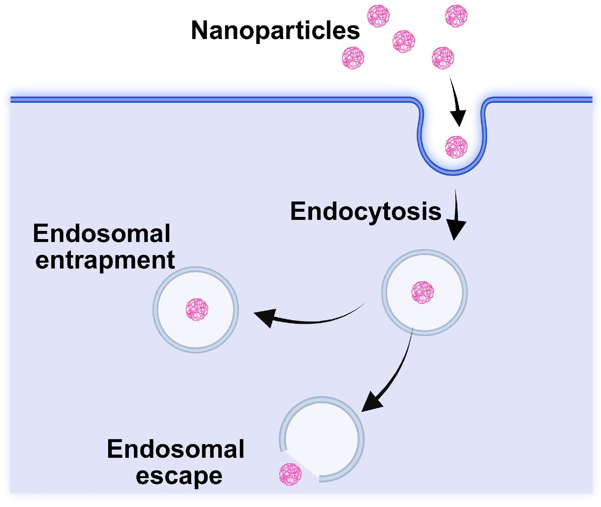
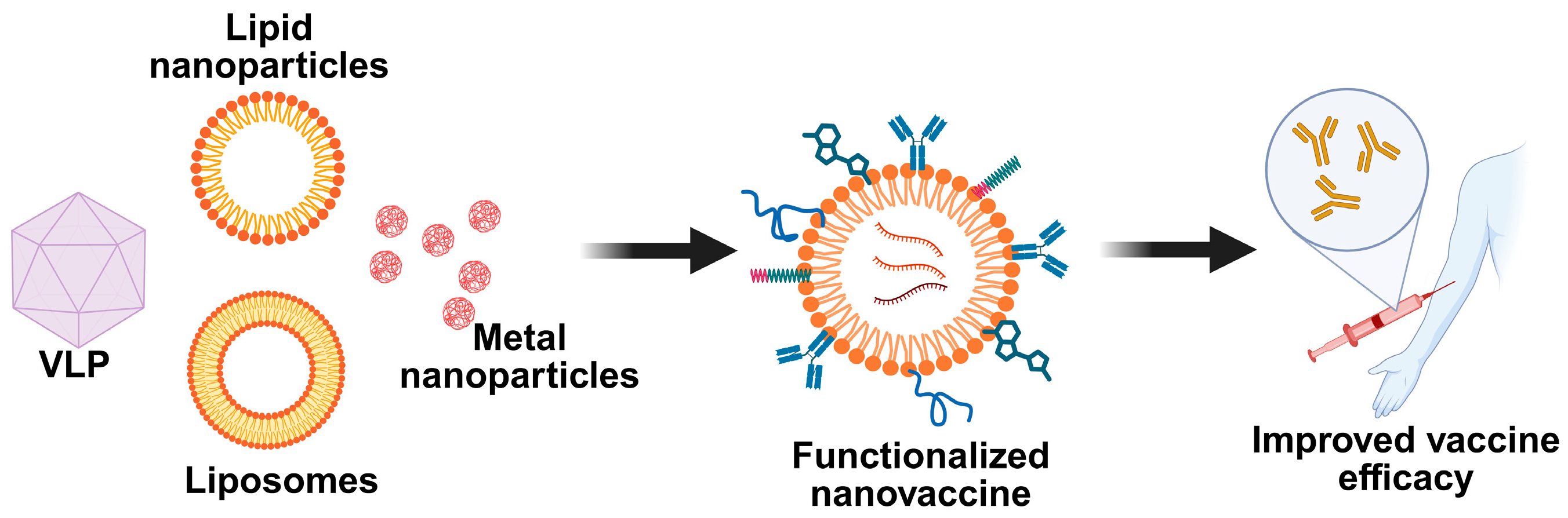
4. Types of Nanocarriers in Cancer Nanovaccines

There are synthetic, semisynthetic, and bio-based nanocarriers in cancer vaccines. Adjuvants and TAAs or neoantigens are combined and encapsulated in nanocarriers or bonded to the vector via interactions before being directed into APCs, therefore significantly enhancing the antigen’s immunogenicity. Proper carrier selection or chemical alteration guarantees the regulated release of vaccine components [49,50]. Many smart nanocarriers for the delivery of various vaccines are manufactured from relatively small protein fragments or genetic material (mRNA) as well as from different nanocarrier materials, such as polymers and liposomes. These nanocarriers have enhanced the effectiveness of immunizations, and further advancements are needed to improve their efficiency in patients.
Furthermore, researchers are thinking of combining these vaccines with other treatments to help patients get better results. One potential strategy is to combine the vaccines with chemotherapeutic drugs, which increase the immune system’s response to the vaccine. Furthermore, combining them with checkpoint inhibitors—a tactic that helps to remove “brakes” on the immune system—is another approach that shows promise for treating cancer [51].

4.1. Synthetic Nanocarriers

4.1.1. Lipid- and Polymer-Based Carriers

Lipid-based NPs, in particular, have the advantages of a straightforward composition and advanced industrial manufacturing technology [38], which presents a significant opportunity for future commercialization and clinical translation of tumor nanovaccines [52]. Many synthetic nanostructures have been proposed for designing vaccines and immunotherapy in previous review articles [53]. Several important concerns still need to be thoroughly considered before the possible clinical use of nanovaccines. Initially, before being used at the clinical level, the biocompatibility and biodegradability of nanocarriers, particularly those that are inorganic, need to be further investigated. Secondly, recognizing the suitable TAA is complicated and challenging. Thirdly, the efficiency of LN-targeting is difficult to ensure due to the difficulties of scale-up production (reproducibility and batch-to-batch consistency). Lastly, studies on the efficacy and possible side effects of combination therapy are desired, particularly in low-dose ratios [54].
Cancer vaccinations are made to deliver the tumor antigens and adjuvants to stimulate APCs and generate strong anti-tumor immune responses. It is possible to manipulate the surface modification of the adjuvant to change its function and change the size, shape, and surface charge of the nanovaccines. Moreover, nanovaccines can regulate the release of payloads in response to normal cues found in the tumor microenvironment, including immune checkpoint inhibitors and biological adjuvants. Furthermore, to prevent the drug from leaking into the bloodstream, which leads to off-target and has toxic effects on the system, when designing nanovaccines, the conjugate interaction effects of vaccine components and nanomaterials should be taken into account. The optimum size for designing the nanovaccine ranges from 20 to 200 nm, which is easy to concentrate in the spleen and lymph nodes, as the size could be easily taken by the APCs. The size is similar to known pathogens and can avoid the problems created by rapid diffusion, which causes low concentration of drug as the extracellular matrix captures it due to interstitial pressure in peripheral blood vessels [55]. Nanovaccines can be customized with targeting ligands that can bind to surface receptors specifically for more targeted delivery expressed by DCs, such as MUC-1, DEC205, CD11c, CD40, mannose receptors, CLEC9A, LOX-1, and Dectin-1. Furthermore, it is necessary to develop nanovaccines with endosomal escape capabilities to enhance the efficient intracellular release and absorption of adjuvants into immune cells. While there are still significant obstacles to overcome before these novel nanovaccine therapy approaches can become widely used, biotechnology and material science advancements will eventually make nanovaccines a viable option for the production of cancer vaccinations that are both highly effective and low-toxic [56]. Figure 3 explains the advantages of nanovaccines for cancer vaccine delivery.

4.1.2. Inorganic Nanocarriers for Nanovaccines

Among other therapies, immunotherapy has been recognized as an innovative approach to cancer treatment because it targets direct immune system modulation rather than an actual tumor. The process of developing peptide-based cancer vaccines begins with antigen and adjuvant selection. These adjuvants primarily stimulate the proliferation and differentiation of lymphocytes and APCs, further enhancing the immune response. The selected antigens can be delivered to lymph nodes specifically with the help of chemically enhanced nanocarriers. More specifically, metal nanomaterials like gold, calcium phosphate, mesoporous silica, silicon, liposomes, dendrimers, hydrogels, micelles, and other polymeric nanomaterials like chitosan, poly(lactic-co-glycolic acid) (PLGA), and polylactic acid are frequently used as vaccine carriers [57]. Researchers demonstrated that immature lymphocytes can be tracked using iron oxide and 19F based probes using magnetic resonance imaging (MRI) [58]. Metal–organic frameworks (MOFs) crystals (MIL-101) can also be directly imaged using HR-TEM [59]. Fe-MIL-101 has been engineered by researchers that exhibited selective ovarian cancer (OC) cell-killing capacity [60]. In a recent study, an airbag-embedded iron-based MIL-101 metal-organic framework (MOFMIL-101(Fe)) was engineered to ignite the tumor microenvironment (TME) in triple-negative breast cancer (TNBC) via bubble-mediated codelivery of and Fe2+/3+ to the tumor [61]. Researchers developed a Fe3O4@MIL-101-OH/Chitosan nanocomposite and utilized it as a carrier to adsorb and release the drug doxorubicin (DOX). The nanocomposite demonstrated a controlled release for 84 h at pH 5, achieving 80% of the DOX release after 60 h [62]. Hence, metal-organic nanovaccines can also be designed and become a promising theragnostic material that can combine immunotherapy with phototherapy and chemotherapy in a synergistic manner to improve anti-tumor efficacy. For a better combination of chemotherapy and metabolic immunotherapy, ZIF-8 was utilized to administer the anticancer medication doxorubicin and the metabolic medicine avasimibe [63]. Furthermore, many researchers have been working on metal-based therapies for the betterment of cancer [57].
A combination of antigens, adjuvants, and delivery mechanisms is a suggestion for successful cancer vaccines, which focus on stimulating the cell-mediated immune response. It also encompasses carbon nanotubes (CNT’s) prospects in cancer vaccine delivery methods, considering the cellular absorption mechanism, biodistribution, biocompatibility, and biodegradability. Zhao and colleagues investigated how CpG ODN transported through single-wall carbon nanotube (SWNT)-mediated delivery could enhance the vaccine’s cellular absorption and immune-stimulating properties, ultimately resulting in a more robust activation of the anti-tumor immune response [64]. Various studies emphasize the immune-stimulating properties of adjuvants and the role of functionalized CNTs in enhancing immune responses against cancer [65].

4.1.3. Dendritic Cell-Based Nanovaccines

Cancer nanovaccine have offered a promising approach in cancer immunotherapy, which stimulates the host immune system to eradicate the tumors either alone or in combination with other therapeutic agents. The therapeutic efficiency of cancer nanovaccines is increased by specific targeting of DCs via modulating the vaccine structure using DC-specific ligands [66]. DCs are indispensable elements for anti-tumor response in anticancer vaccination strategies due to their crucial role in priming CD8+ T cells against TAAs and APCs [67]. In this, the autologous monocytes are obtained from patients, and they are differentiated into DCs in ex-vivo with some maturation-inducing agents and contact with TAAs, and then they are reintroduced into the same patient [68]. Even though there are lots of advantages to DC-based immunotherapy in cancer in pre-clinical studies, there are some disadvantages, like the process of isolation, ex-vivo manipulation, reinfusion of cells, etc. [69]. Furthermore, there is evidence that negative immunoregulatory substances produced by DCs, such as indoleamine 2,3-dioxygenase (IDO), could decrease the proliferation and activity of T-cells, resulting in tumor escape from immune surveillance. To overcome these constraints, scientists are developing agents that target DCs through conjugation of antibodies on NPs or ligands or antigens for binding to DC-specific receptors.
PLGA nanoparticles (PLGA-NPs) have gained significant attention as an effective strategy for cancer vaccine delivery due to their biodegradable nature, biocompatibility, and ability to encapsulate a variety of antigens and immunomodulators. As an FDA-approved polymer, PLGA naturally breaks down into lactic and glycolic acids in aqueous environments, making it highly suitable for medical applications. Studies have highlighted the potential of PLGA-NPs in targeting DCs, which are key players in initiating immune responses, thereby enhancing T cell-mediated immunity against cancer. Compared to soluble formulations, PLGA-NPs provide several benefits, including protecting antigens from enzymatic degradation, ensuring targeted delivery to DCs, and facilitating the co-delivery of immunomodulators like Toll-like receptor (TLR) ligands. The ability to customize the physical and chemical properties of PLGA enables the controlled release of encapsulated substances, potentially leading to less frequent dosing and better treatment plans [70].
Additionally, PLGA-NPs can be modified with poly(ethylene glycol) (PEG) to enhance stability and target specific immune tissues, demonstrating their adaptability in vaccine development. Some research indicates that DCs efficiently take up PLGA-NPs through phagocytosis facilitated by surface receptors like the mannose receptor, DEC-205, and DC-SIGN. Attaching targeting molecules or DC-specific antibodies to PLGA-NPs further improves their specificity for DCs, enhancing vaccine effectiveness [71].
PLGA-NPs have the ability to activate both CD4+ and CD8+ T cells, which are essential for effective cancer immunotherapy [72]. Notably, these nanoparticles can be designed to deliver antigens either to the cytoplasm, promoting MHC class I presentation, or to endosomes, facilitating MHC class II presentation. This adaptability broadens their potential for immune activation. Ongoing research focuses on refining PLGA formulations to further enhance cellular immunity by optimizing key physical properties such as hydrophobicity, polymer composition, particle size, and surface charge. Additionally, PLGA-NPs hold promise for activating innate immune cells like natural killer (NK) cells, providing a targeted approach for stimulating NK cell activity without resorting to systemic cytokine administration. Co-delivering tumor antigens and TLR ligands within PLGA-NPs presents a promising approach to effectively leveraging NK cell-mediated anti-tumor responses [73].
Thus, PLGA-NPs serve as a versatile and powerful platform for cancer vaccine delivery, capable of eliciting strong T cell-mediated immune responses while engaging innate immune cells to enhance anti-tumor immunity. By refining PLGA formulations and exploring their potential in activating various immune cell populations, researchers aim to develop comprehensive cancer vaccines that can target both MHC class I positive and negative tumor cells while minimizing systemic side effects. The diverse functionalities of PLGA-NPs offer exciting prospects for the development of next-generation cancer immunotherapies [74].
An ex vivo study demonstrated that DCs can be extracted from a patient, loaded with tumor antigens and immune stimulants in a lab, and then reintroduced to activate T-cells. However, this approach is expensive and labor-intensive due to the use of autologous cells. Nanovaccines present a promising alternative, acting as nanoscale carriers that deliver tumor antigens and adjuvants directly to DCs. This targeted delivery enhances immune responses, promoting intense anti-tumor activity and potentially improving cancer protection [75].
The effectiveness of nanovaccines depends on key factors such as immunogenicity, cytosolic delivery, and targeting. Immunogenic components stimulate immune responses, while cytosolic delivery ensures optimal T-cell activation. Targeting can be passive or active. Passive targeting depends on nanovaccine sizes—smaller ones (<200 nm) diffuse into lymph vessels, reaching lymph nodes and interacting with DCs, while larger ones remain at the injection site, engaging skin-resident DCs that then migrate to lymph nodes. Active targeting involves functionalizing nanovaccines with ligands or antibodies that bind specifically to DC receptors, ensuring precise cargo delivery [76].
Beyond DC activation, nanovaccines also explore direct T-cell stimulation through artificial antigen-presenting cells (aAPCs), which mimic natural DCs. By optimizing antigen and adjuvant delivery, refining size and targeting strategies, and integrating aAPC technology, nanovaccines hold significant potential for advancing cancer immunotherapy [75,77].

4.2. Semisynthetic Nanocarriers

A personalized cancer vaccine was developed that targets both sex cells and mutated epitopes. They aimed to determine the therapeutic efficacy of the combination of both sex cells and two neoantigens. To expand the therapeutic efficacy, the three sets of multi-targeted vaccines (MTV), mutated epitopes (Mutated-MTV), and germline epitopes (GL-MTV) were developed, and a combination of both (Mix-MTV) was also explored. Positively, the Mix-MTV combination of both provided a significant result, thus inducing cytokine production and increasing therapeutic effect [78]. Interestingly, to permit bedside customization of cancer vaccines for individual patients, Cu-free click chemistry was used for peptide-VLP coupling. This Cu-free click chemistry was non-toxic, highly efficient, and enhanced the immunogenicity that creates coupling of peptides with VLPs at the bedside. These lack cellular toxicity, which leads to selective labeling, tracking, and imaging of cells in vivo. By enhancing immunogenicity, the VLP-based vaccine was the most suitable for effective immunization against cancer progression and prevented the onset of antigen-escape variants [78].

4.3. Biogenic Nanocarriers (Exosome-Based)

The involvement of immunotherapy has been emerging in cancer treatment, which results in the search for an effective nanocarrier. Exosomes are small vesicles that are secreted by different cells in our body. By using tumor cell-derived exosomes (TEX), dendritic cell-derived exosomes (DEX), and ascitic cell-derived exosomes (AEX), immune responses can be triggered [79,80]. These exosomes show promise for preparing the immune system to recognize and kill cancer cells [81]. The immunological response triggered by exosomes includes their purification process and involvement in boosting both cellular and humoral immunity (antibody reaction). It also recognizes the expanding use of nanotechnology in medicine, including the potential of carbon nanotubes (CNTs) and quantum dots (QDs) in cancer immunotherapy. Exosomes have the potential to be used as a nanoscale cancer vaccine due to their unique properties. Phase I clinical studies for colorectal cancer, melanoma, and non-small cell lung carcinoma have produced promising findings [82]. Exosomes produced from several sources (DCs, tumor cells, and ascitic fluid) have been shown to activate immune responses against established cancers. Exosomes appear to activate a variety of immune cells, including T lymphocytes, NK cells, and DCs. This showed that they can train the immune system to recognize and suppress cancer cell development [83]. Another promising option is coupling exosomes and nanotechnology, such as CNTs, to develop novel and perhaps more effective cancer vaccines.
Compared to other types, higher immunogenicity is observed in the tumor cell exosomes, which are treated with heat. Naïve T cells are stimulated when the immune system simulators (CpG adjuvants) are combined with exosome therapy. On the other hand, CNTs have demonstrated promising effects in cancer treatment and imaging, and they can be used in cancer vaccine delivery systems. Some studies showed that CNTs coupled with tumor antigens can activate anticancer responses in animal models. However, there has been little research into combining CNTs with exosomes, which has the potential to be a game-changing strategy for developing innovative cancer vaccines [84].
Even though there are many advantages, disadvantages are also present for nanocarriers. Table 1 shows the advantages and disadvantages of nanocarriers used in vaccine design.

4.4. Functionally Designed Cancer Nanovaccines

4.4.1. Neoantigen-Based Nano Vaccines

In recent years, neoantigen-based nanovaccines have become one of the most promising therapeutic approaches to tumors. But still, it faces minimal cross-presentation and substantial endocytosis pathway degradation. A new thiolated nano-vaccine was developed to improve its efficacy, which enabled the direct cytosolic delivery administration of neoantigen and receptor 9 agonist CpG-ODN. By avoiding endo- or lysosome degradation, this method stimulated APCs and significantly improved T-cell immunity by boosting neoantigen uptake and local concentration [89]. This study reported that in vivo immunization of H22-bearing mice with thiolated nano-vaccine stimulated antigen presentation on DCs. The vaccination also effectively prevented tumor growth. Hence, this study proved that the thiolated nanovaccine, in conjunction with anti-programmed cell death protein-1 antibody (PD-1), effectively reversed immunosuppression and therapeutic approach for cancer [89]. The FDA has approved cell therapies, such as the DC-based vaccination and chimeric antigen receptor (CAR) T-cell therapy. CTLA-4 inhibitors, PDL1 inhibitors, and PD1 inhibitors are some inhibitors that were approved for effective therapeutic use. Moreover, cancer vaccination is an appealing approach because it can be developed to the patient’s mutanome by involving neo-antigens with minimal side effects. In addition, these types of vaccinations were developed to prevent MHC-I downregulation and lack of efficacy due to mutation and thymic tolerance. Combining PAMPs and DAMPs with multiple antigens into vaccine nanoparticles has boosted the possibility of provoking a complete cytotoxic and memory T-cell response [90].
Neoantigens and in vitro transcribed (IVT-mRNA) antigens have been predicted to be excellent candidates for cancer therapeutics. Kranz et al., 2016 [91] created mRNA-lipoplex (RNA-LPX) by combining liposome with neoantigen-based mRNA. Further, it was injected intravenously into both mice and human melanoma patients, efficiently delivering mRNA to lymphoid DCs, which induced strong T-cell responses and improved immunotherapeutic efficacy [91]. Overall, nanovaccines have been explored as potential vaccines to induce anticancer immunity in small animals and can be combined with other therapeutic methods for synergistic cancer treatment [92].

4.4.2. STING Agonist Based Nanovaccines

The remarkable development of cancer immunotherapy examines nano-based interventions such as mRNA cancer vaccines and nanoformulations for activating STING proteins presently undergoing clinical examinations [93]. Several studies have established that STING agonist-based nanovaccines can activate STING signaling and increase cyclic dinucleotides’ bioavailability and immunotherapeutic efficacy for cancer [94]. These proteins help fight against viral infections and cancer by producing interferons. The resistance of cancer immunotherapy and the immunosuppressive nature of the tumor microenvironment have been developed through the utilization of combination therapy [95,96]. An et al., 2018 demonstrated that cytotoxic cationic silica nanoparticles were employed to deliver a STING agonist to the immune cells within the tumor microenvironment and cause necrotic cell death [97]. Several strategies, including inherent nano adjuvants, delivery of antigens and adjuvants in a single nanocarrier, targeted delivery to immune cells, improved immune cell uptake, cross-presentation, and cytosolic delivery, have further improved the efficacy of cancer vaccination. By using RBCs as a source of membrane material, it has been demonstrated that nanoparticles coated with RBC membranes were not cleared by the immune system and circulated for longer periods [98]. Following this RBC membrane-coated nanoparticle work, researchers developed a variety of cell membrane-coated nanoparticles for antibacterial and anticancer vaccines [19].

4.4.3. Self Adjuvant Nanovaccines

The numerous advantages of nanovaccines, such as enhanced lymph node (LN) access, optimum antigen packing and presentation, and generation of a long-lasting anti-tumor immune response, have generated significant interest in cancer therapy. Different kinds of nanoparticles have been developed as a delivery strategy for cancer vaccines to transport adjuvants and antigens to APCs and lymphoid organs. Generally, active cancer vaccines contain tumor cells, exosomes, peptides, proteins, and nucleic acid with TSA and TAA to induce an immune response to inhibit tumor growth. In particular, aluminium-based adjuvant was the first adjuvant used for vaccine development [99]. MF59, aluminium-based adjuvants, and liposomal adjuvants (AS01) are some of the approved adjuvants used for human vaccinations [100,101]. The biomimicking technique improved the vaccination efficacy of nanoparticles. Mimicking natural infections can trigger innate immune responses via pattern recognition receptors (PRRs), which help to produce long-lasting adaptive immunity. Based on this strategy, various cancer vaccines have been developed using recombinant plant viruses, virus particles, and archaeosomes [102].
Nanovaccines activate the dendritic and T cells by nano-scaled complexes of antigens and immunostimulants, which fight against tumor cells. The antibody or peptide design should be more appropriate for CLRs as they stimulate anti-tumor responses through type 1 and type 2 interferon production enhancement, and they can mediate tumor evasion by inducing the IL-10/TGF-β factor production, upregulating FasL expression, and downregulating the MHC molecules. Targeting CLEC9A cannot promote the cross-presentation of antigens without the use of adjuvants or danger signals, although it stimulates strong increases in humoral responses besides CD8+ T-cell responses. Furthermore, targeting TLRs stimulates strong immune responses to fight against tumor cells; by doing so, it may also trigger undesirable cytokine release syndrome or autoimmune reactions [66].
To activate anticancer immune responses, nanovaccines delivering whole tumor lysate or formed from tumor cell lysate have the potential to expand the repertoire of tumor antigens as targets for the immune system while exploiting immunogenic cell death [45]. Nanoparticle-based approaches improve anti-tumor efficacy with minimal toxicity and promote the activation of T cells against tumor cells. Immunogenicity of the tumor antigen and improved anticancer T cell response are obtained in the complete tumor-cell-based nanovaccine as original antigens are provided in this antigen-based nanovaccine. To generate immune checkpoint blockades (ICB), the nanomaterials are employed to administer chemotherapeutics by targeting tumors. On the other hand, phototherapies using nanomedicine could be linked with immunotherapies to increase the anticancer efficiency of immunotherapies using less invasive procedures. Nanovaccines have the potential to be effective, but their synthetic base materials may be reactive, and they are not absorbed through the gastrointestinal tract following subcutaneous (SC), intravenous (IV), or intramuscular (IM) injection, which could result in long-term tissue accumulation or adverse interactions with host tissues. For these reasons, the biosafety of nanovaccines needs to be thoroughly assessed in clinical trials [103].
Relapse and metastasis are the main causes of surgical resection failure in cancer treatment. A generic method for creating specific nanovaccines for post-surgical cancer immunotherapy is completely based on cationic fluoropolymer. The fluoropolymer-model antigen ovalbumin-forming nanoparticles stimulate DC maturation through the TLR4-mediated signaling pathway and facilitate antigen transportation into the DCs’ cytosol, resulting in an efficient antigen cross-presentation [104]. An established ovalbumin-expressing B16-OVA melanoma is inhibited by such nanovaccine. Most significantly, in two subcutaneous tumor models and orthotopic breast cancer, a combination of the fluoropolymer and cell membranes from removed autologous original tumors works in concert with checkpoint blockade treatment to prevent post-surgical tumor recurrence and metastasis. Moreover, immunological memory against tumor rechallenge in the orthotopic cancer model [104].

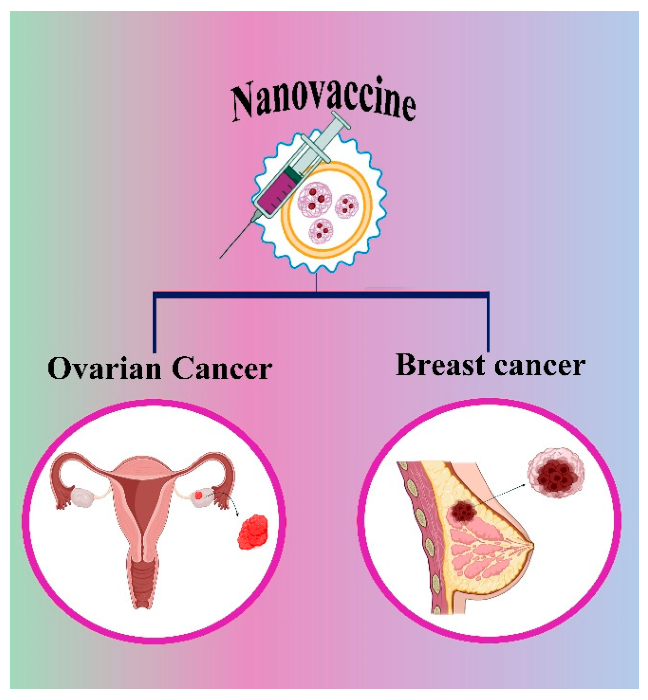
5. Recent Cancer Nanovaccine in the Market

One of the studies reported by Zhang et al., 2022 [105] demonstrated that extracellular degraded antigen proteins have increased the anticancer immune response of cancer vaccines. Before degradation, the antigen peptides are confirmed to have higher capacity upon stimulation from APCs in comparison to proteins while maintaining antigen specificity and major histocompatibility complex (MHC) affinity. The extracellular proteins were degraded by using the intracellular proteasome-mediated protein degradation pathway [105]. A CaP-peptide vaccine (CaP-Pep) was developed when the pre-degraded peptide was conjugated with calcium phosphate nanoparticles (CaP). This strategy played a crucial role in clinical immunology and various cancers [105]. Similarly, radio-immunotherapy was also improved through the nanoformulation of sodium alginate comprising 131 L-labelled catalases, strongly triggering the anticancer immune reactions and providing synergistic effects, thus eliminating metastatic tumors [106,107]. The surface-engineered nanoparticles can capture the cancer-derived protein antigens that improve the contact of APCs and localized radiation therapy. These antigen-capturing nanoparticles attach the cancer antigens via different interactions, enabling passage to lymph nodes and enhancing the immune reaction, thereby initiating the high survival of melanoma patients [107]. Further, the surfactant type of the antigen-associated nanoparticle affects the DCs with various nanoformulations that induce varying levels of T-cell responses and MHC expression. Different approaches, like adsorption and antigen encapsulation of nanoparticles with various surfactants, influence the DC activation and specific antigen T cell responses [108].
So far, a few vaccines have come to the market to offer protection against ovarian and breast cancer. Figure 4 depicts the vaccines developed for ovarian and breast cancer.
Table 2 shows the different types of nanovaccines that are undergoing clinical trials.

5.1. Nanovaccine for Ovarian Cancer

Globally, OC is the most common malignant tumor among women. Over 2,20,000 women are affected by these gynecological malignancies [124]. However, the survival of OC patients has been extended by standard therapies like chemotherapy, radiotherapy, and surgery. The relapse of cancer has been a very serious threat to many lives. So, to overcome these challenges, nanovaccine have been developed. These nanovaccines activate the patient’s immune system and kill the cancer cells. Zhang et al., 2022 [125] revealed that animals treated with fusion cell membrane (FCM)/dendritic nanoparticles had an active Cytotoxic T Lymphocyte immunological response. The fusion process aided in DC maturation, which increased the expression of the costimulatory molecules CD80/CD86. Moreover, FCM-NPs can produce a significant number of tumor-specific cytotoxic CD8+ T lymphocytes by stimulating naive T lymphocytes, as they possess both the immunogenicity of tumor cells and the antigen-presenting capacity of DCs. Both in vitro and in vivo FCM-NPs studies demonstrated a potent immuno-stimulating activity. FCM-NPs were shown to have the effect of postponing the growth and preventing the metastasis of OC by developing subcutaneously transplanted tumor models, patient-derived xenograft tumor models, and abdominal metastatic tumor models. FCM-NPs are anticipated to develop into a novel cancer vaccine for the management of OC [125].

5.2. Nanovaccines for Breast Cancer

Breast cancers are heterogeneous diseases showing different case-based symptoms and severity. There are four different types of breast cancers found according to the expression of estrogen receptor (ER), progesterone receptor (PR), and HER 2; they are Luminal A (ER+/PR+, HER-2-), Luminal B (ER+/PR+, HER-2+), Type HER-2+ (ER-/PR-, HER-2+), and Negative Triad (ER-/PR-, HER-2-) [126]. Active immunotherapies eliminate the cancer cells by triggering the T cells in the patient’s immune system. This reprograms the body’s immune system to target the tumor and protect against the cancer cells. In certain types of breast cancer, immunotherapy elicits a stronger response and demonstrates progressive immune activation, whereas in others, its effectiveness is limited. HER-2 is a protein found in certain breast cancer types, and vaccines are being developed to target it. These vaccines use specific HER-2 peptides to stimulate the immune system against cancer cells. Their effectiveness is enhanced by incorporating polypeptides like LRMK, which improve antigen presentation. Understanding the role of human leukocyte antigens (HLAs) is crucial for maximizing the vaccine-induced immune response [127].

5.3. Applied Strategy for Vaccine Administration

Administration after surgery: A thermo-responsive hydrogel loaded with curcumin-containing polymer nanoparticles and CpG-ODN antigen peptides was used to improve T-cell immunity, which induces immunogenic cell death (ICD), leading to an effective anti-tumor immune response. By promoting the infiltration of cytotoxic T lymphocytes (CTLs), this immunotherapy strategy effectively inhibited local tumor recurrence and pulmonary metastasis [128]. Another study developed an implantable 3D porous scaffold to deplete myeloid-derived suppressor cells and present whole tumor lysates with nanogel-based adjuvants to enhance CTL responses. This immune niche approach modulated the immunosuppressive environment and effectively prevented postoperative tumor recurrence and metastasis. Moreover, for this mode of admission, personalized antigens could be injected for superior results [129].
Admission by injection in the layers of skin: This method of administration has been investigated for cancer treatment. Many studies have shown that subcutaneous immunizations utilizing virus-like particles (VLPs) linked with human EGFR 2 epitopes resulted in increased levels of HER2-specific antibodies targeting HER2-positive tumors [32]. Adaptive microneedle systems have been studied for vaccinating against tumors and infectious diseases in more advanced intradermal administration approaches. A transdermal vaccine could potentially be used for both topical application and intra-tumoral immunotherapy against melanoma [130].
Direct admission to nasal cavity or inhalation: Most respiratory issues are treated by direct delivery of drugs inside the nasal cavity. Lung cancer antigens can be administered this way [131]. Another route to address respiratory infections like TB and lung cancer vaccines can be induced by inhalation methods [132].
Oral administration: Oral vaccines are highly effective due to their formulation, delivery, safety, and storage. Their convenience and high patient compliance make them a widely accepted method of vaccine administration. However, the intestines pose a challenge, as the lymphatic tissue beneath the mucous layer acts as a barrier to antigens. To create an oral vaccine, antigens must be absorbed and transported by epithelial cells before being recognized by immune cells to trigger responses. Throughout this process, antigens may degrade in the gastrointestinal tract, resulting in only a small portion of antigens reaching the mucosal tissue and limited uptake by the intestines [50].

6. Conclusions and Future Direction

Cancer vaccination has become more effective in recent years due to a better understanding of cancer immunology. The ability of these formulations to enhance the immune response has been significantly limited by cancer-induced immunosuppression. Delivering the previously identified vaccine formulation using nanocarriers was one of the attempts used to increase the strength of the immune response against cancers. An important factor in encouraging the use of cancer vaccines is the advancement of nano-delivery technology. Nanocarriers strongly support the stability of storing and transporting vaccines at low temperatures. Many forms of nanocarriers, including those made of polymeric-based nanocarriers, lipid-based nanocarriers, inorganic materials, and protein (DNA/RNA), have been investigated. Among others, the most popular technique for delivering mRNA vaccines is through lipid nanoparticles. So far, many cancer nanovaccines have shown excellent outcomes in treating various types of cancer. The major limitations, such as bioavailability, safety, poor solubility, and stability of vaccines, are improved by nanovaccines. Moreover, the US Food and Drug Administration (FDA) has approved several cancer vaccines. In the future, explorations regarding personalized vaccine design should be more focused.

Funding

This research received no external funding.

Acknowledgments

The authors are grateful to Chettinad Academy of Research and Education for providing the infrastructure. S.U. and D.J.M. acknowledge Chettinad Academy of Research and Education for providing them with PhD fellowship.

Conflicts of Interest

The authors declare no conflicts of interest.

Abbreviations

The following abbreviations are used in this manuscript:
APCAntigen-presenting cells
FDAFood and Drug Administration
CaPcalcium phosphate nanoparticles
DCsDendritic cells
CTLsCytotoxic T lymphocytes
MCTMicrocrystalline tyrosine
VLPsVirus like particle
LNLymph node
NKNatural killer cells
MTVmulti-targeted vaccines
Mutated-MTVmutated epitopes
GL-MTVgermline epitopes
CNTsCarbon nanotubes
PLGA-NPsPoly (lactic-co-glycolic acid) nanoparticles
RNA-LPXmRNA-lipoplex
HLAHuman leukocyte antigens
EREstrogen receptor
DEXDendritic cell-derived exosomes
TEXTumor cell-derived exosomes
AEXAscitic cell-derived exosomes
OCOvarian Cancer

References

  1. Gopal, R.K.; Ganesh, P.S.; Pathoor, N.N. Functional antibody responses associated with effectiveness of RTS, S malaria vaccine in children: New insights and implications. Lancet Microbe 2025, 6, 101001. [Google Scholar] [CrossRef]
  2. Ahmad, M.Z.; Ahmad, J.; Haque, A.; Alasmary, M.Y.; Abdel-Wahab, B.A.; Akhter, S. Emerging advances in synthetic cancer nano-vaccines: Opportunities and challenges. Expert. Rev. Vaccines 2020, 19, 1053–1071. [Google Scholar] [CrossRef] [PubMed]
  3. Kalimuthu, K.; Jagannathan, V.; Rajagopalan, K.; Ganesan, H.; Jeevanandam, M.; Vivekanandam, R.; Christyraj, J.R.S.S.; Mohan, M. Designing of cytotoxic T lymphocyte-based multi-epitope vaccine against SARS-CoV2: A reverse vaccinology approach. J. Biomol. Struct. Dyn. 2022, 40, 13711–13726. [Google Scholar]
  4. Tan, S.; Li, D.; Zhu, X. Cancer immunotherapy: Pros, cons and beyond. Biomed. Pharmacother. 2020, 124, 109821. [Google Scholar] [CrossRef]
  5. Deepika, B.; Pallavi, P.; Gowtham, P.; Girigoswami, A.; Girigoswami, K. Anticancer potential of nanoformulated extract of Passiflora incarnata leaves. Biocatal. Agric. Biotechnol. 2024, 57, 103109. [Google Scholar] [CrossRef]
  6. Janani, G.; Girigoswami, A.; Deepika, B.; Udayakumar, S.; Girigoswami, K. Unveiling the Role of Nano-Formulated Red Algae Extract in Cancer Management. Molecules 2024, 29, 2077. [Google Scholar] [CrossRef] [PubMed]
  7. Deepika, B.; Janani, G.; Jessy Mercy, D.; Udayakumar, S.; Girigoswami, A.; Girigoswami, K. Inhibitory Effect of Nano-Formulated Extract of Passiflora incarnata on Dalton’s Lymphoma Ascites-Bearing Swiss albino Mice. Pharmaceutics 2025, 17, 270. [Google Scholar] [CrossRef]
  8. Metkar, S.K.; Udayakumar, S.; Girigoswami, A.; Girigoswami, K. Natural serine proteases and their applications in combating amyloid formation. ADMET DMPK 2024, 12, 797–820. [Google Scholar] [CrossRef]
  9. Girigoswami, A.; Ghosh, M.; Girigoswami, K. World scenario of earthworms’ prowess as a remedy to a spectrum of diseases–a glimpse on related nanoformulations. Tradit. Med. Res. 2025, 10, 23. [Google Scholar] [CrossRef]
  10. Bhattacharya, J.B.; Jain, S.L.; Devi, S. The Role of Immunocytochemical Markers to Differentiate Primary from Secondary Neoplastic Hepatic Masses: A Diagnostic Challenge on Cytology. Turk. J. Pathol. 2021, 37, 196. [Google Scholar] [CrossRef]
  11. Selvaraj, V.; Sudhakar, S.; Sekaran, S.; Rameshkumar, N.; Krishnan, M.; Sekar, S.K.R. Cutting-edge nanotechnology transforming cancer surgery and recovery. Int. J. Surg. 2024, 110, 8207–8209. [Google Scholar] [CrossRef]
  12. Girigoswami, K.; Prabhu, A.D.; Pallavi, P.; Gowtham, P.; Harini, K.; Thirumalai, A.; Girigoswami, A. Hydrogels of Alginate Derivative-Encased Nanodots Featuring Carbon-Coated Manganese Ferrite Cores with Gold Shells to Offer Antiangiogenesis with Multimodal Imaging-Based Theranostics. Adv. Ther. 2024, 7, 2400054. [Google Scholar]
  13. Girigoswami, A.; Deepika, B.; Udayakumar, S.; Janani, G.; Mercy, D.J.; Girigoswami, K. Peony-shaped zinc oxide nanoflower synthesized via hydrothermal route exhibits promising anticancer and anti-amyloid activity. BMC Pharmacol. Toxicol. 2024, 25, 101. [Google Scholar] [CrossRef]
  14. Jayavardhini, B.; Pravin, Y.R.; Kumar, C.; Murugesan, R.; Vedakumari, S.W. Graphene oxide impregnated sericin/collagen scaffolds–Fabrication and characterization. Mater. Lett. 2022, 307, 131060. [Google Scholar] [CrossRef]
  15. Ameena, M.; Arumugham, M.; Ramalingam, K.; Rajeshkumar, S.; Perumal, E.; Shanmugam, R. Cytocompatibility and wound healing activity of chitosan thiocolchicoside lauric acid nanogel in human gingival fibroblast cells. Cureus 2023, 15, e43727. [Google Scholar]
  16. Rajkumar, M.; Davis Presley, S.; Thiyagarajulu, N.; Girigoswami, K.; Janani, G.; Kamaraj, C.; Madheswaran, B.; Prajapati, B.; Ali, N.; Khan, M.R. Gelatin/PLA-loaded gold nanocomposites synthesis using Syzygium cumini fruit extract and their antioxidant, antibacterial, anti-inflammatory, antidiabetic and anti-Alzheimer’s activities. Sci. Rep. 2025, 15, 2110. [Google Scholar] [CrossRef] [PubMed]
  17. Biswas, K.; Mercy, D.J.; Udayakumar, S.; Girigoswami, A.; Girigoswami, K. Frustrating the Serenity of Bacterial Biofilms by Bristly Reduced Graphene Oxide Sheets. BioNanoScience 2025, 15, 246. [Google Scholar] [CrossRef]
  18. Devi, N.B.; Satyasri, B.; Ramya, R.; Kanmani, R.; Vinuja, G. Nano-structured Molybdenum Trioxide Nano-hybrid based Conductive Platform for Breast Cancer Detection. J. Environ. Nanotechnol. 2024, 13, 418–426. [Google Scholar] [CrossRef]
  19. Kroll, A.V.; Jiang, Y.; Zhou, J.; Holay, M.; Fang, R.H.; Zhang, L. Biomimetic nanoparticle vaccines for cancer therapy. Adv. Biosyst. 2019, 3, 1800219. [Google Scholar] [CrossRef]
  20. Hu, Z.; Ott, P.A.; Wu, C.J. Towards personalized, tumour-specific, therapeutic vaccines for cancer. Nat. Rev. Immunol. 2018, 18, 168–182. [Google Scholar] [CrossRef]
  21. Stolk, D.A.; De Haas, A.; Vree, J.; Duinkerken, S.; Lübbers, J.; Van de Ven, R.; Ambrosini, M.; Kalay, H.; Bruijns, S.; van der Vliet, H.J.; et al. Lipo-based vaccines as an approach to target dendritic cells for induction of T-and iNKT cell responses. Front. Immunol. 2020, 11, 990. [Google Scholar] [CrossRef] [PubMed]
  22. Guo, J.; Ma, Q.; Cheng, R.; Li, K.; Tang, L.; Luo, S.; Liu, C. Application of nanotechnology in therapeutic cancer vaccines. Adv. NanoBiomed Res. 2023, 3, 2200122. [Google Scholar] [CrossRef]
  23. Desai, N.; Chavda, V.; Singh, T.R.R.; Thorat, N.D.; Vora, L.K. Cancer nanovaccines: Nanomaterials and clinical perspectives. Small 2024, 20, 2401631. [Google Scholar] [CrossRef] [PubMed]
  24. Yi, Y.; Yu, M.; Li, W.; Zhu, D.; Mei, L.; Ou, M. Vaccine-like nanomedicine for cancer immunotherapy. J. Control. Release 2023, 355, 760–778. [Google Scholar] [CrossRef]
  25. Schneider, I.C.; Hartmann, J.; Braun, G.; Stitz, J.; Klamp, T.; Bihi, M.; Sahin, U.; Buchholz, C.J. Displaying tetra-membrane spanning Claudins on enveloped virus-like particles for cancer immunotherapy. Biotechnol. J. 2018, 13, 1700345. [Google Scholar] [CrossRef]
  26. Geall, A.J.; Verma, A.; Otten, G.R.; Shaw, C.A.; Hekele, A.; Banerjee, K.; Cu, Y.; Beard, C.W.; Brito, L.A.; Krucker, T.; et al. Nonviral delivery of self-amplifying RNA vaccines. Proc. Natl. Acad. Sci USA 2012, 109, 14604–14609. [Google Scholar] [CrossRef]
  27. Gowtham, P.; Pallavi, P.; Harini, K.; Girigoswami, K.; Girigoswami, A. Hydrogelated Virus Nanoparticles in Tissue Engineering. Curr. Nanosci. 2023, 19, 258–269. [Google Scholar]
  28. Mohsen, M.O.; Bachmann, M.F. Virus-like particle vaccinology, from bench to bedside. Cell. Mol. Immunol. 2022, 19, 993–1011. [Google Scholar] [CrossRef]
  29. Zhou, X.; Zhang, Z.; Zhang, Z.; Zhu, X.; Tian, M.; Zhang, F.; Zhu, B.; Wang, W.; Ge, Q.; Hong, S.; et al. B cells are the dominant antigen-presenting cells that activate naive CD4+ T cells upon immunization with a virus-derived nanoparticle antigen. Immunity 2018, 49, 695–708.e4. [Google Scholar]
  30. Yue, Z.; Chen, Z.; Yang, K.; Cheng, Z.; Zhou, X.; Li, S.; Shen, C. Four ounces can move a thousand pounds: The enormous value of nanomaterials in tumor immunotherapy. Adv. Healthc. Mater. 2023, 12, 2300882. [Google Scholar]
  31. Tornesello, A.L.; Tagliamonte, M.; Buonaguro, F.M.; Tornesello, M.L.; Buonaguro, L. Virus-like particles as preventive and therapeutic cancer vaccines. Vaccines 2022, 10, 227. [Google Scholar] [CrossRef] [PubMed]
  32. Angelicola, S.; Scalambra, L.; Semprini, M.S.; Lollini, P.-L.; Nanni, P.; Palladini, A.; Ruzzi, F.; Cappello, C.; Pittino, O.M. Virus-like particle (VLP) vaccines for cancer immunotherapy. Int. J. Mol. Sci. 2023, 24, 12963. [Google Scholar]
  33. Gül, D.; Önal Acet, B.; Lu, Q.; Stauber, R.H.; Odabaşı, M.; Acet, Ö. Revolution in cancer treatment: How are intelligently designed nanostructures changing the game? Int. J. Mol. Sci. 2024, 25, 5171. [Google Scholar] [CrossRef]
  34. Janani, G.; Girigoswami, A.; Deepika, B.; Udayakumar, S.; Mercy, D.J.; Girigoswami, K. Dual Mechanism of Amphiroa anceps: Antiangiogenic and Anticancer Effects in Skin Cancer. Chem. Biodivers. 2025, 10, e202500626. [Google Scholar] [CrossRef]
  35. Hejabi, F.; Abbaszadeh, M.S.; Taji, S.; O’Neill, A.; Farjadian, F.; Doroudian, M. Nanocarriers: A novel strategy for the delivery of CRISPR/Cas systems. Front. Chem. 2022, 10, 957572. [Google Scholar] [CrossRef]
  36. Hu, Y.; Xia, D.; Bao, H.; Chen, C.; Xu, Y.; Meng, H.; Li, C. Nano-Oncologic Vaccine for Boosting Cancer Immunotherapy: The Horizons in Cancer Treatment. Nanomaterials 2025, 15, 122. [Google Scholar] [CrossRef]
  37. Mamuti, M.; Chen, W.; Jiang, X. Nanotechnology-Assisted Immunoengineering for Cancer Vaccines. Adv. NanoBiomed Res. 2023, 3, 2200080. [Google Scholar] [CrossRef]
  38. Zanganeh, S.; Abbasgholinejad, E.; Doroudian, M.; Esmaelizad, N.; Farjadian, F.; Benhabbour, S.R. The current landscape of glioblastoma biomarkers in body fluids. Cancers 2023, 15, 3804. [Google Scholar] [CrossRef]
  39. Fan, Y.; Moon, J.J. Nanoparticle drug delivery systems designed to improve cancer vaccines and immunotherapy. Vaccines 2015, 3, 662–685. [Google Scholar] [CrossRef]
  40. Urbanavicius, D.; Alvarez, T.; Such, G.K.; Johnston, A.P.; Mintern, J.D. The potential of nanoparticle vaccines as a treatment for cancer. Mol. Immunol. 2018, 98, 2–7. [Google Scholar] [CrossRef]
  41. Mohsen, M.O.; Heath, M.D.; Cabral-Miranda, G.; Lipp, C.; Zeltins, A.; Sande, M.; Stein, J.V.; Riether, C.; Roesti, E.; Zha, L.; et al. Vaccination with nanoparticles combined with micro-adjuvants protects against cancer. J. Immunother. Cancer 2019, 7, 114. [Google Scholar]
  42. Pandey, A.; Karmous, I. Exploring the Potential of Plant-Based Nanotechnology in Cancer Immunotherapy: Benefits, Limitations, and Future Perspectives. Biol. Trace Elem. Res. 2024, 203, 1746–1763. [Google Scholar] [CrossRef] [PubMed]
  43. Chen, J.; Liu, H.; Jehng, T.; Li, Y.; Chen, Z.; Lee, K.-D.; Shen, H.-T.; Jones, L.; Huang, X.-F.; Chen, S.Y. A novel anti-PD-L1 vaccine for cancer immunotherapy and immunoprevention. Cancers 2019, 11, 1909. [Google Scholar] [CrossRef]
  44. Tian, H.; Shi, G.; Wang, Q.; Li, Y.; Yang, Q.; Li, C.; Yang, G.; Wu, M.; Xie, Q.; Zhang, S.; et al. A novel cancer vaccine with the ability to simultaneously produce anti-PD-1 antibody and GM-CSF in cancer cells and enhance Th1-biased antitumor immunity. Signal Transduct. Target. Ther. 2016, 1, 16025. [Google Scholar] [CrossRef]
  45. Meng, Z.; Zhang, Y.; Zhou, X.; Ji, J.; Liu, Z. Nanovaccines with cell-derived components for cancer immunotherapy. Adv. Drug Deliv. Rev. 2022, 182, 114107. [Google Scholar] [CrossRef]
  46. De Charette, M.; Marabelle, A.; Houot, R. Turning tumour cells into antigen presenting cells: The next step to improve cancer immunotherapy? Eur. J. Cancer 2016, 68, 134–147. [Google Scholar] [CrossRef]
  47. Wei, Y.; Tian, H.; Yu, D.; Yang, G.; Yang, Q.; Deng, H.; Wang, Q.; Shi, G.; Xiang, R.; Zhang, S.; et al. Nanomedicinal strategies as efficient therapeutic interventions for delivery of cancer vaccines. Semin. Cancer Biol. 2021, 69, 43–51. [Google Scholar]
  48. Rana, I.; Oh, J.; Baig, J.; Moon, J.H.; Son, S.; Nam, J. Nanocarriers for cancer nano-immunotherapy. Drug Deliv. Transl. Res. 2023, 13, 1936–1954. [Google Scholar] [CrossRef] [PubMed]
  49. Wu, Y.; Zhang, Z.; Wei, Y.; Qian, Z.; Wei, X. Nanovaccines for cancer immunotherapy: Current knowledge and future perspectives. Chin. Chem. Lett. 2023, 34, 108098. [Google Scholar] [CrossRef]
  50. Fang, X.; Lan, H.; Jin, K.; Gong, D.; Qian, J. Nanovaccines for cancer prevention and immunotherapy: An update review. Cancers 2022, 14, 3842. [Google Scholar] [CrossRef]
  51. Liu, J.; Miao, L.; Sui, J.; Hao, Y.; Huang, G. Nanoparticle cancer vaccines: Design considerations and recent advances. Asian J. Pharm. Sci. 2020, 15, 576–590. [Google Scholar] [CrossRef] [PubMed]
  52. Sunoqrot, S.; Abdel Gaber, S.A.; Abujaber, R.; Al-Majawleh, M.; Talhouni, S. Lipid-and polymer-based nanocarrier platforms for cancer vaccine delivery. ACS Appl. Bio Mater. 2024, 7, 4998–5019. [Google Scholar] [CrossRef] [PubMed]
  53. Irvine, D.J.; Hanson, M.C.; Rakhra, K.; Tokatlian, T. Synthetic nanoparticles for vaccines and immunotherapy. Chem. Rev. 2015, 115, 11109–11146. [Google Scholar] [CrossRef] [PubMed]
  54. Sun, X.; He, Z.; Sun, B.; Jiang, Q.; Luo, C.; Wang, Q.; Wang, Z.; Zhang, S.; Sun, J. Lymph node-targeting nanovaccines for cancer immunotherapy. J. Control. Release 2022, 351, 102–122. [Google Scholar]
  55. Dewangan, H.K. Rational application of nanoadjuvant for mucosal vaccine delivery system. J. Immunol. Methods 2020, 481, 112791. [Google Scholar] [CrossRef]
  56. Xie, X.; Song, T.; Feng, Y.; Zhang, H.; Yang, G.; Wu, C.; You, F.; Liu, Y.; Yang, H. Nanotechnology-based multifunctional vaccines for cancer immunotherapy. Chem. Eng. J. 2022, 437, 135505. [Google Scholar] [CrossRef]
  57. Li, J.; Ren, H.; Zhang, Y. Metal-based nano-vaccines for cancer immunotherapy. Coord. Chem. Rev. 2022, 455, 214345. [Google Scholar] [CrossRef]
  58. Ahrens, E.T.; Bulte, J.W. Tracking immune cells in vivo using magnetic resonance imaging. Nat. Rev. Immunol. 2013, 13, 755–763. [Google Scholar] [CrossRef]
  59. Li Li, X.; Zhang, D.; Zhu, Y.; Zheng, X.; Yousef, A.A.; Song, K.; Liu, L.; Cha, D.; Han, Y.; Liu, X.; et al. Direct imaging of tunable crystal surface structures of MOF MIL-101 using high-resolution electron microscopy. J. Am. Chem. Soc. 2019, 141, 12021–12028. [Google Scholar] [CrossRef]
  60. Wang, J.; Chen, D.; Li, B.; He, J.; Duan, D.; Shao, D.; Nie, M. Fe-MIL-101 exhibits selective cytotoxicity and inhibition of angiogenesis in ovarian cancer cells via downregulation of MMP. Sci. Rep. 2016, 6, 26126. [Google Scholar] [CrossRef]
  61. Han, T.; Sun, Y.; Jiang, X.; Gong, C.; Kong, F.; Luo, Y.; Ge, C.; Liu, C.; Liu, Y.; Mou, Y.; et al. Air bag-embedded MIL-101 (Fe) metal-organic frameworks for an amplified tumor microenvironment activation loop through strategic delivery of iron ions and lentinan. Theranostics 2024, 14, 5883. [Google Scholar] [CrossRef] [PubMed]
  62. Najafipour, I.; Emami, N.; Sadeh, P.; Amoli, A.; Mosleh-Shirazi, S.; Amani, A.M.; Kamyab, H.; Chelliapan, S.; Kasaee, S.R.; Vafa, E. Synthesis of Fe3O4@ MIL-101-OH/Chitosan for adsorption and release of doxorubicin. Polym. Test. 2025, 142, 108659. [Google Scholar] [CrossRef]
  63. Zhang, Q.; Zhang, Y.; Chen, H.; Sun, L.-N.; Zhang, B.; Yue, D.-S.; Wang, C.-L.; Zhang, Z.F. Injectable hydrogel with doxorubicin-loaded ZIF-8 nanoparticles for tumor postoperative treatments and wound repair. Sci. Rep. 2024, 14, 9983. [Google Scholar] [CrossRef] [PubMed]
  64. Zhao, D.; Alizadeh, D.; Zhang, L.; Liu, W.; Farrukh, O.; Manuel, E.; Diamond, D.-J.; Badie, B. Carbon nanotubes enhance CpG uptake and potentiate antiglioma immunity. Clin. Cancer Res. 2011, 17, 771–782. [Google Scholar] [CrossRef]
  65. Hassan, H.A.; Diebold, S.S.; Smyth, L.A.; Walters, A.A.; Lombardi, G.; Al-Jamal, K.T. Application of carbon nanotubes in cancer vaccines: Achievements, challenges and chances. J. Control. Release 2019, 297, 79–90. [Google Scholar] [CrossRef]
  66. Achmad, H.; Gabr, G.A.; Ramírez-Coronel, A.A.; Budi, H.S.; Alshahrani, S.H.; Riaz, M.W.; Ibrahim, Y.S.; Al-Taee, M.M.; Sawitri, W.; Jalil, A.T.; et al. Nanovaccines in cancer immunotherapy: Focusing on dendritic cell targeting. Int. Immunopharmacol. 2022, 113, 109434. [Google Scholar] [CrossRef]
  67. Lu, Y.; Shi, Y.; You, J. Strategy and clinical application of up-regulating cross presentation by DCs in anti-tumor therapy. J. Control. Release 2022, 341, 184–205. [Google Scholar] [CrossRef] [PubMed]
  68. Fu, C.; Zhou, L.; Mi, Q.-S.; Jiang, A. DC-Based Vaccines for Cancer Immunotherapy. Vaccines 2020, 8, 706. [Google Scholar] [CrossRef]
  69. Michael, S.G. Immunoengineering: How Nanotechnology Can Enhance Cancer Immunotherapy. Cell 2015, 161, 201–204. [Google Scholar]
  70. Han, H.D.; Byeon, Y.; Kang, T.H.; Jung, I.D.; Lee, J.-W.; Shin, B.C.; Lee, Y.J.; Sood, A.K.; Park, Y.-M. Toll-like receptor 3-induced immune response by poly (d, l-lactide-co-glycolide) nanoparticles for dendritic cell-based cancer immunotherapy. Int. J. Nanomed. 2016, 2, 5729–5742. [Google Scholar] [CrossRef]
  71. Cruz, L.J.; Tacken, P.J.; Fokkink, R.; Joosten, B.; Stuart, M.C.; Albericio, F.; Torensma, R.; Figdor, C.G. Targeted PLGA nano-but not microparticles specifically deliver antigen to human dendritic cells via DC-SIGN in vitro. J. Control. Release 2010, 144, 118–126. [Google Scholar] [CrossRef] [PubMed]
  72. Tang, X.-D.; Lü, K.-L.; Yu, J.; Du, H.-J.; Fan, C.-Q.; Chen, L. In vitro and in vivo evaluation of DC-targeting PLGA nanoparticles encapsulating heparanase CD4+ and CD8+ T-cell epitopes for cancer immunotherapy. Cancer Immunol. Immunother. 2022, 71, 2969–2983. [Google Scholar]
  73. Cruz, L.J.; Tacken, P.J.; Eich, C.; Rueda, F.; Torensma, R.; Figdor, C.G. Controlled release of antigen and Toll-like receptor ligands from PLGA nanoparticles enhances immunogenicity. Nanomedicine 2017, 12, 491–510. [Google Scholar] [CrossRef]
  74. Hamdy, S.; Haddadi, A.; Hung, R.W.; Lavasanifar, A. Targeting dendritic cells with nano-particulate PLGA cancer vaccine formulations. Adv. Drug Deliv. Rev. 2011, 63, 943–955. [Google Scholar] [CrossRef]
  75. Paulis, L.E.; Mandal, S.; Kreutz, M.; Figdor, C.G. Dendritic cell-based nanovaccines for cancer immunotherapy. Curr. Opin. Immunol. 2013, 25, 389–395. [Google Scholar] [CrossRef] [PubMed]
  76. Attia, M.F.; Anton, N.; Wallyn, J.; Omran, Z.; Vandamme, T.F. An overview of active and passive targeting strategies to improve the nanocarriers efficiency to tumour sites. J. Pharm. Pharmacol. 2019, 71, 1185–1198. [Google Scholar] [CrossRef] [PubMed]
  77. Perica, K.; Medero, A.D.L.; Durai, M.; Chiu, Y.L.; Bieler, J.G.; Sibener, L.; Niemöller, M.; Assenmacher, M.; Richter, A.; Edidin, M.; et al. Nanoscale artificial antigen presenting cells for T cell immunotherapy. Nanomed. Nanotechnol. Biol. Med. 2014, 10, 119–129. [Google Scholar] [CrossRef]
  78. Mohsen, M.O.; Vogel, M.; Riether, C.; Muller, J.; Salatino, S.; Ternette, N.; Gomes, A.C.; Cabral-Miranda, G.; El-Turabi, A.; Rued, C.; et al. Targeting mutated plus germline epitopes confers pre-clinical efficacy of an instantly formulated cancer nano-vaccine. Front. Immunol. 2019, 10, 1015. [Google Scholar] [CrossRef]
  79. Al-Hawary, S.I.S.; Almajidi, Y.Q.; Bansal, P.; Ahmad, I.; Kaur, H.; Hjazi, A.; Deorari, M.; Zwamel, A.H.; Hamzah, H.F.; Mohammed, B.A. Dendritic cell-derived exosome (DEX) therapy for digestive system cancers; recent advances and future prospect. Pathol. Res. Pract. 2024, 257, 155288. [Google Scholar] [CrossRef]
  80. Wan, M.; Ning, B.; Spiegel, S.; Lyon, C.J.; Hu, T.Y. Tumor-derived exosomes (TDEs): How to avoid the sting in the tail. Med. Res. Rev. 2020, 40, 385–412. [Google Scholar] [CrossRef]
  81. Tran, T.-H.; Mattheolabakis, G.; Aldawsari, H.; Amiji, M. Exosomes as nanocarriers for immunotherapy of cancer and inflammatory diseases. Clin. Immunol. 2015, 160, 46–58. [Google Scholar] [CrossRef] [PubMed]
  82. Santos, P.; Almeida, F. Exosome-based vaccines: History, current state, and clinical trials. Front. Immunol. 2021, 12, 711565. [Google Scholar] [CrossRef] [PubMed]
  83. Greening, D.W.; Gopal, S.K.; Xu, R.; Simpson, R.J.; Chen, W. Exosomes and their roles in immune regulation and cancer. Semin. Cell Dev. Biol. 2015, 40, 72–81. [Google Scholar] [CrossRef]
  84. Tan, A.; De La Peña, H.; Seifalian, A.M. The application of exosomes as a nanoscale cancer vaccine. Int. J. Nanomed. 2010, 5, 889–900. [Google Scholar]
  85. Smith, D.M.; Simon, J.K.; Baker, J.R. Applications of nanotechnology for immunology. Nat. Rev. Immunol. 2013, 13, 592–605. [Google Scholar] [CrossRef]
  86. Hou, X.; Zaks, T.; Langer, R.; Dong, Y. Lipid nanoparticles for mRNA delivery. Nat. Rev. Mater. 2021, 6, 1078–1094. [Google Scholar] [CrossRef]
  87. Zhu, M.; Wang, R.; Nie, G. Applications of nanomaterials as vaccine adjuvants. Hum. Vaccines Immunother. 2014, 10, 2761–2774. [Google Scholar] [CrossRef] [PubMed]
  88. Mohsen, M.O.; Speiser, D.E.; Knuth, A.; Bachmann, M.F. Virus-like particles for vaccination against cancer. Wiley Interdiscip. Rev. Nanomed. Nanobiotechnol. 2020, 12, e1579. [Google Scholar] [CrossRef]
  89. Zhang, D.; Lin, Z.; Wu, M.; Cai, Z.; Zheng, Y.; He, L.; Li, Z.; Zhou, J.; Sun, L.; Chen, G.; et al. Cytosolic delivery of thiolated neoantigen nano-vaccine combined with immune checkpoint blockade to boost anti-cancer T cell immunity. Adv. Sci. 2021, 8, 2003504. [Google Scholar] [CrossRef]
  90. Lybaert, L.; Vermaelen, K.; De Geest, B.G.; Nuhn, L. Immunoengineering through cancer vaccines–A personalized and multi-step vaccine approach towards precise cancer immunity. J. Control. Release 2018, 289, 125–145. [Google Scholar] [CrossRef]
  91. Kranz, L.M.; Diken, M.; Haas, H.; Kreiter, S.; Loquai, C.; Reuter, K.C.; Meng, M.; Fritz, D.; Vascotto, F.; Hefesha, H.; et al. Systemic RNA delivery to dendritic cells exploits antiviral defence for cancer immunotherapy. Nature 2016, 534, 396–401. [Google Scholar] [CrossRef] [PubMed]
  92. Zhu, G.; Zhang, F.; Ni, Q.; Niu, G.; Chen, X. Efficient nanovaccine delivery in cancer immunotherapy. ACS Nano 2017, 11, 2387–2392. [Google Scholar] [CrossRef]
  93. Amouzegar, A.; Chelvanambi, M.; Filderman, J.N.; Storkus, W.J.; Luke, J.J. STING agonists as cancer therapeutics. Cancers 2021, 13, 2695. [Google Scholar] [CrossRef]
  94. Zhang, Y.; Lin, S.; Wang, X.Y.; Zhu, G. Nanovaccines for cancer immunotherapy. Wiley Interdiscip. Rev. Nanomed. Nanobiotechnol. 2019, 11, e1559. [Google Scholar] [CrossRef] [PubMed]
  95. Jiang, X.; Wang, J.; Zheng, X.; Liu, Z.; Zhang, X.; Li, Y.; Wilhelm, J.; Cao, J.; Huang, G.; Zhang, J.; et al. Intratumoral administration of STING-activating nanovaccine enhances T cell immunotherapy. J. Immunother. Cancer 2022, 10, e003960. [Google Scholar] [CrossRef]
  96. Zhou, S.; Cheng, F.; Zhang, Y.; Su, T.; Zhu, G. Engineering and delivery of cGAS-STING immunomodulators for the immunotherapy of cancer and autoimmune diseases. Acc. Chem. Res. 2023, 56, 2933–2943. [Google Scholar] [CrossRef] [PubMed]
  97. An, M.; Yu, C.; Xi, J.; Reyes, J.; Mao, G.; Wei, W.Z.; Liu, H. Induction of necrotic cell death and activation of STING in the tumor microenvironment via cationic silica nanoparticles leading to enhanced antitumor immunity. Nanoscale 2018, 10, 9311–9319. [Google Scholar] [CrossRef]
  98. Ghosh, S.; Girigoswami, K.; Girigoswami, A. Membrane-encapsulated camouflaged nanomedicines in drug delivery. Nanomedicine 2019, 14, 2067–2082. [Google Scholar] [CrossRef]
  99. Tomljenovic, L.; Shaw, C.A. Aluminum vaccine adjuvants: Are they safe? Curr. Med. Chem. 2011, 18, 2630–2637. [Google Scholar] [CrossRef]
  100. Chen, Z.; Hao, X.; Wang, H.; Zhong, X.; Chen, X.; Zhao, Y.; Zhang, Y.; Du, G.; Sun, X. Smart combination of aluminum hydroxide and MF59 to induce strong cellular immune responses. J. Control. Release 2022, 349, 699–711. [Google Scholar] [CrossRef]
  101. Zhao, T.; Cai, Y.; Jiang, Y.; He, X.; Wei, Y.; Yu, Y.; Tian, X. Vaccine adjuvants: Mechanisms and platforms. Signal Transduct. Target. Ther. 2023, 8, 283. [Google Scholar] [CrossRef] [PubMed]
  102. Liao, Z.; Huang, J.; Lo, P.-C.; Lovell, J.F.; Jin, H.; Yang, K. Self-adjuvanting cancer nanovaccines. J. Nanobiotechnol. 2022, 20, 345. [Google Scholar] [CrossRef]
  103. Aikins, M.E.; Xu, C.; Moon, J.J. Engineered nanoparticles for cancer vaccination and immunotherapy. Acc. Chem. Res. 2020, 53, 2094–2105. [Google Scholar] [CrossRef] [PubMed]
  104. Xu, J.; Lv, J.; Zhuang, Q.; Yang, Z.; Cao, Z.; Xu, L.; Pei, P.; Wang, C.; Wu, H.; Dong, Z.; et al. A general strategy towards personalized nanovaccines based on fluoropolymers for post-surgical cancer immunotherapy. Nat. Nanotechnol. 2020, 15, 1043–1052. [Google Scholar] [CrossRef] [PubMed]
  105. Zhang, Q.-L.; Hong, S.; Dong, X.; Zheng, D.-W.; Liang, J.-L.; Bai, X.-F.; Wang, X.-N.; Han, Z.-Y.; Zhang, X.-Z. Bioinspired nano-vaccine construction by antigen pre-degradation for boosting cancer personalized immunotherapy. Biomaterials 2022, 287, 121628. [Google Scholar] [CrossRef] [PubMed]
  106. Dong, Q.; Xue, T.; Yan, H.; Liu, F.; Liu, R.; Zhang, K.; Chong, Y.; Du, J.; Zhang, H. Radiotherapy combined with nano-biomaterials for cancer radio-immunotherapy. J. Nanobiotechnol. 2023, 21, 395. [Google Scholar] [CrossRef]
  107. Chao, Y.; Xu, L.; Liang, C.; Feng, L.; Xu, J.; Dong, Z.; Tian, L.; Yi, X.; Yang, K.; Liu, Z. Combined local immunostimulatory radioisotope therapy and systemic immune checkpoint blockade imparts potent antitumour responses. Nat. Biomed. Eng. 2018, 2, 611–621. [Google Scholar] [CrossRef]
  108. Mi, Y.; Hagan, I.V.C.T.; Vincent, B.G.; Wang, A.Z. Emerging nano-/microapproaches for cancer immunotherapy. Adv. Sci. 2019, 6, 1801847. [Google Scholar] [CrossRef]
  109. Loquai, C.; Hassel, J.; Brück, P.; Derhovanessian, E.; Cuk, K.; Lörks, V.; Sikorski, J.; Gold, M.; Maurus, D.; Schwarck-Kokarakis, D.; et al. 549 An RNA-lipoplex (RNA-LPX) vaccine demonstrates strong immunogenicity and promising clinical activity in a phase I trial in cutaneous melanoma patients with no evidence of disease at trial inclusion. BMJ Spec. J. 2021, 585, 107–112. [Google Scholar]
  110. Lopez, J.; Powles, T.; Braiteh, F.; Siu, L.L.; LoRusso, P.; Friedman, C.F.; Balmanoukian, A.S.; Gordon, M.; Yachnin, J.; Rottey, S.; et al. Autogene cevumeran with or without atezolizumab in advanced solid tumors: A phase 1 trial. Nat. Med. 2025, 31, 152–164. [Google Scholar] [CrossRef]
  111. Berinstein, N.L.; Karkada, M.; Oza, A.M.; Odunsi, K.; Villella, J.A.; Nemunaitis, J.J.; Morse, M.A.; Pejovic, T.; Bentley, J.; Buyse, M.; et al. Survivin-targeted immunotherapy drives robust polyfunctional T cell generation and differentiation in advanced ovarian cancer patients. Oncoimmunology 2015, 4, e1026529. [Google Scholar] [CrossRef]
  112. Koerner, J.; Horvath, D.; Herrmann, V.L.; MacKerracher, A.; Gander, B.; Yagita, H.; Rohayem, J.; Groettrup, M. PLGA-particle vaccine carrying TLR3/RIG-I ligand Riboxxim synergizes with immune checkpoint blockade for effective anti-cancer immunotherapy. Nat. Commun. 2021, 12, 2935. [Google Scholar] [CrossRef]
  113. Xiong, J.; Huang, J.; Xu, H.; Wu, Q.; Zhao, J.; Chen, Y.; Chen, Y.; Fan, G.; Guan, H.; Xiao, R.; et al. CpG-Based Nanovaccines Enhance Ovarian Cancer Immune Response by Gbp2-Mediated Remodeling of Tumor-Associated Macrophages. Adv. Sci. 2025, 12, e2412881. [Google Scholar] [CrossRef] [PubMed]
  114. Creemers, J.H.; Pawlitzky, I.; Grosios, K.; Gileadi, U.; Middleton, M.R.; Gerritsen, W.R.; Mehra, N.; Rivoltini, L.; Walters, I.; Figdor, C.G.; et al. Assessing the safety, tolerability and efficacy of PLGA-based immunomodulatory nanoparticles in patients with advanced NY-ESO-1-positive cancers: A first-in-human phase I open-label dose-escalation study protocol. BMJ Open 2021, 11, e050725. [Google Scholar] [CrossRef] [PubMed]
  115. Zacharia, A.; Harberts, E.; Valencia, S.M.; Myers, B.; Sanders, C.; Jain, A.; Larson, N.R.; Middaugh, C.R.; Picking, W.D.; Difilippantonio, S.; et al. Optimization of RG1-VLP vaccine performance in mice with novel TLR4 agonists. Vaccine 2021, 39, 292–302. [Google Scholar] [CrossRef] [PubMed]
  116. Das, R.K.; Panda, S.; Bhol, C.S.; Bhutia, S.K.; Mohapatra, S. N-doped carbon quantum dot (NCQD)-Deposited carbon capsules for synergistic fluorescence imaging and photothermal therapy of oral cancer. Langmuir 2019, 35, 15320–15329. [Google Scholar] [CrossRef]
  117. Zou, Y.; Li, S.; Li, Y.; Zhang, D.; Zheng, M.; Shi, B. Glioblastoma Cell Derived Exosomes as a Potent Vaccine Platform Targeting Primary Brain Cancers and Brain Metastases. ACS Nano 2025, 19, 17309–17322. [Google Scholar] [CrossRef]
  118. Kim, H.Y.; Min, H.-K.; Song, H.-W.; Yoo, A.; Lee, S.; Kim, K.-P.; Park, J.-O.; Choi, Y.H.; Choi, E. Delivery of human natural killer cell-derived exosomes for liver cancer therapy: An in vivo study in subcutaneous and orthotopic animal models. Drug Deliv. 2022, 29, 2897–2911. [Google Scholar] [CrossRef]
  119. Zhao, X.; Xuan, F.; Li, Z.; Yin, X.; Zeng, X.; Chen, J.; Fang, C. A KIF20A-based thermosensitive hydrogel vaccine effectively potentiates immune checkpoint blockade therapy for hepatocellular carcinoma. npj Vaccines 2025, 10, 1. [Google Scholar] [CrossRef]
  120. Bhardwaj, U.; Li, Y.; Ma, A.-H.; Mossanen, M.; Lee, J.S.; Lara, P.N.; Lam, K.S.; Lin, T.-Y.; Zhao, J.; Lerner, L.; et al. A phase I first-in-human clinical trial with PLZ4-coated paclitaxel-loaded micelles (PPM) in therapy-resistant non-muscle-invasive bladder cancer (NMIBC). Am. Soc. Clin. Oncol. 2025, 41, TPS4615. [Google Scholar]
  121. Ren, H.; Li, J.; Liu, G.; Sun, Y.; Yang, X.; Jiang, Z.; Zhang, J.; Lovell, J.F.; Zhang, Y. Anticancer vaccination with immunogenic micelles that capture and release pristine CD8+ T-cell epitopes and adjuvants. ACS Appl. Mater. Interfaces 2022, 14, 2510–2521. [Google Scholar] [CrossRef] [PubMed]
  122. An, H.J.; Kang, S.Y.; Kim, I.H.; Jang, J.S.; Sun, D.-S.; Lee, S.-C.; Lee, H.W.; Jin, M. Phase 2 study of weekly polymeric micelle-formulated paclitaxel plus gemcitabine in patients with recurrent and metastatic adenocarcinoma of the pancreas. J. Clin. Oncol. 2023, 41 (Suppl. S16), 465–472. [Google Scholar]
  123. Markovic, M.D.; Panic, V.V.; Pjanovic, R.V. Polymeric Nanosystems: A Breakthrough Approach to Treating Inflammation and Inflammation Related Diseases. Biopolymers 2025, 116, e70012. [Google Scholar] [CrossRef]
  124. Webb, P.M.; Jordan, S.J. Global epidemiology of epithelial ovarian cancer. Nat. Rev. Clin. Oncol. 2024, 21, 389–400. [Google Scholar] [CrossRef]
  125. Zhang, L.; Zhao, W.; Huang, J.; Li, F.; Sheng, J.; Song, H.; Chen, Y. Development of a dendritic cell/tumor cell fusion cell membrane nano-vaccine for the treatment of ovarian cancer. Front. Immunol. 2022, 13, 828263. [Google Scholar] [CrossRef]
  126. Weigelt, B.; Reis-Filho, J.S. Histological and molecular types of breast cancer: Is there a unifying taxonomy? Nat. Rev. Clin. Oncol. 2009, 6, 718–730. [Google Scholar] [CrossRef] [PubMed]
  127. Davodabadi, F.; Sarhadi, M.; Arabpour, J.; Sargazi, S.; Rahdar, A.; Díez-Pascual, A.M. Breast cancer vaccines, New insights into immunomodulatory and nano-therapeutic approaches. J. Control. Release 2022, 349, 844–875. [Google Scholar] [CrossRef]
  128. Herdiana, Y.; Husni, P.; Nurhasanah, S.; Shamsuddin, S.; Wathoni, N. Chitosan-based nano systems for natural antioxidants in breast cancer therapy. Polymers 2023, 15, 2953. [Google Scholar] [CrossRef] [PubMed]
  129. Phuengkham, H.; Song, C.; Um, S.H.; Lim, Y.T. Implantable synthetic immune niche for spatiotemporal modulation of tumor-derived immunosuppression and systemic antitumor immunity: Postoperative immunotherapy. Adv. Mater. 2018, 30, 1706719. [Google Scholar] [CrossRef]
  130. Mauldin, I.S.; Wages, N.A.; Stowman, A.M.; Wang, E.; Olson, W.C.; Deacon, D.H.; Smith, K.T.; Galeassi, N.; Teague, J.E.; Smolkin, M.E.; et al. Topical treatment of melanoma metastases with imiquimod, plus administration of a cancer vaccine, promotes immune signatures in the metastases. Cancer Immunol. Immunother. 2016, 65, 1201–1212. [Google Scholar] [CrossRef]
  131. Maillet, A.; Guilleminault, L.; Lemarié, E.; Lerondel, S.; Azzopardi, N.; Montharu, J.; Congy-Jolivet, N.; Reverdiau, P.; Legrain, B.; Parent, C.; et al. The airways, a novel route for delivering monoclonal antibodies to treat lung tumors. Pharm. Res. 2011, 28, 2147–2156. [Google Scholar] [CrossRef] [PubMed]
  132. Garbuzenko, O.B.; Kuzmov, A.; Taratula, O.; Pine, S.R.; Minko, T. Strategy to enhance lung cancer treatment by five essential elements: Inhalation delivery, nanotechnology, tumor-receptor targeting, chemo-and gene therapy. Theranostics 2019, 9, 8362. [Google Scholar] [CrossRef] [PubMed]
Figure 1. The different types of nanostructures and their role in cancer vaccine designing and action.
Chemistry 07 00097 g001
Figure 2. The endosomal escape mechanism of nanoparticles.
Chemistry 07 00097 g002
Figure 3. The different nanostructures facilitate the design of nanovaccines for enhanced vaccine efficacy.
Chemistry 07 00097 g003
Figure 4. The nanovaccines developed for ovarian and breast cancer.
Chemistry 07 00097 g004
Table 1. The advantages and disadvantages of nanocarriers employed in vaccine design.
Types of NanovaccineAdvantagesDisadvantagesReference
Polymeric nanocarrier
(PLGA, PEG-PLA)
  • These nanocarriers can be easily functionalized
  • They can release an antigen in a controlled and sustained way
  • Polymeric nanocarriers are biodegradable
  • The synthesis of polymeric nanocarriers is a complicated process
  • They can cause inflammatory responses
  • The capacity of loading antigens is lower in this compared to other carriers
[85]
Lipid-based nanocarrier
(liposomes, Lipid nanoparticles)
  • Lipid-based nanovaccines are effective in delivering mRNA or tumor antigens
  • They are highly biodegradable and biocompatible
  • These vaccines strongly trigger the T cell immune responses in the body
  • These nanocarriers carry the risk of rapid clearance in the bloodstream
  • Their physical stability is lower inside the body
[86]
Carbon-based nanocarriers
(CNTs)
  • They can induce a strong immune activation
  • The loading potential of the CNTs are high for antigens
  • They show inconsistency in functionalization
  • They have high toxicity and are poor in biodegradability
[87]
Metal-based nanocarrier
  • They are highly stable
  • They show strong immunostimulatory effects
  • They have a high risk of metal accumulation, and they show toxicity
  • They are poorly biodegradable
[57]
Peptide/protein based nanovaccine
  • They are naturally immunogenic, and they mimic the structure of the virus
  • They enhance their uptake
  • Their capacity is limited for payloads
  • Under physiological conditions they have stability issue
[88]
Table 2. Overview of recently developed nanovaccines for cancer immunotherapy.
Types of NanovaccinesImmunogenicity
(T Cell/DC Activation)
Delivery EfficiencyBiocompatibilityCost EffectivenessClinical Trial NumberClinical Developmental Stages
BNT111Strong T cell responseDelivered via RNA-LPXBiocompatibleModerate scalabilityNCT02410733Phase I trial in patients with cutaneous melanoma is ongoing [109]
Autogene Cevumeran (RNA lipoplex-based neoantigen vaccine)Strong cellular uptake and CD4+/CD8+ T cell responseDelivered via lipoplexNon toxicExpensive due to the synthesis and sequencing processNCT03289962Phase I trial in patients with solid tumors [110]
DPX-survivac (DepoVax-based surviving vaccine)Excellent antigen-specific T cell responseEnhances APC uptake and activationBiocompatible at low dosesLong-term stability and cost-effectivenessNCT01416038Phase I in patients with advanced-stage ovarian, fallopian, and peritoneal cancer [111]
PLGA-Riboxxim-based vaccineEnhanced T cell and dendritic cell responseImproved cellular uptake and activationBiodegradable and biocompatibleCost effective-Preclinical-testes in animal model [112]
PLGA-CpG @1D8-MStrong activationImproved CpG accumulation via PLGABiodegradable and biocompatibleModerately complex due to encapsulation and membrane extraction-Preclinical in ID8 ovarian and 4T1 breast cancer [113]
PRECIOUS-01Activates invariant natural killer cellsIntravenous administrationBiodegradableModerate complexityNCT04751786Phase I clinical trial [114]
RG1-VLPStrong B-cell responseEnhanced lymph node targetingSafety profilesStable formulation and scalable-Preclinical [115]
Carbon quantum dot capivasertibInduces dendritic cell activationEnhanced targeted deliveryBiocompatibleCost effective -Preclinical [116]
ExosomePromotes mature T cell and DC activationEffectively targets both the lymph node and the brain tumor siteBiocompatibleCost effective -Preclinical in mice model [117]
Natural killer cell-derived exosomesActivates immune response and reduces immunosuppressive signallingEffectively targets the tumor siteHighly biocompatibleModerate scalable-Preclinical [118]
KIF20A (kinesin family member 20A) based thermosenstive hydrogelStrong T cell and DC activation Sustained in vivo antigen releaseSafetyModerate complexity-Preclinical study in CDX (cell-derived xenograft) and an immune humanized PDX (patient-derived xenograft) models [119]
PLZ4—coated paclitaxel loaded micellesSynergistically works on post BCGEffectively targets bladder cancer cellsBiocompatible at 25 mg doseCost effectiveNCT05519241Phase I-bladder cancer [120]
MicelleStrong T cell responseEfficient targeting of lymph nodes and active delivery of adjuvants to DCBiocompatibleExpensive-Preclinical [121]
GENEXOL-PMModerate immune responseEnhanced tumor penetrationBiocompatibleCost effectiveNCT02739633Phase II—pancreatic cancer [122]
Gold NPs based vaccinesStrong T cell priming and immune responseTargeted delivery to Dectin-1 expressing APCHighly biocompatibleScalable-Preclinical—mice model [123]
Disclaimer/Publisher’s Note: The statements, opinions and data contained in all publications are solely those of the individual author(s) and contributor(s) and not of MDPI and/or the editor(s). MDPI and/or the editor(s) disclaim responsibility for any injury to people or property resulting from any ideas, methods, instructions or products referred to in the content.

