Article

Synthesis, Reductive Reactivity and Anticancer Activity of Cobalt(III)– and Manganese(III)–Salen Complexes

1
Department of Sciences, John Jay College, The City University of New York, New York, NY 10019, USA
2
Department of Chemistry, Hunter College, The City University of New York, New York, NY 10065, USA
*
Authors to whom correspondence should be addressed.
Chemistry 2025, 7(3), 85; https://doi.org/10.3390/chemistry7030085
Submission received: 4 April 2025 / Revised: 13 May 2025 / Accepted: 20 May 2025 / Published: 23 May 2025
(This article belongs to the Section Inorganic and Solid State Chemistry)

Abstract

:
Mn(III)– and Co(III)–salen complexes (Mn-1 and Co-2) have been synthesized by a simple one-pot procedure through oxidation of Mn(II) and Co(II) precursors in air. X-ray structural analysis reveals that both complexes adopt similar coordination modes, including a typical square planar metal/salen coordination sphere, which is further occupied by two axial ligands, i.e., an acetate anion and a water molecule. Despite their structural similarity, they are not isomorphous given their distinct cell parameters. In the solid-state structures, both complexes exist as hydrogen-bonded dimers through hydrogen bonding interactions between the axially coordinating water molecules and outer O4 cavity from another molecule of the complex. The reductive activity of both complexes has been explored. While the reaction of Mn-1 with potassium triethylborohydride was unsuccessful, leading to a complicated mixture, the use of Co-2 furnished the formation of a novel product (CoK-3) that was isolated as red crystals in reasonable yield. CoK-3 was characterized as a heterometallic dimer involving the coordination of a K+ ion within the O4 cavity of a semi-hydrogenated salen/cobalt complex while the cobalt center has been reduced from Co(III) to Co(II). In addition, an attempt at reducing Co-2 with pinacolborane resulted in the isolation of crystals of Co-4, whose structure was determined as a simple square planar CoII–salen complex. Finally, three complexes (Mn-1, Co-2 and CoK-3) have been investigated for their cytotoxic activities against two human breast cancer cell lines (MCF-7 and MDA-MB 468) and a normal breast epitheliel cell line (MCF-10A), with cisplatin used as a reference in order to discover potential drug candidates that may compete with cisplatin. The results reveal that Co-2 can be a promising drug candidate, specifically for the MCF-7 cancer cells, with minimal damage to healthy cells.

1. Introduction

The design and synthesis of biologically active metal complexes has received considerable interest since the observation of the first metal-based anticancer drug, cisplatin, and its broad-spectrum clinical applications for treatment of many types of cancers, such as ovary, cervix, breast, testis, bladder, colon, rectum, lung, or head and neck cancers [1,2,3,4,5,6]. In addition, structurally modified platinum-based drugs such as oxaliplatin and carboplatin have also been developed for improved performance against certain types of cancers, which inspired further investigations in the field of metal-based antitumor agents in the past decades [7,8]. Although highly effective, platinum-based anticancer drugs may cause several side effects, including nerve and kidney damage, nephrotoxicity, ototoxicity, and bone marrow suppression, as well as drug resistance [9,10,11]. To mitigate the side effect issues, a major approach for scientists was to design metallic drugs by mitigating metal-related toxicity while maintaining good anticancer activity [12,13]. Thus, great efforts have been devoted to observing non-platinum anticancer drugs, in particular, those containing essential trace metals which are much friendlier to normal human cells [14,15,16,17,18,19,20].
In search of new non-platinum anticancer drugs, we were particularly interested in bioactive metal complexes based on essential trace metals such as cobalt, iron, and manganese. We have previously reported a series of cobalt and iron complexes/networks based on 2,2′;6′,2″-terpyridine-derived ligands that displayed promising antiproliferative properties against several human breast cancer cells comparable to cisplatin [21,22]. In this work, we focus on the discovery of new anticancer agents that are less toxic towards normal human cells by using essential trace metals (Mn and Co) and salen-type ligands.
Salen ligands, as a class of tetradentate N2O2 Schiff base ligands, have been popularly employed for the synthesis of metal complexes that have found applications in catalysis, functional materials, as well as bioactive drug candidates [23]. Examples of active anticancer molecules of salen-based metal complexes, mainly explored in the last two decades, have been well documented. Compared to platinum-based drugs, first-row transition metals (such as Fe, Co, Cu, and Mn) have been choices for the synthesis of metallic anticancer drugs due to their low toxicity in normal cells and their natural occurrence in humans [24,25]. It has also been reported that complexes of trace biological metals such as manganese are of interest for their antioxidant properties [26,27,28]. Herein, we wish to report the popularly explored Co(III)– and Mn(III)–salen complexes such as Mn-1 and Co-2 (Scheme 1) with a focus on their reductive reactivities towards potassium triethylborohydride and pinacolborane, which have not been investigated in the literature, to our knowledge. Their in vitro antiproliferative activities against breast cancer cells (MCF-7 and MDA-MB 468), along with a novel heterometallic complex CoK-3 isolated from the reduction reaction, have been further studied.

2. Materials and Methods

2.1. Materials

Unless specified otherwise, the synthesis was carried out under ambient conditions, while the reduction reactions were conducted under an N2 atmosphere using the standard glovebox technique. All chemicals of analytical grade were used as received from Thermo Fisher Scientific (Pittsburgh, PA, USA) without further purification. Human breast cancer cell line (MCF-7), triple-negative breast cancer cell line (MDA-MB-468), and non-tumorigenic epithelial cell line from mammary gland (MCF-10A) were obtained from the American Type Tissue Culture (Manassas, VA, USA). All cell culture reagents were purchased from Thermo Fisher Scientific. FT-IR spectra (Figures S1–S3) were recorded on a Shimadzu 8400S instrument with solid samples using a Golden Gate ATR accessory (Columbia, MD, USA). 1H NMR and 13C NMR spectra were obtained at room temperature on Bruker AV 500 or 600 MHz NMR spectrometers (Billerica, MA, USA), with chemical shifts (δ) referenced to the residual solvent signal. Elemental analyses were performed by Midwest Microlab LLC in Indianapolis in the US. UV-Vis electronic absorption spectroscopy was recorded on a Shimadzu UV-2700 spectrophotometer (Columbia, MD, USA). HR-MS data (Figures S4 and S5) were obtained on an Agilent 6550 QToF coupled to an Agilent 1290 Infinity LC system (Santa Clara, CA, USA). The salen ligand, N,N′-ethylenebis(3-ethoxysalicylaldimine) (H2L), was prepared by a 2:1 condensation of 3-ethoxysalicylaldehyde and eththylenediamine in methanol according to the literature [29]. NMR data for H2L (Figures S6 and S7): 1H NMR (500 MHz, DMSO) δ 13.54 (s, 2H, HOH), 8.57 (s, 2H, HCH=N), 7.06–6.93 (m, 4H, HAr), 6.78 (t, J = 7.9 Hz, 2H, HAr), 4.02 (q, J = 7.0 Hz, 4H, HCH2CH3), 3.93 (s, 4H, HCH2CH2), 1.31 (t, J = 7.0 Hz, 6H, HCH2CH3) ppm; 13C NMR (126 MHz, DMSO) δ 166.98, 151.40, 146.91, 123.09, 118.3, 117.64, 115.92, 63.71, 58.28, 14.59 ppm.

2.2. Synthesis of Mn-1

In a 50-mL flask equipped with a stirring bar, 3-ethoxy-2-hydroxybenzaldehyde (0.166 g, 1.00 mmol) and 1,2-diaminoethane (0.030 g, 0.500 mmol) were dissolved in CH2Cl2/CH3OH (30 mL, v/v, 2:1). Solid Mn(OAc)2·4H2O (0.123 g, 0.500 mmol) was then added in small portions at room temperature upon stirring. The resulting dark brown solution was allowed to stir for 48 h upon exposing to the air. The solution was filtered, and the filtrate was kept at room temperature for slow evaporation over one week, after which time dark-orange block-like crystals suitable for single-crystal X-ray diffraction analysis were harvested by filtration. The crystalline sample was washed with MeOH and dried in vacuo. Yield: 0.21 g (86%). UV/Vis λmax/nm (1.0 × 10⁻⁵ mol dm−3, DMSO): 256 (97.7, ε/103 dm3 mol−1 cm−1), 396 (br, 9.6). FT-IR (solid, cm−1): 3270br, 2924m, 1637s, 1604m, 1575s, 1470s, 1450s, 1392s, 1313s, 1330s, 1225s, 1175w, 1112m, 1087s, 1042m, 1019m, 902w, 732s. 1H NMR (600 MHz, DMSO-d6) δ 8.06 (s, 2H, HCH=N), 6.99 (dd, J = 7.9, 1.7 Hz, 2H, HAr), 6.86 (dd, J = 7.5, 1.7 Hz, 2H, HAr), 6.39 (t, J = 7.6 Hz, 2H, HAr), 4.32 (t, J = 6.4 Hz, 2H, HCH2CH2), 4.30–4.24 (m, 4H, HCH2CH3), 4.06 (t, J = 6.4 Hz, 2H, HCH2CH2), 3.38 (s, 2H, HH2O), 3.18 (s, 3H, HCH3COO), 1.44 (t, J = 7.0 Hz, 6H, HCH2CH3), 1.26 (s, 3H) ppm; 13C NMR (151 MHz, DMSO) δ 177.3, 167.3, 158.8, 153.0, 127.1, 119.8, 118.6, 112.8, 65.1, 59.0, 49.1, 24.4, 15.9 ppm. HR-MS (ESI positive): 409.0951 ([M-2(CH3CO2)-H2O]+, calc. 409.0960, base peak). Anal. Calcd. for C22H27MnN2O7: C 54.33, H 5.60, N 5.76%. Found C 53.96, H 5.84, N 5.49%.

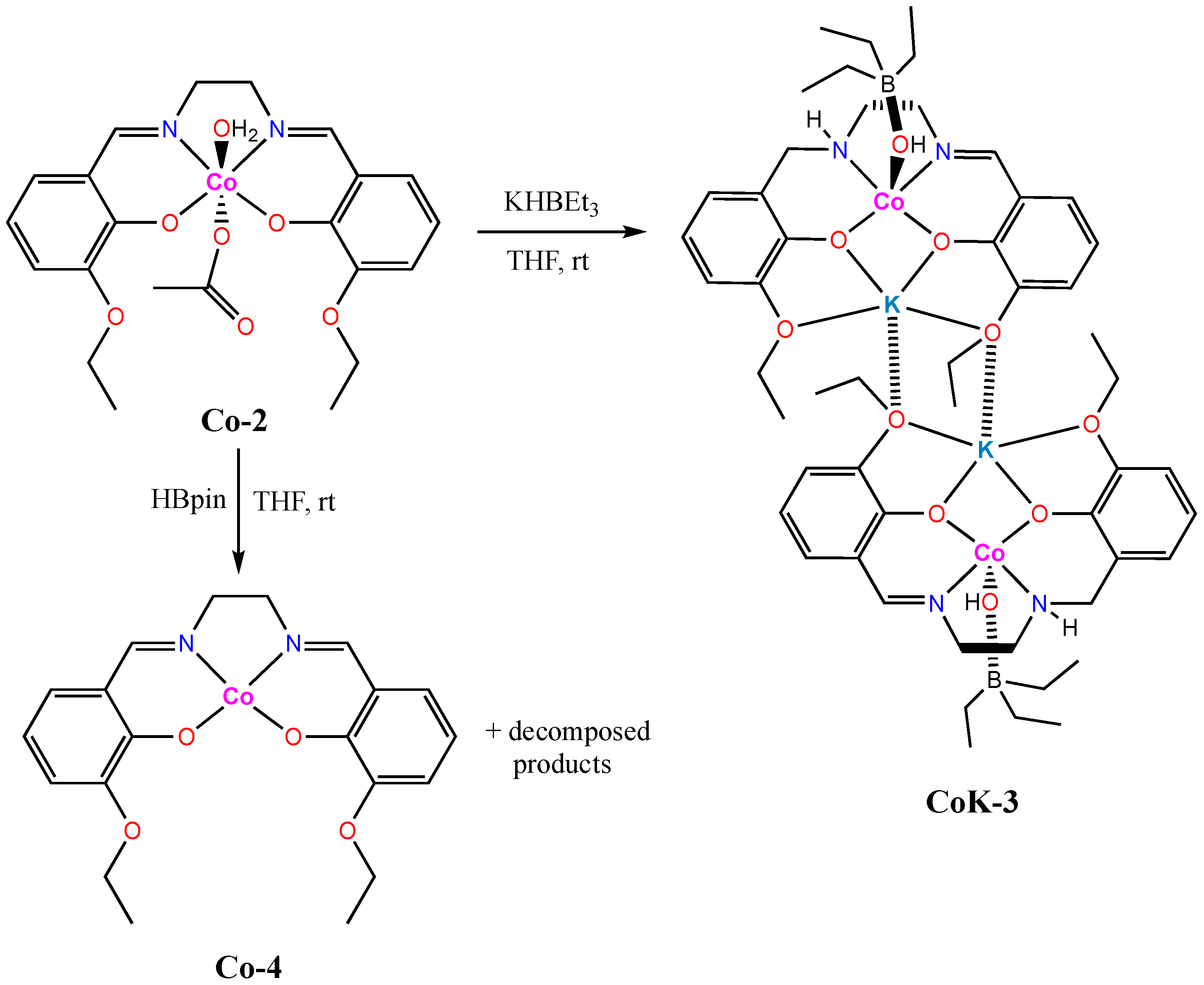
2.3. Synthesis of Co-2

The same procedure was applied as for Mn-1, except the use of Co(OAc)2·4H2O (0.125 g, 0.500 mmol) instead of Mn(OAc)2·4H2O. Dark-red block-like crystals were collected after one week. Like Mn-1, the crystals of Co-2 were poorly soluble in most organic solvents except DMSO. Yield: 0.19 g (78%). UV/Vis λmax/nm (1.0 × 10⁻⁵ mol dm−3, DMSO): 255 (51, ε/103 dm3 mol−1 cm−1, 297 (23.5), 330 (22.4), 422 (br, 8.1). FT-IR (solid, cm−1): 3432br, 2970m, 2925m, 1621s, 1598s, 1551s, 1466s, 1440s, 1388m, 1326s, 1300s, 1220s, 1177w, 1110w, 1080s, 1048w, 1015m, 944w, 892m, 735s. HR-MS (ESI positive): 413.0905 ([M-2(CH3CO2)-H2O]+, calc. 413.0912, base peak). Anal. Calcd. for C22H27CoN2O7: C 53.88, H 5.55, N 5.71%. Found C 53.57, H 5.68, N 5.40%.

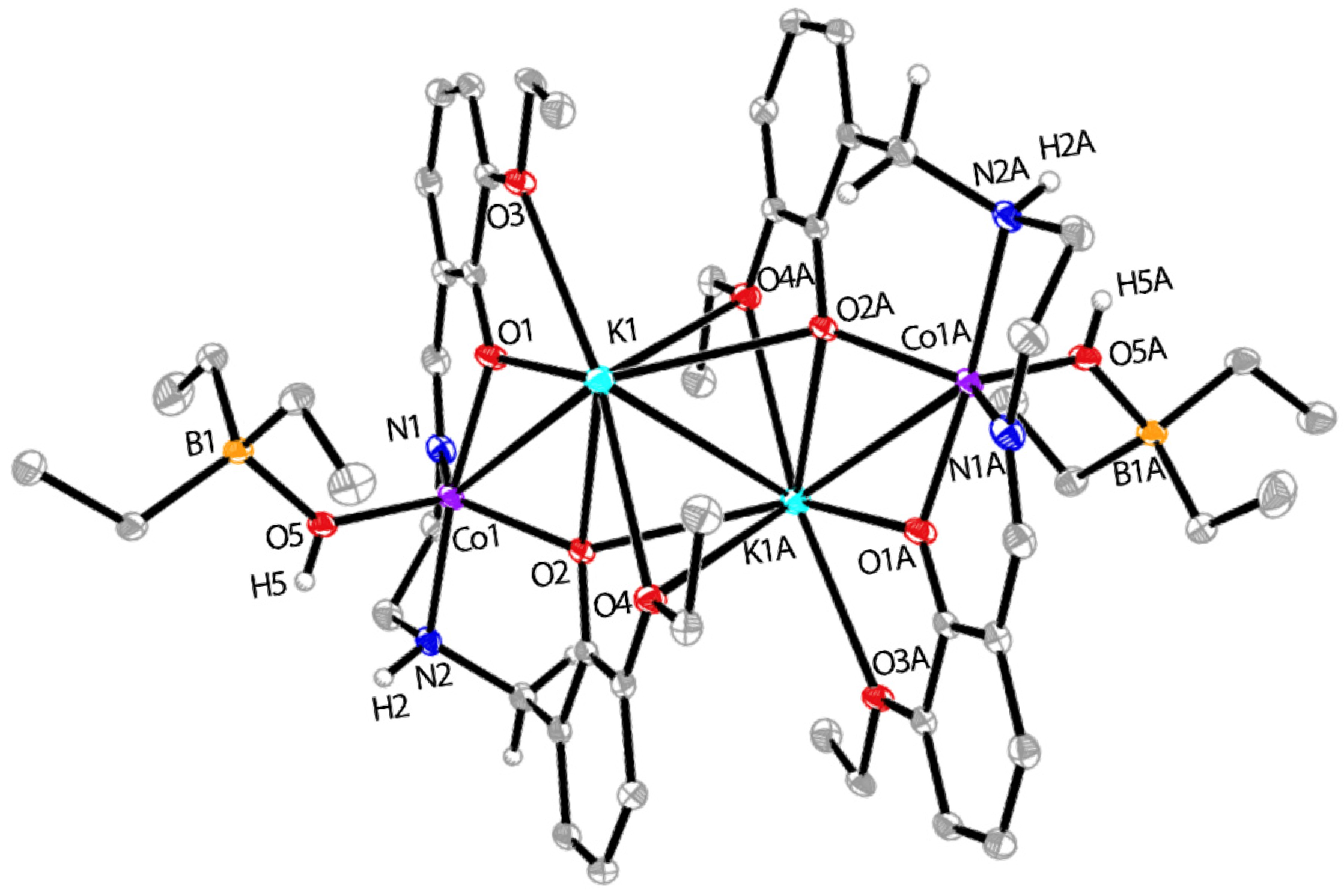
2.4. Synthesis of CoK-3

Under N2 atmosphere, Co-2 (0.049 g, 0.10 mmol) was suspended in THF (8 mL) in a 20 mL disposable vial at room temperature. Three equivalents of potassium triethylborohydride (KHBEt3, 0.3 mL, 1 M solution in THF) were then added dropwise upon stirring. The solid was gradually dissolved upon the addition of KHBEt3. The reaction was allowed to run at room temperature for 2 h until a red solution developed. The solution was filtered, and the filtrate was allowed to slowly evaporate under N2 atmosphere for 3 days, after which time X-ray quality crystals of CoK-3 were isolated as red blocks by decanting the solvent and then washed with diethyl ether. Yield: 0.035 g (62%). UV/Vis λmax/nm (1.0 × 10⁻⁵ mol dm−3, DMSO): 257 (75.5, ε/103 dm3 mol−1 cm−1), 408 (br, 9.3). FT-IR (solid, cm−1): 3400br, 2972m, 2927m, 1601s, 1467s, 1446s, 1393m, 1314s, 1220s, 1176w, 1079s, 1016m, 886w, 845w, 734s. Anal. Calcd. for C52H80B2Co2K2N4O10·(C4H8O)2: C 56.16, H 7.54, N 4.37%. Found C 55.67, H 7.79, N 4.12% (note: the analytic data for carbon is unsatisfactory, probably due to the presence of minor impurities or solvent residue).

2.5. Synthesis of Co-4

Under N2 atmosphere, Co-2 (0.049 g, 0.10 mmol) was suspended in THF (8 mL) in a 20 mL disposable vial at room temperature. Three equivalents of pinacolborane (HBpin, 0.038 g, 0.30 mmol) were then added dropwise upon stirring. The solid was dissolved upon the addition of HBpin. The reaction was allowed to run at room temperature for 2 h until a brownish solution developed. The solution was filtered, and then vapor diffusion of diethyl ether into the filtrate led to the formation of several pieces of red blocks, which were determined by X-ray crystallography to be Co-4, along with the formation of a white solid due to decomposition. Unfortunately, the amount of sample Co-4 was insufficient for further characterization and toxicity studies.

2.6. UV-Vis Absorption Measurement

Samples of compounds Mn-1, Co-2, and CoK-3 for the solution UV-Vis absorption measurements were prepared by the following procedure. A stock solution in DMSO (5 mL) was prepared at a concentration of 1 × 10⁻³ M. A 50 µL stock solution was then taken and added to pure DMSO to make a total 5 mL DMSO solution of each sample at a concentration of 1 × 10⁻⁵ M. In parallel, a 50 µL stock solution was separately taken and added to pure water to make a total 5 mL H2O solution of each sample at a concentration of 1 × 10⁻⁵ M. After the UV-Vis spectrum was recorded, the water solution was kept at room temperature for 72 h after which time its UV-Vis spectrum was recorded again. This solution was then warmed up to 37 °C and kept at this temperature for an additional 2 h, before the third UV-Vis spectrum was recorded.

2.7. Cytotoxicity Measurement

MCF-7 and MDA-MB-468 cells were cultured within completed media containing Dulbecco’s modified Eagle’s medium (DMEM) supplemented with 10% fetal bovine serum (FBS) and 50 µg/mL gentamicin. MCF-10A cells were cultured within completed media containing DMEM supplemented with 5% FBS, 10 µg/mL human insulin, 20 ng/mL hEGF, 100 ng/mL cholera toxin, and 0.5 µg/mL hydrocortisone. All cells were maintained in a 5% humidified CO2 atmosphere at 37 °C. For chemical treatment, cells were subcultured into 96-well plates one day prior to the experiment. Cells were at medium density, 1 × 10⁴ cells per well (~80% confluence) for chemical treatments. Compounds were prepared as a 100 mM stock solution in DMSO, then diluted in the culture media to various concentrations (0 to 100 μM) 15 min prior to chemical treatments. The highest final concentration of DMSO in the cells was 0.1%, which is considered a safe dosage for in vitro studies [30]. The cytotoxicity of samples was determined using a CCK-8 cytotoxicity assay following the manufacturer’s protocol (Sigma-Aldrich, Burlington, MA, USA). The absorbance signals were detected by a BioTek Cytation 7 cell imaging multimode reader (Agilent, Santa Clara, CA, USA) at 450 nm. All treatments were performed in triplicate. The percentage of cell viability for each chemical concentration was used to calculate IC50. Cell images were captured by a BioTek Cytation 7 cell imaging multimode reader.

2.8. X-Ray Crystallography

X-ray diffraction data were collected on a Bruker X8 Kappa Apex II (Mn-1, Co-2, and CoK-3) or Bruker D8 VENTURE (Co-4) diffractometer using Mo Kα radiation (Bruker Scientific LLC, Billerica, MA, USA). Crystal data, data collection, and refinement parameters are summarized in Table S1 (ESI). The structures were solved using a dual-space method and standard difference map techniques and were refined by full-matrix least-squares procedures on F2 with SHELXTL (version 2018/3) [31,32]. All hydrogen atoms bound to carbon were placed in calculated positions and refined with a riding model [Uiso(H) = 1.2–1.5Ueq(C)], while hydrogen atoms bound to nitrogen or oxygen were located on the difference map and freely refined. CCDC Nos. 2419008–2419011 contain the supplementary crystallographic data for this paper. These data can be obtained free of charge via https://www.ccdc.cam.ac.uk/structures/, or from the Cambridge Crystallographic Data Centre, 12 Union Road, Cambridge CB2 1EZ, UK; fax: (+44) 1223–336–033; or e-mail: deposit@ccdc.cam.ac.uk.

3. Results and Discussion

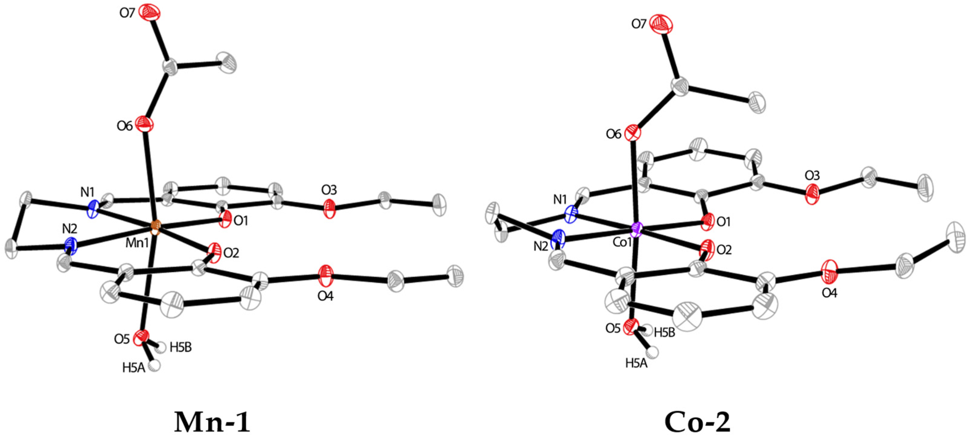
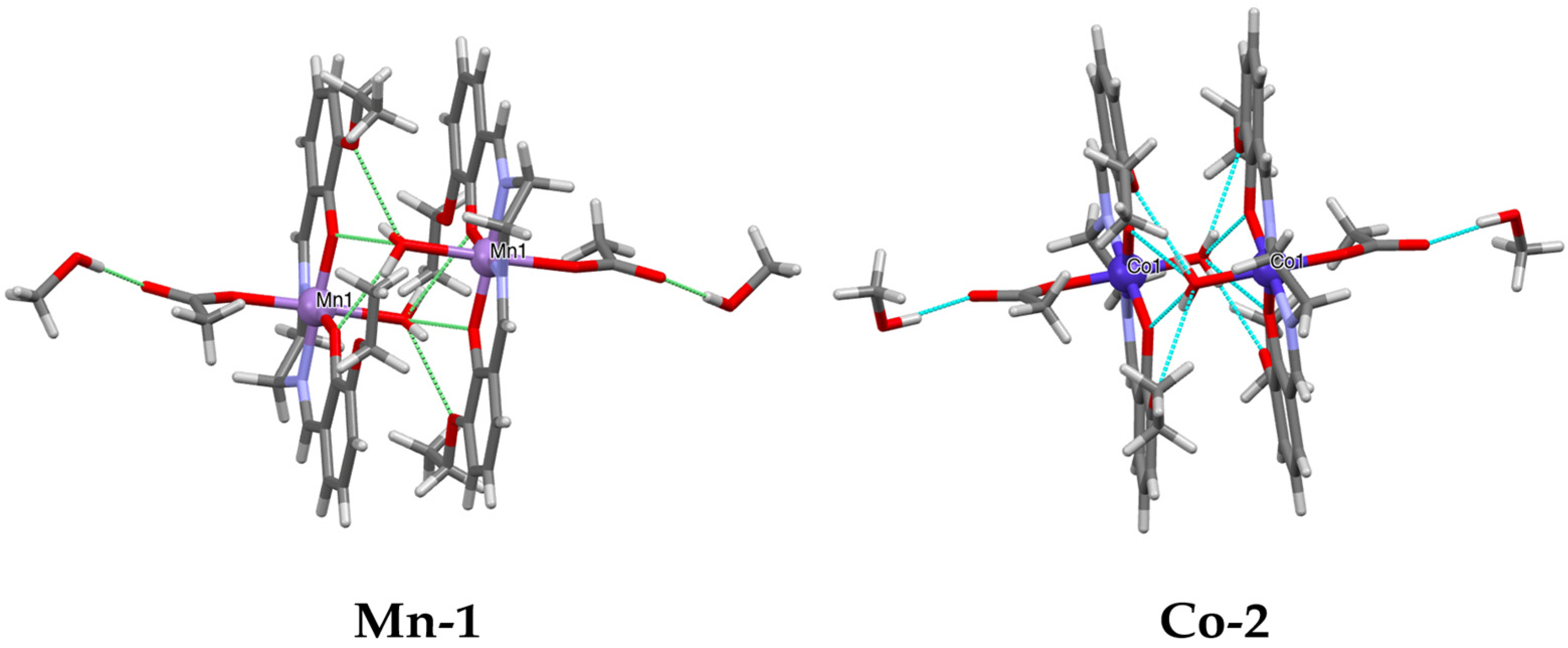
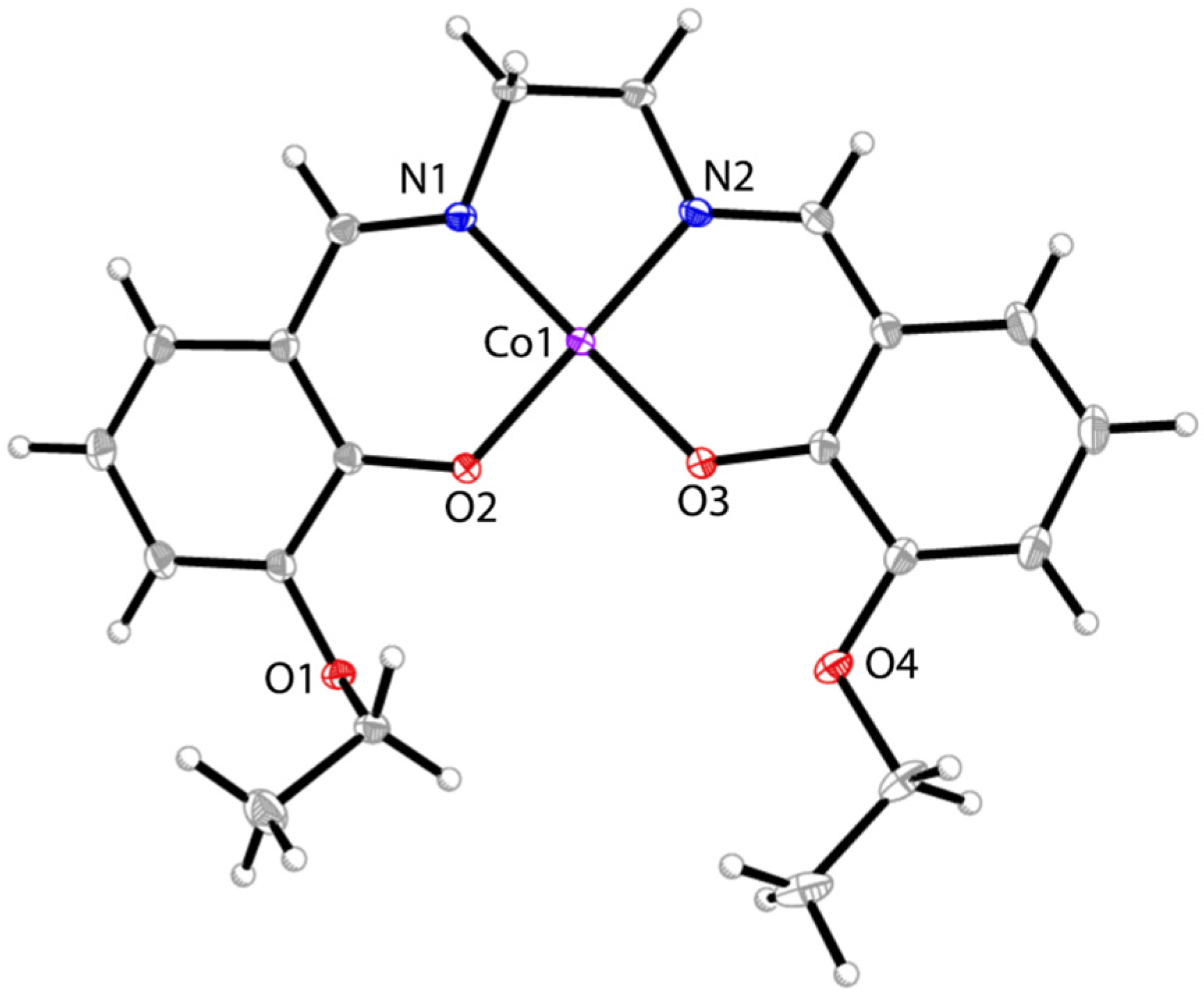
3.1. Synthesis and Crystal Structures of Mn-1 and Co-2

Manganese(III) and cobalt(III) complexes (Mn-1 and Co-2) were readily synthesized in high yields by the one-pot reactions of two equiv. of 3-ethoxysalicylaldehyde and one equiv. of 1,2-diaminoethane with manganese(II) or cobalt(II) acetate at room temperature in the air, respectively. X-ray quality crystals of both complexes were obtained after slow evaporation of a CH2Cl2-MeOH solution over a week. The crystals of both compounds show poor solubility in common organic solvents such as CH2Cl2, CHCl3, MeOH, CH3CN, or THF, yet they are well soluble in DMSO. The complexes were characterized by UV-Vis and FT-IR spectroscopies, high-resolution mass spectrometry, solution NMR spectroscopy, and elemental analysis. Although the 1H NMR spectrum of compound Co-2 in DMSO-d6 shows broad signals due to its paramagnetic characteristics (Figure S8), Mn-1 displays well-resolved NMR signals in solution, and its 1H and 13C NMR spectroscopic data are consistent with the structure as drawn in Scheme 1 (Figures S9 and S10, see Supplementary Materials). The deprotonation of the hydroxyl group in Mn-1 when coordinated to Mn has been proven by its 1H NMR spectrum due to the disappearance of the OH signal at 13.54 ppm, as observed in that of the free ligand. In addition, all other proton signals show significant shifts compared to those of the ligand alone (Figures S6 and S9).
The solid-state structures of both complexes have been unambiguously determined by single crystal X-ray diffraction. The structural analysis reveals similar structures for Mn-1 and Co-2 in which the metal centers were oxidized to the +3 oxidation state during the reactions and bound to the N2O2 cavity of the fully deprotonated salen ligand in a square planar geometry. The metal centers were further saturated in the axial directions with one acetate anion and one water molecule. Despite these structural similarities, their unit cell parameters are notably distinct. Mn-1 crystallizes in the triclinic space group P-1, while Co-2 crystallizes in monoclinic P21/n. In both structures, one independent molecule of mononuclear salen complex was found in the asymmetric unit, along with a co-crystallized methanol molecule stabilized through hydrogen bonding. The molecular structures of Mn-1 and Co-2 are shown in Figure 1, and the relevant bond parameters are given in the caption. It is worth noting that although a large number of similar cobalt and manganese salen complexes with either +2 or +3 metal oxidation states have been reported in the literature, the structures of Mn-1 and Co-2 were thus far unknown. However, closely related structures of MnIII–salen and CoIII–salen were reported [33,34,35,36,37] where the o-vanillin-pendant salen ligand has been used. In addition, analogous complexes containing the same salen ligand yet different axial ligands have been studied [38,39]. The polymorphism of complex Mn-1 has also been observed in earlier reports [40,41]. The introduction of ethoxyl groups provides an extra O4 environment potentially suitable for the incorporation of a larger metal ion (such as K, see below) due to its size, in addition to the N2O2 coordinating cavity.
The bond parameters of Mn-1 and Co-2 are unexceptional and well comparable to those reported [31,32,33,34,35,36,37,38,39,40,41]. In both structures, the M–Osalen bond lengths are significantly shorter than the M–Oaxial bonds, indicating a stronger chelating interaction between the metal ion and the salen ligand. For both complexes, the pendant ethoxyl-O atoms remain uncoordinated; instead, they behave as hydrogen-bond acceptors for the formation of hydrogen-bonded dimers between two centrosymmetric molecules of the complex (Figure 2). Specifically, the axially coordinated water molecule of one complex acts as a hydrogen bond donor to form two pairs of bifurcated hydrogen bonds with the O4 cavity of the salen ligand in the second molecule. In addition, the crystallized methanol molecule is hydrogen bonded with the acetate ligand. Details of all the hydrogen bonding parameters can be found in Tables S2 and S3 (see Supplementary Materials).

3.2. Reductive Reactivity of Co-2

The reduction of salen ligands is a popular way to make new ligands, namely “salan”, with saturated C–N bonds; directly reducing a metal–salen complex is unusual and may lead to unexpected products [42,43,44]. However, we were interested in investigating the reactions of Mn-1 and Co-2 with two reducing agents, potassium triethylborohydride (KHBEt3) and pinacolborane (HBpin), respectively. While the reduction of Mn-1 led to complicated mixtures that could not be isolated and identified, reactions involving Co-2 were carried out with isolable products (see Scheme 2). Thus, reacting a solution of Co-2 with an excess amount of KHBEt3 in THF under inert atmosphere resulted in the switching from a dark red into a light red solution, from which red crystals of CoK-3 were isolated upon evaporation in an appreciable yield (62%). The structure of CoK-3 can be regarded as a novel dimer of Co-K bimetallic complexes with partially reduced salen ligands, as shown in Scheme 2. Its structure was unambiguously identified by X-ray crystallography and also characterized by elemental analysis, UV-Vis absorption, and IR spectroscopies, although its solution NMR spectrum showed a paramagnetic nature (Figure S11). Compound CoK-3 crystallizes in the triclinic space group P-1, and its X-ray structure is presented in Figure 3, which reveals a centrosymmetrical dimer. Two reduction events occurred upon reacting with KHBEt3. First, the salen ligand was partially reduced at only one of the imine bonds by KHBEt3 and water (originally from Co-2). Supporting water as a hydrogen source, the anionic byproduct Et3B(OH) formed during the reduction process acted as a new axial ligand in CoK-3. Second, the original Co(III) center was turned to Co(II) via a one-electron reduction by extra KHBEt3. Although it is well known that KHBEt3 is usually employed to reduce metal complexes for the preparation of metal hydride complexes, [45,46,47] the reduction of Co(II) to Co(I) by KHBEt3 has also been observed during the synthesis of cobalt(I) cyclopentadienyl–phosphine dinitrogen complexes [48]. In addition, K+ sits in the O4 cavity of one ligand and further coordinates to two extra O atoms from another ligand, leading to the formation of a larger dimeric complex.
The bond parameters in CoK-3 appear to be notably different from those in Co-2, due to the different oxidation states and coordination environments of the cobalt centers for the two complexes. In CoK-3, the Co–Nimine and Co–Namine bond lengths are 2.0258(14) and 2.1985(13) Å, respectively, significantly longer than the two Co–Nimine bonds in Co-2 (1.8905(11) and 1.8860(11) Å). The same trend is also observed for the Co–Osalen bonds in two complexes (1.9693(10) and 2.0091(11) Å for CoK-3 vs. 1.8887(8) and 1.8806(9) Å for Co-2). In addition, the ligand N2O2 cavity in CoK-3 is severely distorted due to the semi-reduction of the ligand, compared to the good coplanarity upon coordination in Co-2. Both the Co1-K1 (3.5651(4) Å) and K1-K1A (3.5112(7) Å) distances are less than the sum of the atomic radii of two metals, indicating possible metal/metal contacts.
Previously, it has been well documented that numerous complexes of both salen and salan ligands were applied for catalysis and biomedical science [42]. It was also known that metal-bound salan ligands could undergo an oxidation reaction with O2 to form a mixed salan–salen complex [42]. It has been reported earlier that Ni(II)–salen complex experienced an electroreductive reaction to produce two species, one with metal-centered reduction and another with ligand-centered reduction, respectively [42]. The direct chemical reduction of Co(III)–salen with a hydride source leading to the successful isolation and structural characterization of CoK-3 in this work is unprecedented, paving the way for the direct synthesis of novel metal salen–salan complexes that display interesting physiochemical and biological properties.
Encouraged by the isolation of the novel compound, CoK-3, we conducted another reaction with Co-2 using a different reductant, pinacolborane (HBpin). Thus, mixing complex Co-2 with an excess amount of HBpin in THF under inert atmosphere for 2 h gave rise to the formation of a red solution. The slow diffusion of the resultant solution with diethyl ether at room temperature led to the isolation of red block-like crystals of Co-4 in a very small amount, accompanied by a larger amount of colorless microcrystals, which are possibly due to the decomposition of the salen ligand and/or the reaction between salen and HBpin. The single crystal X-ray diffraction data for Co-4 revealed a simple CoII–salen structure. Apparently, in this case, only the CoIII center was reduced, and the ligand remained intact. The molecular structure, as shown in Figure 4, reveals a mononuclear complex containing a square planar CoIIN2O2 coordination environment. The CoII center adopts no axial coordination, and no solvent molecules are involved in the crystals. The Co–N and Co–O bond lengths in Co-4 are slightly shorter than those found in Co-2. Although the synthesis of complex Co-4 has been reported by metalation of the salen ligand in the literature, [49,50] our approach proves the feasibility of reducing its CoIII precursor, even though the yield could be further improved. Surprisingly, the X-ray structure of Co-4 has not been reported thus far, though analogues with the related OMe–salen have been structurally characterized [51,52].

3.3. Stability of Drug Molecules

To determine whether three drug candidates that have been studied for cytotoxicity are stable enough in aqueous media during the testing period (72 h, see Section 3.4), we examined their solution stability by using UV-Vis absorption spectroscopy. Their UV-Vis spectra were first recorded in pure DMSO, where they showed good solubility (see Figure S12, see Supplementary Materials). Then, the water solution of each drug with a concentration of 1 × 10⁻⁵ M was obtained by diluting the stock solutions for each of the samples in DMSO, similar to the conditions used for cytotoxicity measurement, as described in experimental details. Their UV-Vis spectra were obtained as freshly prepared in the aqueous solution (Figure 5). It was found that all complexes displayed signals of absorption comparable to those recorded in DMSO with very small solvent effect, indicating their good solubility and structural integrity in aqueous solution. Furthermore, the UV-Vis absorption was measured for each sample in the aqueous solution after a period of 72 h, and their spectra were compared to those recorded as prepared (Figures S13–S15). The results demonstrate that all three drugs maintain good stability after 72 h as their UV-Vis absorption peaks remain the same, with only a small decrease in the intensity, reasonably due to slight molecular aggregation and precipitation in water. In addition, their spectra showed no changes after the solutions were warmed up to 37 °C and kept for 2 h.

3.4. In Vitro Anticancer Activities

Next, we sought to determine whether these cobalt and manganese compounds could be promising drug candidates against cancer cells, in comparison with the clinically approved drug cisplatin. Both cobalt and manganese complexes (Mn-1, Co-2, and CoK-3) were highly soluble in DMSO. Therefore, DMSO was used to dissolve the compounds (including cisplatin for comparison), after which cell culture media was added to create a homogeneous aqueous solution (see experimental details).
The in vitro cytotoxicity was assessed using a cell proliferation CCK-8 assay against the human breast cancer cell line MCF-7, the triple-negative breast cancer cell line MDA-MB-468, and the non-tumorigenic epithelial cell line MCF-10A from the mammary gland. All compounds were incubated for 72 h before cell viability was assessed. The results are summarized in Table 1.
The compounds exhibited varying activities in inhibiting the growth of both cancerous and non-cancerous cells. Interestingly, Co-2 displayed a promisingly lower IC50 value (0.425 µM) against MCF-7 cells compared to cisplatin (11.46 µM), despite being less effective against MDA-MB-468 cells (17.44 µM). This discrepancy could be due to the fact that MCF-7 cells have functional tumor suppressor p53, whereas MDA-MB-468 cells have dysfunctional p53. As many anticancer drugs target the p53 pathway, further investigation into the role of p53 in Co-2-induced cell death is necessary to fully elucidate the molecular pharmacological mechanism of this compound. More importantly, this cobalt(III) drug demonstrated notably lower toxicity towards non-cancerous MCF-10A cells, compared to cisplatin under the same conditions (IC50, 214.54 vs. 17.86 µM). Co-2 is also less toxic towards healthy cells than other drugs, Mn-1 and CoK-3, in this study. This makes Co-2 a potentially ideal drug candidate, specifically for the MCF-7 cell treatment. In addition, although CoK-3 shows low toxicity against the non-cancerous MCF-10A cells (IC50 = 116.22 µM), it is less potent against both MCF-7 (31.46 µM) and MDA-MB-468 (14.84 µM) cancer cells. Mn-1, on the other hand, was found to be relatively toxic towards MCF-10A cells (15.10 µM) and less effective against MCF-7 (115.86 µM) and MDA-MB-468 (19.77 µM) cancer cells as compared to cobalt-based drugs. Therefore, this work reveals that among the Mn and Co compounds studied here, Co-2 performs the best as a potent anticancer drug with reduced side effects than cisplatin, making it a promising candidate for further in vivo and clinic experiments.
Based on the current results and relevant literature [53,54,55], a potential mechanism underlying the differential anticancer effects may involve the functional status of the p53 tumor suppressor pathway. MCF-7 cells harbor functional p53, whereas MDA-MB-468 cells contain a dysfunctional variant, which could affect their sensitivity to the compound. Additionally, variations in oxidative stress response, apoptosis signaling, or metal ion homeostasis may also contribute to the observed differences. These hypotheses will be further examined through future studies, including proteomic analyses, to clarify the molecular pharmacological mechanisms involved. Investigations into cellular uptake of compounds using ICP-MS will also be conducted to help elucidate the compound’s detailed anticancer activity.

4. Conclusions

In conclusion, we have synthesized and characterized new Mn(III)– and Cobalt(III)–salen complexes (Mn-1 and Co-2) resulting from a simple one-pot synthesis. Their crystal structures were found to contain a typical MN2O2 coordination sphere for metal–salen complexes with two extra axial ligands. Multiple intermolecular hydrogen bonds exist between the axially coordinating water molecules and the O4 cavity of the salen from another molecule, contributing to the formation of hydrogen-bonded dimers for both complexes. The reductive reactivity of both complexes was further explored with hydride reducing reagents to understand their stability and potential transformation. Thus, a novel heterometallic product (CoK-3) has been isolated and structurally characterized from the reaction between Co-2 and KHBEt3, although the reaction of Mn-1 with the reductant was more complicated. Alternatively, Co-2 could be reduced to a simple square planar CoII–salen (Co-4) without axial coordination using HBpin, despite a rather low yield. In vitro cytotoxicity studies revealed that these compounds showed varying anticancer activities on human breast cancer cells (MCF-7), triple-negative breast cancer cells (MDA-MB 468), and non-cancerous cells (MCF-10A). It was found that Co-2 can be used as a potent anticancer drug that outperforms cisplatin specifically for MCF-7 cell treatment, with minimal damage to healthy cells. Further structural modifications will be required to discover more effective and universal antitumor drug candidates using salen complexes with essential trace metals.

Supplementary Materials

The following supporting information can be downloaded at: https://www.mdpi.com/article/10.3390/chemistry7030085/s1, Figure S1: The FT-IR spectrum of complex Mn-1; Figure S2: The FT-IR spectrum of complex Co-2; Figure S3: The FT-IR spectrum of complex CoK-3; Figure S4: The ESI-MS spectrum of complex Mn-1; Figure S5: The ESI-MS spectrum of complex Co-2; Figure S6: The 1H NMR (500 MHz) spectrum of the free salen ligand in DMSO-d6; Figure S7: The 13C NMR (126 MHz) spectrum of the free salen ligand in DMSO-d6; Figure S8: The 1H NMR (600 MHz) spectrum of complex Co-2 in DMSO-d6; Figure S9: The 1H NMR (600 MHz) spectrum of complex Mn-1 in DMSO-d6. One co-crystallized MeOH molecule is also observed; Figure S10: The 13C NMR spectrum of complex Mn-1 in DMSO-d6; Figure S11: The 1H NMR (600 MHz) spectrum of complex CoK-3 in DMSO-d6; Figure S12: The UV-Vis spectra of compounds Mn-1 (blue), Co-2 (red) and CoK-3 (green) in DMSO (c = 1 × 10⁻⁵ M); Figure S13: The UV-Vis spectra of compound Mn-1 in aqueous solution (concentration: 1 × 10⁻⁵ M) as prepared (blue), after 24 h (red) and after being warmed up to 37 °C for 2 h (black); Figure S14: The UV-Vis spectra of compound Co-2 in aqueous solution (concentration: 1 × 10⁻⁵ M) as prepared (blue), after 24 h (red) and after being warmed up to 37 °C for 2 h (black); Figure S15: The UV-Vis spectra of compound CoK-3 in aqueous solution (concentration: 1 × 10⁻⁵ M) as prepared (blue), after 24 h (red) and after being warmed up to 37 °C for 2 h (black); Table S1: X-ray crystallographic data; Table S2: Hydrogen bonding parameters for Mn-1 [Å and °]; Table S3: Hydrogen bonding parameters for Co-2 [Å and °]; Table S4: Hydrogen bonding parameters for CoK-3 [Å and °].

Author Contributions

Conceptualization, S.-Y.C. and G.Z.; Synthesis, characterization, and cytotoxicity, A.K., H.M. and C.P.; crystallographic analysis, M.C.N.; writing—original draft preparation, S.-Y.C. and G.Z.; writing—review and editing, A.K., H.M., S.-Y.C., M.C.N. and G.Z.; supervision, S.-Y.C. and G.Z.; project administration, G.Z.; funding acquisition, G.Z. and M.C.N. All authors have read and agreed to the published version of the manuscript.

Funding

We are grateful for the financial support from the National Science Foundation (CHE 1900500 and CHE 2247728) for this work. We also thank the support from the American Chemical Society Petroleum Research Fund (#66150-UR1). The PSC-CUNY award (66076–0054) and the PRISM program at John Jay College, the City University of New York, are gratefully acknowledged. The X-ray diffractometer used for this work was provided by the Air Force Office of Scientific Research under award number FA9550–20–1–0158.

Data Availability Statement

Data are contained within the article or Supplementary Material.

Conflicts of Interest

There are no conflicts to declare. The funders had no role in the design of the study, in the collection, analysis, or interpretation of data, in the writing of the manuscript, or in the decision to publish the results.

References

  1. Dilruba, S.; Kalayda, G.V. Platinum-Based Drugs: Past, Present, and Future. Cancer Chemother. Pharmacol. 2016, 77, 1103–1124. [Google Scholar] [CrossRef] [PubMed]
  2. Dasari, S.; Tchounwou, P.B. Cisplatin in Cancer Therapy: Molecular Mechanisms of Action. Eur. J. Pharmacol. 2014, 740, 364–378. [Google Scholar] [CrossRef] [PubMed]
  3. Romani, A.M. Cisplatin in Cancer Treatment. Biochem. Pharmacol. 2022, 206, 115323. [Google Scholar] [CrossRef] [PubMed]
  4. Ghosh, S. Cisplatin: The First Metal-Based Anticancer Drug. Bioorg. Chem. 2019, 88, 102925. [Google Scholar] [CrossRef]
  5. Mjos, K.D.; Orvig, C. Metallodrugs in Medicinal Inorganic Chemistry. Chem. Rev. 2014, 114, 4540–4563. [Google Scholar] [CrossRef]
  6. Gou, Y.; Huang, G.; Li, J.; Yang, F.; Liang, H. Versatile Delivery Systems for Non-Platinum Metal-Based Anticancer Therapeutic Agents. Coord. Chem. Rev. 2021, 441, 213975. [Google Scholar] [CrossRef]
  7. Raymond, E.; Chaney, S.G.; Taamma, A.; Cvitkovic, E. Oxaliplatin: A Review of Preclinical and Clinical Studies. Ann. Oncol. 1998, 9, 1053–1071. [Google Scholar] [CrossRef]
  8. O’Dwyer, P.J.; Stevenson, J.P.; Johnson, S.W. Clinical Status of Cisplatin, Carboplatin, and Other Platinum-Based Antitumor Drugs. In Cisplatin–Chemistry and Biochemistry of a Leading Anticancer Drug; Lippert, B., Ed.; Helvetica Chimica Acta: Zürich, Switzerland, 1999; pp. 29–69. [Google Scholar]
  9. Siddik, Z.H. Cisplatin: Mode of Cytotoxic Action and Molecular Basis of Resistance. Oncogene 2003, 22, 7265–7279. [Google Scholar] [CrossRef]
  10. Florea, A.M.; Büsselberg, D. Cisplatin as an Anti-Tumor Drug: Cellular Mechanisms of Activity, Drug Resistance, and Induced Side Effects. Cancers 2011, 3, 1351–1371. [Google Scholar] [CrossRef]
  11. Oun, R.; Moussa, Y.E.; Wheate, N.J. The Side Effects of Platinum-Based Chemotherapy Drugs: A Review for Chemists. Dalton Trans. 2018, 47, 6645–6653. [Google Scholar] [CrossRef]
  12. Lucaciu, R.L.; Hangan, A.C.; Sevastre, B.; Oprean, L.S. Metallo-Drugs in Cancer Therapy: Past, Present, and Future. Molecules 2022, 27, 6485. [Google Scholar] [CrossRef] [PubMed]
  13. Komeda, S.; Casini, A. Next-Generation Anticancer Metallodrugs. Curr. Top. Med. Chem. 2012, 12, 219–235. [Google Scholar] [CrossRef] [PubMed]
  14. Ott, I.; Gust, R. Non-Platinum Metal Complexes as Anti-Cancer Drugs. Arch. Pharm. 2007, 340, 117–126. [Google Scholar] [CrossRef] [PubMed]
  15. Kar, K.; Ghosh, D.; Kabi, B.; Chandra, A. A Concise Review on Cobalt Schiff Base Complexes as Anticancer Agents. Polyhedron 2022, 222, 115890. [Google Scholar] [CrossRef]
  16. Munteanu, C.R.; Suntharalingam, K. Advances in Cobalt Complexes as Anticancer Agents. Dalton Trans. 2015, 44, 13796–13808. [Google Scholar] [CrossRef]
  17. Tisato, F.; Marzano, C.; Porchia, M.; Pellei, M.; Santini, C. Copper in Diseases and Treatments, and Copper-Based Anticancer Strategies. Med. Res. Rev. 2010, 30, 708–749. [Google Scholar] [CrossRef]
  18. Wani, W.A.; Baig, U.; Shreaz, S.; Shiekh, R.A.; Iqbal, P.F.; Jameel, E.; Ahmad, A.; Mohd-Setapar, S.H.; Mushtaque, M.; Hun, L.T. Recent Advances in Iron Complexes as Potential Anticancer Agents. New J. Chem. 2016, 40, 1063–1090. [Google Scholar] [CrossRef]
  19. Bouché, M.; Hognon, C.; Grandemange, S.; Monari, A.; Gros, P.C. Recent Advances in Iron-Complexes as Drug Candidates for Cancer Therapy: Reactivity, Mechanism of Action, and Metabolites. Dalton Trans. 2020, 49, 11451–11466. [Google Scholar] [CrossRef]
  20. Zhang, P.; Sadler, P.J. Redox-Active Metal Complexes for Anticancer Therapy. Eur. J. Inorg. Chem. 2017, 12, 1541–1548. [Google Scholar] [CrossRef]
  21. Cheng, S.Y.; Zhang, Q.; Tang, Q.; Neary, M.C.; Zheng, S. Diverse Cobalt(II) and Iron(II/III) Coordination Complexes/Polymers Based on 4′-Pyridyl:2,2′;6′,2″-Terpyridine: Synthesis, Structures, Catalytic and Anticancer Activities. Chemistry 2024, 6, 1099–1110. [Google Scholar] [CrossRef]
  22. Zhang, G.; Zeng, H.; Zadori, N.; Marino, C.; Zheng, S.; Neary, M.C. Homoleptic Octahedral CoII Complexes as Precatalysts for Regioselective Hydroboration of Alkenes with High Turnover Frequencies. RSC Adv. 2023, 13, 28089–28096. [Google Scholar] [CrossRef] [PubMed]
  23. Erxleben, A. Transition Metal Salen Complexes in Bioinorganic and Medicinal Chemistry. Inorg. Chim. Acta 2018, 472, 40–57. [Google Scholar] [CrossRef]
  24. Alfonso-Herrera, L.A.; Hernández-Romero, D.; Cruz-Navarro, J.A.; Ramos-Ligonio, Á.; López-Monteon, A.; Rivera-Villanueva, J.M.; Morales-Morales, D.; Colorado-Peralta, R. Transition Metal Complexes with Tetradentate Schiff Bases (N2O2) Obtained from Salicylaldehyde: A Review of Their Possible Anticancer Properties. Coord. Chem. Rev. 2024, 505, 215698. [Google Scholar] [CrossRef]
  25. Alfonso-Herrera, L.A.; Rosete-Luna, S.; Hernández-Romero, D.; Rivera-Villanueva, J.M.; Olivares-Romero, J.L.; Cruz-Navarro, J.A.; Soto-Contreras, A.; Arenaza-Corona, A.; Morales-Morales, D.; Colorado-Peralta, R. Transition Metal Complexes with Tridentate Schiff Bases (ONO and ONN) Derived from Salicylaldehyde: An Analysis of Their Potential Anticancer Activity. ChemMedChem 2022, 17, e202200367. [Google Scholar] [CrossRef]
  26. Doctrow, S.R.; Liesa, M.; Melov, S.; Shirihai, O.S.; Tofilon, P. Salen Mn Complexes Are Superoxide Dismutase/Catalase Mimetics That Protect the Mitochondria. Curr. Inorg. Chem. 2012, 2, 325–334. [Google Scholar] [CrossRef]
  27. Segat, B.B.; Menezes, L.B.; Cervo, R.; Cargnelutti, R.; Tolentino, H.; Latini, A.; Horn, A., Jr.; Fernandes, C. Scavenging of Reactive Species Probed by EPR and Ex-Vivo Nanomolar Reduction of Lipid Peroxidation of Manganese Complexes. J. Inorg. Biochem. 2023, 239, 112060. [Google Scholar] [CrossRef]
  28. Nistri, S.; Boccalini, G.; Bencini, A.; Becatti, M.; Valtancoli, B.; Conti, L.; Lucarini, L.; Bani, D. A New Low Molecular Weight, Mn(II)-Containing Scavenger of Superoxide Anion Protects Cardiac Muscle Cells from Hypoxia/Reoxygenation Injury. Free Radic. Res. 2015, 49, 67–77. [Google Scholar] [CrossRef]
  29. Nayak, M.; Koner, R.; Lin, H.-H.; Flörke, U.; Wei, H.-H.; Mohanta, S. Syntheses, Structures, and Magnetic Properties of Mononuclear Cu(II) and Tetranuclear CuII3MII (M = Cu, Co, or Mn) Compounds Derived from N,N′-Ethylenebis(3-ethoxysalicylaldimine): Cocrystallization Due to Potential Encapsulation of Water. Inorg. Chem. 2006, 45, 10764–10773. [Google Scholar] [CrossRef]
  30. Santos, L.M.; Shimabuko, D.Y.; Sipert, C.R. Dimethyl Sulfoxide Affects the Viability and Mineralization Activity of Apical Papilla Cells in vitro. Braz. Dent. J. 2024, 35, e24-6054. [Google Scholar] [CrossRef]
  31. Sheldrick, G.M. SHELXTL, An Integrated System for Solving, Refining, and Displaying Crystal Structures from Diffraction Data; University of Göttingen: Göttingen, Germany, 1981. [Google Scholar]
  32. Sheldrick, G.M. SHELXT-Integrated Space-Group and Crystal-Structure Determination. Acta Cryst. 2015, 71, 3–8. [Google Scholar] [CrossRef]
  33. Chang, T.; Jing, H.; Jin, L.; Qiu, W. Quaternary Onium Tribromide Catalyzed Cyclic Carbonate Synthesis from Carbon Dioxide and Epoxides. J. Mol. Catal. A Chem. 2007, 264, 241–247. [Google Scholar] [CrossRef]
  34. Casellato, U.; Tamburini, S.; Tomasin, P.; Vigato, P.A. Cyclic and Acyclic Compartmental Schiff Bases, Their Reduced Analogues, and Related Mononuclear and Heterodinuclear Complexes. Inorg. Chim. Acta 2004, 357, 4191–4207. [Google Scholar] [CrossRef]
  35. Costes, J.P.; Tuchagues, J.P.; Vendier, L.; Garcia-Tojal, J. A Strictly Dinuclear MnIII–GdIII Complex: Synthesis and Magnetic Properties. Eur. J. Inorg. Chem. 2013, 19, 3307–3311. [Google Scholar] [CrossRef]
  36. Tripathi, N.; Sharma, V.K. Synthesis, Spectroscopic and Structural Characterization of Trivalent Chromium, Manganese, Iron, and Cobalt Complexes with Schiff Bases Derived from o-Vanillin. Pol. J. Chem. 2008, 82, 523–535. [Google Scholar]
  37. Gutierrez, A.; Perpiñán, M.F.; Sanchez, A.E.; Torralba, M.C.; Gonzalez, V. Water Inclusion Mediated Structural Diversity and the Role of H-Bonds in Molecular Assemblies of Manganese(III) Bicompartmental Schiff-Base Complexes. Inorg. Chim. Acta 2016, 453, 169–178. [Google Scholar] [CrossRef]
  38. Chakraborty, P.; Mohanta, S. Syntheses, Structures, and Catecholase Activity of Two Cobalt(III) Complexes Derived from N,N′-Ethylenebis(3-ethoxysalicylaldiimine): A Special Host–Guest System from a Special Ligand. Inorg. Chim. Acta 2015, 435, 38–45. [Google Scholar] [CrossRef]
  39. Zhang, N.; Huang, C.Y.; Shi, D.H.; You, Z.L. Unprecedented Preparation of Bis-Schiff Bases and Their Manganese(III) Complexes with Urease Inhibitory Activity. Inorg. Chem. Commun. 2011, 14, 1636–1639. [Google Scholar] [CrossRef]
  40. Sutradhar, M.; Carrella, L.M.; Rentschler, E. Mononuclear Mn(III) and Dinuclear Mn(III,III) Schiff Base Complexes: Influence of π–π Stacking on Magnetic Properties. Polyhedron 2012, 38, 297–303. [Google Scholar] [CrossRef]
  41. Chakraborty, P.; Majumder, S.; Jana, A.; Mohanta, S. Syntheses, Structures, Catecholase Activity, Spectroscopy, and Electrochemistry of a Series of Manganese(III) Complexes: Role of Auxiliary Anionic Ligand on Catecholase Activity. Inorg. Chim. Acta 2014, 410, 65–75. [Google Scholar] [CrossRef]
  42. Pessoa, J.C.; Correia, I. Salan vs. Salen Metal Complexes in Catalysis and Medicinal Applications: Virtues and Pitfalls. Coord. Chem. Rev. 2019, 388, 227–247. [Google Scholar] [CrossRef]
  43. Zhang, C.; Sutherland, M.; Herasymchuk, K.; Clarke, R.M.; Thompson, J.R.; Chiang, L.; Walsby, C.J.; Storr, T. Octahedral Co(III) Salen Complexes: The Role of Peripheral Ligand Electronics on Axial Ligand Release upon Reduction. Can. J. Chem. 2018, 96, 110–118. [Google Scholar] [CrossRef]
  44. Miranda, J.A.; Wade, C.J.; Little, R.D. Indirect Electroreductive Cyclization and Electrohydrocyclization Using Catalytic Reduced Nickel(II) Salen. J. Org. Chem. 2005, 70, 8017–8026. [Google Scholar] [CrossRef] [PubMed]
  45. Gehring, H.; Metzinger, R.; Herwig, C.; Intemann, J.; Harder, S.; Limberg, C. Hydride Reactivity of Ni(II)–X–Ni(II) Entities: Mixed-Valent Hydrido Complexes and Reversible Metal Reduction. Chem. Eur. J. 2013, 19, 1629–1636. [Google Scholar] [CrossRef] [PubMed]
  46. Gehring, H.; Metzinger, R.; Braun, B.; Herwig, C.; Harder, S.; Ray, K.; Limberg, C. An Iron(II) Hydride Complex of a Ligand with Two Adjacent β-Diketiminate Binding Sites and Its Reactivity. Dalton Trans. 2016, 45, 2989–2996. [Google Scholar] [CrossRef]
  47. Ding, K.; Brennessel, W.W.; Holland, P.L. Three-Coordinate and Four-Coordinate Cobalt Hydride Complexes That React with Dinitrogen. J. Am. Chem. Soc. 2009, 131, 10804–10805. [Google Scholar] [CrossRef]
  48. Wang, G.X.; Yan, X.; Yin, J.; Yin, Z.B.; Wei, J.; Xi, Z. Cobalt Cyclopentadienyl-Phosphine Dinitrogen Complexes. Chem. Eur. J. 2022, 28, e202202803. [Google Scholar] [CrossRef]
  49. Earnshaw, A.; Hewlett, P.C.; King, E.A.; Larkworthy, L.F. Transition Metal–Schiff’s Base Complexes. Part III. The Magnetic Properties of Some Oxygen-Carrying Cobalt(II) Complexes. J. Chem. Soc. A Inorg. Phys. Theor. 1968, 241–246. [Google Scholar] [CrossRef]
  50. Park, S.; Mathur, V.K.; Planalp, R.P. Syntheses, Solubilities, and Oxygen Absorption Properties of New Cobalt(II) Schiff-Base Complexes. Polyhedron 1998, 17, 325–330. [Google Scholar] [CrossRef]
  51. Jiang, G.B.; Zhang, S.H.; Zeng, M.H. Aqua{6,6′-Dimethoxy-2,2′-[Ethane-1,2-Diylbis(Nitrilomethylidyne)]Diphenolato-κ4O,N,N′,O′}Cobalt(II). Struct. Rep. 2007, 63, m2383. [Google Scholar] [CrossRef]
  52. Reath, A.H.; Ziller, J.W.; Tsay, C.; Ryan, A.J.; Yang, J.Y. Redox Potential and Electronic Structure Effects of Proximal Nonredox Active Cations in Cobalt Schiff Base Complexes. Inorg. Chem. 2017, 56, 3713–3718. [Google Scholar] [CrossRef]
  53. Wang, H.; Guo, M.; Wei, H.; Chen, Y. Targeting p53 Pathways: Mechanisms, Structures and Advances in Therapy. Signal Transduct. Target. Ther. 2023, 8, 92. [Google Scholar] [CrossRef] [PubMed]
  54. Law, B.Y.K.; Qu, Y.Q.; Mok, S.W.F.; Liu, H.; Zeng, W.; Han, Y.; Gordillo-Martinez, F.; Chan, W.K.; Wong, K.M.C.; Wong, V.K.W. New Perspectives of Cobalt Tris(bipyridine) System: Anti-Cancer Effect and Its Collateral Sensitivity towards Multidrug-Resistant (MDR) Cancers. Oncotarget 2017, 8, 55003. [Google Scholar] [CrossRef] [PubMed]
  55. Zhao, P.; Liu, D.; Hu, H.; Qiu, Z.; Liang, Y.; Chen, Z. Anticancer Activity of Four Trinuclear Cobalt Complexes Bearing Bis(salicylidene)-1,3-propanediamine Derivatives. J. Inorg. Biochem. 2022, 233, 111860. [Google Scholar] [CrossRef]
Scheme 1. The molecular structures of manganese(III)– and cobalt(III)–salen complexes studied in this work.
Chemistry 07 00085 sch001
Figure 1. The molecular structure of manganese and cobalt complexes, Mn-1 and Co-2, with atomic displacement parameters drawn at the 30% probability level. The crystalline methanol molecule and H atoms bound to carbon are omitted for clarity. Selected bond parameters for Mn-1: Mn1–N1 = 1.9828(9), Mn1–N2 = 1.9815(8), Mn1–O1 = 1.8877(7), Mn1–O2 = 1.8843(7), Mn1–O5 = 2.3280(8), Mn1–O6 = 2.1474(8) Å; N1–Mn1–N2 = 83.06(4), O1–Mn1–O2 = 92.94(3), O1–Mn1–O5 = 92.21(3), O1–Mn1–O6 = 100.19(3), O2–Mn1–O5 = 90.49(3), O2–Mn1–O6 = 94.64(4), O5–Mn1–O6 = 166.29(3)°; for Co-2: Co1–N1 = 1.8905(11), Co1–N2 = 1.8860(11), Co1–O1 = 1.8887(8), Co1–O2 = 1.8806(9), Co1–O5 = 1.9428(10), Co1–O6 = 1.8953(9) Å; N1–Co1–N2 = 85.54(5), O1–Co1–O2 = 86.67(4), O1–Co1–O5 = 88.87(4), O1–Co1–O6 = 95.03(4), O2–Co1–O5 = 89.94(4), O2–Co1–O6 = 93.92(4), O5–Co1–O6 = 174.66(4)°.
Chemistry 07 00085 g001
Figure 2. Representation of hydrogen-bonded dimers found in both complexes.
Chemistry 07 00085 g002
Scheme 2. The reactions of complex Co-2 with KHBEt3 or HBpin leading to new compounds CoK-3 and Co-4, respectively.
Chemistry 07 00085 sch002
Figure 3. The molecular structure of CoK-3 with atomic displacement parameters drawn at the 30% probability level. The crystalline THF molecules and H atoms bound to carbon are omitted for clarity. Selected bond parameters: Co1–N1 = 2.0258(14), Co1–N2 = 2.1985(13), Co1–O1 = 1.9693(10), Co1–O2 = 2.0091(11), Co1–O5 = 1.9739(11), Co1–K1 = 3.5651(4), K1–O1 = 2.5462(11), K1–O2 = 2.8209(10), K1–O3 = 2.7910(11), K1–O4 = 2.8136(11), K1–O2Ai = 2.8482(11), K1–O4Ai = 3.4017(12), K1–K1Ai = 3.5112(7), C11–N1 = 1.293(2), C21–N2 = 1.478(2), B1–O5 = 1.5707(19) Å; O1–Co1–N1 = 89.25(5), O1–Co1–N2 = 167.19(5), O2–Co1–N2 = 90.35(5), O1–Co1–O5 = 103.60(5), O1–Co1–O2 = 88.77(4), O1–K1–O2 = 62.22(3), O3–K1–O4 = 171.29(3)°. Symmetry code: i = -x, -y, -z.
Chemistry 07 00085 g003
Figure 4. The molecular structure of complex Co-4 with atomic displacement parameters drawn at the 30% probability level. Selected bond parameters: Co1–N1 = 1.8614(11), Co1–N2 = 1.8663(11), Co1–O2 = 1.8538 (9), Co1–O3 = 1.8491(9) Å; O2–Co1–N1 = 93.70(5), O3–Co1–N2 = 93.88(5), O2–Co1–N2 = 171.44(5), O3–Co1–N2 = 93.88(5), N1–Co1–N2 = 85.70(5)°.
Chemistry 07 00085 g004
Figure 5. The UV-Vis spectra of compounds Mn-1 (blue), Co-2 (red), and CoK-3 (green) in H2O/DMSO (v/v = 100:1, c = 1 × 10⁻⁵ M).
Chemistry 07 00085 g005
Table 1. Half-maximal inhibitory concentration (IC50) values (µM) of manganese and cobalt complexes against the breast cancer cell (MCF-7), the triple-negative breast cancer cell (MDA-MB 468), and the non-tumorigenic epithelial cell line from the mammary gland (MCF-10A).
CompoundMCF-7MDA-MB 468MCF-10A
Mn-1115.8619.7715.10
Co-20.42517.44214.54
CoK-331.4614.84116.22
cisplatin11.460.30917.86
Disclaimer/Publisher’s Note: The statements, opinions and data contained in all publications are solely those of the individual author(s) and contributor(s) and not of MDPI and/or the editor(s). MDPI and/or the editor(s) disclaim responsibility for any injury to people or property resulting from any ideas, methods, instructions or products referred to in the content.

