Article

Effect of Gamma Irradiation on Free Radicals and the Antioxidant Properties of Walnuts

1
Centre of EPR Spectroscopy, Institute of Catalysis, Bulgarian Academy of Sciences, Acad. G. Bonchev Str., bldg. 11, 1113 Sofia, Bulgaria
2
Laboratory of Lipid Chemistry, Institute of Organic Chemistry with Centre of Phytochemistry, Bulgarian Academy of Sciences, Acad. G. Bonchev Str., bl. 9, 1113 Sofia, Bulgaria
3
Laboratory of Biologically Active Substances, Institute of Organic Chemistry with Centre of Phytochemistry, Bulgarian Academy of Sciences, 139 Ruski blvd., 4000 Plovdiv, Bulgaria
*
Author to whom correspondence should be addressed.
Chemistry 2025, 7(2), 52; https://doi.org/10.3390/chemistry7020052
Submission received: 7 February 2025 / Revised: 21 March 2025 / Accepted: 31 March 2025 / Published: 1 April 2025
(This article belongs to the Section Molecular Organics)

Abstract

:
The present work represented results from a comprehensive study of free radicals and the antioxidant properties of irradiated walnuts. The effects of gamma irradiation on free radical generation and their stability, as well as on the antioxidant activity in walnuts, were investigated by Electron Paramagnetic Resonance (EPR) spectroscopy, Oxygen Radical Absorbance Capacity (ORAC), and Hydroxyl Radical Antioxidant Capacity (HORAC) assays. Walnut samples were irradiated using 60Co at two different doses: 10 and 25 kGy. As a marker for the identification of high-energy radiation treatment, characteristic cellulose radical signals were detected after irradiation and remained observable for over eight months. A significant increase in antioxidant activity was observed at higher irradiation doses, as measured by DPPH free radical scavenging activity, ORAC and HORAC assays.

Graphical Abstract

1. Introduction

Gamma irradiation is widely used for food sterilization, extending shelf life by reducing microbial and insect contamination, as well as preventing sprouting. It is a fast, efficient, secure, and absolutely safe method initially developed for food products in space missions, military use, and immune-compromised patients. Nowadays, it is increasingly applied to daily food products of the population and has been approved in many countries for preservation. The maximum allowable dose for the gamma irradiation of food is 10 kGy in the European Union, while in China, the USA, and Canada, the limit is 30 kGy for herbs and spices. Various analytical methods have been adopted to prove irradiation, required by global trade and consumer preferences. Among these, Electron Paramagnetic Resonance (EPR) spectroscopy is the leading technique, as it is included in three of the main European standards [1].
It is known that a single (central) signal is observed in the EPR spectra of all plant-based food products containing cellulose before irradiation. There are several suggestions about the origin of the paramagnetic species. According to EN 1787:2000 [2], after the irradiation with high energy radiation, a pair of lines (known as “satellite” signals), one on the left (at a lower magnetic field) and one on the right (at a higher magnetic field), appear in addition to the central signal. These signals are due to cellulose radicals formed by ionizing radiation. The distance between these signals is about 6 mT, and they are characteristic of radiation treatment. Standard EN 1787 is validated for pistachio nut shells, strawberries, and paprika and it deals with irradiated nuts.
It is observed that hydrogen peroxide treatment (known as “bleaching”) of nuts can also induce free radicals, leading to the appearance of two weak lines at the same g-factors, as the satellite lines. However, for walnuts, the intensity ratio of these two signals is significantly higher in bleached samples irradiated with 12 kGy, whereas for those irradiated with 3 kGy, the difference is negligible. Therefore, at higher doses, this ratio can be used to distinguish between bleached and irradiated walnuts [3]. Interlaboratory comparisons of samples have confirmed that EPR spectroscopy can be used for routine official testing of irradiated walnuts on the Italian market. However, further investigations are needed to determine whether false positive results caused by bleached walnuts are a common issue [4].
Gamma-irradiated walnuts (shells and kernels) were tested for the identification of high-energy ionizing irradiation and dosimetry. The dependence of the signal intensity on microwave power, radiation dose, and time stability was analyzed. The results show that EPR is a reliable method for identifying and quantifying irradiation in walnuts using both shells and kernels [5]. A study by Raffi et al. [6], later confirmed by other researchers [7], suggests that the two satellite lines form part of a triplet signal induced by irradiation, while the central line corresponds to the same free radical present before irradiation but with an increased concentration. The single aliquot additive dose method is applied to evaluate the radiation dose in gamma-irradiated nuts, as well as to examine the time stability of the radiation-induced signal [8]. The earlier years of research were focused on identifying gamma sterilization of various food products, including nuts, using different analytical techniques based on European standards [9,10].
Walnuts are recognized as a superfood due to their high content of healthy fats and numerous health benefits. They contain several bioactive compounds, including polyunsaturated fatty acids, plant proteins, dietary fibers, sterols, folates, tannins, and polyphenols [11]. They are also an excellent source of unsaturated fatty acids [12]. On the other hand, walnuts exhibit high antioxidant activity, attributed to their phenolic compounds, which contribute to human health. These antioxidants prevent or delay the oxidation of fatty acids, thereby protecting the integrity of the lipid content of nuts [12]. Roasting treatment affects the phenolic content and antioxidant activity of nuts [13] including walnuts [14]. Previous studies have shown that gamma irradiation has little effect on the antioxidant activity of almonds [15]; whereas, in irradiated walnuts, antioxidant activity was found to increase when measured spectrophotometrically [16]. Different methods are used to assess antioxidant capacity. EPR spectroscopy utilizes the stable free radical 1,1-diphenyl-2-picrylhydrazyl (DPPH). Other methods as Oxygen Radical Absorbance Capacity (ORAC) and Hydroxyl Radical Antioxidant Capacity (HORAC) are fluorometric assays widely used for evaluating the antioxidant potential of foods, beverages, and supplements. An ORAC assay measures the peroxyl radical scavenging capacity of the sample, whereas HORAC is an indicator for its chelating ability and preventing role in hydroxyl radical formation. In previous studies, an increase in polyphenol content and antioxidant activity (measured by ORAC and HORAC methods) was observed in defatted almonds irradiated at 25 kGy [15].
The study aims to investigate the effect of gamma irradiation on free radicals in irradiated walnuts, their stability analyzed via EPR spectroscopy, and their antioxidant activity assessed using EPR, ORAC, and HORAC methods. Additionally, the polyphenol and flavonoid content of walnuts is examined due to its close association with the antioxidant properties. To the best of our knowledge, this is the first comprehensive study on the antioxidant capacity of walnuts, employing multiple analytical methods for assessment.

2. Materials and Methods

2.1. Materials

Walnuts were purchased from the local market. For the purposes of this study, it was essential that the samples had not been previously irradiated as prior gamma irradiation could affect the investigation. To ensure this, the walnuts were tested using EPR spectroscopy. The results confirmed that the walnuts obtained from the local market had not undergone irradiation and were therefore suitable for further analysis. Following this verification, the samples were subjected to subsequent analyses. Walnut shells (of equal weight for all samples) were used to study the stability of free radicals by EPR spectroscopy, while the defatted walnut powder was prepared for antioxidant activity investigations. The preparation process was as follows: accurately weighed portions of approximately 30 g of non-irradiated walnuts and walnuts irradiated at 10 kGy and 25 kGy were ground using a grinder and extracted in a Soxhlet apparatus for 8 h with hexane [17]. The extraction residue was then evaporated to dryness under a stream of nitrogen.
DPPH, high-purity chemical-grade Ethanol Absolute Alcohol 99.9%, and hexane (>99%) were purchased from Sigma Aldrich, Sofia, Bulgaria.

2.2. Sample Irradiation

Walnut samples were packed in polyethylene bags and they were irradiated with gamma rays from a 60Co source (8200 Ci activity and 1.230 kGy/h dose rate) at the National Centre of Radiobiology and Radiation Protection, Sofia, Bulgaria. The irradiation was conducted at room temperature in the air using a semi-industrial “NIGU-7” radiation system. The equipment included four liters of a volume working chamber, which is rotated on its vertical axis during the irradiation. The delivered doses of 10 and 25 kGy were confirmed by Alanine dosimeters (Kodak BioMax, Wesertal, Germany), which were measured by an EPR spectrometer (E-scan Bruker, Bruker, Karlsruhe, Germany).

2.3. EPR Investigations

2.3.1. Preparation of Extracts

Extracts were prepared by adding 0.5 g of dried walnut residue (non-irradiated and irradiated with 10 and 25 kGy) to 7.5 mL absolute ethanol and 2.5 mL distilled water. The mixture was incubated for 24 h at room temperature without air exposure, then it was filtered through 150 mm filter paper, and the filtrate was used for further investigations. Fresh extracts were prepared before each experiment.

2.3.2. Determination of DPPH Free Radical Scavenging Activity (FRSA) by EPR

The stable free radical DPPH was used to estimate the effect of irradiation with different doses of gamma rays on the free radical scavenging activity (FRSA) of walnuts. The solutions were prepared as 1mL of 0.002 M DPPH ethanol solution was mixed with varying amounts of the walnut extracts (0.03, 0.07, 0.1, 0.13, and 0.16 mL). After approximately 50 min of incubation, the EPR spectra were recorded.
During the measurement, the capillary tube was sealed and placed inside a standard EPR quartz tube, which was placed in the EPR cavity. Control samples containing 1 mL DPPH ethanol solution and the same amount of ethanol instead of the extract were used to check the dilution. The experiment was carried out at least three times for each sample to verify the reproducibility of the results.
The amount of scavenged DPPH radicals expressed in a percentage was calculated using the following:
Scavenged DPPH radicals (%) = [(I0 − I)/I0] × 100,
where I0 was the intensity of the second DPPH peak of the control sample and I was the second peak intensity of the same EPR spectrum after the addition of the investigated extract.
In order to characterize and to compare the FRSA of walnut samples, the IC50 parameter was determined. The antioxidant abilities of the investigated sample were inversely proportional to the value of the IC50.

2.3.3. Instrumentation

The EPR spectra were recorded at room temperature using a JEOL JES-FA 100 EPR spectrometer (JEOL, Tokyo, Japan) as the first derivative of the absorption signal. The spectrometer has a standard TE011 cylindrical resonator and it operates in X–band. The walnut shells were cut into small pieces suitable to insert in a quartz EPR tube and they were fixed in the cavity center. The EPR spectra were recorded under the following experimental conditions: modulation frequency 100 kHz, microwave power 0.4 mW, modulation amplitude 0.4 mT, sweep 15 mT, time constant 0.3 s, and sweep time 2 min.

2.4. Determination of the Total Polyphenol and Total Flavonoid Contents, ORAC, and HORAC

2.4.1. Extract Preparation

Approximately 2 g of powdered defatted walnuts were weighed accurately, transferred to an extraction tube, and mixed with 40 mL of 60% (v:v) acetone acidified with 0.5% formic acid. Samples were extracted for 1 h at room temperature using a magnetic stirrer (VELP Scientifica Srl, Usmate, Italy), centrifuged 6000× g, 20 (MPW MED. Instruments, Warszawa, Poland) and supernatants were used for further investigations.

2.4.2. Determination of the Total Polyphenol and Total Flavonoid Contents

Total polyphenols were determined according to the method of Singleton and Rossi, 1965 [18] using gallic acid as a calibration standard. The results were expressed in mg of Gallic acid equivalents (GAE) per 100 g dry weight (DW) ± SD.
The total flavonoid content was determined according to Chang et al., 2002 [19], using AlCl3 reagent and the calibration curve was built with quercetin dihydrate (10–200 mg/L) as a standard compound. The results are expressed as mg quercetin equivalents (QE) per 100 g DW ± SD.

2.4.3. Determination of Antioxidant Activity by ORAC and HORAC Assays

Oxygen Radical Absorbance Capacity and hydroxyl radical averting capacity were measured according to the methodology represented in the literature [20,21,22]. The results were expressed as micromole Trolox equivalents (µmol TE) and micromole gallic acid equivalents (µmol GAE) per gram of dry weight for ORAC and HORAC, respectively. The measurements were carried out on a FLUOstar OPTIMA plate reader (BMG Labtech, Ortenberg, Germany).

2.5. Statistical Analysis

Measurements for polyphenol and flavonoid content and antioxidant activity were carried out in triplicate. The results are represented as a mean value of three parallel determinations ± standard deviation and they were compared by a Students’ t-test (MS Excel 2010 software). The values at p < 0.05 were considered statistically significant.

3. Results and Discussion

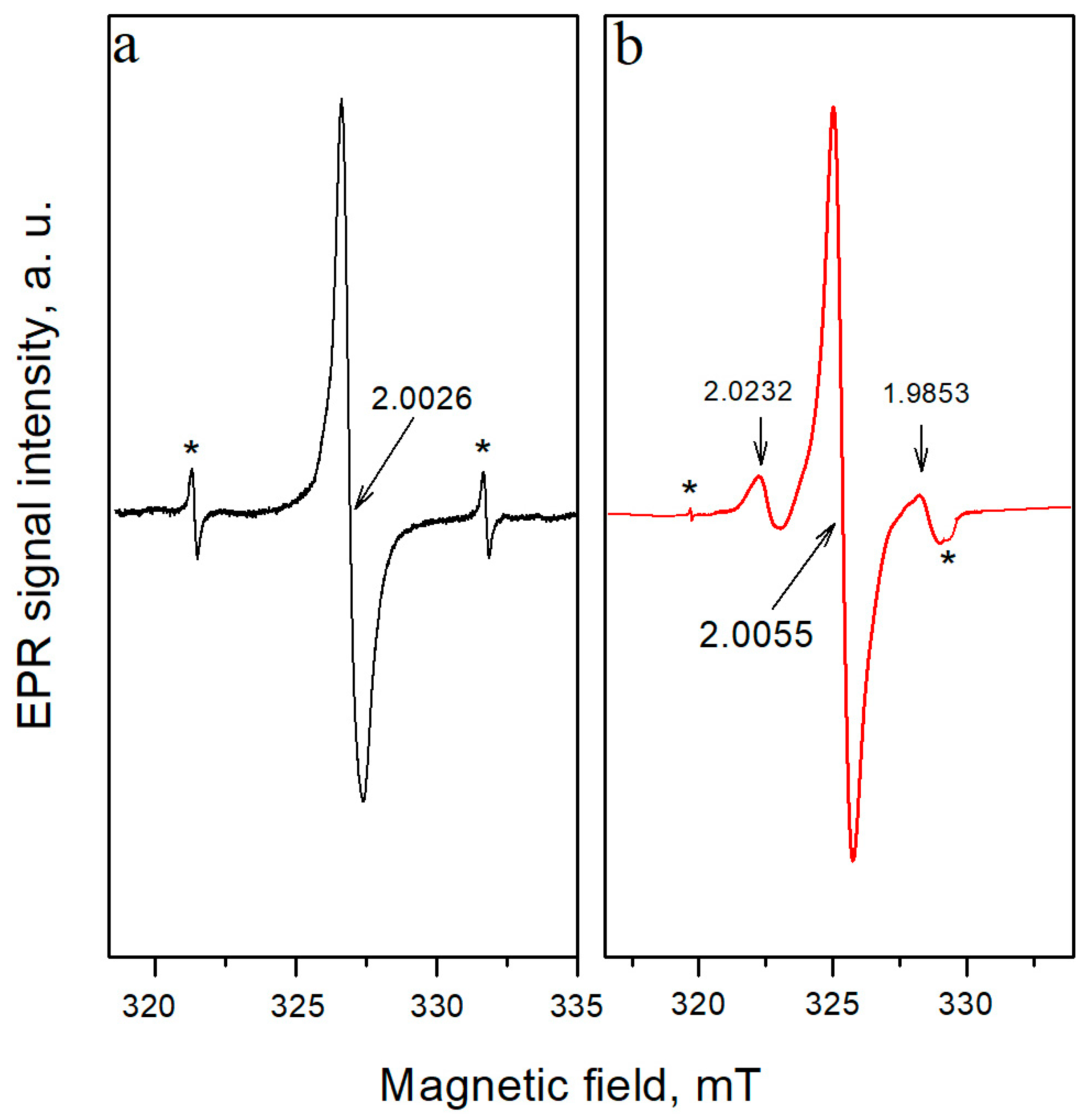
3.1. Characteristics of EPR Spectra
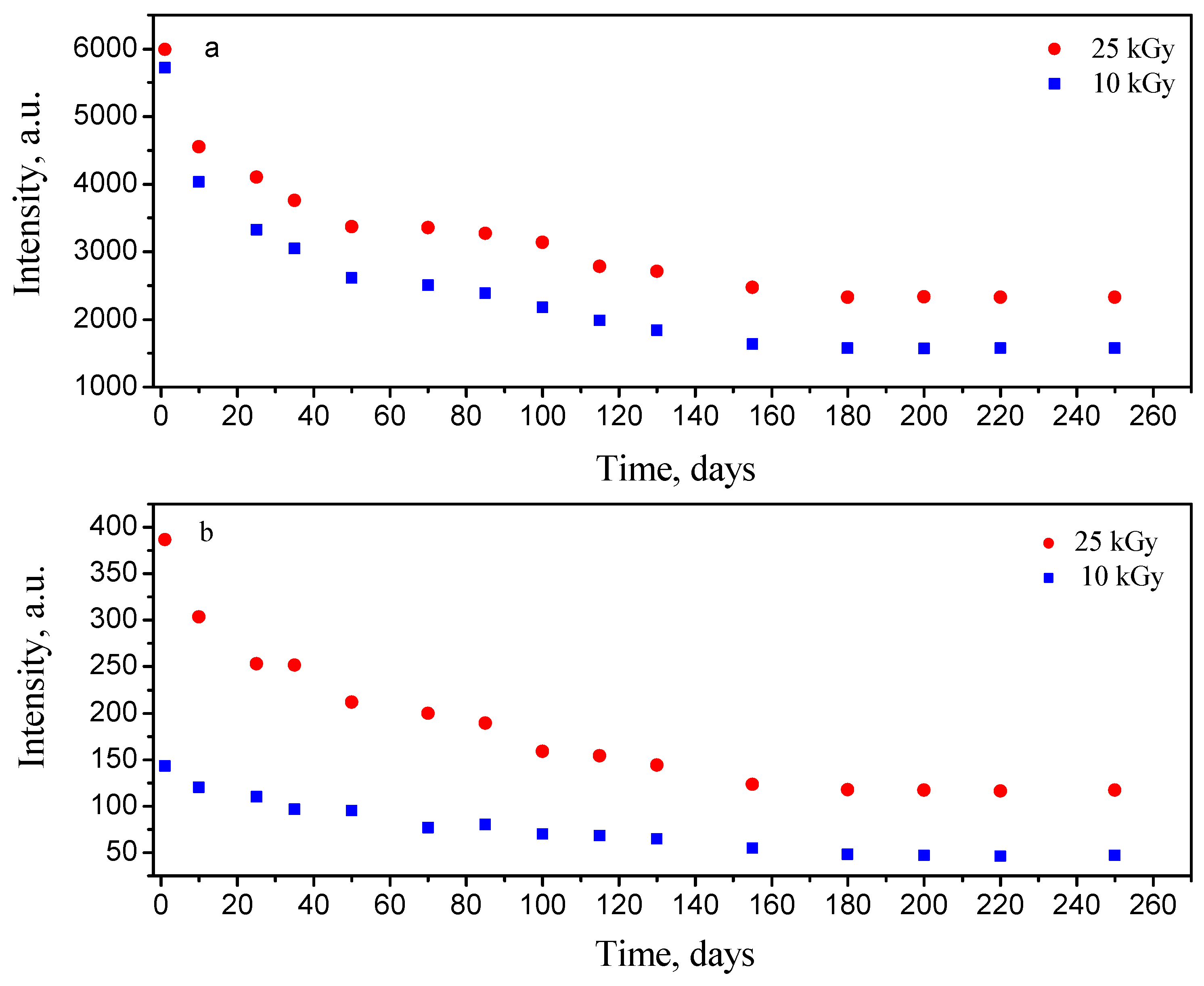
Before irradiation, the EPR spectra of walnut shells displayed a single line signal with a g-factor of 2.0026 and a linewidth ∆Hpp of 0.7 mT (Figure 1a). This signal is commonly observed in non-irradiated plant-based food products and it is attributed to semiquinone free radicals [23]. After gamma irradiation with 10 kGy and 25 kGy, a characteristic “cellulose-like” EPR spectrum was recorded (Figure 1b). It contained the irradiation-specific satellite lines from cellulose radical, spaced ~6 mT from each other (marked by arrows in Figure 1b), and increased intensity of the central line, which has a g factor 2.0055 and a linewidth ∆Hpp 0.8 mT. The left satellite line has g = 2.0232 and the right satellite 1.9853, and the linewidth is ∆Hpp 0.6 mT. The presence of these satellite signals is considered conclusive evidence of gamma irradiation, as defined by EU Standard EN 1787 [2]. This standard is based on interlaboratory tests using pistachio nut shells, paprika powder, and fresh strawberries. Additionally, the EPR spectrum of Mn2+ ions, naturally present in walnuts, was consistently observed (marked by asterisks in Figure 1). As is seen in Figure 1, the right satellite line overlaps the fourth line of Mn2+ ions. They can be used as a marker for the identification of treatment against which to compare the decrease in the radiation-generated signals. This is possible because the Mn spectrum is radiation and thermal insensitive. A detailed study [24] shows that the Mn spectrum can serve as a reference for radiation treatment when the specific satellite lines decrease with time or are completely absent. In the quoted study, significant changes in the ratio of the intensity of the central line to the intensity of the Mn signal of the irradiated samples before and after thermal treatment are observed. In non-irradiated samples after thermal treatment, a small change in the ratio was observed. However, in the present study, this method was not necessary, as the characteristic satellite lines are clearly visible. The quantity of the radiation-induced free radicals was similar between the 10 kGy and 25 kGy doses, with only a ~5% difference in the intensity of the central line. This suggests signal saturation at higher doses, likely due to radical recombination, as previously reported for almonds [15].

3.2. Kinetic Study

The stability of the central line and the satellite lines was monitored over 250 days after irradiation. The results indicate that all studied signals decrease exponentially over time, with the most significant decrease observed in the satellite lines of 25 kGy irradiated walnut shells within the first few days after irradiation (Figure 2). The central line intensity decreased by 73% for the samples irradiated with doses of 10 kGy and by 62% for 25 kGy irradiated samples after 250 days. The satellite lines decreased by ~67% and 70%, respectively, for 10 and 25 kGy doses. In contrast to almonds [15], where the central line intensity decreased by ~90%, the walnut samples decreased its intensity to a much lesser degree, which is visible against the Mn lines. As mentioned above, the quantity of free radicals was similar for both doses, and there was no significant difference in the central line intensity. However, the intensity of the satellite lines is higher for 25 kGy irradiated walnuts, confirming that at least two radiation-induced signals are present after treatment as for almonds [15]. The kinetic behavior of the satellite lines is similar for both doses, and the characteristic satellite signals remained detectable even after 8 months, confirming that irradiation with gamma rays walnuts can still be identified a long time after irradiation.

3.3. Total Polyphenol and Flavonoid Content

Table 1 represents the results for the total polyphenols and flavonoids content in the investigated samples. As is evident from the data, gamma irradiation at a higher dose has a pronounced effect on the phenolic composition of defatted walnuts, increasing the total polyphenols and flavonoid content by approximately 44% and 41%, respectively. Previous studies have also reported that gamma irradiation can enhance the concentration of phenolic compounds in plant matter [25]. However, this effect is quite complex and it depends on multiple factors including the plant matrix, the applied irradiation dose, and the type and quantity of phenolic compounds present in the sample. In general, low to moderate irradiation doses have a less pronounced effect, as observed in the current study [26]. Several mechanisms may explain the increase in phenolic content following gamma irradiation. One of the most significant factors is the breakdown of high-molecular-weight polyphenols, such as tannins, into lower-molecular-weight phenolics, which are more bioavailable and exhibit enhanced antioxidant activity [27]. The observed increase in phenolic compounds was also associated with higher antioxidant activity, as measured by both assays used in this study (Table 1). Besides the higher content of polyphenols in the irradiated samples, the enhanced antioxidant activity may also be attributed to the cleavage of glycosidic bonds in flavonoid molecules, leading to the formation of sugar residues and aglycones. Aglycones typically exhibit higher antioxidant activity than their glycosylated counterparts [28].

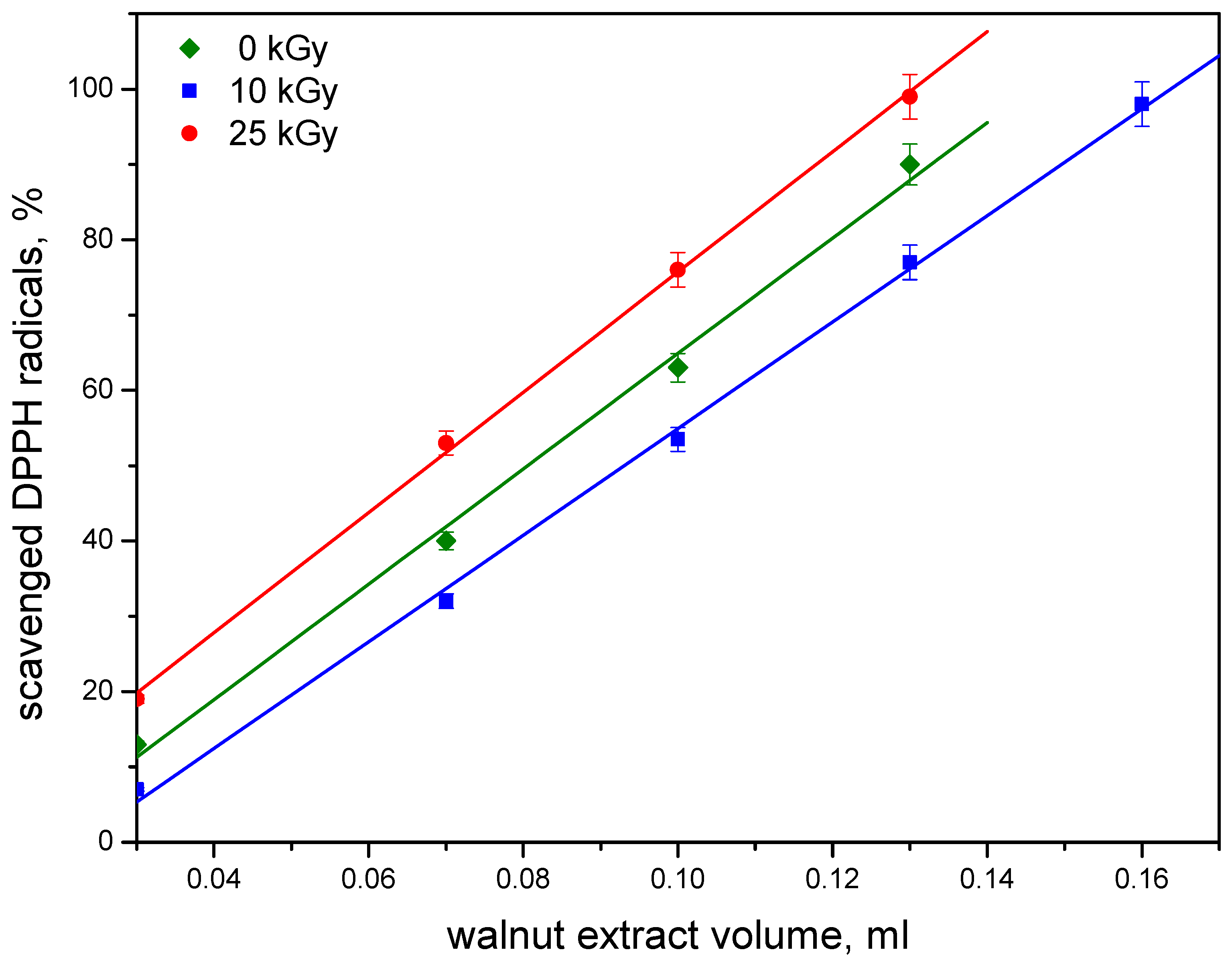
3.4. Free Radical Scavenging Activity (FRSA) Assay by EPR

The relationship between DPPH scavenged radicals and the added walnut extract volume is shown in Figure 3. The results indicate that the ethanolic extract prepared from non-irradiated walnuts exhibited bioactivity for scavenging DPPH radicals. To characterize FRSA, the IC50 parameter was calculated. For the control sample, the IC50 value was 0.081. After the irradiation of defatted walnut samples by 10 kGy gamma rays, the IC50 value increased to 0.093, indicating a decrease in antiradical activity, as FRSA is inversely proportional to IC50.
In contrast, an increase in free radical scavenging activity was observed in walnut samples irradiated with 25 kGy, with an IC50 value of 0.068. As previously mentioned, as a result of irradiation, free radicals are generated. The phenolic compounds present in walnuts, known for their antiradical properties, scavenge these radicals, and during the process, antioxidants are exhausted. This is the reason for the observed approximately 9% decrease in FRSA after walnut irradiation by a dose of 10 kGy. However, the recorded 13% increase in antiradical activity after 25 kGy walnuts irradiation is probably due to a significant increase in phenolic and flavonoid content (data are shown in Table 1). In this work, as in other reports [27], an excellent correlation was found between the total polyphenolic content and the antioxidant capacity of plant products. According to previous investigations [29], we observed different irradiation effects on the antioxidant capacity of walnut samples depending on the applied dose of gamma radiation.
Furthermore, some authors have indicated that plants contain considerable amounts of antioxidant phenolics in bound forms [30]. High-energy irradiation at 25 kGy generates more free radicals, which may break covalently bonded polymeric compounds into their free forms, thereby enhancing FRSA. Another possible explanation is the improved extractability of walnut phenolics after 25 kGy irradiation. Some nut phenolics may be associated with other plant constituents, such as proteins or carbohydrates. Thus, a certain fraction of polyphenols is probably “unextractable” because of bonding with cell wall materials, mostly through hydrogen-bonding interactions. In walnuts, tannins are often found as protein complexes [31]. Probably, after gamma irradiation at 25 kGy, these hydrogen bonds in the complex may loosen and/or break, leading to the release of tannins or their weaker binding to proteins, thus making them more easily extractable.

4. Conclusions

The current study presents a comprehensive analysis of irradiated gamma ray walnuts, focusing on free radical stability and antioxidant capacity. The findings contribute to a deeper understanding of the impact of gamma irradiation on the chemical composition and bioactivity of plant-based food products. EPR spectroscopy can confirm radiation-induced free radical formation for at least 8 months after irradiation. This long-term stability suggests that EPR spectroscopy can serve as a reliable tool for detecting irradiation treatment in walnuts and potentially other plant-based foodstuffs. A significant increase in polyphenol and flavonoid content was observed at 25 kGy, correlating with enhanced antioxidant activity, as evidenced by DPPH, ORAC, and HORAC assays. This indicates that gamma irradiation can alter the chemical structure of bioactive compounds, potentially increasing their accessibility and functionality. Higher irradiation doses improved FRSA, suggesting an increased availability of bioactive compounds. The dose-dependent effects of gamma irradiation demonstrate that while moderate doses (10 kGy) may lead to some antioxidant depletion due to free radical formation, higher doses (25 kGy) can enhance antioxidant potential by promoting the release of phenolic compounds. This suggests that controlled irradiation could be strategically used to optimize the health benefits of walnuts and other plant-based foods.

Author Contributions

Conceptualization—K.A., R.M. and Y.K.; EPR analysis and kinetic study—K.A. and Y.K., Free radical scavenging activity assay—R.M.; ORAC and HORAC assay—P.D.; preparation of walnut defatted powder S.T.; statistical analysis—R.M.; writing—original draft preparation, K.A.; writing—review and editing, Y.K. All authors have read and agreed to the published version of the manuscript.

Funding

This research work received no external funding.

Data Availability Statement

The data presented in this study are available on request from the corresponding author.

Acknowledgments

Research equipment of distributed research infrastructure INFRAMAT (part of the Bulgarian National roadmap for research infrastructures) supported by the Bulgarian Ministry of Education and Science was used in this investigation.

Conflicts of Interest

The authors declare no conflicts of interest.

Abbreviations

The following abbreviations are used in this manuscript:
EPRElectron Paramagnetic Resonance
ORACOxygen Radical Absorbance Capacity
HORACHydroxyl Radical Antioxidant Capacity
DPPH1,1-diphenyl-2-picrylhydrazyl
FRSAFree radical scavenging activity
IC50the amount of antioxidant necessary to decrease the initial DPPH radical concentration by 50%
TEACTrolox equivalent antioxidant capacity
GAEGallic acid equivalents
SDStandard deviation
QEQuercetin equivalents

References

  1. Aleksieva, K.I.; Yordanov, N.D. Various approaches in EPR identification of gamma-irradiated plant foodstuffs: A review. Food Res. Intern. 2018, 105, 1019–1028. [Google Scholar] [CrossRef]
  2. EN 1787; Foodstuffs-Detection of Irradiated Food Containing Cellulose—Method by ESR Spectroscopy. European Committee for Standardization: Brussels, Belgium, 2000.
  3. Aoude-Werner, D.; Straub, I.; Zumsteeg, V.; Kuntz, F. Identification of bleached and irradiated walnuts and hazelnuts by ESR spectroscopy. Rad. Phys. Chem. 2020, 173, 108882. [Google Scholar] [CrossRef]
  4. Bortolin, E.; Cardamone, C.; Chiaravalle, A.E.; Carratu, B.; Deiana, G.; Di Noto, A.; Di Schiavi, M.T.; D’Oca, M.C.; Gargiulo, R.; Mangiacotti, M.; et al. An inter-laboratory comparison to evaluate the suitability of EN 1787 standard to detect irradiation in plant-origin foods with health benefits. Food Control 2020, 117, 107326. [Google Scholar] [CrossRef]
  5. Maghraby, A.; Salama, E.; Sami, A.; Mansour, A.; El-Sayed, M. Identification and dosimetry of irradiated walnuts (Juglans regia) using EPR. Rad. Eff. Def. Solids 2012, 167, 170–178. [Google Scholar] [CrossRef]
  6. Raffi, J.; Yordanov, N.D.; Chabane, S.; Douifi, L.; Gancheva, V.; Ivanova, S. Identification of irradiation treatment of aromatic herbs, spices and fruits by electron paramagnetic resonance and thermoluminescence. Spectrochim. Acta Part A Mol. Biomol. Spectr. 2000, 5, 409–416. [Google Scholar] [CrossRef]
  7. Tomaiuolo, M.; Mangiacotti, M.; Trotta, G.; Marchesani, G.; Chiappinelli, A.; Chiaravalle, A.E. Identification of X-ray irradiated walnuts by ESR spectroscopy. Rad. Phys. Chem. 2018, 150, 35–39. [Google Scholar] [CrossRef]
  8. D’Oca, M.C.; Bartolotta, A. Evaluation of the original dose in irradiated dried fruit by EPR spectroscopy. Rad. Meas. 2011, 46, 813–815. [Google Scholar] [CrossRef]
  9. Esteves, M.P.; Andrade, M.E.; Empis, J. Detection of prior irradiation of dried fruits by electron spin resonance (ESR). Rad. Phys. Chem. 1999, 55, 737–742. [Google Scholar] [CrossRef]
  10. Bayram, G.; Delincee, H. Identification of irradiated Turkish foodstuffs combining various physical detection methods. Food Control 2004, 15, 81–91. [Google Scholar] [CrossRef]
  11. Ganesapillai, M.; Mathew, M.; Singh, A.; Simha, P. Influence of Microwave and Ultrasound pretreatment on Solvent Extraction of Bio-components from Walnut (Julgans regia L.) Shells. Period. Polytechn. Chem. Eng. 2016, 60, 40–48. [Google Scholar] [CrossRef]
  12. Lin, J.-T.; Liu, S.-C.; Hu, C.-C.; Shyu, Y.-S.; Hsu, C.-Y.; Yang, D.-J. Effects of roasting temperature and duration on fatty acid composition, phenolic composition, Maillard reaction degree and antioxidant attribute of almond (Prunus dulcis) kernel. Food Chem. 2016, 190, 520–528. [Google Scholar] [CrossRef] [PubMed]
  13. Chandrasekara, N.; Shahidi, F. Effect of Roasting on Phenolic Content and Antioxidant Activities of Whole Cashew Nuts, Kernels, and Testa. J. Agric. Food Chem. 2011, 59, 5006–5014. [Google Scholar] [CrossRef] [PubMed]
  14. Kita, A.; Figiel, A. Effect of roasting on properties of walnuts. Pol. J. Food Nutr. 2007, 57, 89–94. [Google Scholar]
  15. Momchilova, S.; Kazakova, A.; Taneva, S.; Aleksieva, K.; Mladenova, R.; Karakirova, Y.; Petkova, Z.; Kamenova-Nacheva, M.; Teneva, D.; Denev, P. Effect of Gamma Irradiation on Fat Content, Fatty Acids, Antioxidants and Oxidative Stability of Almonds, and Electron Paramagnetic Resonance (EPR) Study of Treated Nuts. Molecules 2023, 28, 1439. [Google Scholar] [CrossRef]
  16. Masoodi, L.; Masoodi, F.A.; Gull, A.; Gani, A.; Muzaffer, S.; Sidiq, M. Effect of γ-irradiation on the physicochemical and sensory properties of fresh walnut kernels (Juglans regia) during storage. Food Chem. Adv. 2023, 3, 100301. [Google Scholar] [CrossRef]
  17. Latimer, G.W., Jr. (Ed.) Official Methods of Analysis of AOAC International, 22nd ed.; 4.5.06 AOAC Official Method 2003.06 Crude Fat in Feeds, Cereal Grains, and Forages: Randall/Soxtec/Hexanes Extraction-Submersion Method; Oxford University Press: Oxford, UK, 2023; pp. C4-42–C4-43. [Google Scholar] [CrossRef]
  18. Singleton, V.; Rossi, J. Colorimetry of total phenolic with phosphomolybdic-phosphotungstic acid reagents. Am. J. Enol. Vitic. 1965, 16, 144–158. [Google Scholar] [CrossRef]
  19. Chang, C.; Yang, M.; Wen, H.; Chern, J. Estimation of total flavonoid content in propolis by two complementary colorimetric methods. J. Food Drug Anal. 2002, 10, 178–182. [Google Scholar] [CrossRef]
  20. Ou, B.; Hampsch-Woodill, M.; Prior, R.L. Development and validation of an improved oxygen radical absorbance capacity assay using fluorescein as the fluorescence probe. J. Agric. Food Chem. 2001, 49, 4619–4626. [Google Scholar] [CrossRef]
  21. Ou, B.; Huang, D.; Hampsch-Woodill, M.; Flanagan, J.A.; Deemer, E.K. Analysis of antioxidant activities of common vegetables employing oxygen radical absorbance capacity (ORAC) and ferric reducing antioxidant power (FRAP) assays: A comparative study. J. Agric. Food Chem. 2002, 50, 3122–3128. [Google Scholar] [CrossRef]
  22. Denev, P.; Ciz, M.; Ambrozova, G.; Lojek, A.; Yanakieva, I.; Kratchanova, M. Solid-phase extraction of berries’ anthocyanins and evaluation of their antioxidative properties. Food Chem. 2010, 123, 1055–1061. [Google Scholar] [CrossRef]
  23. Polovka, M.; Suhaj, M. The effect of irradiation and heat treatment on composition and antioxidant properties of culinary herbs and spices—A review. Food Rev. Int. 2010, 26, 138–161. [Google Scholar] [CrossRef]
  24. Yordanov, N.D.; Aleskieva, K. X- and Q- band studies on fine powders of irradiated plants. New approach for detection of their radiation history by using Q-band EPR spectrometry. Radiat. Phys. Chem. 2004, 69, 59–64. [Google Scholar] [CrossRef]
  25. Erkan, M.; Wang, S.Y.; Wang, C.Y. Effect of UV-C illumination on antioxidant capacity and postharvest decay in blueberry fruit. J. Food Agric. Environ. 2013, 11, 434–436. [Google Scholar]
  26. Denev, P.; Klisurova, D.; Teneva, D.; Ognyanov, M.; Georgiev, Y.; Momchilova, S.; Kancheva, V. Effect of gamma-irradiation on the chemical composition and antioxidant activity of dried black chokeberry (Aronia melanocarpa) fruits. Bulg. Chem. Comm. 2019, 51, 270–275. [Google Scholar]
  27. Harrison, K.; Were, L.M. Effect of gamma irradiation on total phenolic content yield and antioxidant capacity of Almond skin extracts. Food Chem. 2007, 102, 932–937. [Google Scholar] [CrossRef]
  28. Variyar, P.S.; Limaye, A.; Sharma, A. Radiation-induced enhancement of antioxidant contents of soybean (Glycine max Merrill). J. Agric. Food Chem. 2004, 52, 3385–3388. [Google Scholar] [CrossRef]
  29. Aleksieva, K.I.; Mladenova, R.B.; Solakov, N.Y.; Loginovska, K.K. EPR analysis of free radical components and antioxidant activity of bee pollen before and after gamma-irradiation. J. Radioan. Nucl. Chem. 2021, 327, 713–719. [Google Scholar] [CrossRef]
  30. Lee, S.-C.; Kim, J.-H.; Jeong, S.-M.; Kim, D.-R.; Ha, J.-U.; Nam, K.C.; Ahn, D.U. Effect of far-infrared radiation on the antioxidant activity of rice hulls. J. Agric. Food Chem. 2003, 51, 4400–4403. [Google Scholar] [CrossRef]
  31. Bodoira, R.; Maestri, D. Phenolic compounds from nuts: Extraction, chemical profiles, and bioactivity. J. Agric. Food Chem. 2020, 68, 927–942. [Google Scholar] [CrossRef]
Figure 1. EPR spectra of walnuts: (a) before irradiation (The amplification used in order to record the EPR spectra of non-irradiated samples is 10 times higher than that for irradiated samples); (b) immediately after irradiation with 10 kGy; (*)—Mn signal.
Chemistry 07 00052 g001
Figure 2. The time dependence of the EPR signal intensity of irradiated walnuts: (a) central line; (b) satellite lines.
Chemistry 07 00052 g002
Figure 3. Dependence of scavenged DPPH radicals (%) on walnut extract volume (mL): non-irradiated (), irradiated by 10 kGy (), and irradiated by 25 kGy (). Error bars = SD, n ≥ 3.
Chemistry 07 00052 g003
Table 1. Polyphenol and flavonoid content and antioxidant activity of untreated (control) and γ-irradiated (10 kGy and 25 kGy) defatted walnuts.
Total Polyphenols mg/100 gTotal Flavonoids
mg/100 g
ORAC
µmol TE/g
HORAC
µmol GAE/g
Control2915 ± 29115.5 ± 0.7235 ± 8150 ± 7
10 kGy2948 ± 1915.4 ± 0.6254 ± 9167 ± 9
25 kGy4210 * ± 1226.0 * ± 1.1320 * ± 6178 * ± 8
* Results are listed as mean values ± standard deviations. Values marked by an asterisk are significantly different from the control one (p < 0.05).
Disclaimer/Publisher’s Note: The statements, opinions and data contained in all publications are solely those of the individual author(s) and contributor(s) and not of MDPI and/or the editor(s). MDPI and/or the editor(s) disclaim responsibility for any injury to people or property resulting from any ideas, methods, instructions or products referred to in the content.

