Article

A Novel Galantamine–Curcumin Hybrid Inhibits Butyrylcholinesterase: A Molecular Dynamics Study

by
Evdokiya Salamanova
,
Mariyana Atanasova
and
Irini Doytchinova
*
Faculty of Pharmacy, Medical University of Sofia, 1000 Sofia, Bulgaria
*
Author to whom correspondence should be addressed.
Chemistry 2024, 6(6), 1645-1657; https://doi.org/10.3390/chemistry6060100
Submission received: 17 November 2024 / Revised: 12 December 2024 / Accepted: 14 December 2024 / Published: 16 December 2024
(This article belongs to the Section Theoretical and Computational Chemistry)

Abstract

:
Cholinesterases are enzymes that break down the neurotransmitter acetylcholine in the nervous system. The two main types are acetylcholinesterase (AChE) and butyrylcholinesterase (BChE). AChE inhibitors are used to treat Alzheimer’s disease by increasing acetylcholine levels. BChE activity increases in later stages of Alzheimer’s, suggesting it might contribute to the disease. In previous experiments, it was found that a newly designed hybrid of galantamine (GAL) and curcumin (CCN) (compound 4b) decreases the activity of BChE in murine brain homogenates. Here, we explore this observation using molecular dynamics simulations. GAL and CCN were also studied for comparison. The structures of the complexes between the BChE and the ligands were predicted by molecular docking. Then, molecular dynamics simulations were performed to evaluate the stability of the complexes and the interactions between the ligands and the enzyme over a simulated time of 1 μs. All three ligands formed stable complexes with BChE. Compound 4b formed more hydrogen bonds and other interactions with BChE compared to GAL and CCN, suggesting a stronger binding affinity. The stronger binding of 4b to BChE might explain its superior anti-BChE activity observed in previous experiments.

1. Introduction

Cholinesterases are specific enzymes found in the nervous system which are able to hydrolyze the important neurotransmitter acetylcholine, which facilitates communication between nerve cells and muscles. Cholinesterases can be classified into two primary types: acetylcholinesterase (AChE) and butyrylcholinesterase (BChE). Both are important in the control of acetylcholine concentration but differ in their functions, tissue distribution, and substrate specificities.
AChE is the most well-known cholinesterase, and it is highly specific for acetylcholine. It is primarily found at neuromuscular junctions and cholinergic synapses in the central and peripheral nervous systems. Its main role is to terminate synaptic transmission by rapidly hydrolyzing acetylcholine into acetate and choline after it has been released into the synaptic cleft during nerve signal transmission. The reaction catalyzed by AChE is extremely fast—one of the fastest known enzyme reactions [1]. The speed is critical for precise control of the nerve impulses, particularly in muscle cells, where continuous stimulation causes muscle paralysis or other serious effects. This makes AChE an important target in pharmacology for the treatment of conditions such as myasthenia gravis, Alzheimer’s disease, and certain forms of glaucoma [2].
BChE is a related enzyme that has a broader range of substrates but is less specific for acetylcholine compared to AChE. BChE is found in the plasma, liver, and a variety of tissues but is less abundant in synapses [3]. BChE is considered to undertake the acetylcholine hydrolysis in cases where AChE activity is compromised, such as during exposure to cholinesterase inhibitors. BChE also has clinical importance because of its ability to metabolize succinylcholine—a muscle relaxant used in surgical procedures [4]. Individuals with genetic variations leading to BChE deficiency may experience prolonged paralysis after surgery due to slow metabolism of the drug [5].
Both AChE and BChE are of significant interest in medicine. For instance, Alzheimer’s disease is associated with a decline in cholinergic neurons, and AChE inhibitors, such as galantamine, donepezil, and rivastigmine are used to increase acetylcholine levels in the brain, helping to temporarily improve cognitive function in affected individuals [6]. Research has also shown that BChE activity increases as Alzheimer’s disease progresses, making it a potential biomarker for the condition and a target for future therapies [7].
Galantamine (GAL) is a natural alkaloid used primarily in the treatment of mild to moderate Alzheimer’s disease [8]. Galantamine is derived from the bulbs and flowers of plants like the snowdrop (Galanthus sp.) and daffodil (Narcissus sp.). It is available in oral formulations and is generally well tolerated, though it can cause side effects such as nausea, vomiting, and loss of appetite. It functions as an AChE inhibitor, enhancing the levels of acetylcholine in the brain, which are important for memory and cognition. GAL is a unique AChE inhibitor because of its additional action as an allosteric modulator of nicotinic acetylcholine receptors [9]. This dual mechanism enhances its therapeutic effects by promoting the release of more acetylcholine and modulating neuronal signaling [10]. Although GAL does not cure Alzheimer’s, it can temporarily improve symptoms and slow disease progression, helping patients maintain better daily function and quality of life for a period of time.
Curcumin (CCN) is a naturally occurring polyphenol and the primary bioactive compound found in the spice turmeric (Curcuma longa) [11]. It is widely recognized for its potent anti-inflammatory, antioxidant, and antimicrobial properties. CCN has been used for centuries in Ayurveda and modern research has highlighted its potential therapeutic effects in a variety of health conditions, including cancer, cardiovascular diseases, and neurodegenerative disorders [11]. One of the key benefits of CCN is its ability to neutralize free radicals and reduce oxidative stress, which play a role in the development of chronic diseases [12]. Additionally, CCN inhibits key enzymes and signaling pathways involved in inflammation, such as NF-kB and COX-2, making it valuable in managing inflammatory conditions like arthritis [13].
Recently, we designed a series of GAL–CCN hybrids and conducted an in silico screening for anticholinesterase activity and drug-likeness. The most promising compounds were synthesized and tested in vitro for toxicity and anticholinesterase activity. Among them, the most active and non-toxic compound was identified as the hybrid named 4b (Figure 1). Compound 4b was evaluated for its anticholinesterase, antioxidant, antiamyloid, antidepressant, and neuroprotective activities through in vitro, in vivo, and ex vivo tests [14,15,16,17,18]. In all tests, 4b outperformed its parent compounds GAL and CCN and emerged as a promising multitarget agent for the treatment of neurodegenerative diseases. In one of these studies, it was found that 4b significantly inhibits BChE activity measured ex vivo in brain homogenates from mice with scopolamine-induced dementia [17]. As the present study is a continuation of our previous research [14,15,16,17,18], we preferred to keep the original compound IDs.
To further investigate these experimental findings regarding the effects of GAL, CCN, and 4b on BChE, in the present study, we performed molecular dynamics simulations of their complexes with BChE in saline solution for a duration of 1 μs (1000 ns) at human body temperature (310 K or 37 °C).

2. Models and Methods

2.1. Modeled Ligands and Complexes

The structures of the ligands GAL (CID9651) and CCN (CID 969516) were downloaded from PubChem [19]. Compound 4b was modeled as described elsewhere [14]. CCN was taken in keto-enol form as this form dominates in saline [20]. The crystallographic structure of the human BChE complex with the inhibitor (S)-2-(butylamino)-N-(2-cycloheptylethyl)-3-(1H-indol-3-yl)propanamide (PDB ID: 6QAA) [21] (UniProt ID: P06276) was used as a template to model by homology the murine BChE (UniProt ID: Q03311).

2.2. Molecular Docking Protocol

The molecular docking was conducted using GOLD v.5.2.2 (CCDC Ltd., Cambridge, UK) at the following settings: GoldScore scoring function and binding site set within 6 Å radius around the inhibitor. A total of 100 genetic algorithm (GA) runs were performed, with the protein kept rigid and the ligands set as flexible.

2.3. Molecular Dynamics (MD) Protocol

The mutated protein, in complex with any of the inhibitors, underwent three parallel molecular dynamics simulations of 1 μs with the GROMACS v.2021.3 software package (gromacs.org). The AMBER03 force field was used for the protein [22]. The ligands were parameterized using the GAFF force field [23] at the ACPYPE server [24]. Initially, the system were positioned in a truncated dodecahedral unit cell with PBC conditions and sides that were 1 nm larger than the complex. The systems were solvated with TIP3P water and NaCl was added to maintain a physiological salt concentration and adjusted to neutralize the complexes’ charge. The systems in solution were minimized with the steepest descent algorithm using a 0.01 kcal/mol step size. After that, the complexes were simulated with annealing for 10 ns, heating them from 110 to 310 K with a step of 2 fs. Equilibration and production simulations were the final protocol steps for obtaining the three trajectories. The production runs were held for 1 μs for any of the three systems. Frames were saved every 1 ns for a total of 1000 per trajectory. The trajectories were analyzed by cpptraj V4.24.0 [25].

3. Results

The experimental anti-BChE activities of GAL, CCN, and 4b were determined ex vivo using murine brain homogenates [17]. Since the X-ray structure of murine BChE was unavailable, a homology model was constructed based on the human BChE–inhibitor complex (PDB ID: 6QAA) [21]. A BLAST [26] analysis revealed that human and murine BChE share 79.24% sequence identity. Thirteen amino acids within 10 Å of the ligand in the binding site were mutated through single amino acid substitutions. These mutations were Cys66Tyr, Ser72Ala, His77Gln, Gln223Glu, Leu274Arg, Ala277Arg, Gly283Asp, Trp284Ser, Pro285Ile, Val288Ile, Phe357Tyr, Val393Ile, and Phe398Ile. The mutated protein, in complex with the inhibitor, underwent three parallel molecular dynamics (MD) simulations of 1 μs, as described in Models and Methods. The most populated conformation among the three runs identified through cluster analysis of the trajectories was used for molecular docking studies with the ligands GAL, CCN, and 4b. To validate the docking protocol, the inhibitor from the X-ray structure was redocked with an RMSD of 1.42 Å. The resulting GoldScores for GAL, CCN, 4b, and the inhibitor were 54.85, 66.68, 74.61, and 79.36, respectively. Further, the complexes BChE–GAL, BChE–CCN, and BChE–4b were used as input structures in the MD simulations.
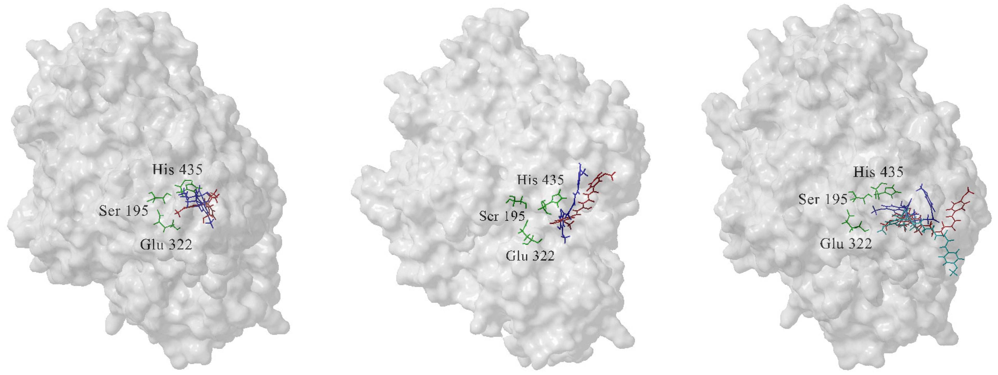
The complexes were solvated in saline, energy-minimized, heated to 310 K, equilibrated, and simulated for 1 μs (1000 ns), as detailed in the Section 2. Each complex was simulated in three independent runs. Figure 2 presents the topologies of the modeled complexes both in the input conformation and the most populated conformation, which was identified through cluster analysis across the three runs for each complex. The resulting trajectories were analyzed to evaluate the stability of the complexes and the interactions between the ligand and enzyme.

3.1. The Most Populated Conformations of the Complexes Were Identified by Cluster Analysis

Clustering molecular conformations from an MD trajectory involves grouping similar conformations into distinct clusters based on differences in coordinates or energies [27]. This method reduces variance and provides valuable insights into the ensemble of conformations. Cluster sizes vary, as higher-energy conformations tend to be less populated than those with lower energy. When the number of clusters is unknown, hierarchical clustering algorithms with average-linkage—using the average distance between members of two clusters—are recommended [27].
In this study, we conducted cluster analysis on the trajectories from the three runs for each complex using the following settings: a hierarchical agglomerative (bottom–up) approach with 10 clusters and average-linkage. The most populated conformations clustered around the lowest RMSD values. For GAL, the most populated conformation comprised 590 frames (19.7%) with an average distance to centroid of 1.5 Å; its representative frame, #733 from run 3, is close to the input conformation (Figure 2, left). The most populated conformation of CCN included 655 frames (21.8%) with an average distance to centroid of 1.6 Å, and its representative frame, #547 from run 3, shows a displacement from the initial conformation (Figure 2, middle). For 4b, the most populated conformation contained 667 frames (22.2%) with an average distance to centroid of 1.7 Å. The GAL moiety in 4b occupies a different position in the binding site and the side chains orient in opposite directions in the initial conformation and in the representative frame (#739 from run 3) (Figure 2, right). The representative frames were used as references in the RMSD calculations. The superimposed representative frames of the most populated conformations of the three compounds are given in Figure 3.

3.2. GAL, CCN, and 4b Form Stable Complexes with the Enzyme BChE

The backbone RMSDs of the enzyme BChE, averaged across the three runs for each of the three studied complexes, are shown over time in Figure 4 (left panels). The average RMSDs of the ligands in these complexes are also provided in Figure 3 (right panels). The representative frame of the most populated conformations for each complex is used as a reference. The backbone atoms of the enzyme exhibit RMSD values in the range of 1.5–2.7 Å, indicating stable structures throughout the simulations. GAL exhibits minimal movement within the binding site, with RMSD values remaining below 1.3 Å. CCN is more flexible, reaching RMSD values up to 2.4 Å. The structural distortion observed in the third run of 4b during the last 100 ns is attributed to the flexibility of the side chain moving toward the lip of the binding site (Figure 2 right, in cyan).

3.3. Compound 4b Forms More Hydrogen Bonds with BChE than GAL and CCN

During the three MD simulations, each lasting 1000 ns, GAL forms a total of 44 hydrogen bonds with BChE, with only eight persisting over 100 ns (Figure 5, top). In one bond, GAL acts as a hydrogen bond acceptor via the hydroxyl oxygen atom O6′ (Figure 1). In the remaining seven, GAL acts as a donor, using the nitrogen atom N12 in two bonds and O6′ in five. Glu322—one of the residues from the catalytic triad—forms long-lived hydrogen bonds with O6′ and N12.
CCN forms 103 hydrogen bonds with BChE across the three simulations. Due to CCN’s flexibility and increased movement within the binding site, only five bonds persist over 100 ns, of which three are unique (Figure 5, middle). In all cases, CCN acts as a hydrogen bond donor through the two hydroxyl oxygens in its aromatic rings (O8′ and O17′). No residue from the catalytic triad of BChE forms long-lived hydrogen bonds with CCN.
Compound 4b establishes 53 hydrogen bonds with BChE in total during the three runs, with eight bonds persisting over 100 ns, six of which are unique (Figure 5, bottom). In three unique bonds, 4b acts as a hydrogen bond acceptor via the carbonyl oxygen in the linker (O20′) and the methoxy oxygen in the distant aromatic ring (O26′). In the other three bonds, 4b acts as a hydrogen bond donor via the nitrogen atom N12 and the hydroxyl oxygen O6′ from the GAL moiety. His435, from the catalytic triad, forms two long-lived hydrogen bonds with O6′. The additional hydrogen bonds with the side-chain atoms in 4b enhance its affinity to BChE, explaining its higher inhibitory activity compared to GAL and CCN.

3.4. Compound 4b Forms More Interactions with BChE than GAL and CCN

In addition to hydrogen bonds, we analyzed other intermolecular interactions between BChE and the ligands in the most populated conformations of the complexes, such as electrostatic and hydrophobic interactions (Figure 6). The hydroxyl oxygen O6′ of GAL forms hydrogen bonds with Gln116 and Thr117 (Figure 6, top), the former being a long-lived bond (>100 ns) (Figure 5, top). Glu322 electrostatically attracts the quaternary nitrogen N12, and Tyr329 engages in π–π stacking with GAL’s aromatic ring. Weak carbon hydrogen bonds form between GAL and Gly113, Ile282, Leu283, Phe326, Met434, and His435. Additionally, hydrophobic interactions occur with Ala325, Phe326, Tyr329, Met434, and His435.
In the representative frame of the most populated conformation of the BChE–CCN complex, Leu270 and His435 form hydrogen bonds with hydroxyl oxygens O8′ and O17′, respectively, while Tyr329 interacts with the oxygen atom in the enol group, O11′ (Figure 6, middle). The bond with His435 is also long-lived (>100 ns) (Figure 5, middle). Additionally, His435 participates in π–π stacking with an aromatic ring of CCN. Carbon hydrogen bonds exist between Thr324 and O17′ and between Met434 and O16′. Hydrophobic interactions involve Ala325, Val328, Trp427, and Met431. Electrostatic attraction occurs between Arg274 and one of CCN’s aromatic rings, while steric interactions are detected with Asn65, Gln68, Phe70, Phe73, Gly72, Gln74, Gln116, Arg271, Glu273, Ser284, Asn286, Val433, and Phe334.
The GAL moiety of 4b is positioned differently in the binding site compared to GAL alone. Here, N12 forms a long-lived hydrogen bond with Ser284, and the methoxy carbon interacts hydrophobically with Trp228 (Figure 6, bottom). Additional hydrogen bonds are formed between Ala69, Phe70, and O20′ in the linker. The distant aromatic ring participates in π–π stacking with Phe275 and is electrostatically attracted to Asp280. Carbon hydrogen bonds are observed between Gln68 and C11, Gln68 and C16, Asn286 and C16, Ser284 and C11, and Gln116 and C13. These additional side-chain interactions stabilize the complex and enhance the affinity of 4b to BChE compared to GAL.

4. Discussion

Typically, Alzheimer’s treatments aim to inhibit AChE, and sometimes BChE, in order to increase acetylcholine levels in the brain and support cognitive functions [28]. However, some recent research suggests that BChE might play a dual role in Alzheimer’s disease. While both AChE and BChE break down acetylcholine, their functions can shift with disease progression. As Alzheimer’s advances, AChE activity tends to decrease while BChE activity increases [29]. Elevated BChE is thought to lead to excessive breakdown of acetylcholine, reducing its availability for neural signaling. By balancing BChE’s activity, it may be possible to prevent the excessive acetylcholine breakdown that contributes to cognitive decline in Alzheimer’s [30].
Some evidence suggests that in early Alzheimer’s stages, BChE may play protective roles, such as reducing the aggregation of beta-amyloid plaques, a hallmark of Alzheimer’s pathology [31]. BChE has been found within amyloid plaques in the brains of Alzheimer’s patients [32]. Research suggests that BChE may contribute to the formation and stability of these plaques, as it binds to Aβ peptides, particularly in the later stages of Alzheimer’s. BChE, when bound to Aβ, may promote the aggregation and deposition of amyloid plaques. This means that increased BChE activity could accelerate the formation of plaques. In an Alzheimer’s disease mouse model with the BChE gene knocked out, there are up to 70% fewer fibrillar Aβ brain plaques, suggesting diminished BChE activity could prove beneficial as a curative approach to Alzheimer’s [26]. Inhibitors that target both AChE and BChE (like rivastigmine) have shown some promise, as they seem to balance acetylcholine levels while potentially reducing amyloid plaque formation [33].
Compound 4b, a hybrid of GAL and CCN, was designed, synthesized, and tested in our laboratory, demonstrating superior anti-AChE, anti-BChE, antioxidant, antiamyloid, antidepressant, and neuroprotective activities compared to GAL and CCN [14,15,16,17,18]. In this study, we used molecular dynamics simulations to investigate the lifespan of complexes formed by GAL, CCN, and 4b with BChE in saline at human body temperature over 1000 ns. Trajectory analysis was conducted to uncover the underlying reasons for the enhanced inhibitory activity of 4b.
The studied inhibitors are also expected to be active against human BChE. The decision to build and refine a homology model of the murine enzyme was guided by the experimental context, where murine models were used to evaluate the inhibitor [17]. Human and murine BChE differ in the dimensions of their active-site gorge, which impact inhibitor binding. The larger active-site gorge of human BChE allows for different orientations and interactions of inhibitors compared to AChE [34]. Additionally, the number and arrangement of aromatic residues lining the active-site gorge vary between human and murine enzymes, influencing substrate specificity and inhibitor interactions [35]. These differences in binding sites can result in distinct inhibitor binding modes between human and murine BChE. For example, compounds like 4,4′-bipyridine and coumarin derivatives have been shown to bind differently to the catalytic and peripheral sites [36]. Structural adaptations observed in murine models provide valuable insights into potential binding modes and inhibitor efficacy which may not be directly extrapolatable to human models due to these structural differences [37].
We found that all three ligands form stable complexes with murine BChE and remain in the binding site throughout the entire simulation. In only one of the three runs, the side chain of 4b extend around the lip of the binding site. In AChE, the lip of the binding site contains the peripheral anionic site (PAS), composed of five aromatic residues—Tyr70, Asp72, Tyr121, Trp279, and Tyr334. The PAS plays a direct role in forming complexes with Aβ fibrils, promoting Aβ assembly [38,39]. This discovery spurred the search for dual-site-binding AChE inhibitors, targeting both the catalytic anionic site (CAS) and the PAS as multitarget agents for Alzheimer’s disease treatment [14,40,41,42,43,44,45,46].
In contrast, BChE’s PAS lacks three of the five aromatic residues present in AChE’s PAS [47] and does not participate in fibril formation. Furthermore, Diamant et al. [39] demonstrated that BChE attenuates fibril formation through a Trp residue at its C-terminus, which forms heteroaromatic complexes with monomeric or low-oligomeric Aβ conformers, thereby disrupting fibril propagation. Mutation of this Trp residue to Arg eliminates BChE’s attenuation effect on Aβ fibril formation [29]. Consequently, while 4b may effectively block AChE’s PAS to prevent Aβ fibril formation, blocking the PAS in BChE has no impact on fibril formation.
The hydrogen bonding analysis from MD simulations of GAL, CCN, and 4b with BChE over 1000 ns reveals distinct interaction patterns that align with the enhanced inhibitory activity of 4b. GAL forms eight hydrogen bonds with BChE that last over 100 ns, involving only two atoms—N12 and O6′. CCN, though forming many hydrogen bonds, has only five that persist beyond 100 ns. This lack of stable interactions, due to CCN’s flexibility, likely reduces its overall binding affinity to BChE. Compound 4b, on the other hand, forms eight long-lived hydrogen bonds that involve not only N12 and O6′ but also the carbonyl oxygen (O20′) from the linker and the distant aromatic methoxy oxygen atom (O26′). These additional side-chain interactions, absent in GAL and CCN, contribute to the unique binding profile of 4b.
In addition to hydrogen bonding, the side chain of 4b engages in various other interactions with residues in the BChE binding site. The linker oxygen O20′ forms additional hydrogen bonds with Ala69 and Phe70. The terminal aromatic ring participates in π–π stacking with Phe275 and is electrostatically attracted to Asp280. This combination of stable and distinctive interactions supports the superior inhibitory activity of 4b against BChE.
Our results align well with data on hybrid inhibitors that target both AChE and BChE, based on classical anti-AChE agents like tacrine [48]. The binding modes of these hybrids frequently involve interactions with both the CAS and PAS regions of AChE, similar to the GAL–CCN derivative [48,49,50]. This dual binding capability is essential for their effectiveness in inhibiting enzyme activity and preventing amyloid aggregation. Studies have shown that CCN–tacrine hybrids are potent dual-site AChE inhibitors, also exhibiting activity against BChE, Aβ aggregation, and β-secretase, highlighting their multifunctional potential [51]. Additionally, research indicates that IAA–tacrine hybrids effectively inhibit both AChE and BChE [49]. Tacrine conjugates with antioxidants demonstrate effective AChE and BChE inhibition, metal chelation, and the potential to block AChE-induced β-amyloid aggregation [50,52].
In conclusion, the direct implications of the presented study could be explored in several directions. The elucidated molecular interactions between BChE and ligands GAL, CCN, and 4b, such as hydrogen bonds, electrostatic attractions, and π–π stacking, explain the superior binding and inhibitory activity of 4b and guide the design of potent BChE inhibitors. The GAL–CCN hybrid shows potential as a multitarget lead for further drug development. The use of murine BChE homology models to predict ligand binding and dynamics offers a valuable tool for studying other enzyme–ligand systems. It demonstrates the reliability of combining molecular docking and MD simulations for predicting enzyme behavior.

Author Contributions

Conceptualization, E.S., M.A. and I.D.; methodology, E.S. and M.A.; software, E.S. and M.A.; formal analysis, I.D.; investigation, E.S., M.A. and I.D.; writing—original draft preparation, I.D.; writing—review and editing, E.S., M.A. and I.D.; visualization, I.D.; supervision, I.D.; project administration, I.D.; funding acquisition, I.D. All authors have read and agreed to the published version of the manuscript.

Funding

This research was funded by the Bulgarian National Roadmap for Research Infrastructure (Grant No. D01-325/2023).

Data Availability Statement

Data is contained within the article.

Acknowledgments

During the preparation of this work, the authors used AI tool for English editing. After using this tool, the authors reviewed and edited the content as needed and take full responsibility for the content of the publication.

Conflicts of Interest

The authors declare no conflicts of interest. The funders had no role in the design of the study; in the collection, analyses, or interpretation of data; in the writing of the manuscript; or in the decision to publish the results.

References

  1. Zhou, Y.; Wang, S.; Zhang, Y. Catalytic Reaction Mechanism of Acetylcholinesterase Determined by Born–Oppenheimer Ab Initio QM/MM Molecular Dynamics Simulations. J. Phys. Chem. B 2010, 114, 8817–8825. [Google Scholar] [CrossRef]
  2. McHardy, S.; Wang, H.; McCowen, S.; Valdez, M. Recent Advances in Acetylcholinesterase Inhibitors and Reactivators: An Update on the Patent Literature (2012–2015). Expert Opin. Ther. Pat. 2017, 27, 455–476. [Google Scholar] [CrossRef] [PubMed]
  3. Mack, A.; Robitzki, A. The Key Role of Butyrylcholinesterase during Neurogenesis and Neural Disorders: An Antisense-5′Butyrylcholinesterase-DNA Study. Prog. Neurobiol. 2000, 60, 607–628. [Google Scholar] [CrossRef] [PubMed]
  4. Hackett, P.; Sakai, T. Pseudocholinesterase Deficiency: A Case Report and Literature Review. Open J. Anesthesiol. 2012, 2, 188–194. [Google Scholar] [CrossRef]
  5. Thomsen, J.; Gätke, M. Patient Information Sheet for Plasma Cholinesterase Deficiency. Anaesthesia 2013, 69, 1287–1288. [Google Scholar] [CrossRef] [PubMed]
  6. Moreta, M.; Burgos-Alonso, N.; Torrecilla, M.; Marco-Contelles, J.; Bruzos-Cidón, C. Efficacy of Acetylcholinesterase Inhibitors on Cognitive Function in Alzheimer’s Disease: Review of Reviews. Biomedicines 2021, 9, 1689. [Google Scholar] [CrossRef]
  7. Rejc, L.; Gómez-Vallejo, V.; Joya, A.; Moreno, O.; Egimendia, A.; Castellnou, P.; Ríos-Anglada, X.; Cossío, U.; Baz, Z.; Passannante, R.; et al. Longitudinal Evaluation of a Novel BChE PET Tracer as an Early In Vivo Biomarker in the Brain of a Mouse Model for Alzheimer Disease. Theranostics 2021, 11, 6542–6559. [Google Scholar] [CrossRef]
  8. Wilcock, G.; Lilienfeld, S.; Gaens, E. Efficacy and Safety of Galantamine in Patients with Mild to Moderate Alzheimer’s Disease: Multicentre Randomised Controlled Trial. BMJ 2000, 321, 1445. [Google Scholar] [CrossRef]
  9. Potasiewicz, A.; Krawczyk, M.; Gzielo, K.; Popik, P.; Nikiforuk, A. Positive Allosteric Modulators of Alpha 7 Nicotinic Acetylcholine Receptors Enhance Procognitive Effects of Conventional Anti-Alzheimer Drugs in Scopolamine-Treated Rats. Behav. Brain Res. 2020, 385, 112547. [Google Scholar] [CrossRef]
  10. Kowal, N.; Ahring, P.; Liao, V.; Indurti, D.; Harvey, B.; O’Connor, S.; Chebib, M.; Olafsdottir, E.; Balle, T. Galantamine Is Not a Positive Allosteric Modulator of Human α4β2 or α7 Nicotinic Acetylcholine Receptors. Br. J. Pharmacol. 2018, 175, 2911–2925. [Google Scholar] [CrossRef] [PubMed]
  11. Sharifi-Rad, J.; Rayess, Y.; Rizk, A.; Sadaka, C.; Zgheib, R.; Zam, W.; Sestito, S.; Rapposelli, S.; Neffe-Skocińska, K.; Zielińska, D.; et al. Turmeric and Its Major Compound Curcumin on Health: Bioactive Effects and Safety Profiles for Food, Pharmaceutical, Biotechnological and Medicinal Applications. Front. Pharmacol. 2020, 11, 1021. [Google Scholar] [CrossRef]
  12. Jakubczyk, K.; Drużga, A.; Katarzyna, J.; Skonieczna-Żydecka, K. Antioxidant Potential of Curcumin—A Meta-Analysis of Randomized Clinical Trials. Antioxidants 2020, 9, 1092. [Google Scholar] [CrossRef]
  13. Buhrmann, C.; Brockmueller, A.; Mueller, A.; Shayan, P.; Shakibaei, M. Curcumin Attenuates Environment-Derived Osteoarthritis by Sox9/NF-κB Signaling Axis. Int. J. Mol. Sci. 2021, 22, 7645. [Google Scholar] [CrossRef] [PubMed]
  14. Stavrakov, G.; Philipova, I.; Lukarski, A.; Atanasova, M.; Zheleva, D.; Zhivkova, Z.D.; Ivanov, S.; Atanasova, T.; Konstantinov, S.; Doytchinova, I. Galantamine-Curcumin Hybrids as Dual-Site Binding Acetylcholinesterase Inhibitors. Molecules 2020, 25, 3341. [Google Scholar] [CrossRef] [PubMed]
  15. Simeonova, R.; Zheleva, D.; Valkova, I.; Stavrakov, G.; Philipova, I.; Atanasova, M.; Doytchinova, I. A Novel Galantamine-Curcumin Hybrid as a Potential Multi-Target Agent Against Neurodegenerative Disorders. Molecules 2021, 26, 1865. [Google Scholar] [CrossRef] [PubMed]
  16. Mladenova, K.; Stavrakov, G.; Philipova, I.; Atanasova, M.; Petrova, S.; Doumanov, J.; Doytchinova, I. A Galantamine-Curcumin Hybrid Decreases the Cytotoxicity of Amyloid-Beta Peptide on SH-SY5Y Cells. Int. J. Mol. Sci. 2021, 22, 7592. [Google Scholar] [CrossRef] [PubMed]
  17. Simeonova, R.; Atanasova, M.; Stavrakov, G.; Philipova, I.; Doytchinova, I. Ex Vivo Antioxidant and Cholinesterase-Inhibiting Effects of a Novel Galantamine–Curcumin Hybrid on Scopolamine-Induced Neurotoxicity in Mice. Int. J. Mol. Sci. 2022, 23, 14843. [Google Scholar] [CrossRef] [PubMed]
  18. Kostadinova, I.; Atanasova, M.; Stavrakov, G.; Philipova, I.; Doytchinova, I. A Galantamine-Curcumin Hybrid Lacks the Depressant Side Effect of Acetylcholinesterase Inhibitors. Biotechnol. Biotechnol. Equip. 2024, 38, 2305903. [Google Scholar] [CrossRef]
  19. Kim, S.; Chen, J.; Cheng, T.; Gindulyte, A.; He, J.; He, S.; Li, Q.; Shoemaker, B.A.; Thiessen, P.A.; Yu, B.; et al. PubChem 2023 Update. Nucleic Acids Res. 2023, 51, D1373–D1380. [Google Scholar] [CrossRef]
  20. Zheng, B.; McClements, D.J. Formulation of More Efficacious Curcumin Delivery Systems Using Colloid Science: Enhanced Solubility, Stability, and Bioavailability. Molecules 2020, 25, 2791. [Google Scholar] [CrossRef] [PubMed]
  21. Meden, A.; Knez, D.; Jukič, M.; Brazzolotto, X.; Gršič, M.; Pišlar, A.; Zahirović, A.; Kos, J.; Nachon, F.; Svete, J.; et al. Tryptophan-Derived Butyrylcholinesterase Inhibitors as Promising Leads Against Alzheimer’s Disease. Chem. Commun. 2019, 55, 3765–3768. [Google Scholar] [CrossRef]
  22. Case, D.A.; Aktulga, H.M.; Belfon, K.; Cerutti, D.S.; Cisneros, G.A.; Cruzeiro, V.W.D.; Forouzesh, N.; Giese, T.J.; Götz, A.W.; Gohlke, H.; et al. AmberTools. J. Chem. Inf. Model. 2023, 63, 6183–6191. [Google Scholar] [CrossRef]
  23. Wang, J.; Wolf, R.M.; Caldwell, J.W.; Kollman, P.A.; Case, D.A. Development and Testing of a General AMBER Force Field. J. Comput. Chem. 2004, 25, 1157–1174. [Google Scholar] [CrossRef] [PubMed]
  24. Kagami, L.; Wilter, A.; Diaz, A.; Vranken, W. The ACPYPE Web Server for Small-Molecule MD Topology Generation. Bioinformatics 2023, 39, btad350. [Google Scholar] [CrossRef] [PubMed]
  25. Roe, D.R.; Cheatham, T.E. PTRAJ and CPPTRAJ: Software for Processing and Analysis of Molecular Dynamics Trajectory Data. J. Chem. Theory Comput. 2013, 9, 3084–3095. [Google Scholar] [CrossRef]
  26. Camacho, C.; Coulouris, G.; Avagyan, V.; Ma, N.; Papadopoulos, J.; Bealer, K.; Madden, T.L. BLAST+: Architecture and Applications. BMC Bioinform. 2009, 10, 421. [Google Scholar] [CrossRef] [PubMed]
  27. Shao, J.; Tanner, S.W.; Thompson, N.; Cheatham, T.E. Clustering Molecular Dynamics Trajectories: 1. Characterizing the Performance of Different Clustering Algorithms. J. Chem. Theory Comput. 2007, 3, 2312–2334. [Google Scholar] [CrossRef]
  28. Xing, S.; Li, Q.; Xiong, B.; Chen, Y.; Feng, F.; Liu, W.; Sun, H. Structure and Therapeutic Uses of Butyrylcholinesterase: Application in Detoxification, Alzheimer’s Disease, and Fat Metabolism. Med. Res. Rev. 2020, 41, 858–901. [Google Scholar] [CrossRef] [PubMed]
  29. Lane, R.M.; Potkin, S.G.; Enz, A. Targeting Acetylcholinesterase and Butyrylcholinesterase in Dementia. Int. J. Neuropsychopharmacol. 2006, 9, 101–124. [Google Scholar] [CrossRef] [PubMed]
  30. Giacobini, E. Cholinesterases: New Roles in Brain Function and in Alzheimer’s Disease. Neurochem. Res. 2003, 28, 515–522. [Google Scholar] [CrossRef]
  31. Zhang, J.; Zhang, Y.; Wang, J.; Xia, Y.; Zhang, J.; Chen, L. Recent Advances in Alzheimer’s Disease: Mechanisms, Clinical Trials, and New Drug Development Strategies. Sig. Transduct. Target Ther. 2024, 9, 211. [Google Scholar] [CrossRef] [PubMed]
  32. Darvesh, S. Butyrylcholinesterase as a Diagnostic and Therapeutic Target for Alzheimer’s Disease. Curr. Alzheimer Res. 2016, 13, 1173–1177. [Google Scholar] [CrossRef] [PubMed]
  33. Sun, T.; Zhen, T.; Harakandi, C.H.; Wang, L.; Guo, H.; Chen, Y.; Sun, H. New Insights into Butyrylcholinesterase: Pharmaceutical Applications, Selective Inhibitors and Multitarget-Directed Ligands. Eur. J. Med. Chem. 2024, 275, 116569. [Google Scholar] [CrossRef]
  34. Saxena, A.; Redman, A.; Jiang, X.; Lockridge, O.; Doctor, B. Differences in active-site gorge dimensions of cholinesterases revealed by binding of inhibitors to human butyrylcholinesterase. Chem. Biol. Interact. 1999, 119–120, 61–69. [Google Scholar] [CrossRef] [PubMed]
  35. Rosenberry, T.; Brazzolotto, X.; Macdonald, I.; Wandhammer, M.; Trovaslet-Leroy, M.; Darvesh, S.; Nachon, F. Comparison of the binding of reversible inhibitors to human butyrylcholinesterase and acetylcholinesterase: A crystallographic, kinetic, and calorimetric study. Molecules 2017, 22, 2098. [Google Scholar] [CrossRef] [PubMed]
  36. Simeon-Rudolf, V.; Kovarik, Z.; Radić, Z.; Reiner, E. Reversible inhibition of acetylcholinesterase and butyrylcholinesterase by 4,4′-bipyridine and by a coumarin derivative. Chem. Biol. Interact. 1999, 119–120, 119–128. [Google Scholar] [CrossRef] [PubMed]
  37. Kharlamova, A.; Lushchekina, S.; Petrov, K.; Kots, E.; Nachon, F.; Villard-Wandhammer, M.; Zueva, I.; Krejci, E.; Reznik, V.; Zobov, V.; et al. Slow-binding inhibition of acetylcholinesterase by an alkylammonium derivative of 6-methyluracil: Mechanism and possible advantages for myasthenia gravis treatment. Biochem. J. 2016, 473, 1225–1236. [Google Scholar] [CrossRef]
  38. Alvarez, A.; Opazo, C.; Alarcon, R.; Garrido, J.; Inestrosa, N.C. Acetylcholinesterase promotes the aggregation of amyloid-beta-peptide fragments by forming a complex with the growing fibrils. J. Mol. Biol. 1997, 272, 348–361. [Google Scholar] [CrossRef]
  39. Diamant, S.; Podoly, E.; Friedler, A.; Ligumsky, H.; Livnah, O.; Soreq, H. Butyrylcholinesterase Attenuates Amyloid Fibril Formation In Vitro. Proc. Natl. Acad. Sci. USA 2006, 103, 8628–8633. [Google Scholar] [CrossRef]
  40. Camps, P.; Formosa, X.; Galdeano, C.; Gómez, T.; Muñoz-Torrero, D.; Ramírez, L.; Viayna, E.; Gómez, E.; Isambert, N.; Lavilla, R.; et al. Tacrine-Based Dual Binding Site Acetylcholinesterase Inhibitors as Potential Disease-Modifying Anti-Alzheimer Drug Candidates. Chem. Biol. Interact. 2010, 187, 411–415. [Google Scholar] [CrossRef]
  41. Gupta, S.; Mohan, C.G. Dual Binding Site and Selective Acetylcholinesterase Inhibitors Derived from Integrated Pharmacophore Models and Sequential Virtual Screening. Biomed Res. Int. 2014, 2014, 291214. [Google Scholar] [CrossRef]
  42. Chierrito, T.P.C.; Pedersoli-Mantoani, S.; Roca, C.; Requena, C.; Sebastian-Perez, V.; Castillo, W.O.; Moreira, N.C.S.; Pérez, C.; Sakamoto-Hojo, E.T.; Takahashi, C.S.; et al. From Dual Binding Site Acetylcholinesterase Inhibitors to Allosteric Modulators: A New Avenue for Disease-Modifying Drugs in Alzheimer’s Disease. Eur. J. Med. Chem. 2017, 139, 773–791. [Google Scholar] [CrossRef] [PubMed]
  43. Pisani, L.; Catto, M.; De Palma, A.; Farina, R.; Cellamare, S.; Altomare, C.D. Discovery of Potent Dual Binding Site Acetylcholinesterase Inhibitors via Homo- and Heterodimerization of Coumarin-Based Moieties. ChemMedChem 2017, 12, 1349. [Google Scholar] [CrossRef]
  44. Shi, Y.; Huang, W.; Wang, Y.; Zhang, R.; Hou, L.; Xu, J.; Qiu, Z.; Xie, Q.; Chen, H.; Zhang, Y. Bis(9)-(−)-Meptazinol, a Novel Dual-Binding AChE Inhibitor, Rescues Cognitive Deficits and Pathological Changes in APP/PS1 Transgenic Mice. Transl. Neurodegener. 2018, 7, 21. [Google Scholar] [CrossRef] [PubMed]
  45. Zueva, I.; Dias, J.; Lushchekina, S.; Semenov, V.; Mukhamedyarov, M.; Pashirova, T.; Babaev, V.; Nachon, F.; Petrova, N.; Nurullin, L.; et al. New Evidence for Dual Binding Site Inhibitors of Acetylcholinesterase as Improved Drugs for Treatment of Alzheimer’s Disease. Neuropharmacology 2019, 155, 131–141. [Google Scholar] [CrossRef]
  46. Suwanhom, P.; Nualnoi, T.; Khongkow, P.; Tipmanee, V.; Lomlim, L. Novel Lawsone–Quinoxaline Hybrids as New Dual Binding Site Acetylcholinesterase Inhibitors. ACS Omega 2023, 8, 32498–32511. [Google Scholar] [CrossRef] [PubMed]
  47. Bajda, M.; Wieckowska, A.; Hebda, M.; Szałaj, N.; Sotriffer, C.; Malawska, B. Structure-Based Search for New Inhibitors of Cholinesterases. Int. J. Mol. Sci. 2013, 14, 5608–5632. [Google Scholar] [CrossRef]
  48. Hamuľaková, S.; Janovec, L.; Soukup, O.; Jun, D.; Janocková, J.; Hrabinová, M.; Sepsová, V.; Kuča, K. Tacrine-coumarin and Tacrine-7-chloroquinoline Hybrids with Thiourea Linkers: Cholinesterase Inhibition Properties, Kinetic Study, Molecular Docking, and Permeability Assay for Blood–Brain Barrier. Curr. Alzheimer Res. 2018, 15, 1096–1105. [Google Scholar] [CrossRef] [PubMed]
  49. Cheng, Z.; Zhu, K.; Zhang, J.; Song, J.; Muehlmann, L.; Jiang, C.; Liu, C.; Zhang, H. Molecular-Docking-Guided Design and Synthesis of New IAA-Tacrine Hybrids as Multifunctional AChE/BChE Inhibitors. Bioorg. Chem. 2019, 83, 277–288. [Google Scholar] [CrossRef]
  50. Elkina, N.; Grishchenko, M.; Shchegolkov, E.; Makhaeva, G.; Kovaleva, N.; Rudakova, E.; Boltneva, N.; Lushchekina, S.; Astakhova, T.; Radchenko, E.; et al. New Multifunctional Agents for Potential Alzheimer’s Disease Treatment Based on Tacrine Conjugates with 2-Arylhydrazinylidene-1,3-Diketones. Biomolecules 2022, 12, 1551. [Google Scholar] [CrossRef] [PubMed]
  51. Sun, Q.; Peng, D.; Yang, S.; Zhu, X.; Yang, W.; Yang, G. Syntheses of Coumarin-Tacrine Hybrids as Dual-Site Acetylcholinesterase Inhibitors and Their Activity Against Butyrylcholinesterase, Aβ Aggregation, and β-Secretase. Bioorg. Med. Chem. 2014, 22, 4784–4791. [Google Scholar] [CrossRef] [PubMed]
  52. Makhaeva, G.; Kovaleva, N.; Rudakova, E.; Boltneva, N.; Grishchenko, M.; Lushchekina, S.; Astakhova, T.; Serebryakova, O.; Timokhina, E.; Zhilina, E.; et al. Conjugates of Tacrine and Salicylic Acid Derivatives as New Promising Multitarget Agents for Alzheimer’s Disease. Int. J. Mol. Sci. 2023, 24, 2285. [Google Scholar] [CrossRef] [PubMed]
Figure 1. Structures of galantamine (GAL), curcumin (CCN), and their hybrid, 4b.
Chemistry 06 00100 g001
Figure 2. Superimposed conformations of the three complexes in the input conformation (blue) and in the most populated conformation (red). The enzyme is given in grey. The catalytic triad residues Ser195, Glu322, and His435 are given in green. (Left): BChE–GAL, (middle): BChE–CCN, (right): BChE–4b. The final conformation (cyan) is also given for 4b. Ser195, Glu322, and His435 correspond to Ser198, Glu325, and His438 from human BChE (PDB ID: 6QAA).
Chemistry 06 00100 g002
Figure 3. Superimposed representative frames of the most populated conformations of GAL (blue), CCN (red), and 4b (green).
Chemistry 06 00100 g003
Figure 4. RMSDs of the enzyme (left panels) and the ligands (right panels) in the studied complexes averaged over the three runs. The representative frame of the most populated conformation for each complex was used as a reference.
Chemistry 06 00100 g004
Figure 5. Intermolecular hydrogen bonds between BChE and ligands GAL (top), CCN (middle), and 4b (bottom) with lifetime ≥ 100 ns. The former atom corresponds to the H-bond acceptor and the latter to the H-bond donor. Atom numbering aligns with Figure 1 for reference.
Chemistry 06 00100 g005
Figure 6. Intermolecular interactions between BChE and ligands GAL (top), CCN (middle), and 4b (bottom) detected in the most populated conformations of the complexes. Conventional hydrogen bonds (between heteroatoms) are given in dark green dashes, carbon hydrogen bonds—in light green, electrostatic attractions—in orange, π–π interactions—in dark purple, hydrophobic interactions—in light purple, and van der Waals interactions—without dashes.
Chemistry 06 00100 g006aChemistry 06 00100 g006b
Disclaimer/Publisher’s Note: The statements, opinions and data contained in all publications are solely those of the individual author(s) and contributor(s) and not of MDPI and/or the editor(s). MDPI and/or the editor(s) disclaim responsibility for any injury to people or property resulting from any ideas, methods, instructions or products referred to in the content.