Article

Divalent Europium, NIR and Variable Emission of Trivalent Tm, Ho, Pr, Er, Nd, and Ce in 3D Frameworks and 2D Networks of Ln–Pyridylpyrazolates

1
Institute of Inorganic and Analytical Chemistry, Justus-Liebig-University Giessen, Heinrich-Buff-Ring 17, 35392 Giessen, Germany
2
Department of Chemistry, Faculty of Science, Mansoura University, El Gomhouria, Mansoura Qism 2, Dakahlia Governorate, Mansoura 11432, Egypt
3
Fachbereich Chemie, Philipps-University Marburg, Hans-Meerwein-Straße, 35032 Marburg, Germany
4
Lebedev Physical Institute of the Russian Academy of Sciences, Leninskiy pr-t, 53, 119991 Moscow, Russia
5
Basic Department of Chemistry of Innovative Materials and Technologies, G.V. Plekhanov Russian University of Economics, Stremyanny Per. 36, 117997 Moscow, Russia
6
Center of Materials Research (LAMA), Justus-Liebig-University Giessen, Heinrich-Buff-Ring 16, 35392 Giessen, Germany
*
Author to whom correspondence should be addressed.
Chemistry 2023, 5(2), 1006-1027; https://doi.org/10.3390/chemistry5020069
Submission received: 27 March 2023 / Revised: 20 April 2023 / Accepted: 26 April 2023 / Published: 28 April 2023
(This article belongs to the Special Issue Commemorating 150 Years of Justus von Liebig’s Legacy)

Abstract

:
The redox reactions of various lanthanide metals with 3-(4-pyridyl)pyrazole (4-PyPzH) or 3-(3-pyridyl)pyrazole (3-PyPzH) ligands yield the 2D network 2 [Eu(4-PyPz)2(Py)2] containing divalent europium, the 3D frameworks 3 [Ln(4-PyPz)3] and 3 [Ln(3-PyPz)3] for trivalent cerium, praseodymium, neodymium, holmium, erbium, and thulium as well as 3 [La(4-PyPz)3], and the 2D networks 2 [Ln(4-PyPz)3(Py)] for trivalent cerium and thulium and 2 [Ln2(4-PyPz)6]·Py for trivalent ytterbium and lutetium. The 18 lanthanide coordination polymers were synthesized under solvothermal conditions in pyridine (Py), partly acting as a co-ligand for some networks. The compounds exhibit a variety of luminescence properties, including metal-centered 4f–4f/5d–4f emission in the visible and near-infrared spectral range, metal-to-ligand energy transfer, and ligand-centered fluorescence and phosphorescence. The anionic ligands 3-PyPz and 4-PyPz serve as suitable antennas for lanthanide-based luminescence in the visible and near-infrared range through effective sensitization followed by emission through intra–4f transitions of the trivalent thulium, holmium, praseodymium, erbium, and neodymium. 2 [Ce(4-PyPz)3(Py)], 3 [Ce(4-PyPz)3], and 3 [Ce(3-PyPz)3] exhibit strong degrees of reduction in the 5d excited states that differ in intensity compared to the ligand-based emission, resulting in a distinct emission ranging from pink to orange. The direct current magnetic studies show magnetic isolation of the lanthanide centers in the crystal lattice of 3 [Ln(3-PyPz)3], Ln = Dy, Ho, and Er.

Graphical Abstract

1. Introduction

Divalent europium, the mildest reducing agent of the redox-sensitive divalent lanthanide ions, has been successfully used in a wide variety of material applications such as medical imaging [1,2], photochemistry [3,4], lanthanide-activated phosphors [5,6], and sensing [7,8]. Trivalent lanthanides are known for their luminescence properties, with f–f based emission covering the spectrum from the ultraviolet (UV) to the near-infrared (NIR) spectral region, characteristic for each metal ion [9,10]. Several ions of typical NIR emitters also have possible transitions in the visible range [11], but these are usually too weak to be readily observed, especially for Tm3+ and Ho3+ [12,13,14,15,16]. NIR emitters have played an important role in many modern technologies such as organic light-emitting diodes (OLEDs) [17,18] and photovoltaics [19,20], which encouraged us to further study the photophysical properties of NIR emitters such as Tm, Ho, Nd, and Er [11,12,21,22].
In addition to the forbidden f–f transitions, 5d–4f transitions can also be detected among the trivalent and the divalent lanthanides such as Ce3+ and Eu2+. The 5d–4f transitions have also been studied for decades on the luminescent mechanism and potential applications in various fields [23,24,25]. The emission occurs in the UV and/or in the blue spectral regions but can be shifted to a much longer wavelength depending on the environment of the Ln3+ ion [26,27]. Mostly, the 5d–4f transitions are absent due to thermal quenching by fast intersystem crossing from 4fn−15d1 to 4fn configuration [4,28]. Pink-emitting cerium has rarely been detected in doped materials such as cerium-doped single-crystal aluminum nitride [29] and cerium–manganese-activated phosphor [30] and not for undoped systems. Furthermore, undoped red-emitting 1 [Ce(2-PyPz)3] and orange-emitting [Ce(2-PyPzH)3Cl3] (2-PyPzH = 3-(2-pyridyl)pyrazole) were just recently reported for orange emission [31,32]. These results inspired further investigations on the influence of changing the position of the nitrogen of the pyridyl ring in 3-(4-pyridyl)pyrazole (4-PyPzH) and 3-(3-pyridyl)pyrazole (3-PyPzH), which are presented in this work. The ligands 4-PyPzH and 3-PyPzH were used to synthesize homoleptic and highly luminescent trivalent lanthanide 3D coordination polymers with the formulas 3 [Ln(3-PyPz)3] and 3 [Ln(4-PyPz)3], Ln = Sm, Eu, Gd, Tb, Dy [33]. Neither 3-PyPzH nor 4-PyPzH as ligands have been explored for complexing divalent lanthanide ions. Following the reaction of europium metal with 4-PyPzH, a 2D network based on divalent europium was synthesized and presented in this work.
3-PyPzH was used to synthesize a wide variety of structures, from 3D and 2D networks to complexes of lanthanide trichlorides [14]. The weak ferromagnetic interaction for 2 [Ho2(3-PyPzH)3Cl6]·2MeCN encouraged us to study the magnetic properties of the presented Ln metal-based series.

2. Results and Discussion

Elemental lanthanides together with 3-(4-pyridyl)pyrazole (4-PyPzH) or 3-(3-pyridyl)pyrazole (3-PyPzH) in solvothermal synthesis-based reactions were used to obtain eighteen 3D frameworks and 2D networks (Scheme 1).

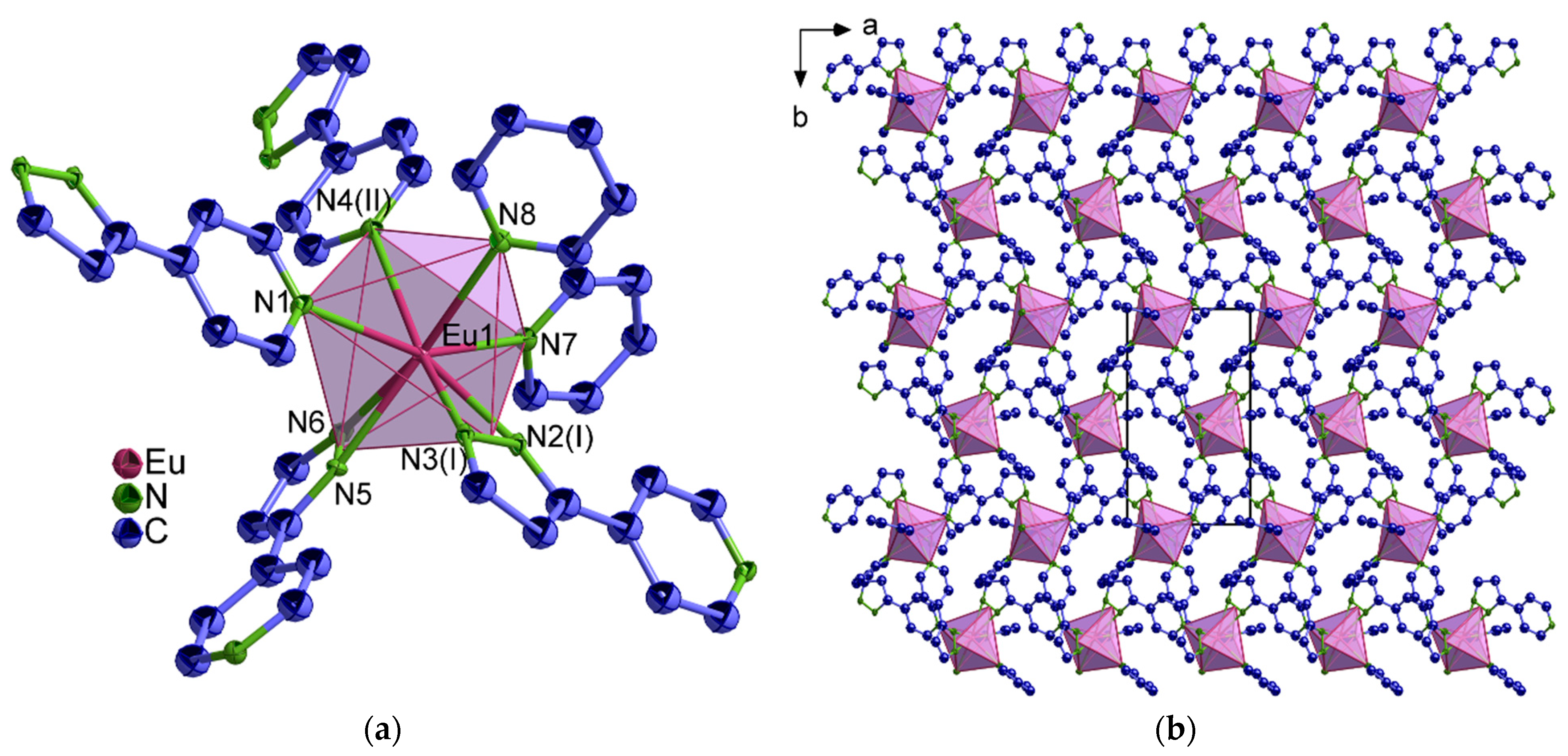
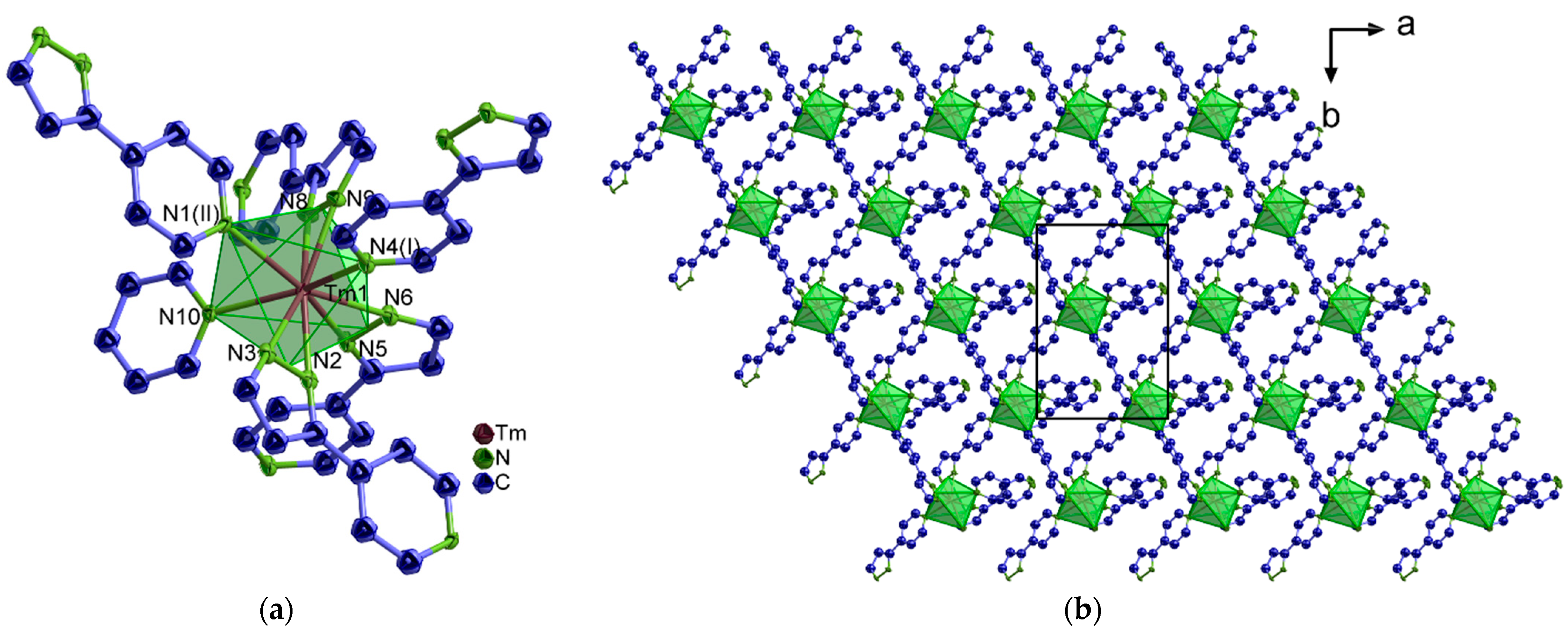
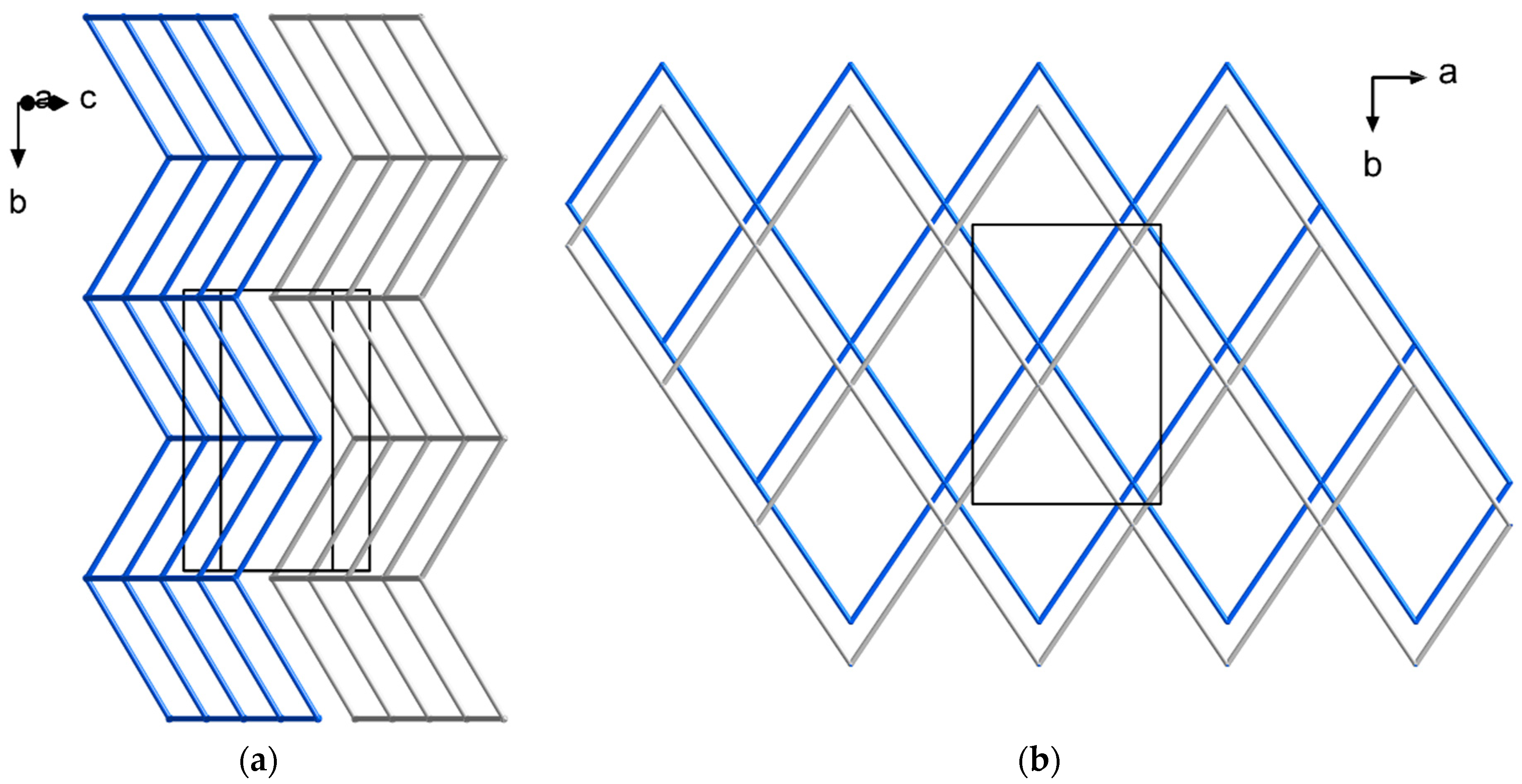
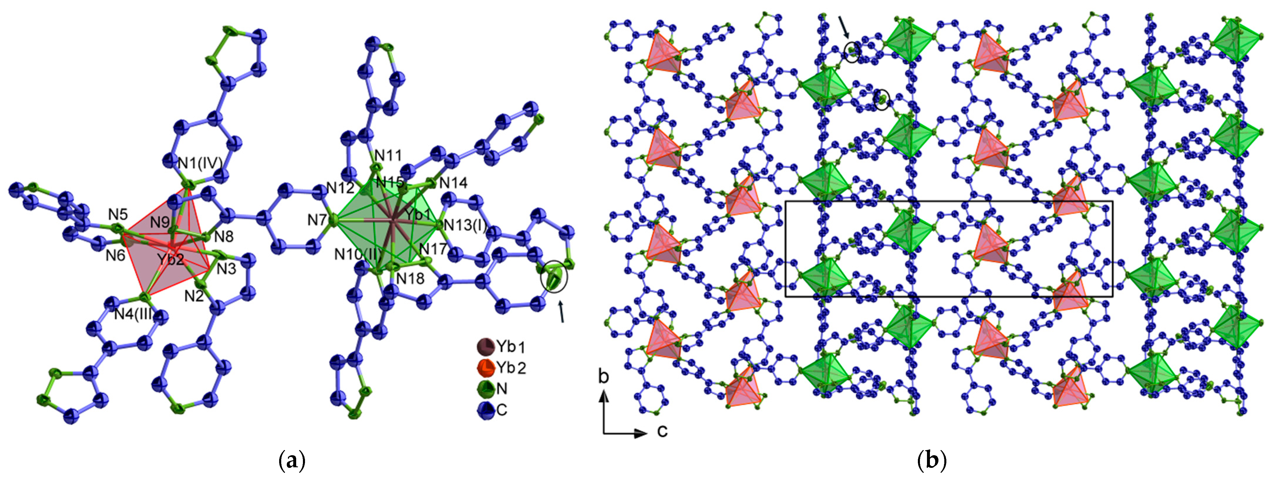
2.1. Structural Analysis

Structural diversity is observed along the series based on 4-PyPz, depending on the content of both 4-PyPz and pyridine (Py) as linkers, all of which crystallize in the monoclinic crystal system and mostly with the space group P21/n. Exceptions are 2 [Eu(4-PyPz)2(Py)2] (4-Eu2+) and 2 [Ln(4-PyPz)3(Py)], Ln = Ce (4- 2 Ce), Tm (4- 2 Tm), which crystallize with the space groups P21 and Cc, respectively. The other 3-PyPz based series 3 [Ln(3-PyPz)3], (3, Ln = Ce, Pr, Nd, Ho, Er, Tm) further crystallizes in the cubic crystal system with the space group Pa 3 ¯ .
In 2 [Eu(4-PyPz)2(Py)2] (4-Eu2+), containing divalent europium, each Eu2+ ion coordinates to eight nitrogen atoms, six nitrogen atoms from four pyrazolate anions, and two nitrogen atoms from two pyridine molecules in a distorted pseudo-octahedral assembly (Figure 1), if the two nitrogen atoms of the pyrazolate anion are considered as one corner of the octahedron. The four pyrazolate anions act as bridges to the neighboring Eu2+ ions, forming a 2D coordination polymer. The Eu–N interatomic distances for divalent europium in 2 [Eu(4-PyPz)2(Py)2] (4-Eu2+) (254.9(4)–274.5(2) pm) are longer than those reported for the trivalent europium 3 [Eu(4-PyPz)3] (240.8–258.8 pm) [33], consistent with the difference in charge density and ionic radius [34]. Another comparison of the Eu–N of 4-Eu2+ with the divalent europium complex [Eu(Ph2pz)2(Py)4]⋅2Py (Ph2pz = 3,5-diphenylpyrazolate, Eu–N = 253.8–274.1 pm) resulted in good agreement [35].
In 2 [Ln(4-PyPz)3(Py)], Ln = Ce3+ (4- 2 Ce), Tm3+ (4- 2 Tm), each Ln3+ ion coordinates to nine nitrogen atoms, eight nitrogen atoms from five pyrazolate anions, and a nitrogen atom from a pyridine molecule in a distorted pseudo-octahedral arrangement (Figure 2), if the two nitrogen atoms of the pyrazolate anion are viewed as one corner of the octahedron. The pyrazolate anions act as bridges to the neighboring Ln3+ ions, forming a 2D coordination polymer. Due to the lack of Tm3+-nitrogen-based complexes and coordination polymers in the literature, only one example was comparable to 4- 2 Tm, [Tm(L1)3]3+ (L1 = 2,6-bis(5,6-dipropyl-1,2,4-triazin-3-yl)pyridine, Tm–N = 248.3–252.2 pm) [36], which is longer than 4- 2 Tm (Tm–N = 232.4–259.2 pm) due to the anionic character of the ligands, in the latter 4-PyPz.
The topology of the 4-Eu2+ and 4- 2 Tm networks was determined according to the Reticular Chemistry Structure Resource (RCSR) and the Wells terminology [37,38] to result in an sql topology (Figure 3) with the Schläfli symbol 44·62 for both cases. This topology distinguishes from the rest of the series 3 [Ln(4-PyPz)3] (4, Ln = La, Ce, Pr, Nd, Ho, Er, Tm), which represent the pcu topology with the Schläfli symbol 412·63 [33]. The pcu topology was also found for the isotypic series 3 [Ln(3-PyPz)3] (3, Ln = Ce, Pr, Nd, Ho, Er, Tm).
The crystal structure of 2 [Ln2(4-PyPz)6]ꞏPy, Ln = Yb (4-Yb), Lu (4-Lu) contains two lanthanide sites. One site coordinates to nine nitrogen atoms from six pyrazolate anions in a distorted pseudo-octahedron, while the other coordinates to eight nitrogen atoms from five pyrazolate anions in a distorted trigonal bipyramid (Figure 4), if the two nitrogens of the pyrazolate anion are considered as a corner of the polyhedron. The two lanthanide sites are bridged through a pyrazolate anion, while each lanthanide site is simultaneously bridged through pyrazolate anions to adjacent identical sites to form a 2D layer extending along the bc plane. An anion coordinated to Ln1 does not act as a bridge through the nitrogen atom of its pyridine ring to another neighboring Ln ion (its position is pointed by arrows in Figure 4), making some ligands infinite and, thus, a correct topological analysis with the central atoms impossible.
The different coordination numbers are also reflected by the Yb–N distances, which range from 227.7(6)–247.9(6) pm for CN = 8 (Yb2) to 234.1(6)–257.5(5) pm for CN = 9 (Yb1). Comparison of the Yb–N distances with [Yb(Ph2pz)3(Py)2]·2(thf) (Ph2pz = 3,5-diphenylpyrazolate, CN = 8, Yb–N = 225.7–244.3 pm) [39] and [YbL3]CH3OH (HL = 2-(tetrazol-5-yl)-1,10-phenanthroline, CN = 9, Yb–N = 240.9–259.9) [40] shows good agreement for Yb–N. The Yb2–Npz (pz = pyrazolate nitrogen atom) is slightly shorter than the reported range, indicating the strength of the electrostatic interaction between the metal cation and the anionic pyrazolate ring.
The crystal structures of 3 [Ln(4-PyPz)3] (4, Ln = La, Ce, Pr, Nd, Ho, Er, Tm) and 3 [Ln(3-PyPz)3] (3, Ln = Ce, Pr, Nd, Ho, Er, Tm) are isotypic to the respective reported series of 3 [Ln(4-PyPz)3] and 3 [Ln(3-PyPz)3], Ln = Sm, Eu, Gd, Tb, Dy, respectively [33]. The extended coordination sphere of Ce3+ (CN = 9) in the two isotypic series 3 [Ln(4-PyPz)3] and 3 [Ln(3-PyPz)3] are shown in Figures S1 and S2. The volume of the unit cell and the average of Ln–N decrease with the increasing charge density along the two series 3 [Ln(4-PyPz)3] and 3 [Ln(4-PyPz)3] (Tables S10 and S11) as a direct consequence of the lanthanide contraction [41]. Tables with detailed crystallographic data and selected interatomic distances (pm) and angles (◦) of the studied compounds are given in the Supplementary Materials (Tables S1–S11).
The crystal structures were mostly determined by single-crystal X-ray diffraction (SCXRD), while the structures of 2 [Ce(4-PyPz)3(Py)] (4- 2 Ce) and 3 [Tm(4-PyPz)3] (4-Tm) were characterized from microcrystalline products by powder X-ray diffraction (PXRD) and subsequent Pawley refinements (Figure 5a,b), confirming the isotypic character based on the SCXRD of 2 [Tm(4-PyPz)3(Py)] (4- 2 Tm) and 3 [Er(4-PyPz)3] (4-Er), respectively.
All bulk products were investigated by PXRD. For 3 [Ln(3-PyPz)3] (3, Ln = Ce, Pr, Nd, Ho, Er), the experimental diffraction patterns agree with the diffraction patterns simulated from the single-crystal data with no observation of additional reflections indicating the absence of crystalline byproducts (Figure S3). To account for the different measurement conditions of PXRD (298 K) and SCXRD (100 K), Pawley refinements for 2 [Eu(4-PyPz)2(Py)2] (4-Eu2+) (Figure 5c), 3 [Ce(4-PyPz)3] (4-Ce) (Figure 5d), and 3 [Ln(4-PyPz)3] (4, Ln = La, Pr, Nd, Ho, Er) (Figures S4−S8) were carried out, confirming the phase purity of the respective series of coordination polymers. The resulting difference plots show no significant deviations, and the refinement results (Rwp, GOF) are shown in Table S12. Other crystalline phases were found in the PXRD of 2 [Tm(4-PyPz)3(Py)] (4- 2 Tm) and 2 [Ln2(4-PyPzH)6]ꞏPy, Ln = Yb (4-Yb), Lu (4-Lu) (Figure S9). Isolation of 3 [Tm(3-PyPz)3] (3-Tm) as single crystals was also possible.

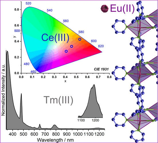
2.2. Photophysical Properties

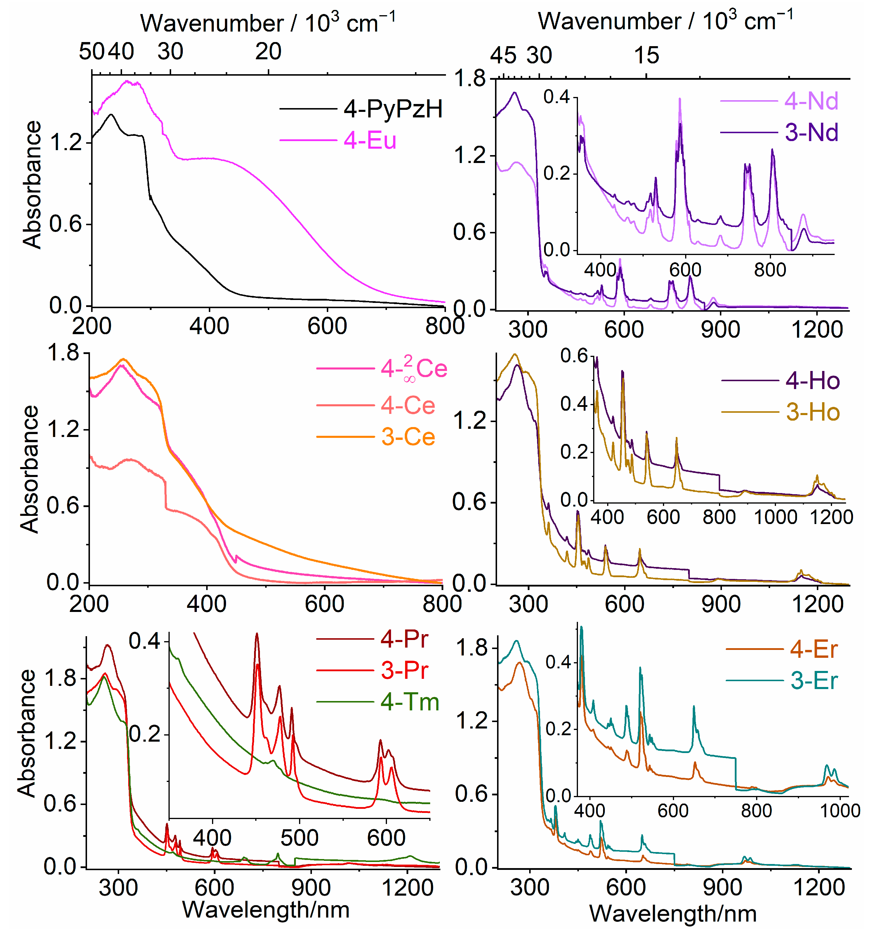
2.2.1. UV–VIS–NIR Absorption Spectra

Electronic absorption spectra were recorded in the solid state at room temperature (RT) for 4-PyPzH, 2 [Eu(4-PyPz)2(Py)2] (4-Eu2+), 2 [Ce(4-PyPz)3(Py)] (4- 2 Ce), 3 [Ln(4-PyPz)3] (4, Ln = Ce, Pr, Nd, Ho, Er, Tm), and 3 [Ln(3-PyPz)3] (3, Ln = Ce, Pr, Nd, Ho, Er) (Figure 6). The absorption spectra of the free ligand 3-PyPzH was shown for the solid state in a range from about 200–270 and 570–305 nm corresponding to the intra-ligand transitions π–π* and/or n–π* [14]. The free ligand 4-PyPzH shows a broad band from 200 to 280 nm corresponding to the intra-ligand transitions. In the investigated coordination polymers, the intense wide absorption band corresponding to either ligand appears in the UV region. For 2 [Eu(4-PyPz)2(Py)2] (4-Eu2+), a broad absorption shoulder from 356–640 nm is associated with a metal-to-ligand charge transfer (MLCT) transition from the Eu2+ 4f orbitals to the π* orbitals of the coordinated ligands. For 2 [Ce(4-PyPz)3(Py)] (4- 2 Ce), 3 [Ce(4-PyPz)3] (4-Ce), and 3 [Ce(3-PyPz)3] (3-Ce), the formation of shoulders at a higher wavelength from 320–470 nm is observed due to the transition from 4f to 5d. These absorption shoulders are compatible with the shoulders observed for the orange and the red emitters [Ce(2-PyPzH)3Cl3] and 1 [Ce(2-PyPz)3] [31]. Moreover, sharp and weak to medium bands can be assigned to the respective ff transitions in both the VIS and NIR regions for 3 [Ln(4-PyPz)3], Ln = Pr (4-Pr), Nd (4-Nd), Ho (4-Ho), Er (4-Er), Tm (4-Tm) and 3 [Ln(3-PyPz)3], Ln = Pr (3-Pr), Nd (3-Nd), Ho (3-Ho), Er (3-Er), as assigned in Table 1 [11,42,43,44,45].

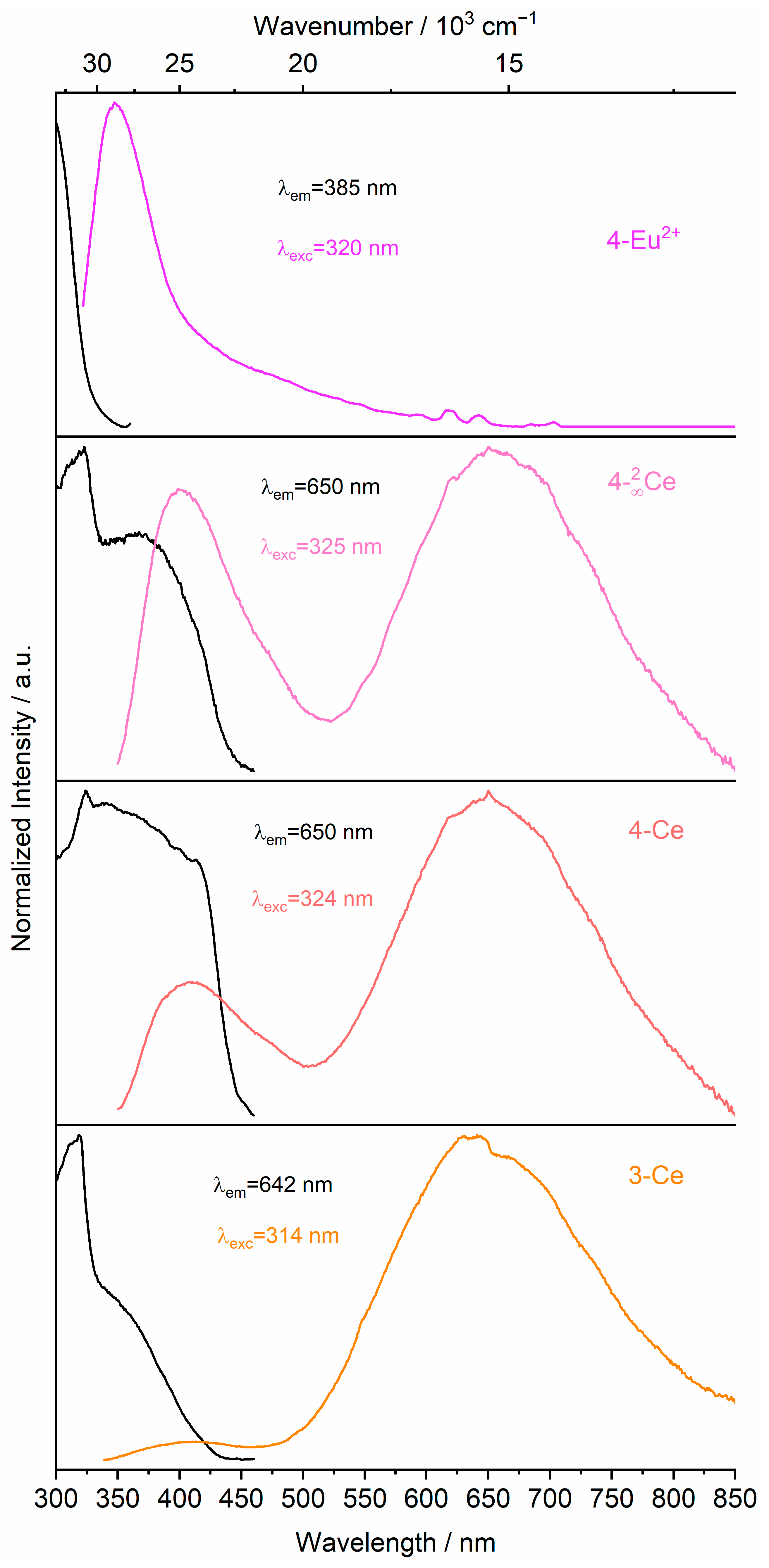
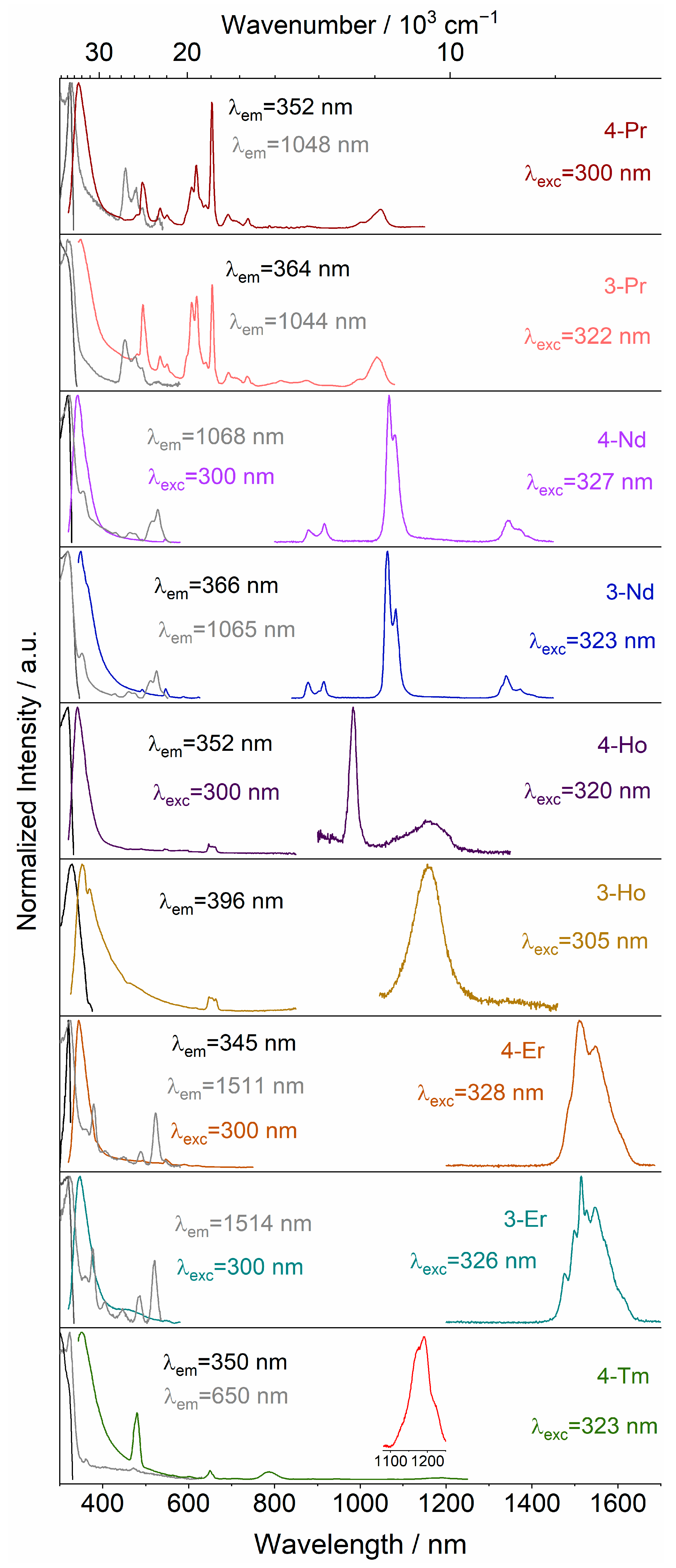
2.2.2. Emission and Excitation Spectra

The photoluminescence properties were recorded for all bulk products, 2 [Eu(4-PyPz)2(Py)2] (4-Eu2+), 2 [Ce(4-PyPz)3(Py)] (4- 2 Ce), 3 [Ln(4-PyPz)3] (4, Ln = La, Ce, Pr, Nd, Ho, Er, Tm), and 3 [Ln(3-PyPz)3] (3, Ln = Ce, Pr, Nd, Ho, Er) in the solid state at RT and 77 K. The photoluminescence spectroscopy determinations for 2 [Ce(4-PyPz)3(Py)] (4- 2 Ce), 3 [Ce(4-PyPz)3] (4-Ce), and 3 [Ce(3-PyPz)3] (3-Ce) (Figure 7) show interesting 5d−4f transitions with Ce3+-centered light emission in the VIS range. Broad emission bands appear for 4- 2 Ce, 4-Ce, and 3-Ce from 520, 500, and 460 to 850 nm centered at 650, 650, and 641 nm, respectively, at RT, indicating large crystal field splitting and a large redshift for the emission wavelength reaches the red–orange visible region. The intensity of the ligand-based emission decreases from 4- 2 Ce through 4-Ce to 3-Ce which shifts the emission color from pink through orange pink to orange, the emission colors are represented in the CIE 1931 chromaticity diagram (Figure S24), and the color coordinates are listed in Table S13. In agreement with the absorption spectra, the excitation spectra show shoulders at higher wavelengths, which correlate with the lowest energy levels of the crystal field splitting bands of the 5d excited state of the Ce3+ ions.
The maximum excitation bands are at about 400 nm, corresponding to the respective coordinated pyrazolate anions. The lifetimes of 4- 2 Ce (1.08(2) ns), 4-Ce (1.16(2) ns), and 3-Ce (1.26(2) ns) are expected to be nanoseconds due to the parity allowed nature of the 5d−4f transition. These lifetimes are slightly shorter than the lifetimes of the reported red-emitting cerium 1 [Ln(2-PyPz)3] (2 ns, 2-PyPzH = 3-(2-pyridyl)pyrazole) [32] and orange-emitting cerium [Ce(2-PyPzH)3Cl3] (2.83 ns) [31].
The emission spectrum of 2 [Eu(4-PyPz)2(Py)2] (4-Eu2+) shows a ligand-based transition at 350 nm (Figure 7) along with some weak f−f transitions that can be assigned to a low content of trivalent Eu emission features. The ligand-based excitation band at around 335 nm for 4-PyPzH and 3-PyPzH [33] shows a hypochromic shift upon coordination to the investigated compounds to around 325 nm in 4-Pr, 4-Nd, 3-Nd, 3-Ho, 4-Er, 3-Er, and 4-Tm. The blue shift for the ligand-based excitation band increases, reaching 318 nm for 3-Pr and 4-Ho and even below 300 nm for 4-Eu2+. For Pr3+, Nd3+, and Er3+, additional direct f−f excitations from the ground states 3H4, 4I9/2, and 4I15/2, respectively, were also observed. After coordination, the ligand-based emission band for both ligands at 405 nm shows a hypochromic shift to a value between 341 and 352 nm.
For 3 [La(4-PyPz)3] (4-La), an additional resolved broad band with λonset = 423 nm (~23,640 cm−1), corresponding to the triplet state of the pyrazolate anion, was observed in the emission spectrum (Figure S27) at 77 K, which agrees well with the previously reported value [33].
Although the excitation and emission spectra can provide a wealth of information, particularly about the coordination environment of the Ln3+ ions, it is uncommon to find the luminescence spectra for the Tm3+ and Ho3+-based compounds. Nine-coordinated Tm3+ was only reported in three examples, and none of them investigated the photophysical properties, they mainly focused on the structural aspects [36,46,47]. In other cases, poor ligand-to-metal sensitization or back energy transfer occurs to allow only ligand-based luminescence, as in [Tm2(C15H11N3)2(C7H4BrO2)4(C2O4)], (C15H11N3 = 2,2′:6′2″-terpyridine and C7H4BrO2 = p-bromobenzoic acid) [15], Tm(bfa)3phen, (bfa = 4,4,4-trifluoro-1-phenyl-1,3-butanedione, phen = 1,10-phenanthroline) [12], and Tm(ppa)3·2H2O, (ppa = 3-phenyl-2,4-pentanedionate) [13], where a significant ligand emission dominates the spectrum in addition to a single spectral band for the Tm3+. Even nonefficient ligand sensitization with only a ligand emission band in the emission spectra was shown for [(Tm-(TC)3(H2O)2)·(HPy·TC)]n, (TC = 2-thiophenecarboxylate and HPy = pyridinium cation) [16]. In contrast, very good ligand-to-metal sensitization is observed for 3 [Tm(4-PyPz)3] (4-Tm) (Figure 8). The transitions 1G43H6, 3F4, 3H5, and 3H4 are readily observable at 480, 650, 787, and 1192 nm, respectively.
For 3 [Ho(4-PyPz)3] (4-Ho) and 3 [Ho(3-PyPz)3] (3-Ho), the 5F55I8 is observed at 648 nm in addition to the NIR transition 5I65I8 at 1155 nm. An additional NIR transition appears for 4-Ho at 983, corresponding to the transition 5F55I7 and indicating more efficient ligand sensitization than in reported cases, such as [(Ho-(TC)3(H2O)2)·(HPy·TC)]n with only ligand emission observable in the emission spectra [16]. The Ho3+-based emission observed for both 3-Ho and 4-Ho is stronger than that of 2 [Ho2(3-PyPzH)3Cl6]·2MeCN [14], which may be due to the absence of the vibrational energy of the chloride ligands.
For 3 [Pr(4-PyPz)3] (4-Pr) and 3 [Pr(3-PyPz)3] (3-Pr) (Figure 8), the highest intensity for the Pr3+-based emission is found at 655 nm, corresponding to 3P03F2. NIR emission bands can also be observed at 738 and 1048 for 4-Pr and at 736 and 1038 nm for 3-Pr, corresponding to the transitions 3P03F4 and 1D23F4. Despite the ligand-based emission in 4-Pr and 3-Pr being more dominated than for the reported 1 [Pr(2-PyPz)3] [32], the Pr3+-based transitions are more characteristic than for other published cases, such as 1 [PrCl3(ptpy)] and [PrCl3(ptpy)(py)], ptpy = 4′-phenyl-2,2′:6′,2″-terpyridine) [48].
For 3 [Er(4-PyPz)3] (4-Er) and 3 [Er(3-PyPz)3] (3-Er), the NIR transition 4I13/24I15/2 is observed at about 1510 nm and the VIS transition at about 545 nm can also be observed for 4-Er at RT and 77 K, while at 77 K for 3-Er. Both 4-Er and 3-Er show Stark-level splitting in the emissive transitions in contrast, e.g., to the reported 1 [LnCl3(bipy)(py)2]py, which shows no fine splitting [48].
For the NIR emitters 3 [Nd(4-PyPz)3] (4-Nd) and 3 [Nd(3-PyPz)3] (3-Nd), the transitions 4F3/24IJ/2, (J = 9, 11, 13) are observed at about 915, 1065, and 1345 nm [11].
Generally, for the VIS–NIR and the NIR emitters, the emissive energy levels of the Ln3+-based transitions are populated by an antenna effect between the pyridylpyrazolate-based ligands and the lanthanide ions, which leads to ligand-to-metal energy transfer, as observed for 2-PyPzH [32] and 4,4`-bipyridine (bipy) [49].
The ligands’ fluorescence emission band has lifetimes of 6.07 ns for 4-PyPzH and 3.4 ns for 3-PyPzH [33] which are shortened by coordination with different Ln3+ (τ = 0.93–1.04; Table S14). The lifetime increases with decreasing the temperature to 77 K, especially for 4-Nd, which increases from 0.93(2) ns at RT to 3.04(9) ns at 77 K due to the decrease in thermal quenching. See the Supplementary Materials for half-page size absorption and photoluminescence spectra with designated 4f–4f transitions for the investigated compounds (Figures S10–S37).

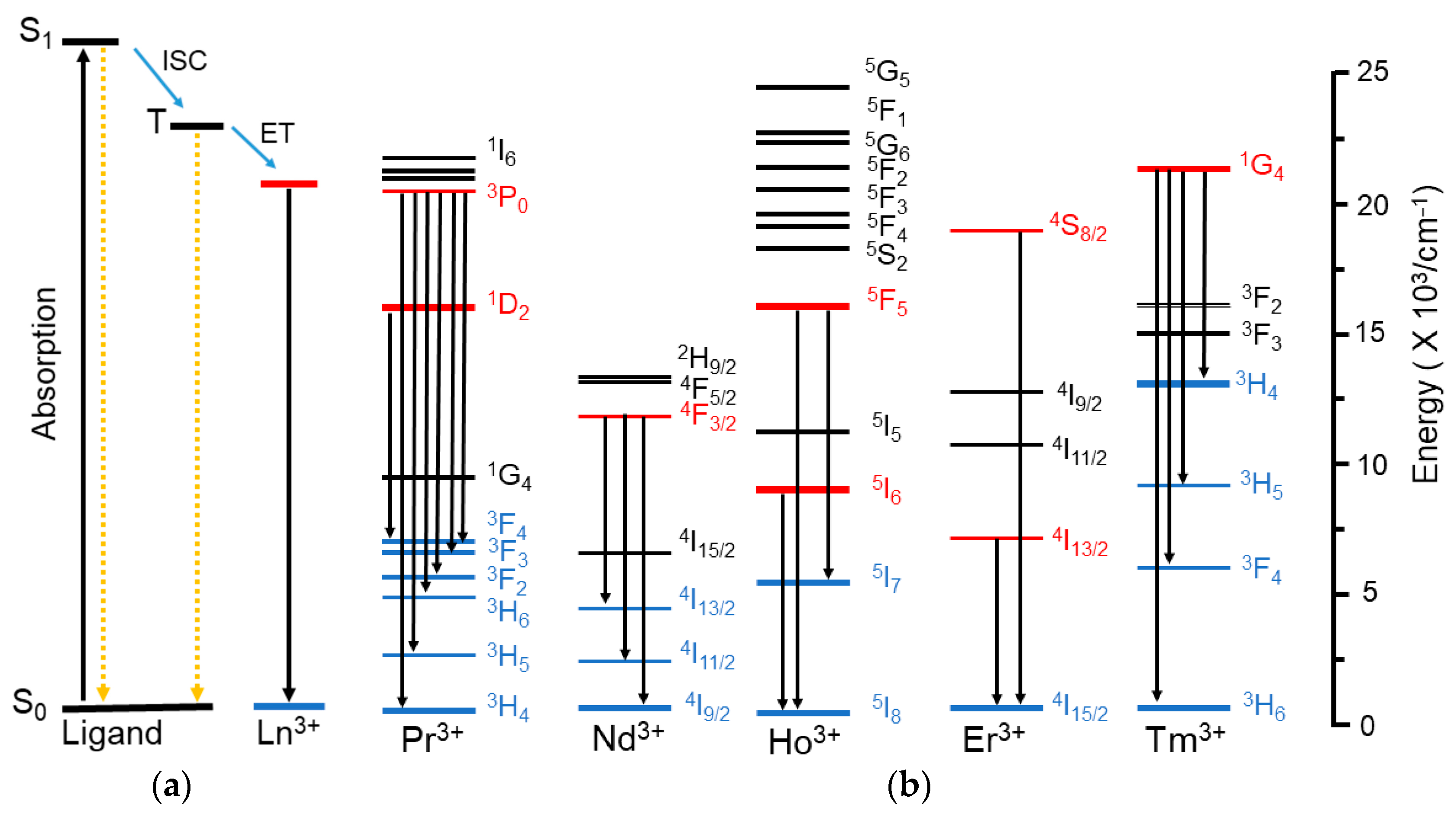
2.2.3. Mechanism of Energy Transfer

To explain and understand the observed spectral results, a schematic diagram (Scheme 2) is shown, depicting the primary energy levels involved and the main energy transfer and relaxation pathways during the sensitization of lanthanide luminescence via the ligands. The ligands absorb energy and become excited from the singlet S0 ground state to the singlet S1 excited state by the absorption of visible light. The energy of the S1 excited state is then transferred to the triplet-excited state (T) of the ligands through intersystem crossing (ISC). Competing processes include ligand fluorescence and nonradiative deactivation of the excited singlet state. Subsequently, the excitation energy is transferred to the excited 4f levels of the Ln3+ ions, resulting in the respective lanthanide ion emission to the respective 4f ground state [50]. According to Dexter’s theory [51], the energy gap between the first excited energy level of the Ln3+ ions and the energy level of the triplet state of the respective ligand is important for an efficient energy transfer. If the energy gap is too large, the overlap between the ligand and the Ln3+ is reduced, and as a result, the energy transfer decreases sharply. On the other hand, if the energy gap is too small, energy transfer also occurs from the Ln3+ back to the resonance levels of the triplet states of the ligands, which also reduces the 4f-based emission.
In this study, the triplet-state energy levels of 4-PyPz and 3-PyPz were investigated by deduction from the phosphorescence spectra of the Gd-based coordination polymers and calculated to be 23,640 and 23,250 cm−1, respectively [33]. This analogy is confirmed for 4-PyPz through the spectra of 4-La, as discussed before. The discussed 4f emission bands of the respective Ln-based CPs indicate that the triplet states of the ligands are suitable for a transfer of the absorbed light to the lanthanide ions via such an antenna effect. For instance, the energy difference (ΔE) between the ligand triplet state of 4-PyPz (~23,640 cm−1) and the energetic positions of Tm3+ (1G4 = ~21,300 cm−1) results in an ΔE value in the optimal range. Pr3+ is slightly more complicated because it has two emission levels (3P0 and 1D2). By considering the 3P0 level as the main acceptor level with an energetic position of ~20,475 cm−1 [11], the ΔE values are calculated as 3165 and 2775 for 4-Pr and 3-Pr, respectively, both also being in the optimal range. For Ho3+, the 5F4, 5S2, and 5F5 levels are the main acceptor levels, and the emission from the 5F5 and 5I6 levels can partially be the result of a relaxation of the upper levels followed by transitions to the lower levels to give the characteristic NIR emission of the Ho3+ ion [52].

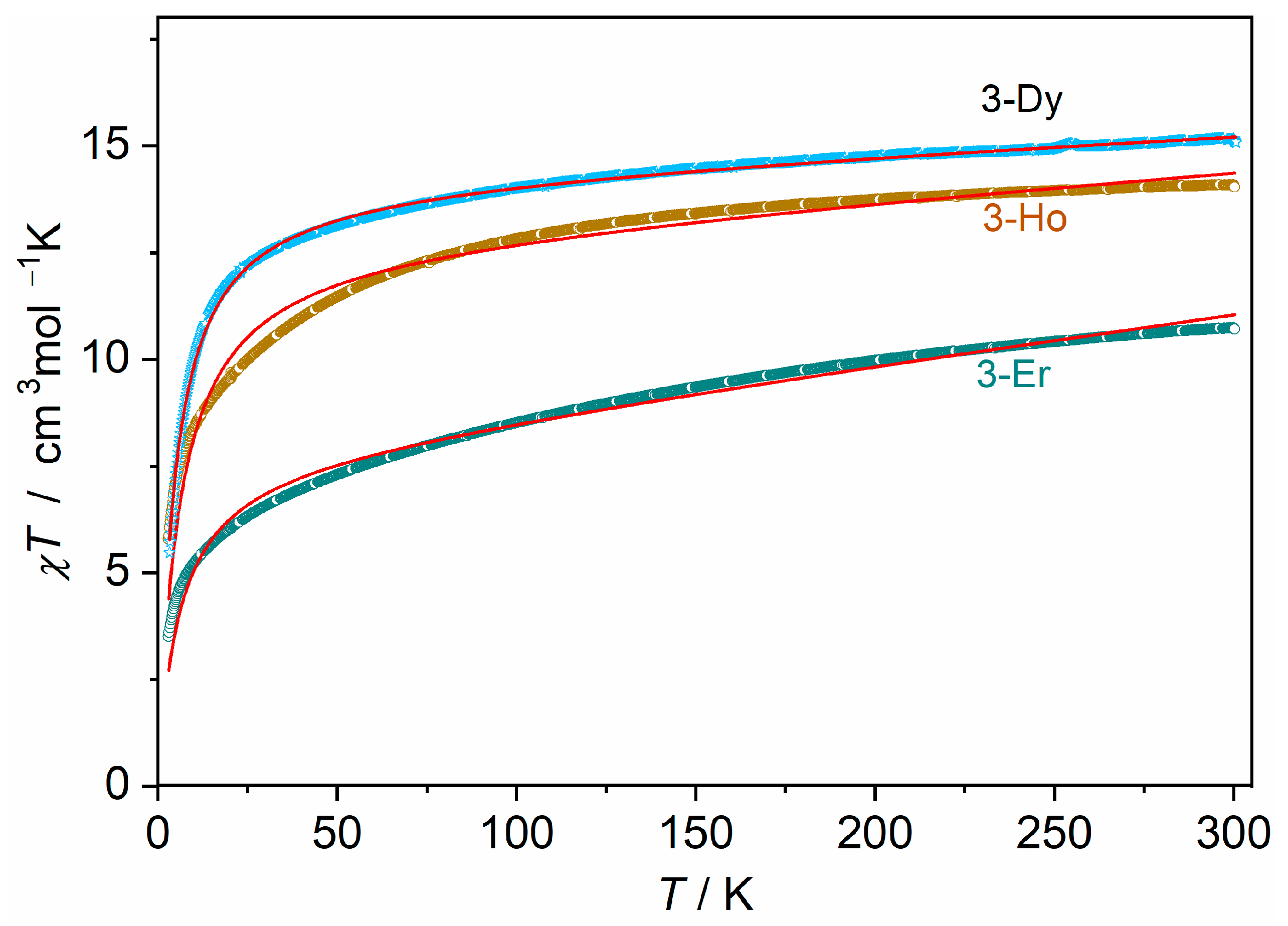
2.3. Magnetic Properties

Direct current (DC) magnetic susceptibility measurements were performed for 3 [Ln(3-PyPz)3], Ln = Ho (3-Ho), Er (3-Er) in a temperature range of 3 to 300 K and a magnetic field of 1T. As a link to the reported isotypic series of 3 [Ln(3-PyPz)3], Ln = Sm, Eu, Gd, Tb, Dy [33], the DC magnetic susceptibility measurements of 3 [Dy(3-PyPz)3] (3-Dy) were also performed.
The temperature dependence of the product of χT for all samples can be observed in Figure 9. At room temperature, the χMT (χM = molar magnetic susceptibility) values are 15.03, 14.05, and 10.70 for 3-Dy, 3-Ho, and 3-Er, respectively. These experimental data are in satisfactory agreement with the theoretical values for the corresponding noninteracting Dy3+ (6H15/2, S = 5/2, L = 5, g = 4/3, χT = 14.17 cm3 K mol−1), Ho3+ (5I8, S = 5/2, L = 6, g = 5/4. χT = 14.07 cm3 K mol−1), and Er3+ (4I15/2, S = 3/2, L = 6, g = 6/5, χT = 11.48 cm3 K mol−1) [53].
For 3-Dy, 3-Ho, and 3-Er, a monotonic slow decrease in the χMT product was observed upon cooling, which could be related to thermal depopulation within the mJ levels of the ground 6H15/2, 5I8, and 4I15/2 multiplet, respectively. In addition, the χMT vs T plot did not display abrupt changes, which suggests a lack of magnetic interactions down to 45, 75, and 80 K, with χMT reaching 3.01, 5.79, and 3.49 cm3 K mol−1, respectively, because of the efficient magnetic isolation of lanthanide centers in the crystal lattice.
The data were fitted for 3-Dy, 3-Ho, and 3-Er in the given temperature range with an effective magnetic moment μeff of 10.651(2), 10.09(1), and 7.855(9) μB and a Weiss constant θ of −4.41(1), −5.77(7), and −5.72(8) K, as well as a temperature-independent paramagnetic susceptibility χ0 of 4.03(3) × 10−3, 6.18(2) × 10−3, and 11.52(8) × 10−3 cm3 mol−1. The small, negative Weiss constants θ are the results of spin–orbit coupling as well as the crystal field effect [54,55].

3. Materials and Methods

3.1. General Procedures

3-(4-pyridyl)pyrazole (4-PyPzH) and 3-(3-pyridyl)pyrazole (3-PyPzH) were synthesized as reported in the literature [56,57]. Lanthanide metals (holmium: 99.9%, Chempur, Karlsruhe, Germany; rest: >99.99%, Smart Elements, Vienna, Austria) were purchased and used as received. Pyridine (Py), dichloromethane (DCM), and cyclohexane (Cy) were purified by distillation and dried by standard procedures. All syntheses involving lanthanide elements were performed under argon or using vacuum lines, gloveboxes (MBraun Labmaster SP, Innovative Technology PureLab, Garching, Germany), Schlenk tubes, and Duran® glass ampoules (outer Ø 10 mm, wall thickness 1.5 mm). The solid reactants for the solvothermal reactions were mixed and sealed together with the solvent in an ampoule under reduced pressure (p = 1.0 × 10⁻³ mbar) after freezing the solvent with liquid nitrogen. Subsequently, the prepared ampoules were placed in heating furnaces based on Al2O3 tubes with Kanthal wire resistance heating and NiCr/Ni (Eurotherm 2416) temperature control elements, for which temperature programs and working steps according to the specific synthesis methods were used. After the solvents had been removed, the solid raw products were dried at RT in a dynamic vacuum (p = 1.0 × 10⁻³ mbar) before further steps. The bulk materials were characterized by PXRD and CHN analysis. The prepared 3D frameworks and 2D networks are air sensitive due to the known oxophilic behavior of the Ln-based CPs. It is expected that the CPs are insoluble in common organic solvents. We think the photostability tests are not significant in the possible applications of these synthesized CPs.

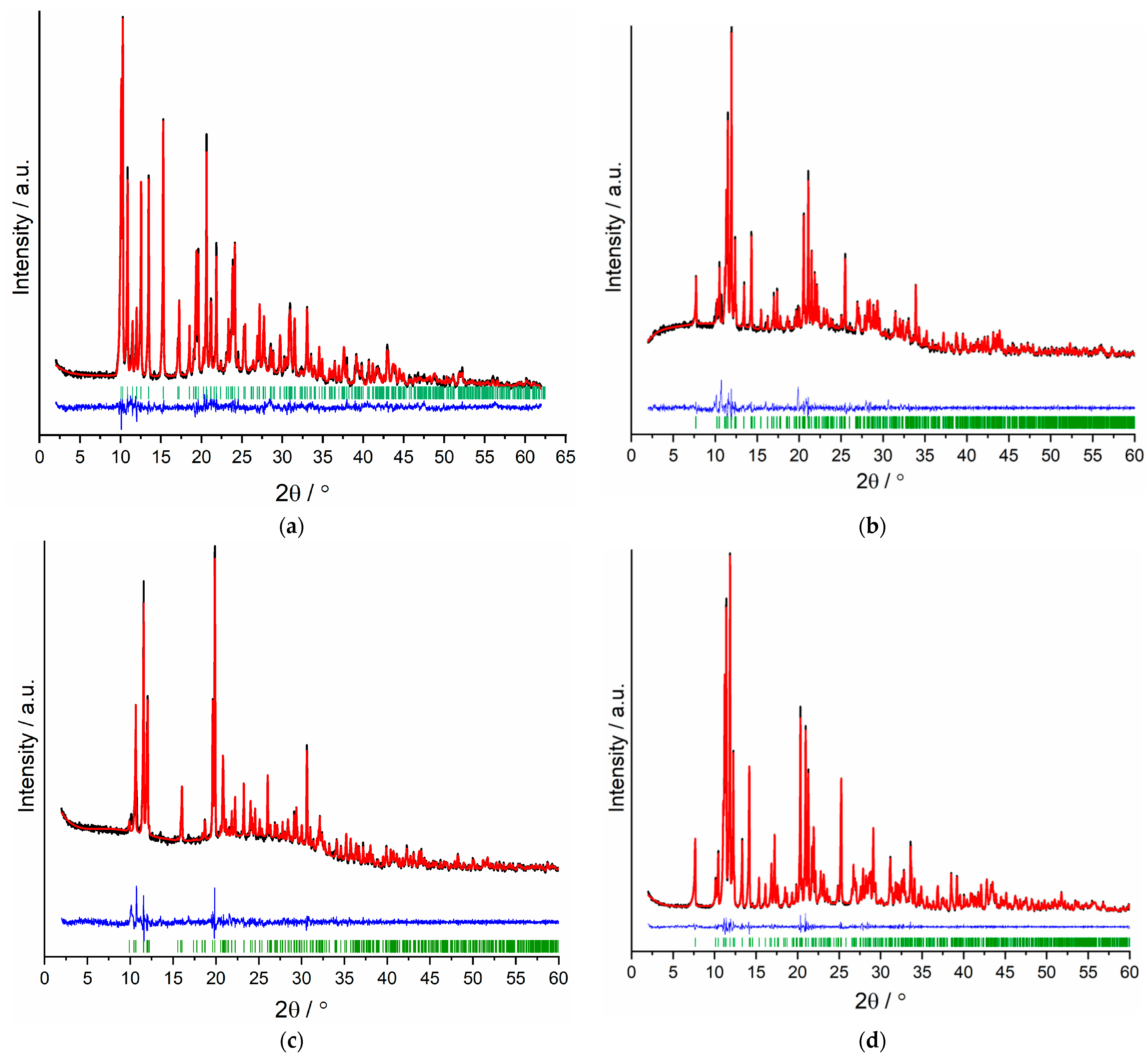
3.2. X-ray Crystallography

SCXRD determinations were performed on a Bruker AXS D8 Venture diffractometer (Karlsruhe, Germany) equipped with dual IµS microfocus sources, a collimating Quazar multilayer mirror, a Photon 100 detector, and an Oxford Cryosystems 700 low-temperature system (Mo–Kα radiation; λ = 71.073 pm). The structures were solved with direct methods and refined with the least squares method implemented in ShelX [58,59]. All nonhydrogen atoms were refined anisotropically. Hydrogen atoms were assigned to idealized geometric positions and included in structure factor calculations. Further, a ligand anion (4-PyPz) and the pyridine solvent in the asymmetric unit of 4-Yb and 4-Lu were found to be fully disordered and were refined with the help of restraints to achieve a proper structural model. For polymers 4-La, 4-Ce, 4-Pr, 4-Nd, 4-Ho, and 4-Er, the SQUEEZE [60] algorithm in PLATON [61,62,63,64] was used to include a bulk solvent model in the refinement. Two voids per unit cell were identified with SQUEEZE. The average volume was found to be 135 × 106 pm3 for each void. The equivalent of 8 electrons for (4-La, 4-Ce, 4-Pr), 10 for (4-Ho), and 12 for (4-Nd) electrons per unit cell was also identified. ToposPro program package was used to determine the topology of the polymers [65]. Depictions of the crystal structures were created with Diamond [66]. The crystal structures have been deposited to the Cambridge Crystallographic Data Center (CCDC) as supplementary publication No. 2237763 (4-Eu2+), 2237764 (4- 2 Tm), 2237765 (4-Yb), 2237766 (4-Lu), 2237767 (4-La), 2237768 (4-Ce), 2237769 (4-Pr), 2237770 (4-Nd), 2237771 (4-Ho), 2237772 (for 4-Er), 2237773 (3-Ce), 2237774 (3-Pr), 2237775 (3-Nd), 2237776 (3-Ho), 2237777 (for 3-Er), and 2237778 (3-Tm). Crystallographic data and selected interatomic distances are listed in Tables S1–S11 for the investigated compounds.
PXRD analyses of the investigated compounds were carried out on a Stoe Stadi P diffractometer (Darmstadt, Germany) with a focusing Ge(111) monochromator and a Dectris Mythen 1K strip detector in Debye–Scherrer geometry. All powder samples were ground in a mortar and filled into Lindemann glass capillaries with 0.3 mm diameter under an inert gas atmosphere. All samples were measured in transmission geometry with Cu–Kα radiation (λ = 154.056 pm). Data collection was performed using the Stoe Powder Diffraction Software Package WinXPOW V3.0.2.1 and Pawley fits on the data were performed using TOPAS Academic V7 [67]. The data are presented in Figure 5 and Figures S3–S9 in addition to Table S12.

3.3. Spectroscopical Investigations

3.3.1. Absorption Spectra

The UV–Vis–NIR absorption spectra were measured on solid-state products using a standard Agilent Cary 5000 UV–VIS–NIR spectrophotometer (Agilent Technologies, Waldbronn, Germany) with a Praying Mantis accessory (Harrick Scientific Instruments, New York, NY, USA), which had been mounted and aligned for use with the DRP-ASC ambient chamber. The source, detector, and grating changeovers were at the standard position of 350, 800, and 800 nm, respectively, for all studied compounds except 4- 2 Ce, 4-Nd, 4-Tm, and 3-Nd, the detector and grating changeovers were set to 850 nm, while 750 nm for 3-Er. For 4-Eu2+, 4- 2 Ce, 4-Ce, 4-Nd, 4-Tm, 3-Ce, and 3-Nd, the source was set to 320, 450, 330, 340, 335, 320, and 340 nm, respectively. The reference spectrum was collected on PTFE and the reference and samples were packed in the ambient chamber within the glovebox under inert conditions.

3.3.2. Photoluminescence Spectroscopy

The excitation and emission spectra were recorded for ground solid samples after filling them in quartz glass tubes under argon. The measurements were performed at room temperature as well as 77 K (latter using the liquid nitrogen-filled assembly FL-1013 of HORIBA) with a HORIBA Jobin Yvon Spex Fluorolog 3 spectrometer (Horiba-Jobin Yvon, Oberursel, Germany) equipped with a 450 W Xe short-arc lamp (USHIO INC., Tokyo, Japan), double-grated excitation, and emission monochromators, and a photomultiplier tube (R928P) using the FluoroEssence™ software V3.9. Excitation and emission spectra were corrected for the spectral response of the monochromators and the detector using spectral corrections provided by the constructor. In addition, a photodiode reference detector was used to correct the excitation spectra for the spectral distribution of the lamp intensity. An R5509-73 detector was used to collect the data in the NIR region. When required, the collection of the data was performed using an edge filter (Newport 20CGA-345, 395, 495 for the visible region and Reichmann Optics RG 830 long pass for the NIR region). Emission spectra with gating were recorded using a xenon flashlamp with a pulse repetition rate of 41 ms.
Photoluminescence overall decay process times were determined using the above-mentioned HORIBA Jobin Yvon Spex Fluorolog 3 spectrometer equipped with a dual lamp housing (FL-1040A), a UV xenon flashlamp (Exelitas FX-1102), and a TCSPC (time-correlated single-photon counting) upgrade, or picosecond pulsed laser diode. Emission decays were recorded using DataStation software V2.7. Exponential tail fitting was used for the calculation of resulting intensity decay using Decay Analysis Software 6. The quality of the fit was confirmed by χ2 values being below 1.2.

3.4. PPMS Magnetic Measurements
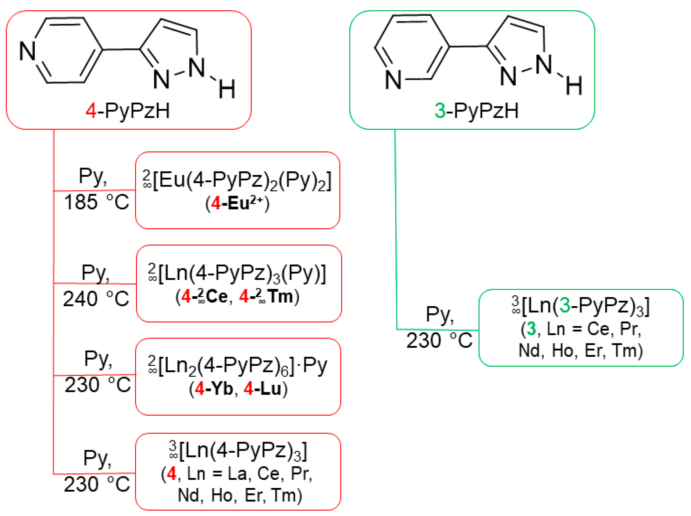
Magnetic data were obtained with the application of the VSM option of a Quantum Design physical property measurement system (ppms). The data were corrected with respect to the contribution of the polypropylen sample holder as well as the diamagnetic contribution of the sample through utilization of both experimental data and Pascal constants (increment method). The total magnetic susceptibility is comprised of different parts: the diamagnetic contribution χDiam., the Curie paramagnetic contribution χCW, and a temperature-independent paramagnetic contribution χ0.
χ t o t . = χ D i a m . + χ P a r a m . = χ D i a m . + χ C W + χ 0
The Curie paramagnetic part is the ratio of the Curie constant C and the modified temperature (Tθ). θ is the Weiss temperature
χ C W = C T θ
The Curie constant is given by the formula:
C = μ 0 N A μ B 2 n e f f 2 3 k B
with µ0 = magnetic constant, NA = Avogadro number, µB = Bohr magneton, neff = effective magnetic moment, kB = Boltzmann constant.
The molar Curie paramagnetic contribution of the susceptibility is:
χ m o l C W = N A μ B 2 n e f f 2 3 k B T θ
Or:
χ m o l C W = 0.1250   n e f f 2 ˙ T Θ   /   cm 3 mol 1
To calculate the molar paramagnetic contribution of the susceptibility, we used the equation:
χ m o l = 0.1250   n e f f 2 ˙ T Θ + χ 0   /   cm 3 mol 1
For the analysis of the data the OriginPro software V2021b (Academic) was used.

3.5. Synthesis

3.5.1. Synthesis of 2 [Eu(4-PyPz)2(Py)2] (4-Eu2+)

Freshly filed Eu metal (108.6 μmol) and an excess of 4-PyPzH (C8H7N3, 220 μmol) were mixed with Py (0.6 mL) and sealed in an evacuated ampoule. The ampoule was heated to 185 °C in 1 h and maintained at this temperature for 72 h. The reaction mixture was then cooled to room temperature within 4 h. The excess ligand was washed away using a mixture of DCM and Cy. Suitable single crystals were selected for a SCXRD measurement. C26H22N8Eu (598.47 g·mol–1): C 51.59 (calcd. 52.18); H 3.04 (3.71); N 19.49 (18.72)%. Yield: 83%. FT-IR (ATR, Figure S39): Chemistry 05 00069 i001 = 3036 (w), 1065 (s), 1552 (w), 1522 (w), 1456 (w), 1439 (m), 1418 (w), 1404 (w), 1346 (w), 1293 (w), 1213 (m), 1187 (w), 1065 (w), 1044 (m), 999 (s), 963 (w), 951 (w), 925 (w), 871 (w), 830 (s), 761 (s), 690 (s), 659 (w), 614 (m), 532 (m), 451 (m) cm–1.

3.5.2. Synthesis of 2 [Ln(4-PyPz)3(Py)] (4- 2 Ce, 4- 2 Tm)

The respective freshly filed Ln metal (81.4 μmol) and an excess of 4-PyPzH (C8H7N3, 327.6 μmol) were mixed with Py and sealed in an evacuated ampoule. The ampoule was heated to 180 °C in 24 h then the temperature was raised to 240 °C within 48 h and maintained at this temperature for 72 h. The reaction mixture was then cooled to room temperature within 48 h. The excess ligand was washed away using a mixture of DCM and Cy. Suitable single crystals were selected for a SCXRD measurement. C29H23N10Ce (651.67 g·mol–1): C 52.63 (calcd. 53.45); H 2.67 (3.06); N 22.16 (21.49)%. Yield: 83%. FT-IR (ATR, Figure S40): Chemistry 05 00069 i001 = 3085 (w), 1698 (w), 1606 (s), 1523 (w), 1459 (w), 1440 (m), 1418 (w), 1347 (w), 1330 (w), 1295 (w), 1212 (s), 1099 (w), 1066 (w), 1047 (m), 1003 (s), 992 (m), 968 (w), 929 (m), 857 (w), 830 (m), 773 (s), 741 (w), 692 (s), 652 (m), 622 (w), 572 (m), 457 (s) cm–1.

3.5.3. Synthesis of 2 [Ln2(4-PyPz)6]ꞏPy (4-Yb, 4-Lu)

Freshly filed Yb (68.6 μmol) and an excess of 4-PyPzH (C8H7N3, 275.6 μmol) were mixed with Py and sealed in an evacuated ampoule. The ampoule was heated to 180 °C in 24 h then the temperature was raised to 230 °C within 48 h and maintained at this temperature for 72 h. The reaction mixture was then cooled to room temperature within 48 h. Suitable single crystals were selected for a SCXRD measurement.

3.5.4. Synthesis of 3 [Ln(4-PyPz)3] (4, Ln = La, Ce, Pr, Nd, Ho, Er, Tm)

A mixture of the respective freshly filed Ln metal (91.1 μmol) and excess 4-PyPzH (C8H7N3, 275.6 μmol), in 0.3 mL pyridine, was sealed in an evacuated ampoule. The temperature was raised to 230 °C in 48 h, held for 96 h, and then lowered to room temperature over a further 24 h. The excess ligand was washed away using a mixture of DCM and Cy. Colorless crystals were selected for SCXRD measurements.
3 [La(4-PyPz)3] (4-La): C24H18N9La (571.38 g·mol–1): C 49.53 (calcd. 50.45); H 2.89 (3.18); N 21.50 (22.06)%. Yield: 80%. FT-IR (ATR, Figure S41): Chemistry 05 00069 i001 = 3096 (w), 1698 (w), 1607 (s), 1550 (w), 1526 (w), 1460 (w), 1447 (m), 1420 (w), 1348 (w), 1329 (w), 1215 (m), 1099 (w), 1066 (w), 1046 (m), 1005 (s), 968 (w), 927 (m), 831 (m), 763 (s), 739 (w), 694 (s), 652 (m), 527 (m), 459 (s) cm–1.
3 [Ce(4-PyPz)3] (4-Ce): C24H18N9Ce (572.58 g·mol–1): C 49.81 (calcd. 50.34); H 3.26 (3.17); N 21.73 (22.02)%. Yield: 85%. FT-IR (ATR, Figure S42): Chemistry 05 00069 i001 = 3101 (w), 1608 (s), 1526 (w), 1459 (w), 1447 (w), 1420 (w), 1348 (m), 1215 (m), 1066 (w), 1046 (m), 1006 (s), 969 (w), 927 (m), 831 (m), 763 (m), 739 (w), 695 (s), 652 (m), 527 (m), 460 (s) cm–1.
3 [Pr(4-PyPz)3] (4-Pr): C24H18N9Pr (573.37 g·mol–1): C 49.39 (calcd. 50.27); H 3.07 (3.16); N 21.16 (21.99)%. Yield: 84%. FT-IR (ATR, Figure S43): Chemistry 05 00069 i001 = 3095 (w), 1607 (s), 1526 (w), 1460 (w), 1446 (w), 1420 (w), 1348 (w), 1328 (w), 1215 (m), 1099 (w), 1067 (w), 1046 (m), 1006 (s), 969 (w), 927 (m), 831 (m), 762 (s), 739 (w), 694 (s), 652 (m), 527 (m), 460 (s) cm–1.
3 [Nd(4-PyPz)3] (4-Nd): C24H18N9Nd (576.70 g·mol–1): C 49.33 (calcd. 49.98); H 2.73 (3.15); N 21.11 (21.86)%. Yield: 83%. FT-IR (ATR, Figure S44): Chemistry 05 00069 i001 = 3093 (w), 1609 (s), 1526 (w), 1459 (w), 1447 (m), 1420 (w), 1348 (w), 1215 (m), 1074 (w), 1047 (m), 1007 (s), 969 (w), 928 (m), 831 (m), 771 (s), 763 (s), 740 (w), 696 (s), 652 (w), 527 (w), 461 (s) cm–1.
3 [Ho(4-PyPz)3] (4-Ho): C24H18N9Ho (597.39 g·mol–1): C 47.44 (calcd. 48.25); H 2.75 (3.04); N 20.14 (21.10)%. Yield: 86%. FT-IR (ATR, Figure S45): Chemistry 05 00069 i001 = 3113 (w), 1697 (w), 1609 (s), 1549 (w), 1526 (w), 1460 (w), 1446 (w), 1419 (w), 1348 (w), 1328 (w), 1297 (w), 1214 (m), 1101 (w), 1075 (w), 1048 (m), 1007 (s), 970 (w), 929 (m), 845 (w), 831 (m), 770 (s), 760 (s), 739 (m), 696 (s), 652 (m), 528 (m), 461 (s) cm–1.
3 [Er(4-PyPz)3] (4-Er): C24H18N9Er (599.72 g·mol–1): C 48.85 (calcd. 48.07); H 3.34 (3.03); N 20.69 (21.02)%. Yield: 89%. FT-IR (ATR, Figure S46): Chemistry 05 00069 i001 = 3062 (w), 1696 (w), 1609 (s), 1549 (w), 1526 (w), 1460 (w), 1446 (w), 1419 (w), 1348 (w), 1328 (w), 1214 (m), 1101 (w), 1076 (m), 1048 (m), 1008 (s), 971 (w), 929 (m), 831 (m), 770 (s), 759 (s), 739 (m), 697 (s), 652 (m), 528 (m), 461 (s) cm–1.
3 [Tm(4-PyPz)3] (4-Tm): C24H18N9Tm (601.39 g·mol–1): C 48.90 (calcd. 47.93); H 3.11 (3.02); N 19.97 (20.96)%. Yield: 82%. FT-IR (ATR, Figure S47): Chemistry 05 00069 i001 = 3117 (w), 1697 (w), 1609 (s), 1550 (w), 1527 (m), 1460 (m), 1447 (m), 1419 (w), 1349 (m), 1329 (w), 1296 (w), 1214 (s), 1102 (w), 1077 (m), 1049 (m), 1008 (s), 971 (w), 930 (m), 845 (w), 830 (s), 770 (s), 759 (s), 740 (m), 696 (s), 652 (m), 528 (m), 461 (s) cm–1.

3.5.5. Synthesis of 3 [Ln(3-PyPz)3] (3, Ln = Ce, Pr, Nd, Ho, Er)

A mixture of the respective freshly filed Ln metal (78.8 μmol) and an excess of 3-PyPzH (C8H7N3, 240 μmol) together with 0.3 mL pyridine were sealed in an evacuated ampoule. The oven was heated to 180 °C in 24 h. Subsequently, the temperature was raised to 230 °C in 48 h. The temperature was held for 72 h and then lowered to room temperature over a further 48 h. Single crystals were selected for SCXRD measurements.
3 [Ce(3-PyPz)3] (3-Ce): C24H18N9Ce (572.58 g·mol–1): C 50.50 (calcd. 50.34); H 3.11 (3.17); N 21.16 (22.02)%. Yield: 83%. FT-IR (ATR, Figure S48): Chemistry 05 00069 i001 = 3085 (w), 1596 (w), 1576 (m), 1509 (w), 1464 (m), 1453 (m), 1407 (m), 1359 (w), 1346 (w), 1250 (w), 1206 (m), 1186 (m), 1122 (w), 1099 (w), 1072 (m), 1039 (s), 963 (m), 928 (m), 859 (w), 818 (m), 779 (s), 716 (w), 702 (s), 656 (w), 635 (s), 510 (w), 461 (s) cm–1.
3 [Pr(3-PyPz)3] (3-Pr): C24H18N9Pr (573.37 g·mol–1): C 49.35 (calcd. 50.27); H 2.63 (3.16); N 22.89 (21.99)%. Yield: 81%. FT-IR (ATR, Figure S49): Chemistry 05 00069 i001 = 2897 (w), 1683 (m), 1596 (w), 1576 (w), 1508 (w), 1453 (m), 1407 (m), 1359 (w), 1345 (w), 1260 (w), 1249 (w), 1207 (m), 1186 (m), 1099 (w), 1074 (m), 1041 (s), 963 (m), 928 (m), 817 (w), 781 (s), 702 (s), 657 (w), 636 (s), 515 (w), 462 (s) cm–1.
3 [Nd(3-PyPz)3] (3-Nd): C24H18N9Nd (576.70 g·mol–1): C 48.28 (calcd. 48.98); H 2.84 (3.15); N 21.39 (21.86)%. Yield: 80%. FT-IR (ATR, Figure S50): Chemistry 05 00069 i001 = 3085 (w), 1576 (m), 1509 (w), 1465 (m), 1452 (m), 1408 (m), 1360 (m), 1347 (m), 1330 (w), 1249 (w), 1207 (m), 1186 (s), 1122 (w), 1099 (w), 1072 (m), 1041 (s), 963 (m), 928 (s), 818 (m), 779 (s), 717 (w), 702 (s), 656 (m), 636 (s), 509 (w), 463 (s) cm–1.
3 [Ho(3-PyPz)3] (3-Ho): C24H18N9Ho (597.39 g·mol–1): C 47.95 (calcd. 48.25); H 2.94 (3.04); N 20.16 (21.10)%. Yield: 86%. FT-IR (ATR, Figure S51): Chemistry 05 00069 i001 = 3086 (w), 1597 (w), 1577 (m), 1508 (w), 1465 (m), 1452 (m), 1408 (m), 1348 (m), 1248 (w), 1210 (m), 1187 (s), 1100 (w), 1075 (m), 1044 (s), 964 (m), 931 (m), 818 (m), 778 (s), 700 (s), 657 (m), 637 (s), 467 (s) cm–1.
3 [Er(3-PyPz)3] (3-Er): C24H18N9Er (599.72 g·mol–1): C 48.86 (calcd. 48.07); H 2.69 (3.03); N 20.77 (21.02)%. Yield: 87%. FT-IR (ATR, Figure S52): Chemistry 05 00069 i001 = 3086 (w), 1577 (w), 1508 (w), 1465 (w), 1453 (w), 1408 (w), 1348 (w), 1248 (w), 1210 (w), 1187 (m), 1100 (w), 1077 (m), 1045 (s), 964 (m), 931 (m), 818 (w), 780 (s), 700 (m), 657 (w), 637 (m), 513 (w), 468 (s) cm–1.

3.5.6. Single Crystal of 3 [Tm(3-PyPz)3] (3-Tm):

Freshly filed Tm (68.7 μmol) and excess of 4-PyPzH (C8H7N3, 210.5 μmol) were mixed with Py and sealed in an evacuated ampoule. The ampoule was heated to 180 °C in 1 h then the temperature was raised to 240 °C within 48 h and maintained at this temperature for 168 h. The reaction mixture was then cooled to room temperature within 48 h. Suitable single crystals were selected for a SCXRD measurement.

4. Conclusions

Divalent europium in the 2D network 2 [Eu(4-PyPz)2(Py)2] and the trivalent lanthanide containing 3D frameworks 3 [Ln(4-PyPz)3] and 3 [Ln(3-PyPz)3], Ln = Ce3+, Pr3+, Nd3+, Ho3+, Er3+, Tm3+, 3 [La(4-PyPz)3], as well as the 2D networks 2 [Ln(4-PyPz)3(Py)], Ln = Ce3+, Tm3+ and 2 [Ln2(4-PyPz)6]ꞏPy, Ln = Yb3+, Lu3+ were synthesized by redox reactions between the elemental lanthanide and the ligand 3-(4-pyridyl)pyrazole (4-PyPzH) or 3-(3-pyridyl)pyrazole (3-PyPzH). The 18 coordination polymers were synthesized in a solvothermal processes in pyridine, in which the latter can act as a co-ligand. Uncommon NIR emission for Tm3+ and Ho3+ was detected along with additional Pr3+, Er3+, and Nd3+ NIR emission benefited from a good ligand sensitizing effect. In addition, Ce3+-based coordination polymers showed strong reductions in the 5d excited state, resulting in a distinctive pink to orange emission. Magnetic studies conducted with direct current (DC) showed magnetic isolation of the lanthanide centers in 3 [Ln(3-PyPz)3], Ln = Dy, Ho, Er. In summary, coordination polymers with pyridylpyrazolate ligands as N-donors can display a wide range of photoluminescent properties.

Supplementary Materials

The following supporting information can be downloaded at: https://www.mdpi.com/article/10.3390/chemistry5020069/s1, additional experimental details; Tables S1–S11: Crystallographic data and selected interatomic distances (pm) and angles (◦) of 2 [Eu(4-PyPz)2(Py)2] (4-Eu2+), 2 [Ln2(4-PyPzH)6]ꞏPy, Ln = Yb (4-Yb) and Lu (4-Lu), 2 [Tm(4-PyPz)3(Py)] (4- 2 Tm), 3 [Ln(4-PyPz)3], Ln = La (4-La), Ce (4-Ce), Pr (4-Pr), Nd (4-Nd), Ho (4-Ho), Er (4-Er), and 3 [Ln(3-PyPz)3], Ln = Ce (3-Ce), Pr (3-Pr), Nd (3-Nd), Ho (3-Ho), Er (3-Er), Tm (3-Tm); Figure S1: Extended coordination sphere of Ce3+ in 3 [Ce(4-PyPz)3] (4-Ce) representing the series of isotypic framework compounds (4, Ln = La, Ce, Pr, Nd, Ho, Er, Tm). The coordination polyhedra around Ce3+ is indicated in green and the thermal ellipsoids describe a 50% probability level of the atoms. Symmetry operations: I x + 1/2, −y + 1/2, z + 1/2 II −x + 3/2, y − 1/2, −z + 3/2 III x + 1, y, z; Figure S2: Extended coordination sphere of Ce3+ in 3 [Ce(3-PyPz)3] (3-Ce) representing the series of isotypic framework compounds (3, Ln = Ce, Pr, Nd, Ho, Er, Tm). The coordination polyhedra around Ce3+ is indicated in green and the thermal ellipsoids describe a 50% probability level of the atoms. Symmetry operations: I −z + 1, x + 1/2, −y + 3/2 II y − 1/2, −z + 3/2, −x + 1 III x − 1/2, y, −z + 3/2 IV y − 1/2, z, −x + 3/2 V z − 1/2, x, −y + 3/2; Figure S3: Comparison of the experimental X-ray powder diffraction pattern of 3 [Ln(3-PyPz)3] (3, Ln = Pr, Nd, Ho, Er, Tm) at RT with the simulated pattern from the single-crystal X-ray data of 3 [Er(3-PyPz)3] (3-Er) at 100 K; Figures S4–S8: Pawley refinement results for PXRD of 3 [Ln(4-PyPz)3], Ln = La (4-La), Pr (4-Pr), Nd (4-Nd), Ho (4-Ho), and Er (4-Er) showing the experimental data (black) together with the Pawley fit (red), the corresponding difference plot (blue), as well as hkl position markers (green); Table S12: Pawley refinement results for 2 [Eu(4-PyPz)2(Py)2] (4-Eu2+), 2 [Ce(4-PyPz)3(Py)] (4- 2 Ce), and 3 [Ln(4-PyPz)3] (4, Ln = La, Pr, Nd, Ho, Er, Tm); Figure S9: Comparison of the experimental X-ray powder diffraction pattern of 2 [Tm(4-PyPz)3(Py)] (4- 2 Tm), 2 [Ln2(4-PyPzH)6]ꞏPy, Ln = Yb (4-Yb) and Lu (4-Lu) at RT with the respective simulated pattern from single-crystal X-ray data at 100 K; Figures S10–S23: Absorption spectra of 4-PyPzH, 2 [Eu(4-PyPz)2(Py)2] (4-Eu2+), 2 [Ce(4-PyPz)3(Py)] (4- 2 Ce), 3 [Ln(4-PyPz)3], (4, Ln = Ce, Pr, Nd, Ho, Er, Tm) and 3 [Ln(3-PyPz)3], (3, Ln = Ce, Pr, Nd, Ho, Er) in the solid state at room temperature; Figure S24: Chromaticity coordinate diagram (CIE 1931) of the emission colors of 2 [Ce(4-PyPz)3(Py)] (4- 2 Ce), 3 [Ce(4-PyPz)3] (4-Ce) and 3 [Ce(3-PyPz)3] (3-Ce); Table S13: Chromaticity coordinates (x,y) for 2 [Ce(4-PyPz)3(Py)] (4- 2 Ce), 3 [Ce(4-PyPz)3] (4-Ce), and 3 [Ce(3-PyPz)3] (3-Ce); Figures S25–S38: Normalized excitation and emission spectra of 2 [Eu(4-PyPz)2(Py)2] (4-Eu2+), 2 [Ce(4-PyPz)3(Py)] (4- 2 Ce), 3 [Ln(4-PyPz)3], (4, Ln = La, Ce, Pr, Nd, Ho, Er, Tm) and 3 [Ln(3-PyPz)3], (3, Ln = Ce, Pr, Nd, Ho, Er) at room temperature (top) and 77 K (bottom). Wavelengths at which the spectra were recorded are reported in the legends; Table S14: Photophysical data of 2 [Eu(4-PyPz)2(Py)2] (4-Eu2+), 2 [Ce(4-PyPz)3(Py)] (4- 2 Ce), 3 [Ln(4-PyPz)3], (4, Ln = La, Ce, Pr, Nd, Ho, Er, Tm) and 3 [Ln(3-PyPz)3], (3, Ln = Ce, Pr, Nd, Ho, Er) in the solid state at room temperature and 77 K; Figures S39–S54: The infrared spectrum (ATR) of 2 [Eu(4-PyPz)2(Py)2] (4-Eu2+), 2 [Ce(4-PyPz)3(Py)] (4- 2 Ce), 3 [Ln(4-PyPz)3], (4, Ln = La, Ce, Pr, Nd, Ho, Er, Tm), 3 [Ln(3-PyPz)3], (3, Ln = Ce, Pr, Nd, Ho, Er), 3-PyPzH, and 4-PyPzH.

Author Contributions

Conceptualization, K.M.-B. and H.Y.; methodology, H.Y.; software, H.Y. and J.B.; validation, H.Y.; formal analysis, H.Y. and C.P.; investigation, H.Y.; resources, I.V.T., F.K. and K.M.-B.; data curation, H.Y. and J.B.; writing—original draft preparation, H.Y.; writing—review and editing, H.Y., J.B., I.V.T., C.P., F.K. and K.M.-B.; visualization, H.Y.; supervision, K.M.-B.; project administration, K.M.-B. All authors have read and agreed to the published version of the manuscript.

Funding

This research was funded by the Deutsche Forschungsgemeinschaft DFG, grant No. MU-1562/7-2. H.Y. was awarded a PhD fellowship by the Egyptian Ministry of Higher Education (MoHE) and the German Academic Exchange Service (DAAD) within the German Egyptian Research Long-term Scholarship (GERLS) Program, 2017 (57311832), the funding agency is the German Academic Exchange Service Cairo. In the 14th round of applications, H.Y. was awarded a dissertation completion grant offered on the basis of JLU’s Gender Equality Concept. The synthesis of the studied ligand was funded by the Russian Science Foundation (project No. 19–13–00272).

Data Availability Statement

CCDC 2237763 (4-Eu2+), 2237764 (4- 2 Tm), 2237765 (4-Yb), 2237766 (4-Lu), 2237767 (4-La), 2237768 (4-Ce), 2237769 (4-Pr), 2237770 (4-Nd), 2237771 (4-Ho), 2237772 (for 4-Er), 2237773 (3-Ce), 2237774 (3-Pr), 2237775 (3-Nd), 2237776 (3-Ho), 2237777 (for 3-Er), and 2237778 (3-Tm) contain the supplementary crystallographic data for this paper. These data can be obtained free of charge via http://www.ccdc.cam.ac.uk/conts/retrieving.html (or from the CCDC, 12 Union Road, Cambridge, CB2 1EZ, UK; Fax: +44 1223 336033; email: [email protected]).

Conflicts of Interest

The authors declare no conflict of interest.

References

  1. Millward, J.M.; Ariza de Schellenberger, A.; Berndt, D.; Hanke-Vela, L.; Schellenberger, E.; Waiczies, S.; Taupitz, M.; Kobayashi, Y.; Wagner, S.; Infante-Duarte, C. Application of europium-doped very small iron oxide nanoparticles to visualize neuroinflammation with MRI and fluorescence microscopy. Neuroscience 2017, 403, 136–144. [Google Scholar] [CrossRef]
  2. Lenora, C.U.; Carniato, F.; Shen, Y.; Latif, Z.; Haacke, E.M.; Martin, P.D.; Botta, M.; Allen, M.J. Structural features of europium(II)-containing cryptates that influence relaxivity. Chem. Eur. J. 2017, 23, 15404–15414. [Google Scholar] [CrossRef]
  3. Acharjya, A.; Corbin, B.A.; Prasad, E.; Allen, M.J.; Maity, S. Solvation-controlled emission of divalent europium salts. J. Photochem. Photobiol. A 2022, 429, 113892–113899. [Google Scholar] [CrossRef]
  4. Dorenbos, P. Anomalous luminescence of Eu2+ and Yb2+ in inorganic compounds. J. Phys. Condens. Matter 2003, 15, 2645–2665. [Google Scholar] [CrossRef]
  5. Qin, X.; Liu, X.; Huang, W.; Bettinelli, M.; Liu, X. Lanthanide-activated phosphors based on 4f-5d optical transitions: Theoretical and experimental aspects. Chem. Rev. 2017, 117, 4488–4527. [Google Scholar] [CrossRef] [PubMed]
  6. Zurawski, A.; Mai, M.; Baumann, D.; Feldmann, C.; Müller-Buschbaum, K. Homoleptic imidazolate frameworks 3 [Sr1−xEux(Im)2]―hybrid materials with efficient and tuneable luminescence. Chem. Commun. 2011, 47, 496–498. [Google Scholar] [CrossRef]
  7. Kajdas, C.; Furey, M.J.; Ritter, A.L.; Molina, G.J. Triboemission as a basic part of the boundary friction regime. Lubr. Sci. 2002, 14, 223–254. [Google Scholar] [CrossRef]
  8. Galimov, D.I.; Yakupova, S.M.; Vasilyuk, K.S.; Bulgakov, R.G. A novel gas assay for ultra-small amounts of molecular oxygen based on the chemiluminescence of divalent europium. J. Photochem. Photobiol. A 2021, 418, 113430–113437. [Google Scholar] [CrossRef]
  9. Eliseeva, S.V.; Bünzli, J.-C.G. Lanthanide luminescence for functional materials and bio-sciences. Chem. Soc. Rev. 2010, 39, 189–227. [Google Scholar] [CrossRef]
  10. Bünzli, J.C.G.; Piguet, C. Taking advantage of luminescent lanthanide ions. Chem. Soc. Rev. 2005, 34, 1048–1077. [Google Scholar] [CrossRef]
  11. Carnall, W.T.; Fields, P.R.; Rajnak, K. Electronic energy levels in the trivalent lanthanide aquo Ions. I. Pr3+, Nd3+, Pm3+, Sm3+, Dy3+, Ho3+, Er3+, and Tm3+. J. Chem. Phys. 1968, 49, 4424–4442. [Google Scholar] [CrossRef]
  12. Feng, J.; Zhang, H.-J.; Song, S.-Y.; Li, Z.-F.; Sun, L.-N.; Xing, Y.; Guo, X.-M. Syntheses, crystal structures, visible and near-IR luminescent properties of ternary lanthanide (Dy3+, Tm3+) complexes containing 4,4,4-trifluoro-1-phenyl-1,3-butanedione and 1,10-phenanthroline. J. Lumin. 2008, 128, 1957–1964. [Google Scholar] [CrossRef]
  13. Serra, O.A.; Nassar, E.J.; Calefi, P.S.; Rosa, I.L.V. Luminescence of a new Tm3+ β-diketonate compound. J. Alloys Compd. 1998, 275–277, 838–840. [Google Scholar] [CrossRef]
  14. Youssef, H.; Schäfer, T.; Becker, J.; Sedykh, A.E.; Basso, L.; Pietzonka, C.; Taydakov, I.V.; Kraus, F.; Müller-Buschbaum, K. 3D-Frameworks and 2D-networks of lanthanide coordination polymers with 3-pyridylpyrazole: Photophysical and magnetic properties. Dalton Trans. 2022, 51, 14673–14685. [Google Scholar] [CrossRef]
  15. Ridenour, J.A.; Carter, K.P.; Butcher, R.J.; Cahill, C.L. RE-p-halobenzoic acid–terpyridine complexes, Part II: Structural diversity, supramolecular assembly, and luminescence properties in a series of p-bromobenzoic acid rare-earth hybrid materials. CrystEngComm 2017, 19, 1172–1189. [Google Scholar] [CrossRef]
  16. Batrice, R.J.; Adcock, A.K.; Cantos, P.M.; Bertke, J.A.; Knope, K.E. Synthesis and characterization of an isomorphous lanthanide-thiophenemonocarboxylate series (Ln = La–Lu, except Pm) amenable to color tuning. Cryst. Growth Des. 2017, 17, 4603–4612. [Google Scholar] [CrossRef]
  17. Kawamura, Y.; Wada, Y.; Hasegawa, Y.; Iwamuro, M.; Kitamura, T.; Yanagida, S. Observation of neodymium electroluminescence. Appl. Phys. Lett. 1999, 74, 3245–3247. [Google Scholar] [CrossRef]
  18. Curry, R.; Gillin, W.P. 1.54 μm electroluminescence from erbium (III) tris(8-hydroxyquinoline) (ErQ)-based organic light-emitting diodes. Appl. Phys. Lett. 1999, 75, 1380–1382. [Google Scholar] [CrossRef]
  19. Mehrdel, B.; Nikbakht, A.; Aziz, A.A.; Jameel, M.S.; Dheyab, M.A.; Khaniabadi, P.M. Upconversion lanthanide nanomaterials: Basics introduction, synthesis approaches, mechanism and application in photodetector and photovoltaic devices. Nanotechnology 2021, 33, 082001. [Google Scholar] [CrossRef] [PubMed]
  20. Fischer, S.; Ivaturi, A.; Fröhlich, B.; Rüdiger, M.; Richter, A.; Krämer, K.W.; Richards, B.S.; Goldschmidt, J.C. Upconverter silicon solar cell devices for efficient utilization of sub-band-gap photons under concentrated solar radiation. IEEE J. Photovolt. 2013, 4, 183–189. [Google Scholar] [CrossRef]
  21. Bünzli, J.-C.G.; Eliseeva, S.V. Basics of lanthanide photophysics. In Springer Series on Fluorescence: Lanthanide Luminescence: Photophysical, Analytical and Biological Aspects; Wolfbeis, O.S., Hof, M., Eds.; Springer: Berlin/Heidelberg, Germany, 2011; Volume 7, pp. 1–46. [Google Scholar]
  22. Davies, G.M.; Aarons, R.J.; Motson, G.R.; Jeffery, J.C.; Adams, H.; Faulkner, S.; Ward, M.D. Structural and near-IR photophysical studies on ternary lanthanide complexes containing poly (pyrazolyl) borate and 1,3-diketonate ligands. Dalton Trans. 2004, 4, 1136–1144. [Google Scholar] [CrossRef]
  23. Bao, S.; Liang, Y.; Wang, L.; Wang, L.; Xu, L.; Wang, Y.; Liang, X.; Xiang, W. Superhigh-luminance Ce:YAG phosphor in glass and phosphor-in-glass film for laser lighting. ACS Sustain. Chem. Eng. 2022, 10, 8105–8114. [Google Scholar] [CrossRef]
  24. Li, J.; Wang, L.; Zhao, Z.; Sun, B.; Zhan, G.; Liu, H.; Bian, Z.; Liu, Z. Highly efficient and air-stable Eu(II)-containing azacryptates ready for organic light-emitting diodes. Nat. Commun. 2020, 11, 5218–5225. [Google Scholar] [CrossRef] [PubMed]
  25. Meyer, L.V.; Schönfeld, F.; Zurawski, A.; Mai, M.; Feldmann, C.; Müller-Buschbaum, K. A blue luminescent MOF as a rapid turn-off/turn-on detector for H2O, O2 and CH2Cl2, MeCN: 3 [Ce(Im)3ImH]·ImH. Dalton Trans. 2015, 44, 4070–4079. [Google Scholar] [CrossRef]
  26. Matthes, P.R.; Müller-Buschbaum, K. Synthesis and characterization of the cerium(III) UV-emitting 2D-coordination polymer 2 [Ce2Cl6(4,4′-bipyridine)4]·py. Z. Anorg. Allg. Chem. 2014, 640, 2847–2851. [Google Scholar] [CrossRef]
  27. Zhao, Z.; Wang, L.; Zhan, G.; Liu, Z.; Bian, Z.; Huang, C. Efficient rare earth cerium(III) complex with nanosecond df emission for blue organic light-emitting diodes. Natl. Sci. Rev. 2021, 8, nwaa193. [Google Scholar] [CrossRef] [PubMed]
  28. Frey, S.T.; Horrocks Jr., W.D. Complexation, luminescence, and energy transfer of Ce3+ with a series of multidentate amino phosphonic acids in aqueous solution. Inorg. Chem. 1991, 30, 1073–1079. [Google Scholar] [CrossRef]
  29. Wang, Q.; Wu, W.; Zhang, J.; Zhu, G.; Cong, R. Formation, photoluminescence and ferromagnetic characterization of Ce doped AlN hierarchical nanostructures. J. Alloys Compd. 2019, 775, 498–502. [Google Scholar] [CrossRef]
  30. Vadan, M.; Popovici, E.J.; Ungur, L.; Vasilescu, M.; Macarovici, D. Synthesis of luminescent strontium-magnesium orthophosphate activated with cerium and manganese. In Proceedings of the SIOEL’99: Sixth Symposium on Optoelectronics, Bucharest, Romania, 22–24 September 1999; pp. 111–116. [Google Scholar]
  31. Youssef, H.; Sedykh, A.E.; Becker, J.; Taydakov, I.V.; Müller-Buschbaum, K. 3-(2-pyridyl)pyrazole based luminescent 1D-coordination polymers and polymorphic complexes of various lanthanide chlorides including orange-emitting cerium(III). Inorganics 2022, 10, 254. [Google Scholar] [CrossRef]
  32. Youssef, H.; Becker, J.; Sedykh, A.E.; Schäfer, T.; Taydakov, I.V.; Müller-Buschbaum, K. Red emitting cerium(III) and versatile luminescence chromaticity of 1D-coordination polymers and heterobimetallic Ln/AE pyridylpyrazolate complexes. Z. Anorg. Allg. Chem. 2022, 648, e202200295. [Google Scholar] [CrossRef]
  33. Youssef, H.; Sedykh, A.E.; Becker, J.; Schäfer, T.; Taydakov, I.V.; Li, H.R.; Müller-Buschbaum, K. Variable luminescence and chromaticity of homoleptic frameworks of the lanthanides together with pyridylpyrazolates. Chem. Eur. J. 2021, 27, 16634–16641. [Google Scholar] [CrossRef]
  34. Shannon, R.D. Revised effective ionic radii and systematic studies of interatomic distances in halides and chalcogenides. Acta Crystallogr. Sect. A Cryst. Phys. Diffr. Theor. Gen. Crystallogr. 1976, 32, 751–767. [Google Scholar] [CrossRef]
  35. Guo, Z.; Blair, V.L.; Deacon, G.B.; Junk, P.C. Europium is different: Solvent and ligand effects on oxidation state outcomes and C-F activation in reactions between europium metal and pentafluorophenylsilver. Chem. Eur. J. 2022, 28, e202103865. [Google Scholar] [CrossRef] [PubMed]
  36. Drew, M.G.B.; Guillaneux, D.; Hudson, M.J.; Iveson, P.B.; Russell, M.L.; Madic, C. Lanthanide(III) complexes of a highly efficient actinide(III) extracting agent—2,6-bis(5,6-dipropyl-1,2,4-triazin-3-yl)pyridine. Inorg. Chem. Commun. 2001, 4, 12–15. [Google Scholar] [CrossRef]
  37. O’Keeffe, M.; Peskov, M.A.; Ramsden, S.J.; Yaghi, O.M. The reticular chemistry structure resource (RCSR) database of, and symbols for, crystal nets. Acc. Chem. Res. 2008, 41, 1782–1789. [Google Scholar] [CrossRef]
  38. Wells, A.F. Three-Dimensional Nets and Polyhedra; Wiley-Interscience: New York, NY, USA, 1977. [Google Scholar]
  39. Guo, Z.; Luu, J.; Blair, V.; Deacon, G.B.; Junk, P.C. Replacing mercury: Syntheses of lanthanoid pyrazolates from free lanthanoid metals, pentafluorophenylsilver, and pyrazoles, aided by a facile synthesis of polyfluoroarylsilver compounds. Eur. J. Inorg. Chem. 2019, 2019, 1018–1029. [Google Scholar] [CrossRef]
  40. Jiménez, J.R.; Díaz-Ortega, I.F.; Ruiz, E.; Aravena, D.; Pope, S.J.A.; Colacio, E.; Herrera, J.M. Lanthanide tetrazolate complexes combining single-molecule magnet and luminescence properties: The effect of the replacement of tetrazolate N3 by β-diketonate ligands on the anisotropy energy barrier. Chem. Eur. J. 2016, 22, 14548–14559. [Google Scholar] [CrossRef]
  41. Hughes, I.D.; Däne, M.; Ernst, A.; Hergert, W.; Lüders, M.; Poulter, J.; Staunton, J.B.; Svane, A.; Szotek, Z.; Temmerman, W.M. Lanthanide contraction and magnetism in the heavy rare earth elements. Nature 2007, 446, 650–653. [Google Scholar] [CrossRef]
  42. Carnall, W.T.; Fields, P.R.; Rajnak, K. Electronic energy levels of the trivalent lanthanide aquo ions. IV. Eu3+. J. Chem. Phys. 1968, 49, 4450–4455. [Google Scholar] [CrossRef]
  43. Carnall, W.T.; Fields, P.R.; Rajnak, K. Electronic energy levels of the trivalent lanthanide aquo ions. III. Tb3+. J. Chem. Phys. 1968, 49, 4447–4449. [Google Scholar] [CrossRef]
  44. Seidel, C.; Lorbeer, C.; Cybińska, J.; Mudring, A.-V.; Ruschewitz, U. Lanthanide coordination polymers with tetrafluoroterephthalate as a bridging ligand: Thermal and optical properties. Inorg. Chem. 2012, 51, 4679–4688. [Google Scholar] [CrossRef]
  45. Huskowska, E.; Turowska-Tyrk, I.; Legendziewicz, J.; Riehl, J.P. The structure and spectroscopy of lanthanide(III) complexes with 2,2′-bipyridine-1,1′-dioxide in solution and in the solid state: Effects of ionic size and solvent on photophysics, ligand structure and coordination. New J. Chem. 2002, 26, 1461–1467. [Google Scholar] [CrossRef]
  46. Fernández-Fernández, M.d.C.; Bastida, R.; Macías, A.; Pérez-Lourido, P.; Platas-Iglesias, C.; Valencia, L. Lanthanide(III) complexes with a tetrapyridine pendant-armed macrocyclic ligand: 1H NMR structural determination in solution, X-ray diffraction, and density-functional theory calculations. Inorg. Chem. 2006, 45, 4484–4496. [Google Scholar] [CrossRef] [PubMed]
  47. Bochkarev, M.N.; Khoroshenkov, G.V.; Schumann, H.; Dechert, S. A novel bis(imino)amine ligand as a result of acetonitrile coupling with the diiodides of Dy(II) and Tm(II). J. Am. Chem. Soc. 2003, 125, 2894–2895. [Google Scholar] [CrossRef]
  48. Sedykh, A.E.; Kurth, D.G.; Müller-Buschbaum, K. Two series of lanthanide coordination polymers and complexes with 4′-phenylterpyridine and their luminescence properties. Eur. J. Inorg. Chem. 2019, 2019, 4564–4571. [Google Scholar] [CrossRef]
  49. Matthes, P.R.; Eyley, J.; Klein, J.H.; Kuzmanoski, A.; Lambert, C.; Feldmann, C.; Müller-Buschbaum, K. Photoluminescent one-dimensional coordination polymers from suitable pyridine antenna and LnCl3 for visible and near-IR emission. Eur. J. Inorg. Chem. 2015, 2015, 826–836. [Google Scholar] [CrossRef]
  50. Bünzli, J.-C.G.; Comby, S.; Chauvin, A.-S.; Vandevyver, C.D.B. New Opportunities for Lanthanide Luminescence. J. Rare Earths 2007, 25, 257–274. [Google Scholar] [CrossRef]
  51. Dexter, D.L. A theory of sensitized luminescence in solids. J. Chem. Phys. 1953, 21, 836–850. [Google Scholar] [CrossRef]
  52. Dang, S.; Yu, J.; Wang, X.; Sun, L.; Deng, R.; Feng, J.; Fan, W.; Zhang, H. NIR-luminescence from ternary lanthanide [HoIII, PrIII and TmIII] complexes with 1-(2-naphthyl)-4,4,4-trifluoro-1,3-butanedionate. J. Lumin. 2011, 131, 1857–1863. [Google Scholar] [CrossRef]
  53. Kahn, O. Molecular Magnetism; Wiley-VCH: Weinheim, Germany, 1993. [Google Scholar]
  54. Pham, Y.H.; Trush, V.A.; Carneiro Neto, A.N.; Korabik, M.; Sokolnicki, J.; Weselski, M.; Malta, O.L.; Amirkhanov, V.M.; Gawryszewska, P. Lanthanide complexes with N-phosphorylated carboxamide as UV converters with excellent emission quantum yield and single-ion magnet behavior. J. Mater. Chem. C 2020, 8, 9993–10009. [Google Scholar] [CrossRef]
  55. Benelli, C.; Gatteschi, D. Magnetism of lanthanides in molecular materials with transition-metal ions and organic radicals. Chem. Rev. 2002, 102, 2369–2388. [Google Scholar] [CrossRef] [PubMed]
  56. Del Giudice, M.R.; Mustazza, C.; Borioni, A.; Gatta, F.; Tayebati, K.; Amenta, F.; Tucci, P.; Pieretti, S. Synthesis of 1-methyl-5-(pyrazol-3-and-5-yl- and 1,2,4-triazol-3- and 5-yl)-1,2,3,6-tetrahydropyridine derivatives and their evaluation as muscarinic receptor ligands. Arch. Pharm. Pharm. Med. Chem. 2003, 336, 143–154. [Google Scholar] [CrossRef] [PubMed]
  57. Bauer, V.J.; Dalalian, H.P.; Fanshawe, W.J.; Safir, S.R.; Tocus, E.C.; Boshart, C.R. 4-[3(5)-Pyrazolyl]pyridinium salts. A new class of hypoglycemic agents. J. Med. Chem. 1968, 11, 981–984. [Google Scholar] [CrossRef] [PubMed]
  58. Sheldrick, G.M. SHELXT—Integrated space-group and crystal-structure determination. Acta Crystallogr. Sect. A Found. Crystallogr. 2015, 71, 3–8. [Google Scholar] [CrossRef] [PubMed]
  59. Sheldrick, G.M. Crystal structure refinement with SHELXL. Acta Crystallogr. Sect. C Struct. Chem. 2015, 71, 3–8. [Google Scholar] [CrossRef]
  60. Spek, A.L. PLATON SQUEEZE: A tool for the calculation of the disordered solvent contribution to the calculated structure factors. Acta Crystallogr. Sect. C Struct. Chem. 2015, 71, 9–18. [Google Scholar] [CrossRef]
  61. Spek, A.L. Structure validation in chemical crystallography. Acta Crystallogr. Sect. D Biol. Crystallogr. 2009, 65, 148–155. [Google Scholar] [CrossRef]
  62. Spek, A.L. What makes a crystal structure report valid? Inorg. Chim. Acta 2018, 470, 232–237. [Google Scholar] [CrossRef]
  63. Spek, A.L. CheckCIF validation ALERTS: What they mean and how to respond. Acta Crystallogr. Sect. E Crystallogr. Commun. 2020, 76, 1–11. [Google Scholar] [CrossRef]
  64. Spek, A.L. Single-crystal structure validation with the program PLATON. J. Appl. Crystallogr. 2003, 36, 7–13. [Google Scholar] [CrossRef]
  65. Blatov, V.A.; Shevchenko, A.P.; Proserpio, D.M. Applied topological analysis of crystal structures with the program package topospro. Cryst. Growth Des. 2014, 14, 3576–3586. [Google Scholar] [CrossRef]
  66. Pennington, W.T. DIAMOND—Visual crystal structure information system. J. Appl. Crystallogr. 1999, 32, 1028–1029. [Google Scholar] [CrossRef]
  67. Coelho, A.A. TOPAS and TOPAS-Academic: An optimization program integrating computer algebra and crystallographic objects written in C++. J. Appl. Crystallogr. 2018, 51, 210–218. [Google Scholar] [CrossRef]
Scheme 1. Synthetic scheme for reactions of lanthanide metals with 4-PyPzH and 3-PyPzH to obtain 3D frameworks and 2D networks.
Chemistry 05 00069 sch001
Figure 1. (a) Extended coordination sphere of the Eu2+ ion in 2 [Eu(4-PyPz)2(Py)2] (4-Eu2+); (b) crystal structure of 4-Eu2+ with a view along [001]. The coordination polyhedra around Eu2+ are indicated in violet with thermal ellipsoids depicted at the 50% probability level. Symmetry operations: I x + 1, y, z II −x + 1, y + 1/2, −z. In all figures, the hydrogen atoms are omitted for clarity and the unit cell is depicted when required.
Chemistry 05 00069 g001
Figure 2. (a) Extended coordination sphere of Tm3+ in 2 [Tm(4-PyPz)3(Py)] (4- 2 Tm); (b) crystal structure of 4- 2 Tm with a view along [001]. The coordination polyhedra around Tm3+ are indicated in green with thermal ellipsoids depicted at the 50% probability level. Symmetry operations: I x − 1/2, y + 1/2, z II x + 1/2, y + 1/2, z.
Chemistry 05 00069 g002
Figure 3. Topological representation of (a) 2 [Eu(4-PyPz)2(Py)2] (4-Eu2+); (b) 2 [Tm(4-PyPz)3(Py)] (4- 2 Tm) as a uninodal 4-c net with sql topology.
Chemistry 05 00069 g003
Figure 4. (a) Extended coordination sphere of Yb3+ in 2 [Yb2(4-PyPz)6] (4-Yb); (b) crystal structure of 4-Yb with a view along [100]. The coordination polyhedra around Yb1 and Yb2 are indicated in green and red, respectively. The arrows point to the uncoordinated nitrogen atom. The solvent molecules are omitted for clarity. Symmetry operations: I −x + 1/2, y−1/2, −z + 1/2 II x, y−1, z III −x + 1/2,y − 1/2, − z + 3/2 IV x, y + 1,z.
Chemistry 05 00069 g004
Figure 5. Pawley refinement results for PXRD of (a) 2 [Ce(4-PyPz)3(Py)] (4- 2 Ce), (b) 3 [Tm(4-PyPz)3] (4-Tm), (c) 2 [Eu(4-PyPz)2(Py)2] (4-Eu2+), and (d) 3 [Ce(4-PyPz)3] (4-Ce), showing the experimental data (black) together with the Pawley fit (red), the corresponding difference plot (blue), as well as hkl position markers (green).
Chemistry 05 00069 g005
Figure 6. Solid-state absorption spectra of 4-PyPzH, 2 [Eu(4-PyPz)2(Py)2] (4-Eu2+), 2 [Ce(4-PyPz)3(Py)] (4- 2 Ce), 3 [Ln(4-PyPz)3], (4, Ln = Ce, Pr, Nd, Ho, Er, Tm) and 3 [Ln(3-PyPz)3], (3, Ln = Ce, Pr, Nd, Ho, Er) in the solid state at room temperature.
Chemistry 05 00069 g006
Figure 7. Normalized solid-state excitation (black) and emission (colored) spectra of 2 [Eu(4-PyPz)2(Py)2] (4-Eu2+), 2 [Ce(4-PyPz)3(Py)] (4- 2 Ce), 3 [Ce(4-PyPz)3] (4-Ce), and 3 [Ce(3-PyPz)3] (3-Ce) at RT. Wavelengths for which the spectra were recorded are reported in the legends.
Chemistry 05 00069 g007
Figure 8. Normalized solid-state excitation (black) and emission spectra (colored) of 3 [Ln(4-PyPz)3], (4, Ln = Pr, Nd, Ho, Er, Tm) and 3 [Ln(3-PyPz)3], (3, Ln = Pr, Nd, Ho, Er) at RT. Wavelengths, for which the spectra were recorded, are reported in the legends.
Chemistry 05 00069 g008
Scheme 2. (a) The Energy transfer mechanism; (b) energy level diagrams of Pr3+, Nd3+, Ho3+, Er3+, and Tm3+ ions.
Chemistry 05 00069 sch002
Figure 9. Variable temperature magnetic susceptibilities of 3 [Ln(4-PyPz)3], (4, Ln = Dy, Er), and 3 [Ho(3-PyPz)3] (3-Ho) in a temperature range from 3 to 300 K and a magnetic field of 1T.
Chemistry 05 00069 g009
Table 1. Absorption wavelengths of transitions of 3 [Ln(4-PyPz)3], (4, Ln = Pr, Nd, Ho, Er, Tm) and 3 [Ln(3-PyPz)3], (3, Ln = Pr, Nd, Ho, Er) in the solid state at room temperature.
IDIntra–4f Absorption Transitionsλmax (nm)
Ground StateExcited States
4-Pr3H43P2, 3P1, 3P0, 1D2451, 477, 491, 593 nm
3-Pr3H43P2, 3P1, 3P0, 1D2451, 477, 492, 593 nm
4-Nd4I9/24D3/2, 2P1/2, 2K15/2, 2K13/2, 4G5/2, 4F9/2, 4F7/2, 4F5/2, 4F3/2353, 432, 478, 529, 586, 681, 746, 803, 878 nm
3-Nd4I9/22I11/2, 2P1/2, 2K15/2, 2K13/2, 4G5/2, 4F9/2, 4F7/2, 4F5/2, 4F3/2353, 433, 479, 529, 588, 682, 740, 804, 878 nm
4-Ho5I8(5G, 3H)5, (5G, 3G)5, 5G6, 5F2, 5F3, 5F4, 5F5, 5I5, 5I6362, 419, 452, 474, 486, 539, 646, 892, 1150 nm
3-Ho5I8(5G, 3H)5, (5G, 3G)5, 5G6, 5F2, 5F3, 5F4, 5F5, 5I5, 5I6362, 420, 455, 475, 486, 539, 646, 891, 1149 nm
4-Er4I15/24G11/2, (2G, 4F)9/2, 4F5/2, 4F7/2, 2H11/2, 4S8/2, 4F9/2, 4I11/2379, 408, 451, 488, 521, 543, 652, 971 nm
3-Er4I15/24G11/2, (2G, 4F)9/2, 4F5/2, 4F7/2, 2H11/2, 4S8/2, 4F9/2, 4I11/2379, 408, 450, 487, 520, 543, 649, 968 nm
4-Tm3H61D2, 1G4, 3F3, 3H4, 3H5360, 470, 691, 796, 1209 nm
Disclaimer/Publisher’s Note: The statements, opinions and data contained in all publications are solely those of the individual author(s) and contributor(s) and not of MDPI and/or the editor(s). MDPI and/or the editor(s) disclaim responsibility for any injury to people or property resulting from any ideas, methods, instructions or products referred to in the content.

