Article

Promising Anticancer Activity of β-Carboline Derivatives: Design, Synthesis, and Pharmacological Evaluation

by
Ravindra Kumar Chourasiya
1,
Ram Kishore Agrawal
1 and
Ankur Vaidya
2,*
1
Department of Pharmaceutical Sciences, Dr. Harisingh Gour University, Sagar 470003, MP, India
2
Faculty of Pharmacy, Uttar Pradesh University of Medical Sciences, Saifai, Etawah 206130, UP, India
*
Author to whom correspondence should be addressed.
Chemistry 2022, 4(4), 1395-1406; https://doi.org/10.3390/chemistry4040091
Submission received: 21 September 2022 / Revised: 22 October 2022 / Accepted: 27 October 2022 / Published: 28 October 2022

Abstract

:
β-carboline consists of a pyridine ring fused to an indole skeleton; it possesses numerous pharmacological activities, including anticancer. Previously, we reported a satisfactory 2D and 3D QSAR study on β-carboline derivatives. Based on QSAR studies, we designed, synthesized, characterized, and screened fourteen β-carboline derivatives for anticancer activity. Eleven of them demonstrated potent anticancer activity against both liver (HepG2) and adenocarcinoma (A549) cell lines. Compound 1-(N, N-dimethylbenzenamine)-3-(4-(p-tolylmethanimine)-5-thio-1, 2, 4-triazol-3-yl) β-carboline (9) was found to be most potent against both cancer cell lines and equipotent towards standard drug Adriamycin. Compounds 1-(p-tolyl)-3-(4-(p-(iminomethyl)-N, N-dimethylbenzenamine) -5-thio-1, 2, 4-triazol-3-yl) β-carboline (4) and 1-(N, N-dimethylbenzenamine)-3-(4-(m-tolylmethanimine)-5-thio-1, 2, 4-triazol-3-yl) β-carboline (10) were found to be 7 to 10 times less potent as compared to Adriamycin against the HepG2 cell line. Molecular docking was also performed with the Glide docking program to explore the binding mode between the synthesized β-carboline derivatives and the receptor CDK2 [1AQ1] protein.

1. Introduction

With over 19 million new cases and 9.9 million deaths in 2020, cancer remains a leading cause of premature mortality [1]. Numerous novel strategies, including targeted therapies, have been introduced for cancer treatment, but they are also associated with serious limitations, and, therefore, there is still a great need for the discovery and development of new lead small-molecule compounds with increased activity and reduced toxicity towards nonmalignant cells [2,3].
Natural and synthetic β-carboline alkaloids are well-known planar tricyclic ring structures, possess potential antitumor activity, and can act through multiple mechanisms, including intercalating into DNA [4,5,6] and inhibiting topoisomerase I and II [7], cyclin-dependent kinases (CDKs) [8,9], mitogen-activated protein kinase-2 (MK-2) [10], kinesin-like protein Eg5 [11], and I-kappa-B kinase (IKK) [3]. DNA intercalation and topoisomerase I inhibition were thought to be the primary mechanisms of carbolines’ antitumor activity [12,13]. To date, numerous researchers have reported a number of β-carboline derivatives that possess anticancer activity [14,15,16,17].
Rational drug design identifies new bioactive compounds with favorable properties from the total chemical space. This often implies knowledge of the target, usually a protein, to find new ligands. These ligands do not necessarily originate from a design process but can also branch from a virtual screening of compound libraries [18]. The present research group performs and reports a number of QSARs and drug design studies, and reports the numbers of compounds that possess considerable biological activities, including anticancer activities [19,20,21,22]. We previously conducted 2D and 3D QSAR studies on β-carboline derivatives using the V-Life Science molecular design software and PHASE (Schrödinger) [13]. The study revealed highly predictive 2D QSAR and atom-based 3D QSAR models. The 2D QSAR studies signify the positive contribution of the hydrogen count (-NH2, -SH, groups) and SaaaCE index (thiadiazole, oxadiazole groups) towards the biological activity. Moreover, 3D QSAR studies suggested the favorability of bulky groups (naphthyl, 4-dimethylaminobenzyl, benzotriazole groups) in the R1, R2 positions for producing potent compounds for better activity (Figure 1).
Studies were further extended by our research group and 2D and 3D QSAR studies on different data sets of β-carboline derivatives were reported [23]. The results revealed that the 2D QSAR studies signify a positive contribution of the carbon count (benzyl, naphthyl, octyl groups) and SsCH3 count (methyl, acetyl groups) towards the biological activity, whereas there is a negative contribution of the oxygen count (hydroxy groups) towards anticancer activity. Moreover, 3D QSAR studies suggested the favorability of bulky groups (3-benzyl-4H-pyrazole, naphthyl groups) at R2 positions for producing potent compounds for better activity (Figure 1). These new 2D QSAR and atom-based 3D QSAR models provide us with valuable information and insights into the structural requirements of novel β-carboline derivatives as antitumor agents. Based on these QSAR results, we have reported the synthesis and anticancer activity of some novel β-carboline derivatives. Furthermore, docking studies are also reported, which explore the binding mode between the synthesized compounds and the protein molecules.

2. Materials and Methods

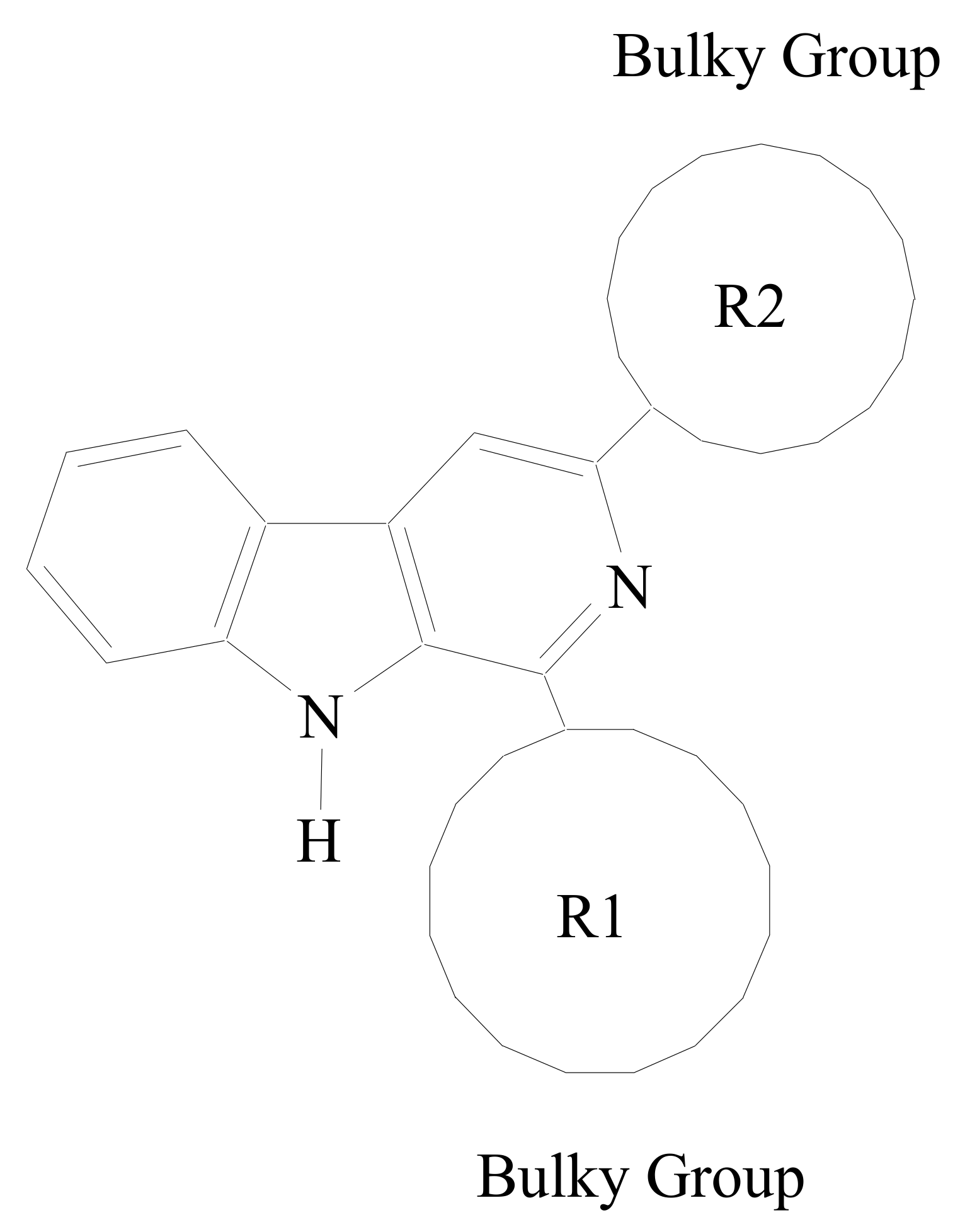
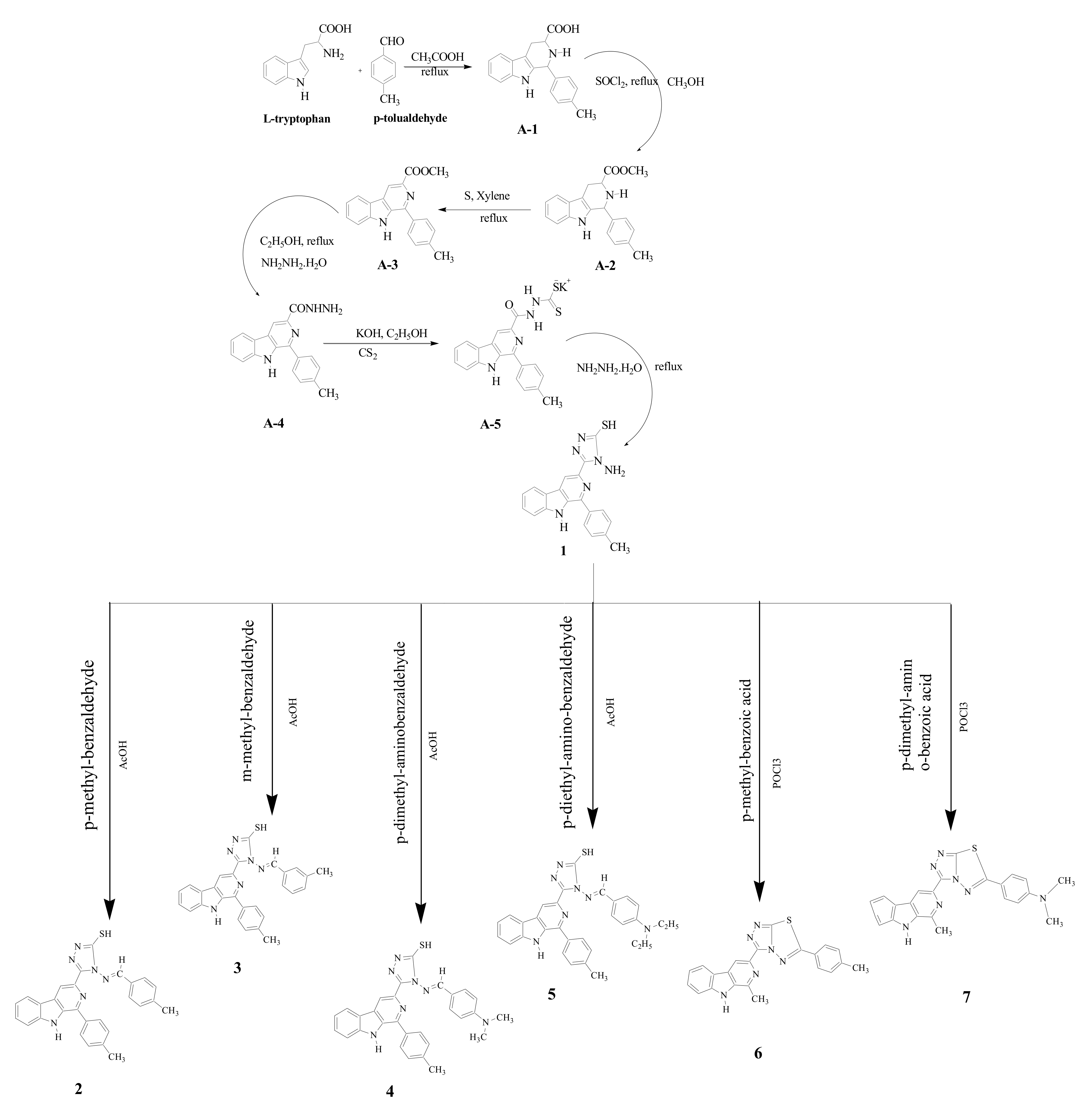
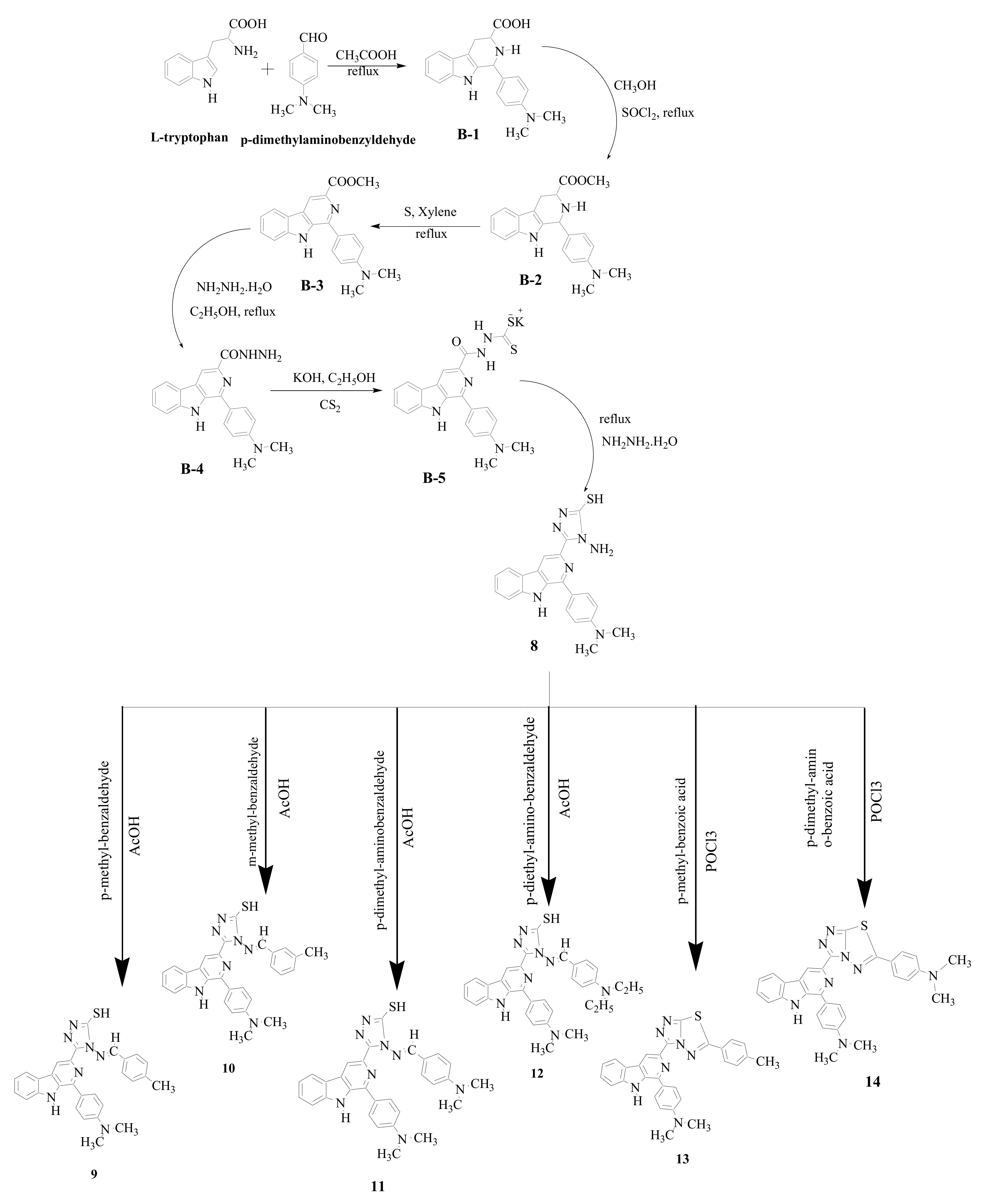
2.1. Synthesis of β-Carboline Derivatives

In this work, a number of new β-carboline derivatives were designed and synthesized. All the chemicals for synthesis were purchased in the highest available quality from commercial suppliers (Sigma-Aldrich, Merck Ltd., Mumbai, India) and used without further purification.
The synthetic routes for the preparation of β-carboline derivatives are presented in Scheme 1 and Scheme 2.
In Scheme 1, the methyl tetrahydro- β-carboline-3-carboxylates (A-2) were prepared through the Pictet–Spengler condensation of L-tryptophan with p-tolualdehyde in acid medium and subsequent esterification of the carboxylic acids with methanol and thionyl chloride. The conversion of the derivatives to the corresponding β-carboline-3-carbohydrazides (A-3) was carried out by oxidation with sulfur in refluxing xylene of methyl-1,2,3,4-tetrahydro-9H-β-carboline-3-carboxilates, followed by the reaction of methyl-β-carboline-3-carboxylates with hydrazine hydrate, in ethanol under reflux (yield A-4). The acid hydrazides (A-4) were allowed to react with carbon disulfide in the presence of potassium hydroxide in ethanol to afford the corresponding intermediate potassium dithiocarbazinate (A-5). This salt underwent ring closure with an excess of 99% hydrazine hydrate to give a 4-amino-3-substituted-5-mercapto-(4H)-1,2,4-triazole β-carboline derivative (1). The resulting triazole-carboline derivatives were then converted to 1,2,4-triazolo-[3,4-b]-1,3,4-thiadiazoles (27) in a one-pot reaction with aromatic acids and phosphorus oxychloride.
Similar to Scheme 1, in Scheme 2, the reaction is carried out through the Pictet–Spengler condensation of L-tryptophan with another aldehyde (i.e., p-dimethylaminobenzyldehyde). Furthermore, all the compounds reported in Scheme 2 were synthesized in a similar fashion as in Scheme 1. The detailed synthesis procedures of compounds 1–14 are given in the Supplementary Materials.
All the synthesized compounds were tested for their purity by TLC using the solvent system CHCl3:CH3OH (15:1) as a mobile phase and silica gel G precoated aluminum sheets (60 F254, Merck) as a stationary phase. The melting points of the synthesized compounds were determined by the open capillary method using the Toshniwal melting point apparatus. The proton NMR and 13C NMR spectra of the synthesized compounds were recorded on the Bruker Avance II 400 NMR spectrometer as solutions in CDCl3 or DMSO-d6 using TMS as an internal reference, and chemical shift values are expressed in δ units. The IR spectra of the synthesized compounds were recorded on an IR spectrophotometer (Jasco, FT/IR-4100 type A) in KBr phase. The mass spectra of the synthesized compounds were recorded on an FAB mass spectrometer (Jeol SX102-FAB).

2.2. Biological Screening

In the present study, all the synthesized compounds were subjected to growth inhibition assays in different cancer cell lines (A-549 and HepG2). At 540 nm, the optical density (OD) was measured using an ELISA reader. The optical density (OD) of sulforhodamine B (SRB) in each well was directly proportional to the cell number, so the OD values could be plotted against the concentration and the IC50 determined by using a program such as Graph-Pad PRISM [24].

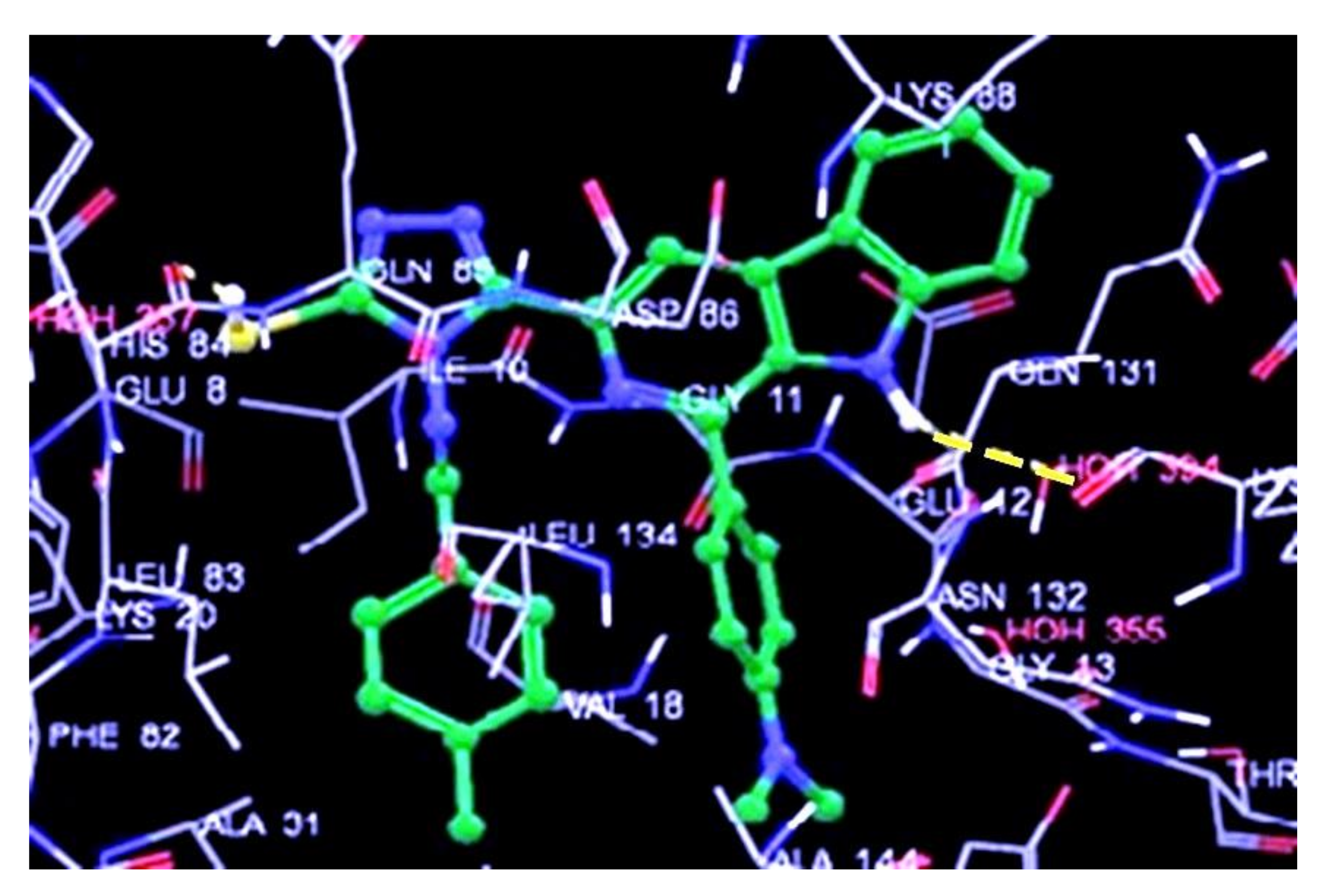
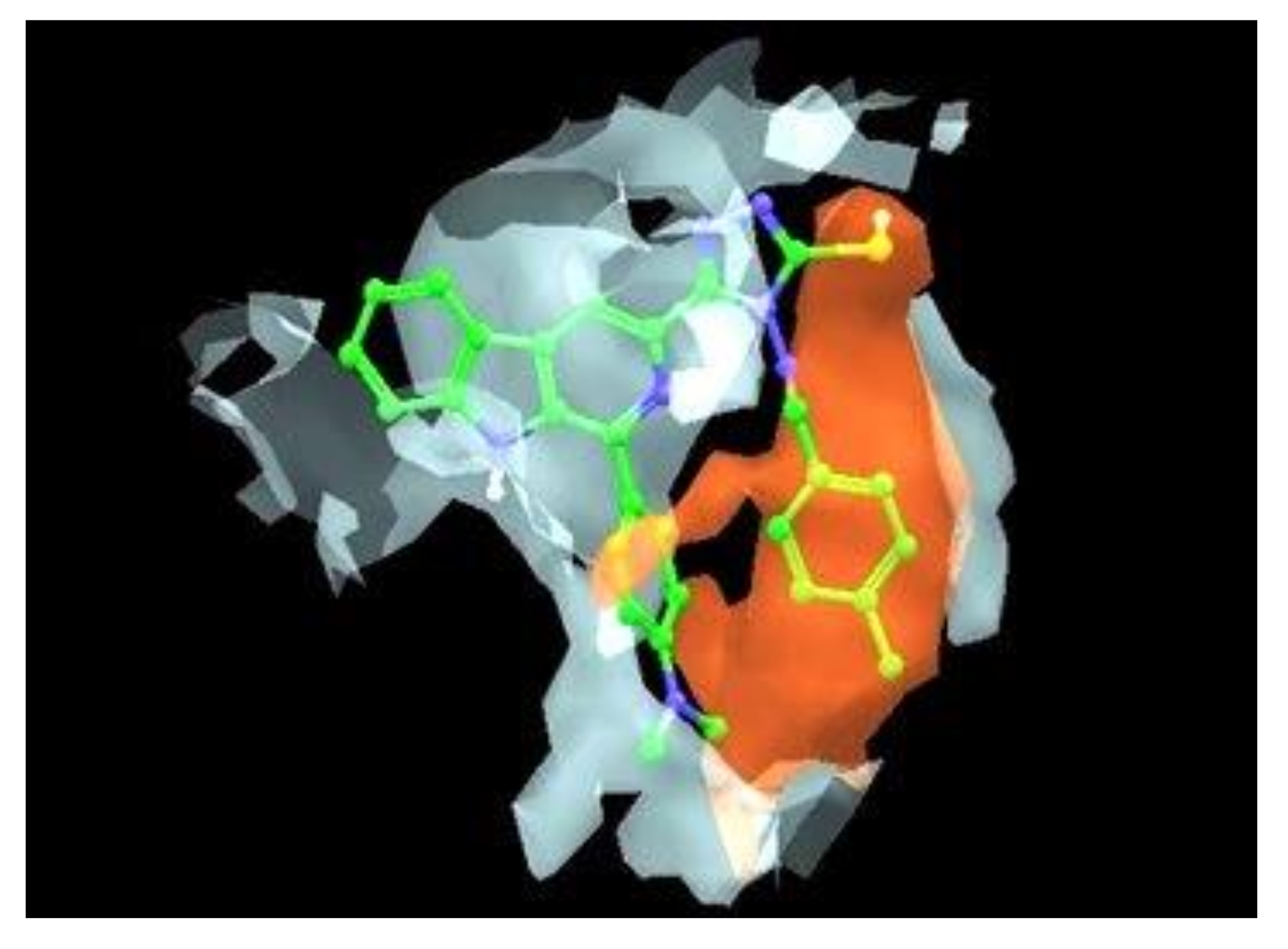
2.3. Docking Studies of Designed Compounds

Using the docking program Glide, the researchers investigated the appropriate binding orientations and conformations of the synthesized β-carboline derivatives interacting with cyclin-dependent kinases (CDKs). Glide is a fast, flexible docking method that uses an incremental construction algorithm to place ligands into active sites. By default, the docking program produces 10 docked structures for each β-carboline derivative. The conformation with the lowest docking energy in the most populated cluster is selected as the possible “active” conformation against the CDK2 [PDB: 1AQ1] active site [25,26,27]. In the present study, 14 compounds were successfully docked into the 1AQ1 site. The detailed procedure of docking studies is reported in the Supplementary Materials.

3. Results and Discussion

The novel β-carboline derivatives designed are shown in Table 1. These designed compounds were synthesized and characterized via spectroscopic techniques, and evaluated for their anticancer activity. The spectroscopic data revealed the successful synthesis of the designed compounds, and the anticancer activity was shown to be significant as compared to that of marketed ones.
The synthesis procedures are depicted in Scheme 1 and Scheme 2. Compounds 114 were synthesized by the reaction of starting material L-tryptophan with p-tolualdehyde or p-dimethylaminobenzyldehyde in the presence of acetic acid. The products were obtained in a 65–80% yield. These were found to be stable toward air and moisture at room temperature. All the synthesized derivatives showed moderate to high solubility in various organic solvents, such as methanol, chloroform, acetone, and dimethyl sulfoxide, but were insoluble in water. The spectral data matched the predicted structures of the synthesized compounds. In the 1H NMR, all protons were in their predictable regions, with integral area ratios per group conforming to the predicted number of protons per group. In the 13C NMR, the peaks of each group were consistent with the theoretical prediction of the number of carbon atoms in the structure. Elemental analysis confirmed the elemental composition of C, H, and N in the synthesized compounds.

3.1. Biological Screening Results

Except for (6), (7), and (13), all of the synthesized β-carboline derivatives inhibited various cancer cell lines effectively (Table 2). Compound (9) showed the utmost activity against both liver (HepG2) and adenocarcinoma (A549) cancer cell lines and was found to be roughly as equipotent as Adriamycin. Compounds (4) and (10) were found to be approximately 7–10 times less potent as compared to Adriamycin against the HepG2 cell line. The majority of compounds were active but approximately 50–100 times less potent than Adriamycin against both the HepG2 and A549 cancer cell lines.
For most compounds, drug sensitivity for both cell lines (HepG2 and A549 cells) was nearly equal, and the anticancer activity (IC50 value) was almost equally dependent on the type of aromatic ring on the ligand. The presence of the 4-methanamine group promotes biological activity, whereas β-carboline substituted with 1,4 triazolo (3,4-b)-1,3,4-thiadiazole is either inactive or less potent. It has been reported that 1-(N,N-dimethylbenzenamine)-substituted β-carboline derivatives are more potent than 1-(p-tolyl)-substituted β-carbolines. Compounds (i.e., 6, 7, and 13) containing a 1,3,4-thiadiazole-fused ring are biologically inactive (except compound 14). The imino moiety is conducive to biological activity and the three most active compounds (i.e., 4, 9, and 10) are imino derivatives.

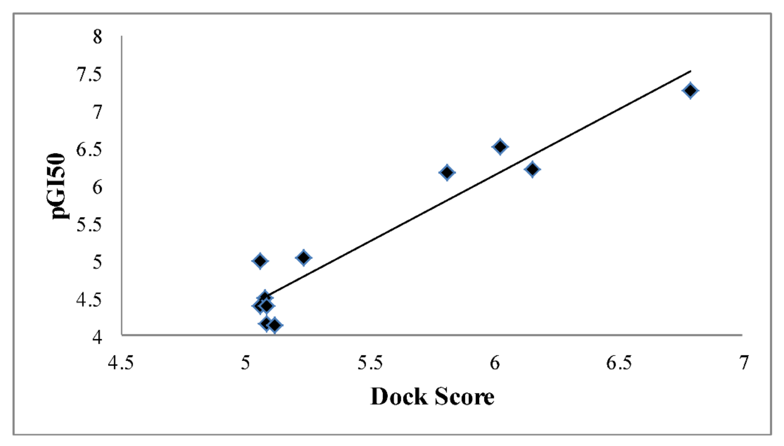
3.2. Docking Results

The in silico (docking) studies distinguished the compounds’ hypothetical binding modes using the X-ray crystal structure of CDK2 [PDB ID: 1AQ1] and G-score, as shown in Table 2. The top docked conformations (poses) closely resembled the co-crystallized conformation, with a root-mean-square deviation (RMSD) of 1.07–1.70 in the non-hydrogen atomic positions of the ligand.
Hydrogen bonding is an important factor that causes bonding with hetero atoms, so the docking interactions of the most active compound (9) with 1AQ1 were shown through hydrogen bonding, as seen in Figure 2. The hydrogen bond was found between the residue of compound (9) and the 1AQ1 in the R2 position. Furthermore, the sulfur atom of the triazole ring of compound (9) exhibited a van der Waals interaction with the amino acid residues, such as His84, at a distance of 2.68895 Å. Docking interactions were also discovered between compounds (114)’s residues and the 1AQ1. The docking results showed that the binding mode of β-carbolines of compound (9) with CDK2, dock score, and hydrophobic cavity included His-84, Gln-131, and Asp-86 amino acid residues; results are shown in Figure 2 and Figure 3. These interaction results revealed the possible binding of a target molecule to CDK2 and the further development of novel compounds for antitumor activity.
Compound (9) showed the highest dock score or G-score and this suggested that the docking interactions of compound (9) in 1AQ1 binding sites may be responsible for its highest biological interaction, followed by compounds (4) and (10). A linear correlation between G-score and biological activity was observed.
The correlation between the biological activities (pGI50 for HepG2 cell line) of the synthesized compounds and their dock scores in Glide docking is shown in Figure 4, which shows a linear correlation. The docking study gives only a rough approximation of the kinase inhibition activities of the synthesized compounds, and enzyme-based kinase (CDK2) inhibition experiments are required in the future.

4. Conclusions

With the aim of developing potent anticancer compounds, we have previously performed and reported 2D and 3D QSAR models on β-carboline derivatives, which provided useful information and insights into the structural requirement for anticancer activity. On the basis of QSAR outcomes, new, potent compounds were designed, synthesized, and characterized using FT-IR, 1HNMR, 13CNMR, FAB-MS, and elemental analysis techniques. These synthesized compounds were assayed for their in vitro biological activities, which showed that compound (9) was the most potent against the HepG2 and A549 cancer cell lines as compared to Adriamycin. All the synthesized compounds docked well into the binding pocket of the target protein CDK2 [1AQ1] and interacted with the crucial amino residues. The docking studies revealed a linear correlation between the docking score and anticancer activity, which suggested that the binding interaction of compounds with the active site of target protein 1AQ1 may be responsible for their anticancer activity.

Supplementary Materials

The following supporting information can be downloaded at https://www.mdpi.com/article/10.3390/chemistry4040091/s1. Supplementary File contains detailed procedures for synthesis of compounds and spectroscopic spectra of final compounds, and also contains detailed procedure for docking studies.

Author Contributions

Conceptualization, A.V.; methodology, R.K.A.; software, R.K.A.; validation, R.K.C.; formal analysis, R.K.C.; investigation, R.K.C.; resources, R.K.A.; data curation, R.K.C.; writing—original draft preparation, R.K.C.; writing—review and editing, A.V.; visualization, R.K.C.; supervision, A.V.; project administration, A.V.; funding acquisition, A.V. All authors have read and agreed to the published version of the manuscript.

Funding

This research received no external funding.

Data Availability Statement

Data available in article and raw data are available from the corresponding authors upon request.

Acknowledgments

The authors are grateful to SAIF, Punjab University Chandigarh, for the spectroscopic analysis.

Conflicts of Interest

There is no conflict of interest.

References

  1. Sung, H.; Ferlay, J.; Siegel, R.L.; Laversanne, M.; Soerjomataram, I.; Jemal, I.; Bray, A.F. Global Cancer Statistics 2020: GLOBOCAN Estimates of Incidence and Mortality Worldwide for 36 Cancers in 185 Countries. CA Cancer J. Clin. 2021, 713, 209–249. [Google Scholar] [CrossRef] [PubMed]
  2. Lopez, J.S.; Banerji, U. Combine and conquer: Challenges for targeted therapy combinations in early phase trials. Nat. Rev. Clin. Oncol. 2017, 141, 57–66. [Google Scholar] [CrossRef] [PubMed]
  3. Jain, S.; Chandra, V.; Jain, P.K.; Pathak, K.; Pathak, D.; Vaidya, A. Comprehensive review on current developments of quinoline-based anticancer agents. Arab. J. Chem. 2019, 128, 4920–4946. [Google Scholar] [CrossRef]
  4. Xiao, S.; Lin, W.; Wang, C.; Yang, M. Synthesis and Biological Evaluation of DNA Targeting Flexible Side-Chain Substituted β-Carboline Derivatives. Bioorg. Med. Chem. Lett. 2001, 11, 437–441. [Google Scholar] [CrossRef]
  5. Cao, R.; Peng, W.; Chen, H.; Hou, X.; Guan, H.; Chen, Q.; Ma, Y.; Xu, A. Synthesis and in vitro cytotoxic evaluation of 1,3-bisubstituted and 1,3,9-trisubstituted β-carboline derivatives. Eur. J. Med. Chem. 2005, 40, 249–257. [Google Scholar] [CrossRef]
  6. Castro, A.C.; Dang, L.C.; Soucy, F.; Grenier, L.; Mazdiyasni, H.; Hottelet, M.; Parent, L.; Pien, C.; Palombella, V.; Adams, J. Novel IKK Inhibitors: β-Carbolines. Bioorg. Med. Chem. Lett. 2003, 13, 2419–2422. [Google Scholar] [CrossRef]
  7. Deveau, A.M.; Labroli, M.A.; Dieckhaus, C.M.; Barthen, M.T.; Smith, K.S.; Macdonald, T.L. The Synthesis of Amino-Acid Functionalized β-Carbolines as Topoisomerase II Inhibitors. Bioorg. Med. Chem. Lett. 2001, 11, 1251–1255. [Google Scholar] [CrossRef]
  8. Cao, R.; Guan, X.; Shi, B.; Chen, Z.; Ren, Z.; Peng, W.; Song, H. Design, synthesis and 3D-QSAR of β-carboline derivatives as potent antitumor agents. Eur. J. Med. Chem. 2010, 45, 2503–2515. [Google Scholar] [CrossRef]
  9. Cao, R.; Yi, W.; Wu, Q.; Guan, X.; Feng, M.; Ma, C.; Chen, Z.; Song, H.; Peng, W. Synthesis and cytotoxic activities of 1-benzylidine substituted β-carboline derivatives. Bioorg. Med. Chem. Lett. 2008, 18, 6558–6561. [Google Scholar] [CrossRef]
  10. Trujillo, J.I.; Meyers, M.J.; Anderson, D.R.; Hegde, S.; Mahoney, M.W.; Vernier, W.F.; Buchler, I.P.; Wu, K.K.; Yang, S.; Hartmann, S.J.; et al. Novel tetrahydro-β-carboline-1-carboxylic acids as inhibitors of mitogen activated protein kinase-activated protein kinase 2 (MK-2). Bioorg. Med. Chem. Lett. 2007, 17, 4657–4663. [Google Scholar] [CrossRef]
  11. Barsanti, P.A.; Wang, W.; Ni, Z.; Duhl, D.; Brammeier, N.; Martin, E.; Bussiere, D.; Walter, A.O. The discovery of tetrahydro-β-carbolines as inhibitors of the kinesin Eg5. Bioorg. Med. Chem. Lett. 2010, 20, 157–160. [Google Scholar] [CrossRef] [PubMed]
  12. Cao, R.; Peng, W.; Chen, H.; Ma, Y.; Liu, X. DNA binding properties of 9-substituted harmine derivatives. Biochem. Biophys. Res. Commun. 2005, 3383, 1557–1563. [Google Scholar] [CrossRef]
  13. Chourasiya, K.R.; Rao, R.A.; Agrawal, K.R. Pharmacophore Modeling and QSAR Analysis of Novel β-carboline Derivatives as Antitumor Agents. Lett. Drug Des. Discov. 2013, 107, 572–584. [Google Scholar]
  14. Formagio, A.S.; Tonin, L.T.; Foglio, M.A.; Madjarof, C.; de Carvalho, J.E.; da Costa, W.F.; Cardoso, F.P.; Sarragiotto, M.H. Synthesis and antitumoral activity of novel 3-(2-substituted-1,3,4-oxadiazol-5-yl) and 3-(5-substituted-1,2,4-triazol-3-yl) beta-carboline derivatives. Bioorg. Med. Chem. 2008, 16, 9660–9667. [Google Scholar] [CrossRef] [PubMed]
  15. Ishida, J.; Wang, H.K.; Bastow, K.F.; Hu, C.Q.; Lee, K.H. Antitumor agents 201. Cytotoxicity of harmine and beta-carboline analogs. Bioorg. Med. Chem. Lett. 1999, 9, 3319–3324. [Google Scholar] [CrossRef]
  16. Cao, R.; Chen, H.; Peng, W.; Ma, Y.; Hou, X.; Guan, H.; Liu, X.; Xu, A. Design, synthesis and in vitro and in vivo antitumor activities of novel beta-carboline derivatives. Eur. J. Med. Chem. 2005, 40, 991–1001. [Google Scholar] [CrossRef] [PubMed]
  17. Zhao, M.; Bi, L.; Wang, W.; Wang, C.; Baudy-Floc’h, M.; Ju, J.; Peng, S. Synthesis and cytotoxic activities of beta-carboline amino acid ester conjugates. Bioorg. Med. Chem. 2006, 14, 6998–7010. [Google Scholar]
  18. Vaidya, A.; Jain, S.; Jain, A.K.; Prashanthakumar, B.R.; Kashaw, S.K.; Agrawal, R.K. Computational Analysis of Quinoline Derivatives as Potent Topoisomerase-II Inhibitors. Med. Chem. Res. 2015, 24, 383–393. [Google Scholar] [CrossRef]
  19. Vaidya, A.; Jain, A.K.; Kumar, P.; Kashaw, S.K.; Agrawal, R.K. Predicting anti-cancer activity of quinoline derivatives: CoMFA and CoMSIA approach. J. Enzyme Inhib. Med. Chem. 2011, 26, 854–861. [Google Scholar]
  20. Agrawal, R.K.; Jain, A.K.; Veerasamy, R.; Vaidya, A.; Kashaw, S.; Mourya, V.K.; Agrawal, R.K. QSAR analysis of B-ring-modified diaryl ether derivatives as a InhA inhibitors. Med. Chem. Res. 2012, 21, 145–151. [Google Scholar]
  21. Jain, S.; Vaidya, A.; Jain, A.K.; Agrawal, R.K.; Kashaw, S.K. Computational analysis of benzyl vinylogous derivatives as potent PDE3B inhibitors. Arab. J. Chem. 2017, 10, S109–S113. [Google Scholar] [CrossRef]
  22. Vaidya, A. Discovery of novel 1,2,4-oxadiazole derivatives as potent caspase-3 activator for cancer treatment. Chemistry 2021, 3, 373–381. [Google Scholar] [CrossRef]
  23. Chourasiya, R.K.; Rao, A.R.; Agrawal, R.K. QSAR and docking studies of novel β-carboline derivatives as anticancer. Med. Chem. Res. 2013, 22, 2991–3001. [Google Scholar] [CrossRef]
  24. Vaidya, A.; Jain, S.; Kumar, B.R.P.; Singh, S.K.; Kashaw, S.K.; Agrawal, R.K. Synthesis of 1.;2.;4-oxadiazole derivatives: Anticancer and 3D QSAR studies. Mon. Chem. 2020, 151, 385–395. [Google Scholar] [CrossRef]
  25. Shankaraiah, N.; Jadala, C.; Nekkanti, S.; Senwar, K.R.; Nagesh, N.; Shrivastava, S.; Naidu, V.G.; Sathish, M.; Kamal, A. Design and synthesis of C3-tethered 1,2,3-triazolo-β-carboline derivatives: Anticancer activity, DNA-binding ability, viscosity and molecular modeling studies. Bioorg. Chem. 2016, 64, 42–50. [Google Scholar] [CrossRef]
  26. Abdelsalam, M.A.; AboulWafa, O.M.; Badawey, M.E.A.; El-Shoukrofy, M.S.; El-Miligy, M.M.; Gouda, N.; Elaasser, M.M. Design, synthesis, anticancer screening, docking studies and in silico ADME prediction of some β-carboline derivatives. Future Med. Chem. 2018, 10, 1159–1175. [Google Scholar] [CrossRef]
  27. Vaidya, A.; Jain, A.K.; Kumar, B.R.P.; Sastry, G.N.; Kashaw, S.K.; Agrawal, R.K. CoMFA, CoMSIA, kNN MFA and Docking studies of 1,2,4-Oxadiazole derivatives as potent Caspase-3 activators. Arab. J. Chem. 2017, 10, S3936–S3946. [Google Scholar] [CrossRef]
Figure 1. Structure of β-carboline with bulky groups at R1 and R2 positions for anticancer activity.
Chemistry 04 00091 g001
Scheme 1. Synthetic scheme for β-carboline derivatives via the reaction of starting material L-tryptophan and p-tolualdehyde.
Chemistry 04 00091 sch001
Scheme 2. Synthetic scheme for β-carboline derivatives via the reaction of starting material L-tryptophan and p-dimethylaminobenzyldehyde.
Chemistry 04 00091 sch002
Figure 2. Docking conformation in the active site of CDK2 of most active compound (9) in the context of hydrogen bonding is displayed as dotted yellow lines.
Chemistry 04 00091 g002
Figure 3. Docking conformation in the active site of CDK2 of most active compound (9) in the context of hydrophobic region.
Chemistry 04 00091 g003
Figure 4. Correlation between the biological activity of synthesized compounds and their dock scores in Glide docking.
Chemistry 04 00091 g004
Table 1. Newly designed, substituted β-carboline derivatives.
Chemistry 04 00091 i001
Compound No.R1R2
1Chemistry 04 00091 i002Chemistry 04 00091 i003
2Chemistry 04 00091 i002Chemistry 04 00091 i005
3Chemistry 04 00091 i002Chemistry 04 00091 i007
4Chemistry 04 00091 i002Chemistry 04 00091 i009
5Chemistry 04 00091 i002Chemistry 04 00091 i011
6Chemistry 04 00091 i002Chemistry 04 00091 i013
7Chemistry 04 00091 i002Chemistry 04 00091 i015
8Chemistry 04 00091 i016Chemistry 04 00091 i017
9Chemistry 04 00091 i016Chemistry 04 00091 i019
10Chemistry 04 00091 i016Chemistry 04 00091 i021
11Chemistry 04 00091 i016Chemistry 04 00091 i023
12Chemistry 04 00091 i016Chemistry 04 00091 i025
13Chemistry 04 00091 i016Chemistry 04 00091 i027
14Chemistry 04 00091 i016Chemistry 04 00091 i029
Table 2. In vitro cytotoxic activity of synthesized compounds by SRB assay with their dock score or G-score.
Compound No.Growth Inhibitory Effects, GI50 (µM)Dock Score
(G-Score)
HepG2 Cell LineA549 Cell Line
18.147.214.15
28.348.874.48
38.738.224.98
40.944.106.52
58.648.054.39
6>100>1003.27
7>100>1003.98
88.127.354.38
90.160.147.28
100.696.766.21
115.816.525.03
127.614.224.12
13>100>1004.05
141.541.426.17
Adriamycin0.10.1
Publisher’s Note: MDPI stays neutral with regard to jurisdictional claims in published maps and institutional affiliations.

