Review

Approaching Electroencephalographic Pathological Spikes in Terms of Solitons

Center for Nonlinear Science, Department of Physics, University of North Texas, 1155 Union Circle, #311427, Denton, TX 76203, USA
Signals 2024, 5(2), 281-295; https://doi.org/10.3390/signals5020015
Submission received: 27 February 2024 / Revised: 22 April 2024 / Accepted: 26 April 2024 / Published: 1 May 2024
(This article belongs to the Special Issue Advancing Signal Processing and Analytics of EEG Signals)

Abstract

:
A delicate balance between dissipative and nonlinear forces allows traveling waves termed solitons to preserve their shape and energy for long distances without steepening and flattening out. Solitons are so widespread that they can generate both destructive waves on oceans’ surfaces and noise-free message propagation in silica optic fibers. They are naturally observed or artificially produced in countless physical systems at very different coarse-grained scales, from solar winds to Bose–Einstein condensates. We hypothesize that some of the electric oscillations detectable by scalp electroencephalography (EEG) could be assessed in terms of solitons. A nervous spike must fulfill strict mathematical and physical requirements to be termed a soliton. They include the proper physical parameters like wave height, horizontal distance and unchanging shape; the appropriate nonlinear wave equations’ solutions and the correct superposition between sinusoidal and non-sinusoidal waves. After a thorough analytical comparison with the EEG traces available in the literature, we argue that solitons bear striking similarities with the electric activity recorded from medical conditions like epilepsies and encephalopathies. Emerging from the noisy background of the normal electric activity, high-amplitude, low-frequency EEG soliton-like pathological waves with relatively uniform morphology and duration can be observed, characterized by repeated, stereotyped patterns propagating on the hemispheric surface of the brain over relatively large distances. Apart from the implications for the study of cognitive activities in the healthy brain, the theoretical possibility to treat pathological brain oscillations in terms of solitons has powerful operational implications, suggesting new therapeutical options to counteract their detrimental effects.

1. Introduction

Solitons are single, spatially localized waves that travel without changing their shape and size [1]. Due to a delicate balance between dissipative effects and nonlinear dynamics, solitons are coherent structures propagating in space and time at a constant velocity without attenuation, distortion, or energy loss [2,3]. At least in an idealized framework, the stability and stationarity of soliton’s analytical solutions are confirmed against non-integrable perturbations [4] (Ye et al., 2020) so that frictional effects like viscosity do not cause solitons to decay over time [5]. The energy of solitons is carried adiabatically through the medium, i.e., without transferring heat or mass [6]. Since the energy transfer to shorter wavelength regions is difficult, solitons reach a state of thermodynamic equilibrium that allows them to evolve for a long time without damping [7,8]. Solitons are naturally observed or artificially produced in different macro-, meso- and microscopic settings, playing key roles in the context of out-of-equilibrium nonlinear systems. They can be found in space plasmas, solar winds, planetary magnetospheres, marine and fluvial surface gravity waves, telegraph lines [9], high-speed silica fiber-optic telecommunication, optical coherence tomography, photorefractive crystals [10], micromechanical systems [11], magnetic matter, superconductors [12], Bose–Einstein condensates [13], etc. Their occurrence in ionic crystals, crystalline solids and nematic liquid crystals [14] suggests a role for solitons in the physical properties of solids at high temperatures [11]. It has been conjectured that soliton-like mechanisms could also underlie biological phenomena like electron/proton transport along proteins’ helices [15,16] as well as cardiac action potentials [17].
Still, attempts have been made to describe the MICROSCOPIC nervous activity of the central nervous system in terms of solitons [18]. It has been conjectured that the standard Hodgkin–Huxley model of the neurons’ action potential could be piezoelectrically coupled with synchronized soliton pressure pulses generated inside the lipids of biological membranes [19] and influenced by thermodynamic variables like voltage, temperature, pressure, chemical potentials [17]. According to this controversial model, the traveling solitons are localized in the membrane, do not spread out in the surrounding medium, do not release heat, and can modify the local membrane density and thickness [20,21].
Here, we take a different turn, suggesting another potential approach to assess the nervous electric spikes in the context of solitary waves. Looking at the central nervous system’s nervous activity at the MACROSCOPIC scale, we argue that some of the brain electric oscillations detected by electroencephalography (EEG) could be mathematically and biophysically approached in terms of solitons’ motion. We contend that the electric oscillation spikes attained by scalp EEG monitoring techniques resemble, in many respects, solitary waves. Since an electric wave must fulfill strict mathematical and physical requirements in order to be termed a soliton, we will analytically examine the manifold physical features of solitary waves and will look for their feasible counterparts in the cortical electric activity measurable on the scalp. In sum, by delving into the potential existence of solitons within EEG signals, we explore various nonlinear medium features and physical properties, such as energy expression, wavelength, phase velocity, and wave height. We conclude that soliton-related analytical methods could be effectively employed in the study of EEG signals.

2. Nonlinear Media and Solitons

Solitons are naturally robust to perturbations as they travel with little dissipation over long timeframes [22] (Rowley et al., 2022). The peculiar coherence of these highly localized wave packets can be attributed to the fact that the tendency for a wave pulse to disperse is exactly balanced by nonlinear effects occurring in the propagation medium [5]. While the linear and nonlinear effects acting alone would cause the wave to decay, in the right combination, they lead to waves that persist stably and compactly as they propagate, periodically returning to the initial state.
Numerous natural and artificial phenomena are characterized by the slight nonlinear dynamics required to generate solitons. Looking for a model of soliton-like EEG oscillations, we must first look for the possible presence of nonlinear dynamics in the central nervous system.

Nervous Nonlinear Medium

Solitons require a dissipative physical medium characterized by a certain degree of nonlinearity. In keeping with this claim, nervous nonlinear dynamics have been ubiquitously detected in both experimental settings and ecologically valid conditions [23,24]. Using brain network analysis, data-driven approaches and machine learning [25], various types of nonlinear dynamic behavior have been found in the wave series extracted from EEG data sets [23]. Nonlinear dynamics in the central nervous system consist of phase transitions, branching processes, metastability, limit cycles, self-oscillations, non-stationarity, neuronal avalanches, chaotic behavior and collective phenomena with the emergence of order [26,27]. Increasing evidence suggests that cortical neuronal networks operate near a critical state where balanced activity patterns support optimal information processing, scale-free correlations and the emergence of adaptive collective behavior [26].
Therefore, it can be stated that the nonlinear dynamics required by a nervous soliton-like model are widely diffused in the brain.

3. Equations for Solitons

In mathematical terms, solitons stand for one of the countless possible solutions for different types of nonlinear wave equations [6]. In particular, the Korteweg-de Vries equation (KdVE), originally developed to describe weakly nonlinear wave propagation in shallow water, admits analytic solutions representing solitons. KdVE equation can be written as
tΦ + ∂3xΦ − 6Φ∂xΦ = 0
where ∂x and ∂t are partial derivatives of x and t, Φ stands for the height displacement of the water surface from its equilibrium height. KdVE implies an equilibrium between dispersion ∂3xΦ and advection Φ∂xΦ, the latter standing for the particle transport provided by bulk motion and velocity of the fluid. KdVE can describe soliton systems in which the particles are driven by fluid pressure [28] as well as soliton turbulence in ocean waves [29]. The Benjamin–Bona–Mahony equation (BBME) is a KdVE variant for modeling long surface gravity waves of small amplitude, propagating uni-directionally in 1+1 dimensions. Yet, a particle-based fluid trajectory description of multi-soliton interactions can be achieved by the Kadomtsev–Petviashvili equation (KPE), i.e., a two-dimensional version of KdVE.
Apart from KdVE, BBME, and KPE, other wave equations display soliton solutions. The most prominent is the one-dimensional nonlinear Schrödinger equation (NLSE), an example of integrable systems of nonlinear partial differential equations owning an infinite set of conservation laws. NLSE stands for a universal tool to model the dynamical evolution in dispersive media of weakly nonlinear, varying packets of quasi-monochromatic waves [30]. Among the manifold versions, the linearly damped and driven NLSE can be expressed in the dimensionless form:
iu t + 1 2   u xx + ׀ u ׀ 2   u = f i γ u
where f = f(x,t) is the driving (or forcing) of the system, iγu is the linear damping of strength γ > 0 [31]. Depending on the chosen physical setting, different NLSE formulations provide different results according to the values of the dispersive nonlinear coefficients [30]. NLSE has a wide range of applications, including, e.g., the slow evolution of wave amplitude in shallow waters of uniform depth, the irregular wave trains propagating in deep water, superconductivity devices, nonlinear optics, etc. [32]. Concerning Bose–Einstein condensates, the soliton’s behavior can be described by a variant of the NLSE referred to as the Gross–Pitaevskii equation (GPE) [30,33]. In this case, the velocities of single particles are determined by the phase of the wave function. Figure 1 illustrates a few examples of soliton solutions to various nonlinear wave equations.
Therefore, every attempt to look for hints of nervous solitons requires electric solitary waves to obey explicit nonlinear wave equations.

Solitons in the Electrical Domain and in EEG Spikes

Solitons may be produced in the context of the electrical domain, both at the macro-, meso- and micro-levels of observation. The occurrence of electrostatic solitons is ubiquitous in macroscopic contexts such as two-fluid astrophysical plasmas, planetary magnetospheres and lunar wakes [3]. In solar wind, the interplanetary magnetic fields provide favorable conditions for solitons formation [36]. In space plasmas, nonlinear electrostatic solitary waves can be interpreted in terms of ion- and electron-acoustic solitons propagating parallel to the ambient magnetic field. Concerning the micro-levels of observation, electromagnetic fields and saturable nonlinear dielectrics may influence solitary waves’ shape and behavior [37]. Self-reinforcing magnetic solitons have been generated in Bose–Einstein condensates from atoms with different spins [38]. In nematic and cholesteric liquid crystals, external electric fields drive the collective reorientation of nematic molecules. This process generates solitons representing self-trapped “bullets” that move with high speed, perpendicularly to the electric field and to the initial alignment direction [14,39]. The above-mentioned nonlinear wave equations have been widely used to cope with electric as well as external uniform magnetic fields inside the discrete nonlinear electrical lattices used for electrical transmission [40].
Once the solitons can be generated in an electromagnetic milieu, the question is whether they could occur in the hemispheric electric field detected by scalp EEG. The fact that NLSE may govern soliton propagation on spherical surfaces of electromagnetic fields traveling in coiled optical fibers [41] provides an indirect hint that solitons can be uncovered inside spherical electric fields [42]. Further, it has been shown that electric or magnetic monopole pairs can interact with Coulombic fields equipped with the SO(3) rotations typical of spherical surfaces [43].
Therefore, the nervous electric activity detected on the scalp could be assessed by using nonlinear wave equations with solitons solutions.

4. The Remarkable Physical Features of Solitons

Solitons’ solutions can be given analytically by using mathematical devices such as first-order Fourier analysis and statistical analysis [6,44]. Different solutions of KdVE, BBME, NLSE, KPE and GPE generate different types of propagating solitons characterized by distinct physical features and various wave shapes. The physical parameters to assess the presence and the dynamics of solitons are manifold. They include surface elevation, wave height, phase velocity (i.e., the rate at which the wave phase propagates), wavelength, spatiotemporal evolution of the normalized force as a function of a fixed or infinite horizontal distance, breathing period and maximum intensity, trajectories, velocities, acceleration, medium depth, material parameters [2,45]. In fluid media like seas or channels, also surface tension, density, gravity, channel height, water depth, carrier frequency and amplitude of the background wave [44] conspire to generate the solitons’ features.
Therefore, when looking for the theoretical occurrence of solitons in EEG traces, the above-mentioned physical parameters must be taken into account.

4.1. Different Shapes for Different Solitary Waves

One of the most distinguished features of solitons is the shape, so different from the usual sine waves (Figure 1). Solitary waves include W-shaped, bright, dark, kink-dark, singular, kink antikink solitons [34,40], cnoidal waves (Figure 1A), abnormally large waves (also termed “freak” or “rogue” waves), “thick” or “top-table” solitons, algebraic solitons, solitons of different polarities [46], multi-solitons with doubly-localized peaks [1], etc. Unlike “bright” solitons, which locally amplify the surrounding medium, “dark” solitons stand for a local decrease in the wave amplitude able to cause a depression [44]. Dark solitons on the surface of water might influence extreme events like tsunamis, such that an almost flat ocean surface may evolve into a singular, destructive rogue wave with high amplitude, as energy is conveyed in the central zone from the surrounding areas [44] (Figure 1B). Contrary to the usual solitons that keep their profile unchanged during propagation, the Peregrine soliton displays a double spatio-temporal localization characterized by progressive, standardized and cyclic changes in shape [47] (Figure 1D). Apart from the ones described above, other parameters may influence the large variety of solitons’ shapes. These parameters include hyperbolic function solutions, elliptic parameters for cnoidal waves, variable–coefficient functions, modulation instability gain [34], the Sine–Gordon expansion method [40], etc. Furthermore, periodic traveling-wave solutions may give rise to “trains” of many solitons (Figure 1F). This occurs in the solar wind; whereas weak shock waves produce only single soliton pulses, strong shocks produce trains of solitons [36] (Figure 1G).
Therefore, when looking for the possible occurrence of solitary waves in EEG traces, it is mandatory to carefully evaluate whether the shape of the electric spikes matches the features of solitons.

4.2. Solitons in Healthy EEG Traces

The occurrence of solitons in EEG traces from healthy individuals has been conjectured. Sen [48] generated action potential-like solitons resembling EEG-like surface waves on a synthetic brain connectome in response to surface deformations produced by local pinch. Yet, part of the electric activity of the healthy human brain is characterized by nervous electric spikes that are reminiscent of solitons. For instance, the neuronal activity in the medial entorhinal cortex displays peculiar ultraslow periodic oscillations from tens of seconds to minutes. Further, electrocorticographic studies suggest that alpha and theta oscillations generate traveling waves propagating in a posterior-to-anterior direction at ∼0.25–0.75 m/s [49,50]. The last, but not the least significant, electrical recordings and spectral analysis from many brain regions exhibit neural oscillations at multiple spatial scales that, like solitons, are non-sinusoidal [50].
Therefore, theoretical mathematical findings, as well as implicit cues from neurophysiological studies, suggest the possibility that at least part of the normal electric EEG activity might involve solitons.

4.3. Solitons in Pathological EEG Traces

Reviewing the neuroscientific literature, it is easy to notice that quite a few EEG electric spikes detectable in disease states look like solitons [51]. Emerging from the noisy background of normal electric activity, high-amplitude, low-frequency EEG solitary waves of different shapes can be observed in various human diseases. A few EEG snapshots are illustrated in Figure 2.
  • Figure 2A illustrates a case of Herpesvirus-6 limbic encephalitis with lateralized periodic discharges over the left temporal region, with no apparent inter-discharge interval between the spike-and-wave complexes (modified from [52] Gélisse et al. (2023).
  • Figure 2B illustrates a bilateral synchronous symmetrical delta burst pattern with almost flat amplitude during burst intervals, recorded during wakefulness in the advanced stages of subacute sclerosing panencephalitis in an adult patient; modified from [53].
  • Figure 2C illustrates an example of a 3 to 4 Hz generalized spike-wave in a child affected by juvenile absence epilepsy; modified from https://www.epilepsy.com/what-is-epilepsy/syndromes/juvenile-absence-epilepsy (accessed on 23 March 2024).
  • Figure 2D illustrates a typical EEF in a case of sporadic Creutzfeldt–Jacob disease. Note the disease-typical periodic sharp wave complexes that occur in the middle and late stages; modified from https://www.eurocjd.ed.ac.uk/images/typical-eeg-sporadic-cjd (accessed on 23 March 2024).
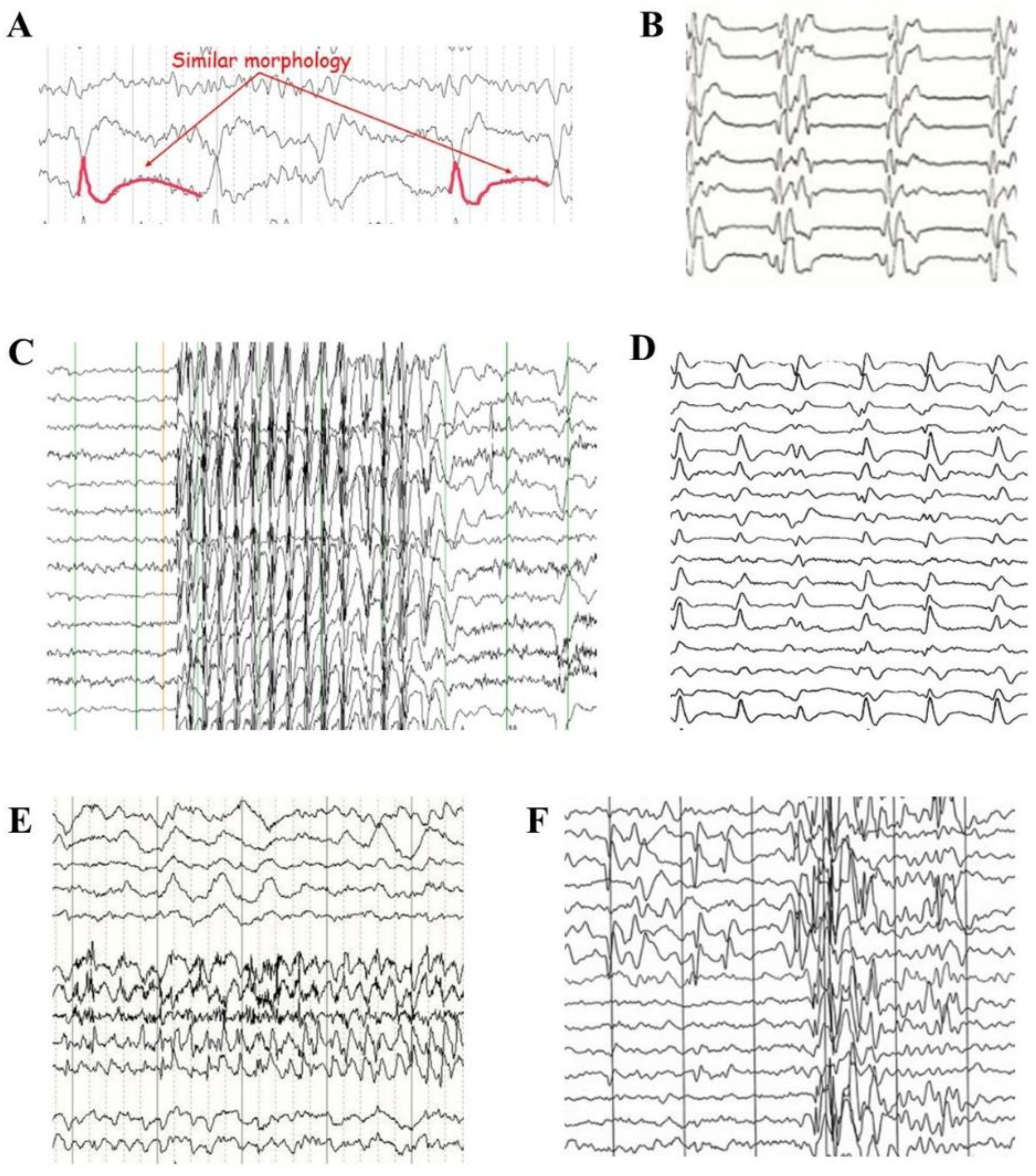
  • Figure 2E illustrates an EEG snapshot showing a right temporal focal seizure in an adult. A focal slowing occurs in the temporal region; modified from [54].
  • Figure 2F illustrates the coexistence of focal and generalized epileptiform discharges in a 7-year-old boy with benign childhood epilepsy with occipital paroxysms [55].
Figure 2. Pathological EEG patterns resembling trains of solitons. (A) Herpesvirus-6 limbic encephalitis; modified from [52]. (B) Typical “metronomic” delta burst pattern with almost flat amplitude during burst intervals, recorded during advanced stages of subacute sclerosing panencephalitis; modified from [53]. (C) Juvenile absence epilepsy; modified from https://www.epilepsy.com/what-is-epilepsy/syndromes/juvenile-absence-epilepsy (accessed on 23 March 2024) (D) Creutzfeldt–Jacob disease; modified from https://www.eurocjd.ed.ac.uk/images/typical-eeg-sporadic-cjd (accessed on 23 March 2024) (E) EEG snapshot showing a right temporal focal seizure; modified from [54]. (F) Coexistence of focal and generalized epileptiform discharges in a 7-year-old boy with benign childhood epilepsy with occipital paroxysms [55]. Similarities between solitons and pathological EEG patterns can be identified by comparing the waves illustrated in Figure 1 and Figure 2.
Signals 05 00015 g002
Distinct medical conditions, including critically ill patients, encephalitis and epilepsy, are characterized by EEG traces with features that remind solitons trains, i.e., repeated patterns or stereotyped activity with or without interchange intervals, at an approximately regular rate or intervals [55]. Just as solitons, pathological EEG waveforms may display relatively uniform morphology and duration. Like solitons, pathological EEG oscillations can travel on the hemispheric surface of the brain over relatively large distances, and, like solitons, neither tend to flatten out nor steepen and topple over. Further, some pathological EEG waves display double spatio-temporal localization with cyclic changes in shape, just as Peregrine solitons.
Summarizing, the occurrence in EEG traces of features and shapes corresponding to various types of solitary waves suggests that some pathological brain electric activities might represent the biological counterpart of physical solitons.

5. Dynamics of Solitary Waves

Solitons travel long distances with no need for leading or trailing waves. The soliton’s wave crest can be much higher than the surrounding waves [29]. Since the velocity depends on the wave’s amplitude and height, a soliton is slower than its carrier waves. In addition, the velocity of solitons can be increased by an acceleration induced by an external water flow. When the distance along the surface approaches zero, a sharp rise of the wave crest can be detected above the rest of the deep fluid medium. The deeper the water, the more negative the nonlinearity parameter and the higher the possibility of generating rogue solitons, characterized by large-amplitude oscillations with height two times larger than the wave train [32]. When the soliton disappears, a defocusing process takes place in which the instantaneous frequencies in front become lower than those behind it [32].

5.1. Interactions between Solitons

Time-reversible interactions among multiple solitons, carriers, and media occur in both natural and artificial settings. When a carrier wave enters the soliton, its amplitude drops to zero; when it exits, its amplitude returns to the previous value [44]. Two colliding solitons penetrate through each other, exchange their amplitude and velocity and emerge fully intact as the exact pulses that entered the collision [2,45] (Figure 1H). At first, the two waves would align to temporarily reproduce the initial sine wave before separating again and repeating the same cycle. The higher amplitude wave is narrower and faster than the wave with the minor amplitude. Heart- and bell-shaped-cusp optical solitons can be produced by two-wave superposition (see the Figures in [28]). Though 90% of the soliton collisions occur between pairs of solitons, the remnant collisions involve multiple solitons [46]. Multi-spot soliton packets bound together with the appropriate relative phase can be achieved in saturable Kerr nonlinear media by simply shifting the refractive index with harmonic transverse modulations [56].
Therefore, theoretical multiple soliton-like waves detectable in EEG traces are required to preserve their shape after collision.

5.2. Dynamics of Solitary Waves

Viable examples of interactions between solitons can be found in pathological EEG traces. For instance
(a)
Figure 2E illustrates the coexistence of generalized normal and focal pathological discharges that are spatially separated.
(b)
Figure 2F illustrates the coexistence of generalized and focal epileptiform discharges that are spatially mixed.
Hence, it is theoretically possible that EEG waves could be produced by the transient superposition of diverse solitary waves from different brain areas. In touch with this claim, models of soliton conduction in microscopic branchlets with polarized microstructure suggest that linear superposition of two oppositely directed traveling waves might occur [2,45]. The resulting wave made of superimposed solitons can be resolved at late times into separate single solitons, making it possible to uncover the cortical source of every one of the solitary oscillations [32].
Therefore, clues from the physical literature and pathological EEG traces let us argue that the superposition of solitons might occur in the electrical fields produced by cortical activity.

6. Solitons’ Generation

Due to their inability to form spontaneously from noisy and/or dissipative systems, solitons’ generation relies on the occurrence of external perturbations (Figure 1I) [23]. Artificial solitons can be engineered via different techniques in a wide range of media. A steady train of water waves with a well-defined shape can be produced in a shallow water regime by driving a piston at one end of the tank. The solitary waves may either ricochet back and forth or be absorbed at the other end with a porous material acting as an artificial beach [6,44]. In contrast to homogeneous media in which stable multi-peaked solitons do not exist, localized “lumps” of light can be produced in slightly nonlinear diffractive media such as glass, photonic lattices and optical fibers [33,56] by manipulating the light in miniaturized semiconductor chips [57]. In nonlinear silica optical fiber systems, the spreading due to dispersion and the intensity-dependent refraction due to nonlinearity are extremely balanced to generate solitons traveling in the waveguide for long distances [34]. In this case, the production of compact soliton beams is driven by femtosecond pulses oriented towards the transverse dimension [5], allowing ultrafast switching rates of several terahertz. Yet, solitons made of repulsively interacting atoms have been produced in microcavities and Bose–Einstein condensates [58]. Every soliton generated at each boundary of the box propagates in a uniform background density, colliding with one another in a way that can be controlled [59].
Therefore, a model that predicts soliton propagation in the mammalian central nervous system must carefully describe how solitary waves could be produced in the electromagnetic milieu produced by the neural tissue.

Generation of Neuronal Solitons

We stated that surface solitons might emerge from (and may interact with) electromagnetic fields like the ones detected on the scalp, a question arises: how could solitary waves be physically produced in the nervous tissue? Different theoretical possibilities might be considered:
(a)
The nervous solitons detectable on the scalp are electric waves produced by the underlying neuronal tissue. In this case, solitons are produced INSIDE the electric fields.
(b)
The nervous solitons detectable on the scalp are pressure waves that only later become electric currents. In this case, solitons are produced OUTSIDE the electric fields. This can be accomplished in several ways, including the piezoelectric effect, e.g., a simple electromechanical interaction between the mechanical stress and the electrical fields.
Another distinction could be made concerning the anatomical source of these theoretical nervous solitons:
(c)
The nervous solitons can be naturally produced in the healthy brain.
(d)
The nervous solitons can be produced in the pathological brain, e.g., by the spared tissue adjacent to injured areas.
In summation, further studies are needed to elucidate the possible mechanisms underlying solitons generation in the central nervous system.

7. Conclusions

We suggested that the central nervous system’s nervous macroscopic activity could be assessed in terms of solitons. Particularly, we argued that the brain electric oscillations detected by scalp EEG monitoring could be described as solitary waves. Going through many examples, we claimed that solitons resemble, in many respects, the cortical electric spikes. Solitons are very versatile, being able to generate both destructive waves in the ocean and noise-free messages in optic fibers. They have the potential to achieve powerful and reliable information processing in different fields, including telecommunications networks in electronics, optical computing and signal control [60], as well as practical applications in ultra-sharp pulse/edge generation and high-speed metrology [37]. The main questions are as follows: what does the potential relationship between solitons and cortical electric field bring to the table? What are the beneficial outcomes, operational qualities and methodological advantages?
Considering that solitons may carry just a modest amount of information when viewed as a code, what extent of information can be extracted from them in the context of brain activity? Solitons would allow the electric, noise-free transport of messages without dissipation. The low signal-to-noise ratio (S/N ratio) of scalp EEG often acts as a bottleneck, limiting the widespread application of EEG technology. If solitons do occur in EEG traces, they might be utilized to enhance the S/N ratio of scalp EEG recordings. Indeed, solitons stand for bits of information that are robust to perturbations and noise, highly stable over long timeframes and able to recover spontaneously even after complete disruption [23]. Solitons in Bose–Einstein condensates could function as qubits with long lifetimes of the order of a few seconds [61]. While isolated solitons can only encode single bits, two or more coupled solitons could boost transmission capacity by encoding more than one bit of information at a time. Also, charged soliton pulses could be used as a substrate for memory storage since they carry details of the input deformation parameters [48]. Further, multidimensional solitons in nematic and cholesteric liquid crystals can be used as vehicles for the two-dimensional delivery of micro-cargos [38]. Therefore, the theoretical occurrence of EEG soliton-like waves in the brain might provide benefits from a physiological and evolutionary standpoint.
The functional contributions of solitary waves to brain activity in healthy individuals could be manifold. It has been suggested that soliton pulses and surface waveforms might cooperate to identify incoming signals, help focus attention and retrieve memory [48]. Traveling waves could be behaviorally relevant in supporting brain connectivity and correlation between task events [49]. Ultraslow sequences of solitary traveling oscillations might couple circuits across extended time scales, serving as a template for new sequence formation during navigation. Since the mutual attraction of multiple solitons traveling in optical fibers can generate bound states, we speculate that solitary waves could help the phase synchronization of cortical networks [50]. Solitons’ production could compensate for maladjustments of one variable by fine-tuning another one, providing a theoretical explanation for the effects of anesthetics [17,19,20]. Further, cortical traveling waves might contribute to the hierarchical organization of each brain region by unidirectional communication between arbitrarily paired areas [50].
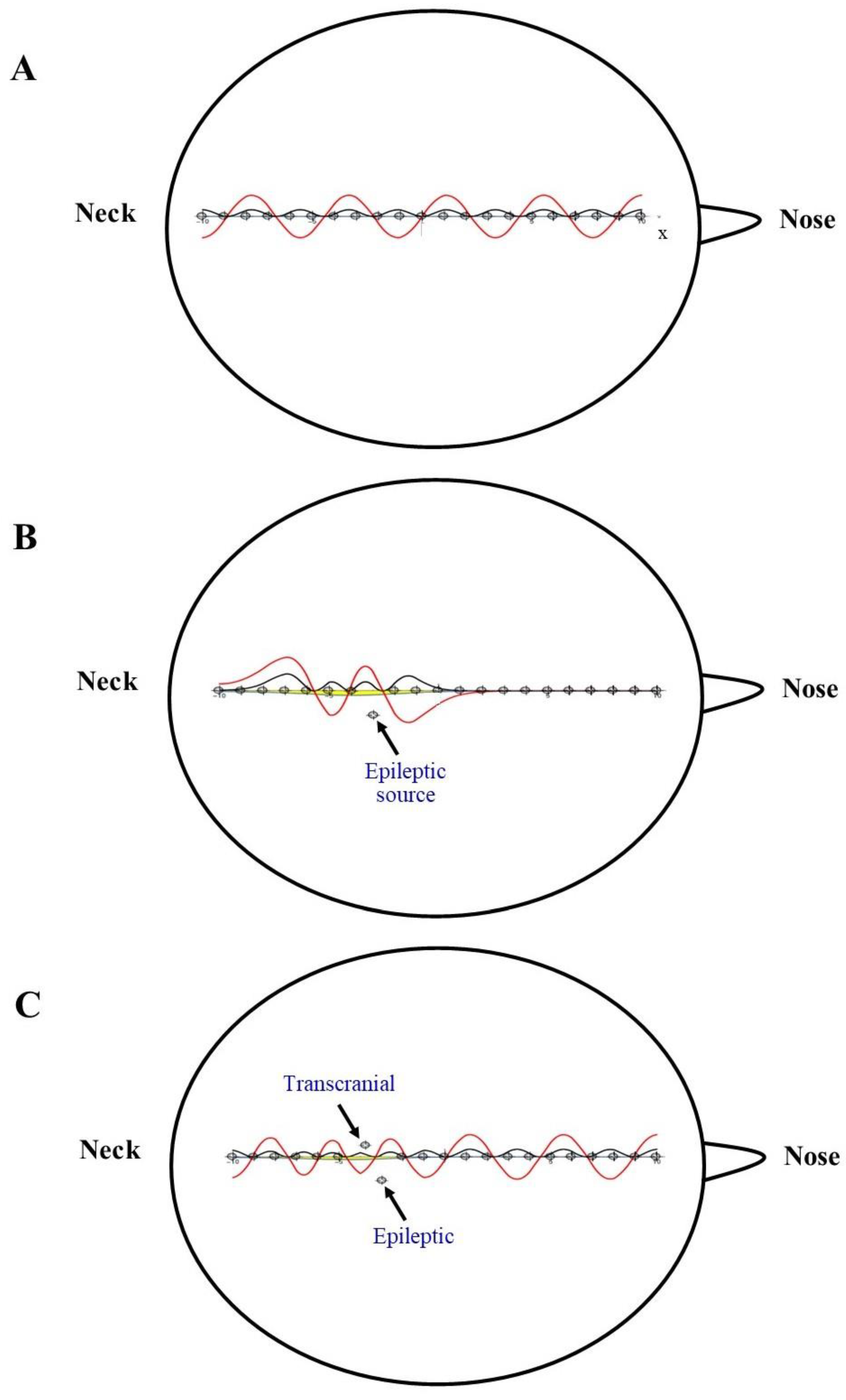
We argued that if a correlation does exist between solitons and pathological EEG patterns, this similarity could be harnessed to identify certain diseases through the detection and analysis of solitons. Therefore, if the soliton-like EEG modalities could be somehow disrupted. The elimination of the soliton that the proper procedures might lead to a recovery of the brain’s healthy state. Potential detrimental solitons correlated with medical conditions might be countered and neutralized in various ways. Since solitons are produced by a delicate balance between nonlinear and dissipative forces like viscosity, a pharmacologically induced modification of the brain viscosity might lead to the derangement of pathological solitons. Further, solitons display peculiar physical features like non-sinusoidal waves that could be theoretically manipulated by focal electrical brain stimulation induced by external magnetic fields, namely, by transcranial magnetic stimulation (TMS) [62,63]. In pathological states, the ordinary EEG oscillations deviate from the norm. In this case, our model of nervous solitons based on Schrodinger-like equations predicts that pathological spikes could be correlated with changes in the anatomical/functional location of the electric sources. Figure 3 provides a proof-of-concept example of brain EEG waves treated in terms of the Schrodinger equation. In the healthy brain, the energy sources are arranged in a regular order along the straight line connecting two scalp electrodes (Figure 3A). This generates a regular wave with well-defined periodicity and rhythm. When a disease occurs, e.g., an epileptic focus, one of the energy sources turns off its location on the straight line (Figure 3B). This divergence perturbs the wave profile: even slight changes in the location of energy sources can vary the potential, leading to waves with fully different shapes. To restore the healthy wave, an appropriately located external current could be given via TMS to counteract the effects of the displaced energy source (Figure 3C). Note that the profile wave in Figure 3 is drawn from the classical Schrodinger equation that holds just for microscopic systems subject to the laws of quantum mechanics. Nevertheless, the example can be generalized to macroscopic systems like EEG traces when considering NLSE instead of the classical Schrodinger equation.
Therefore, artificial manipulation of harmful solitons in the nervous tissue can be achieved by superimposing TMS external currents to counteract the pathological electric waves. This might have consequences for medical treatment. For instance, the proper external current might contribute to modify the harmful solitons produced during epileptic discharge, bringing healing to the convulsive symptoms.
The possible relationships between solitary waves and brain activities pave the way to novel lines of research. We focused on the spikes extracted from EEG traces, but the universe of the discourse could also be extended to the oscillations detected by other experimental procedures like fMRI and local field potential electrophysiology. When nonlinear effects occur in silica optical fibers, the momentum and the medium dispersion might coincide. This generates a “phase matching” that allows solitons to emit a special kind of low-intensity radiation called resonant radiation [33]. It could be speculated that this radiation could be found also in EEG traces. The property of solitons to interact with each other in a nonlinear medium suggests that they could also be analyzed in terms of elastically colliding particles that do not interact with each other and do not perturb the wave [46,64]. When the soliton propagates through the medium, the particles are accelerated until the wave has left the region. After the interaction, the particles become motionless again, but their positions have shifted. The particles’ single trajectories are influenced only by wave velocity, while their movement is governed by the current flow. This suggests that solitons at the interface of two biological fluid mediums might be useful to move or mix particles, contributing to the intra- and extra-cellular transport mechanisms.
In summation, research concerning relationships and interactions between solitons and the electric waves produced by the central nervous system still requires attention, but the premises are so promising that the field would be worth exploring.

Funding

This research did not receive any specific grant from funding agencies in the public, commercial or not-for-profit sectors.

Data Availability Statement

All data and materials generated or analyzed during this study are included in the manuscript. The author had full access to all the data in the study and takes responsibility for the integrity of the data and the accuracy of the data analysis.

Conflicts of Interest

The author declares no conflict of interest.

References

  1. Chabchoub, A.; Slunyaev, A.; Hoffmann, N.; Dias, F.; Kibler, B.; Genty, G.; Dudley, J.; Akhmediev, N. The Peregrine Breather on the Zero-Background Limit as the Two-Soliton Degenerate Solution: An Experimental Study. Front. Phys. 2021, 9, 633549. [Google Scholar] [CrossRef]
  2. Poznanski, R.R.; Cacha, L.A.; Al-Wesabi, Y.M.; Ali, J.; Bahadoran, M.; Yupapin, P.P.; Yunus, J. Solitonic conduction of electrotonic signals in neuronal branchlets with polarized microstructure. Sci. Rep. 2017, 7, 2746. [Google Scholar] [CrossRef]
  3. Sharma, A.; Malik, H.K.; Kumar, H.; Goyal, S. Effect of magnetic field on electromagnetic soliton evolution by different pulses. J. Theor. Appl. Phys. 2019, 13, 31–37. [Google Scholar] [CrossRef]
  4. Ye, Y.; Bu, L.; Wang, W.; Chen, S.; Baronio, F.; Mihalache, D. Peregrine Solitons on a Periodic Background in the Vector Cubic-Quintic Nonlinear Schrödinger Equation. Front. Phys. 2020, 8, 596950. [Google Scholar] [CrossRef]
  5. Minardi, S.; Eilenberger, F.; Kartashov, Y.V.; Szameit, A.; Röpke, U.; Kobelke, J.; Schuster, K.; Bartelt, H.; Nolte, S.; Torner, L.; et al. Three-dimensional light bullets in arrays of waveguides. Phys. Rev. Lett. 2010, 105, 263901. [Google Scholar] [CrossRef]
  6. Redor, I.; Barthélemy, E.; Michallet, H.; Onorato, M.; Mordant, N. Experimental Evidence of a Hydrodynamic Soliton Gas. Phys. Rev. Lett. 2019, 122, 214502. [Google Scholar] [CrossRef]
  7. Krylov, S.F.; Yankov, V.V. The role of solitons in strong turbulence. Zhurnal Eksperimental Teor. Fiz. 1980, 79, 8246. [Google Scholar]
  8. Diebel, F.; Timotijević, D.V.; Jović Savić, D.M.; Denz, C. Soliton formation by interacting Airy beams. In Complex Light and Optical Forces X; Proceedings Volume 9764; SPIE: Basel, Switzerland, 2016; p. 97640S. [Google Scholar] [CrossRef]
  9. Kayum, A.; Ara, A.; Barman, H.K.; Ali Akbar, M. Soliton solutions to voltage analysis in nonlinear electrical transmission lines and electric signals in telegraph lines. Results Phys. 2020, 18, 103269. [Google Scholar] [CrossRef]
  10. Bouchet, T.; Marsal, N.; Sciamanna, M.; Wolfersberger, D. Two dimensional Airy beam soliton. Sci. Rep. 2022, 12, 9064. [Google Scholar] [CrossRef] [PubMed]
  11. Manley, M.E.; Sievers, A.J.; Lynn, J.W.; Kiselev, S.A.; Agladze, N.I.; Chen, Y.; Alatas, A. Intrinsic localized modes observed in the high-temperature vibrational spectrum of NaI. Phys. Rev. B 2009, 79, 134304. [Google Scholar] [CrossRef]
  12. Kachulin, D.; Dyachenko, A.; Zakharov, V. Soliton Turbulence in Approximate and Exact Models for Deep Water Waves. Fluids 2020, 5, 67. [Google Scholar] [CrossRef]
  13. Strecker, K.E.; Partridge, G.B.; Truscott, A.G.; Hulet, R.G. Formation and propagation of matter-wave soliton trains. Nature 2002, 417, 150–153. [Google Scholar] [CrossRef] [PubMed]
  14. Li, B.-X.; Borshch, V.; Xiao, R.-L.; Paladugu, S.; Turiv, T.; Lavrentovich, O.D. Electrically driven three-dimensional solitary waves as director bullets in nematic liquid crystals. Nat. Commun. 2018, 9, 2912. [Google Scholar] [CrossRef] [PubMed]
  15. Ciblis, P.; Cosic, I. The Possibility of Soliton/Exciton Transfer in Proteins. J. Theor. Biol. 1997, 184, 331–338. [Google Scholar] [CrossRef]
  16. Sinkala, Z. Soliton/exciton transport in proteins. J. Theor. Biol. 2006, 241, 919–927. [Google Scholar] [CrossRef]
  17. Heimburg, T. The thermodynamic soliton theory of the nervous impulse and possible medical implications. Prog. Biophys. Mol. Biol. 2022, 173, 24–35. [Google Scholar] [CrossRef]
  18. Heimburg, T.; Jackson, A.D. On soliton propagation in biomembranes and nerves. Proc. Natl. Acad. Sci. USA 2005, 102, 9790–9795. [Google Scholar] [CrossRef]
  19. Andersen Søren, S.L.; Jackson, A.D.; Heimburg, T. Towards a thermodynamic theory of nerve pulse propagation. Prog. Neurobiol. 2009, 88, 104–113. [Google Scholar] [CrossRef]
  20. Heimburg, T.; Jackson, A.D. On the action potential as a propagating density pulse and the role of anesthetics. arXiv 2006, arXiv:physics/0610117. [Google Scholar] [CrossRef]
  21. Johnson, A.S.; Winlow, W. The Soliton and the Action Potential—Primary Elements Underlying Sentience. Front. Physiol. 2018, 9, 357787. [Google Scholar] [CrossRef]
  22. Rowley, M.; Hanzard, P.H.; Cutrona, A.; Bao, H.; Chu, S.T.; Little, B.E.; Morandotti, R.; Moss, D.J.; Oppo, G.L.; Totero Gongora, J.S.; et al. Self-emergence of robust solitons in a microcavity. Nature 2022, 608, 303–309. [Google Scholar] [CrossRef]
  23. Hernandez, C.I.; Kargarnovin, S.; Hejazi, S.; Karwowski, W. Examining electroencephalogram signatures of people with multiple sclerosis using a nonlinear dynamics approach: A systematic review and bibliographic analysis. Front. Comput. Neurosci. 2023, 17, 1207067. [Google Scholar] [CrossRef]
  24. Poikonen, H.; Zaluska, T.; Wang, X.; Magno, M.; Kapur, M. Nonlinear and machine learning analyses on high-density EEG data of math experts and novices. Sci. Rep. 2023, 13, 8012. [Google Scholar] [CrossRef]
  25. Luo, C.; Li, F.; Li, P.; Yi, C.; Li, C.; Tao, Q.; Zhang, Y.; Yao, D.; Yin, G.; Song, P.; et al. A survey of brain network analysis by electroencephalographic signals. Cogn. Neurodyn. 2022, 16, 17–41. [Google Scholar] [CrossRef]
  26. Meisel, C.; Olbrich, E.; Shriki, O.; Achermann, P. Fading Signatures of Critical Brain Dynamics during Sustained Wakefulness in Humans. J. Neurosci. 2013, 33, 17363–17372. [Google Scholar]
  27. Mahgoub, A.; Qaraqe, M. Automatic detection of ictal activity in EEG using synchronization and chaos-based attributes. Med. Biol. Eng. Comput. 2023, 61, 3387–3396. [Google Scholar] [CrossRef]
  28. Alquran, M.; Jaradat, I.; Yusuf, A.; Sulaiman, T.A. Heart-cusp and bell-shaped-cusp optical solitons for an extended two-mode version of the complex Hirota model: Application in optics. Opt. Quant. Electron. 2021, 53, 26. [Google Scholar] [CrossRef]
  29. Costa, A.; Osborne, A.R.; Resio, D.T.; Alessio, S.; Chrivì, E.; Saggese, E.; Bellomo, K.; Long, C.E. Soliton Turbulence in Shallow Water Ocean Surface Waves. Phys. Rev. Lett. 2014, 113, 108501. [Google Scholar] [CrossRef]
  30. Karjanto, N. The nonlinear Schrödinger equation: A mathematical model with its wide-ranging applications. arXiv 2019, arXiv:1912.10683. [Google Scholar]
  31. Fotopoulos, G.; Frantzeskakis, D.J.; Karachalios, N.I.; Kevrekidis, P.G.; Koukouloyannis, V.; Vetas, K. Extreme wave events for a nonlinear Schrödinger equation with linear damping and Gaussian driving. Commun. Nonlinear Sci. Numer. Simul. 2020, 82, 105058. [Google Scholar] [CrossRef]
  32. Xia, W.; Ma, Y.; Dong, G.; Zhang, J.; Ma, X. Emergence of Solitons from Irregular Waves in Deep Water. J. Mar. Sci. Eng. 2021, 9, 1369. [Google Scholar] [CrossRef]
  33. Rubino, E.; McLenaghan, J.; Kehr, S.C.; Belgiorno, F.; Townsend, D.; Rohr, S.; Kuklewicz, C.E.; Leonhardt, U.; König, F.; Faccio, D. Negative-Frequency Resonant Radiation. Phys. Rev. Lett. 2012, 108, 253901. [Google Scholar] [CrossRef] [PubMed]
  34. Liu, F.-F.; Lü, X.; Wang, J.-P. Dynamical behavior and modulation instability of optical solitons with spatio-temporal dispersion. Phys. Lett. A 2024, 496, 129317. [Google Scholar] [CrossRef]
  35. Bilman, D.; Buckingham, R. Large-Order Asymptotics for Multiple-Pole Solitons of the Focusing Nonlinear Schrödinger Equation. J. Nonlinear Sci. 2019, 29, 2185–2229. [Google Scholar] [CrossRef]
  36. Shah, A.; Rehman, S.-U.; Haque, Q.-U.L.; Mahmood, S. Solitons and Turbulence in Solar Wind at 1 au: Multi-satellite Data and FDTD Simulations. Astrophys. J. 2020, 890, 1. [Google Scholar] [CrossRef]
  37. Bradley, P.J.; De Angelis, C. Soliton dynamics and surface waves at the interface between saturable nonlinear dielectrics. Opt. Commun. 1996, 130, 205–218. [Google Scholar] [CrossRef]
  38. Chai, X.; Lao, D.; Fujimoto, K.; Ramas, C. Magnetic soliton: From two to three components with SO(3) symmetry. Phys. Rev. Res. 2020, 3, L012003. [Google Scholar] [CrossRef]
  39. Shen, Y.; Dierking, I. Dynamics of electrically driven solitons in nematic and cholesteric liquid crystals. Commun. Phys. 2020, 3, 14. [Google Scholar] [CrossRef]
  40. Yamgoué, S.B.; Deffo, G.R.; Tala-Tebue, E.; Pelap, F.B. Exact solitary wave solutions of a nonlinear Schrödinger equation model with saturable-like nonlinearities governing modulated waves in a discrete electrical lattice. Chin. Phys. B 2018, 27, 126303. [Google Scholar] [CrossRef]
  41. Körpınara, T.; Demirkola, R.C.; Körpınara, Z. Soliton propagation of electromagnetic field vectors of polarized light ray traveling along with coiled optical fiber on the unit 2-sphereS2. Rev. Mex. Fis. 2020, 65, 626. [Google Scholar] [CrossRef]
  42. Lima, F.M.S. What Exactly is the Electric Field at the Surface of a Charged Conducting Sphere? Reason 2018, 23, 1215–1223. [Google Scholar] [CrossRef]
  43. Wabnig, J.; Resch, J.; Theuerkauf, D.; Anmasser, F.; Faber, M. Numerical Evaluation of a Soliton Pair with Long Range Interaction. arXiv 2022, arXiv:2210.13374. [Google Scholar]
  44. Chabchoub, A.; Kimmoun, O.; Branger, H.; Hoffmann, N.; Proment, D.; Onorato, M.; Akhmediev, N. Experimental Observation of Dark Solitons on the Surface of Water. Phys. Rev. Lett. 2013, 110, 124101. [Google Scholar] [CrossRef] [PubMed]
  45. Xiao, Y.; Wang, P.; Zhang, J.; Guo, T.; Gao, R.; Ren, S. Controllable soliton propagation of Airy-Gaussian beams under the fractional effect. Optik 2021, 243, 167431. [Google Scholar] [CrossRef]
  46. Didenkulova, E. Soliton turbulence in weakly nonlinear and weakly dispersive media. In Proceedings of the EGU General Assembly 2021, Online, 19–30 April 2021; p. EGU21-552. [Google Scholar] [CrossRef]
  47. Peregrine, D.H. Water waves, nonlinear Schrödinger equations and their solutions. J. Austral. Math. Soc. B 1983, 25, 16–43. [Google Scholar] [CrossRef]
  48. Sen, S. A global method of generating action potentials and EEG oscillations in a topological surface network. BiorXiv 2022. biorXiv:2022-06. [Google Scholar] [CrossRef]
  49. Zhang, H.; Watrous, A.J.; Patel, A.; Jacobs, J. Theta and Alpha Oscillations Are Traveling Waves in the Human Neocortex. Neuron 2018, 98, P1269–P1281.e4. [Google Scholar] [CrossRef]
  50. Cole, S.R.; Voytek, B. Brain Oscillations and the Importance of Waveform Shape. Trends Cogn. Sci. 2017, 21, 137–149. [Google Scholar] [CrossRef]
  51. Handa, P.; Mathur, M.; Goel, N. Open and free EEG datasets for epilepsy diagnosis. arXiv 2021, arXiv:2108.01030. [Google Scholar]
  52. Gélisse, P.; Tatum, W.O.; Crespel, A.; Jallon, P.; Kaplan, P.W. Determining ICU EEG periodic patterns and why it matters. J. Neurol. 2023, 270, 4744–4752. [Google Scholar] [CrossRef]
  53. Gadoth, N. Subacute Sclerosing Pan-Encephalitis (SSPE)—Past and Present. In Pathogenesis of Encephalitis; Hayasaka, D., Ed.; IntechOpen: London, UK, 2011. [Google Scholar] [CrossRef]
  54. Chari, G.; Yadav, K.; Nishijima, D.; Omurtag, A.; Zehtabchi, S. Improving the ability of ED physicians to identify subclinical/electrographic seizures on EEG after a brief training module. Int. J. Emerg. Med. 2019, 12, 11. [Google Scholar] [CrossRef] [PubMed]
  55. Fernández-Torre, J.L. First Chapter: Interictal EEG. In Atlas of Epilepsies; Panayioutopoulos, C., Ed.; Springer: Berlin/Heidelberg, Germany, 2010. [Google Scholar]
  56. Kartashov, Y.V.; Vysloukh, V.A.; Torner, L. Soliton trains in photonic lattices. Opt. Express 2004, 12, 2831–2837. [Google Scholar] [CrossRef] [PubMed]
  57. Genevet, P.; Barland, S.; Giudici, M.; Tredicce, J.R. Cavity Soliton Laser Based on Mutually Coupled Semiconductor Microresonators. Phys. Rev. Lett. 2008, 101, 123905. [Google Scholar] [CrossRef]
  58. Fritsch, A.R.; Guo, S.; Koh, S.M. Dark solitons in Bose–Einstein condensates: A dataset for many-body physics research. Mach. Learn.: Sci. Technol. 2022, 3, 047001. [Google Scholar] [CrossRef]
  59. Halperin, E.J.; Bohn, J.L. Quench-produced solitons in a box-trapped Bose-Einstein condensate. Phys. Rev. Res. 2020, 2, 043256. [Google Scholar] [CrossRef]
  60. Gaur, D.S.; Purohit, A.; Mishra, A.K. Trapping and guiding soliton through Airy pulse. Opt. Commun. 2024, 556, 130300. [Google Scholar] [CrossRef]
  61. Shaukat, M.I.; Castro, E.V.; Terças, H. Quantum dark solitons as qubits in Bose-Einstein condensates. Phys. Rev. A 2017, 95, 053618. [Google Scholar] [CrossRef]
  62. Chung, S.W.; Sullivan, C.; Rogasch, N.C.; Hoy, K.E.; Bailey, N.W. The effects of individualized intermittent theta burst stimulation in the prefrontal cortex: A TMS-EEG study. Hum Brain Mapp 2019, 40, 608–627. [Google Scholar] [CrossRef]
  63. Kohli, S.; Casson, A. Machine learning validation of EEG tACS artefact removal. J. Neural. Eng. 2019, 17, 016034. [Google Scholar] [CrossRef]
  64. Hirota, R. Exact Solution of the Korteweg–de Vries Equation for Multiple Collisions of Solitons. Phys. Rev. Lett. 1971, 27, 1192–1194. [Google Scholar] [CrossRef]
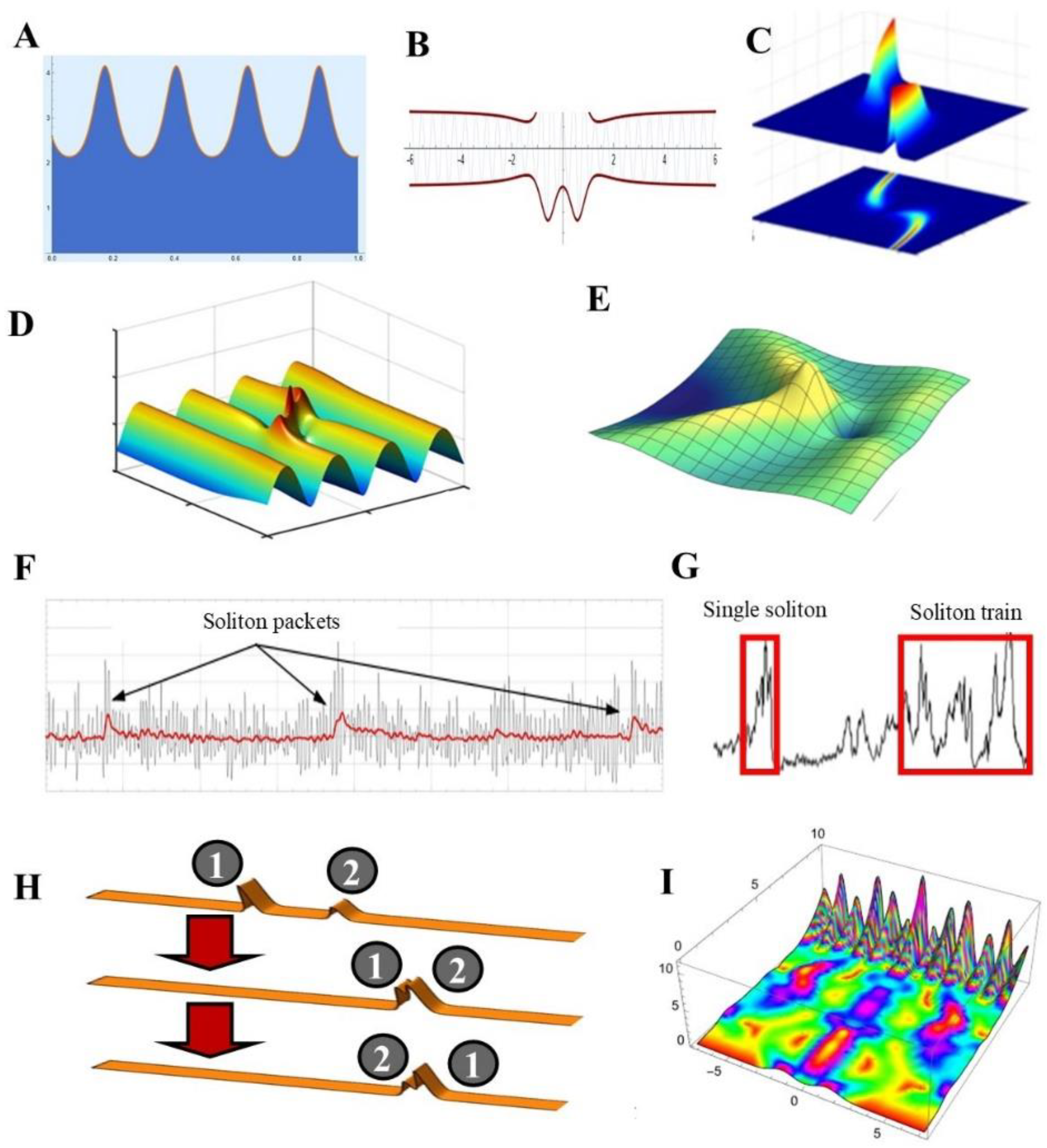
Figure 1. Features of different types of solitons. (A) A cnoidal wave is a periodic traveling-wave solution of KdVE. It refers to surface waves whose wavelength is large compared to the water depth; modified from https://demonstrations.wolfram.com/CnoidalWavesFromKortewegDeVriesEquation/ (accessed on 4 March 2024) (B) Formation of a rogue wave in the deep ocean, modeled using LNSE; modified from https://demonstrations.wolfram.com/RogueOceanWaves/ (accessed on 4 March 2024) (C) Top figure: example of dynamic behavior of an optical soliton with spatio-temporal dispersion, based on variable–coefficient NLSE. Bottom Figure: Time evolution of the soliton position x(t) interpreted in terms of wave streamlines obeying NLSE; modified from [34]. (D) Peregrine soliton solution based on a periodic-wave background generated by an NLSE variant; modified from [4]. (E) Soliton solution of the integrable focusing NLSE with spectral data consisting of a pair of conjugate poles of order 2n; modified from [35]. (F) Soliton trains (packets) in shallow water wind waves. The wave train (black) is achieved by low-pass filtering of the data (red); modified from [29]. (G) Single soliton and soliton trains detected for a month in the solar wind’s pressure; modified from [36]. (H) Weakly nonlinear two-soliton propagation in shallow water described by KdVE; modified from https://demonstrations.wolfram.com/SolitonsFromTheKortewegDeVriesEquation/ (accessed on 4 March 2024) (I) NLSE-derived soliton profile perturbed by a periodic potential; modified from https://demonstrations.wolfram.com/SolutionOfANonlinearSchroedingerEquation/ (accessed on 4 March 2024).
Signals 05 00015 g001
Figure 3. Schrödinger wavefunctions in a continuously varying potential field. The red oscillations stand for the wavefunctions, and the black ones stand for the probability density. The plot of the Schrödinger equation is embedded in a head contour. The plot represents an EEG wave spreading across the scalp electrodes between the neck and the nose. Solutions of the one-dimensional Schrödinger equation with quantum number 10 are provided in a potential-energy field V(x), which can be varied by moving a series of locators (the little circles). Even small changes in position of one or more locators modify the potential energy field, generating waves with fully different heights, frequencies, shapes and periodicities. In the example, when the elementary periodic wave (A) is disrupted by a change in position of one locator (B), it can be restored by changing the position of another locator (C). In the context of a human disease like epilepsy, this means that the healthy oscillation (A) disrupted by epileptic foci (B) could be restored by the proper transcranial current (C); modified from https://demonstrations.wolfram.com/SchroedingerWavefunctionsInAContinuouslyVaryingPotential/ (accessed on 3 March 2024).
Signals 05 00015 g003
Disclaimer/Publisher’s Note: The statements, opinions and data contained in all publications are solely those of the individual author(s) and contributor(s) and not of MDPI and/or the editor(s). MDPI and/or the editor(s) disclaim responsibility for any injury to people or property resulting from any ideas, methods, instructions or products referred to in the content.