1. Introduction
Gastroparesis is a disorder in which individuals experience dysfunctional gastric motor symptoms, but the underlying etiologies are not clear. Autoimmune factors have long been suspected of playing a role in GI motility, patients’ symptoms and perhaps the pathophysiology of gastroparesis [
1]. It is understood that the gastric mucosal immune system modulates many gastrointestinal processes, including regulating enteric nervous system and smooth muscle contractility [
2]. Therefore, dysregulation of the mucosal immune system can possibly be a component of the etiology of gastroparesis symptoms. Furthermore, autoimmune diseases cause damage, resulting in metabolic dysfunction and often chronic inflammation [
3]. For example, a chronic inflamed state can manifest in individuals as elevated C-reactive protein (CRP) and erythrocyte sedimentation rate (ESR). A previous study demonstrated that inflammatory markers including interleukin-17, interleukin-10, interferon-γ, interleukin-8, and interleukin-6 correlated with disease status in individuals with ulcerative colitis [
4]. Possibly an inflamed state may drive the pathogenesis of gastroparesis similarly to other disorders like ulcerative colitis. It has also been noted that dysfunction in metabolism has been shown to affect immune cell function, indicating connections between autoimmune, inflammatory, as well as metabolic factors possibly being components of the pathophysiology of gastroparesis symptoms.
Inflammatory markers have connections to other systems including the enteric nervous system, and inflammation can result in functional and structural alterations in neurons and therefore resulting in dysfunction [
5]. Inflammation is also known to impact the activity of the autonomic nervous system and perhaps may lead indirectly to gastrointestinal dysfunction. Therefore, there can be connections between inflammatory, autonomics, and enteric markers as well.
Overall, this exploratory study aimed to assess the relationships between autoimmune, inflammatory, and metabolic markers, as well as enteric measurements in relation to autonomic nervous system measures and electrogastrography (EGG) in a cohort of patients with gastroparetic symptoms. This study aimed to assess the gastric emptying test (GET) and several questionnaire scores in the context of autoimmune, inflammatory, metabolic, autonomics, and enteric measures.
We hypothesized that the measurements of antibodies and presence of autoimmune symptoms would potentially reveal existing relationships with inflammatory and metabolic markers. We hypothesized that measurements of inflammatory markers would yield insight and potentially have relationships with autonomic and enteric measurements.
3. Discussion
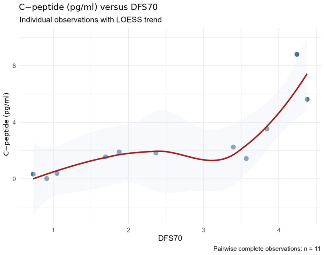
In this pilot study, there were several significant associations noted between different components of AIMAE. Of note, DFS-70 was positively associated with C-peptide. C-peptide is primarily associated with insulin production; C-peptide at low levels acts as an anti-inflammatory, but when excessively elevated, C-peptide can indicate inflammation. In addition, DFS-70 has been noted in previous studies to possibly be associated with organ-specific autoimmune diseases [
6]. The association between DFS-70 antibody and C-peptide may indicate an interaction between metabolic regulation and chronic inflammation. Therefore, anti-DFS-70 antibody may identify an immunometabolic relationship that warrants further evaluation.
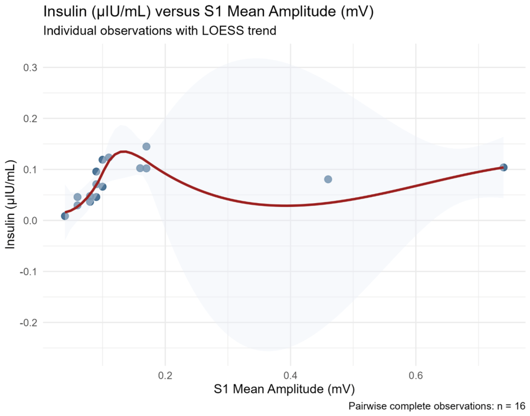
The analysis revealed that there were significant associations between LR-EGG measurements and inflammatory in addition to metabolic markers. S1 mean amplitude was positively associated with insulin. S1 mean amplitude refers to the pre-prandial amplitude of gastric myoelectrical activity which is often lower in individuals with Gp. Insulin resistance has been demonstrated to damage the interstitial cells of Cajal (ICC), which are pacemaker cells of the gastrointestinal muscles leading to gastric dysmotility [
7]. Furthermore, hyperinsulinemia is associated with autonomic dysfunction, which is a part of the regulation of gastric motor activity. Therefore, impaired autonomic function would negatively affect gastric smooth muscle contractility, also resulting in irregular gastric motility. This association indicates a possible relationship between the enteric nervous system and biomarkers that requires further investigation.
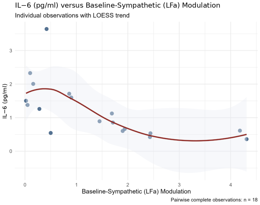
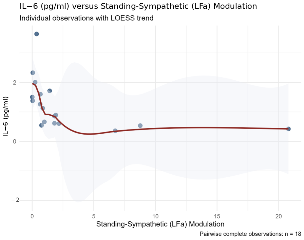
Furthermore, there were also notable negative associations between ANSAR measurements and IL-6. ANSAR-baseline-sympathetic (LFa) modulation and ANSAR-standing-sympathetic (LFa) modulation specifically were noted to be negatively associated with IL-6. Sympathetic (LFa) modulation indicates the level of sympathetic activity. IL-6 is a cytokine that contributes to the immune response and leads to the release of acute phase reactions such as CRP. It has also been found that increased IL-6 levels delay gastric emptying [
8]. Therefore, IL-6 was inversely associated with sympathetic modulation; however, causality cannot be determined from this data.
Gastroparesis is classically defined by delayed gastric emptying and symptom burden. Following this pilot study, the relationship between these two components and biomarkers remains uncertain. The study demonstrated no significant associations between the GET or symptom scores with antibodies, inflammatory and metabolic markers, EEG and autonomic measurements. The cumulative findings suggest that though these markers may provide insights into gastroparesis, their utility for reflecting delayed gastric emptying and patient-reported symptoms is limited. Given the small sample size of this study, these results should be carefully interpreted.
Overall, there were several associations between different components of AIMAE. However, larger prospective studies are needed to confirm and expand these findings to elucidate the pathophysiology of gastroparesis.
Gastroparesis is increasingly recognized as a disorder that involves multiple interacting physiologic systems rather than isolated gastric dysmotility. The present analyses identified associations among autoimmune, inflammatory, metabolic, autonomic, and electrogastrographic measurements, supporting this broader conceptual framework. Nevertheless, these observations should not be interpreted as evidence of causal mechanisms. Rather, they identify biologically plausible relationships that merit further investigation using larger, prospectively designed studies incorporating longitudinal follow-up and mechanistic experimentation.
In this study, the principal estimates were not materially altered by sequential removal of individual participants, and their directions were generally preserved after excluding participants with diabetes or a gastric electrical stimulator. These findings reduce concern that the associations were attributable solely to one influential observation, diabetes status, or stimulator status. However, the restricted analyses included only 8–13 pairwise-complete observations and therefore remain imprecise and descriptive.
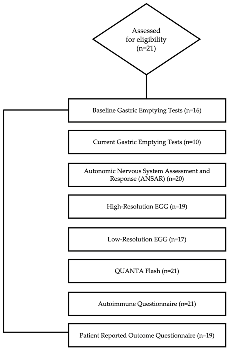
This study has several important limitations. First, the cohort consisted of only 21 patients and was intended as an exploratory pilot study. Although bootstrap confidence intervals and false-discovery-rate correction were used to improve statistical robustness, the relatively small sample size limits statistical power and increases uncertainty around individual correlation estimates. Consequently, these findings should be interpreted as hypothesis-generating rather than confirmatory. Furthermore, it is also important to note that given the small sample size, these findings are not applicable to the broader public.
Additionally, the study lacked a healthy control group. Therefore, the observed associations cannot be assumed to be unique to patients with gastroparesis symptoms and may reflect broader physiologic relationships. Future studies that include healthy controls and disease-comparison cohorts will be necessary to determine disease specificity. In addition, because this prospective pilot study relied on clinically available data, not every patient underwent every laboratory or physiologic assessment, resulting in variable sample sizes across analyses. Sample sizes are therefore reported for every correlation to facilitate interpretation.
Also, the autoimmune questionnaire is an investigator-developed tool used in this study. However, it is worth noting that it does not have formal validation which may limit the interpretation of the reported findings from the autoimmune questionnaire.
Furthermore, diabetes and gastric electrical stimulator status were available for all participants and were examined in restricted-cohort sensitivity analyses. However, insulin therapy, GLP-1 receptor agonist therapy, fasting status, contemporaneous glucose and HbA1c, prokinetic use, opioid use, and immunomodulatory therapy, as well as specific timing of biomarker, autonomic, EGG, and GET measurements, were not available as variables in the analysis dataset. These factors may influence biomarker concentrations, gastric electrical activity, autonomic measures, or their observed associations. The small cohort also precluded stable simultaneous multivariable adjustment for several confounders. Residual confounding therefore remains likely, and future prospective studies should standardize fasting and glucose assessment and collect complete medication histories. Finally, because this study is observational and based entirely on correlation analyses, causal relationships cannot be inferred. The observed associations identify candidate biologic relationships that warrant investigation in larger prospective mechanistic studies.
Understanding the pathophysiology of Gp symptoms is important for diagnostic and therapeutic implications. All in all, this exploratory pilot study investigated many serologic and physiologic markers and revealed possible existing interactions between the separate components of AIMAE. The analysis demonstrated associations between antibodies and metabolic markers. It also revealed associations between LR-EGG measurements and metabolic as well as inflammatory markers. It also demonstrated relationships between inflammatory markers and ANSAR measurements of the autonomic nervous system. These cumulative findings support the hypothesis that interactions between different physiological systems may interact in Gp, but do not establish the causal mechanisms of Gp. Rather, this exploratory pilot study may help work toward building a foundation in understanding the underlying components of Gp symptoms and may be useful in directing larger and more expansive studies in the future investigation of Gp symptoms, whether with delayed or non-delayed GET.




 ,
,