Case Report

Triple Synchronous Colorectal Cancer: An Extremely Rare Case Underscoring the Need for Careful Perioperative Evaluation

1
Department of Surgery, Can Tho General Hospital, 04 Chau Van Liem Street, Tan An Ward, Ninh Kieu Disctrict, Can Tho 900000, Vietnam
2
Faculty of Medicine, Can Tho University of Medicine and Pharmacy, 179 Nguyen Van Cu Street, An Khanh Ward, Ninh Kieu District, Can Tho 900000, Vietnam
3
Department of Pathology, Can Tho General Hospital, 04 Chau Van Liem Street, Tan An Ward, Ninh Kieu Disctrict, Can Tho 900000, Vietnam
4
Faculty of Medical Laboratory Science, Da Nang University of Medical Technology and Pharmacy, 99 Hung Vuong Street, Hai Chau 1 Ward, Hai Chau Disctrict, Da Nang 550000, Vietnam
*
Author to whom correspondence should be addressed.
Gastrointest. Disord. 2025, 7(2), 36; https://doi.org/10.3390/gidisord7020036
Submission received: 29 April 2025 / Revised: 15 May 2025 / Accepted: 19 May 2025 / Published: 23 May 2025

Abstract

:
Synchronous colorectal cancer (SCRC) is characterized by the simultaneous occurrence of two or more primary colorectal malignancies, diagnosed either preoperatively, intraoperatively, or within six months postoperatively. The rare prevalence of SCRC makes it an uncommon scenario among colorectal malignancies. Since the majority of SCRC patients have been reported to have two concurrent malignancies, triple synchronous malignancies are extremely rare. We report the case of a 65-year-old male individual presenting with a history of abdominal pain, anemia, anorexia, and unintentional weight loss. He was diagnosed with synchronous colorectal cancer with three distinct tumors: two located in the splenic flexure and sigmoid colon, respectively, and another in the rectum that caused partial obstruction. This case highlights the importance of intraoperative evaluation and an appropriate choice of surgical intervention in colorectal cancer. The early identification and proper management of multiple colorectal cancers remain essential for better survival rates.

1. Introduction

Synchronous colorectal cancer (SCRC) is characterized by the simultaneous occurrence of multiple primary malignancies in the same patient. The additional lesions should be diagnosed preoperatively, intraoperatively, or within six months post-surgery. Furthermore, the additional lesions must be distinctly isolated from the initially identified tumor by a minimum distance of 4 cm and should not exhibit submucosal spread or qualify as satellite lesions of the primary tumor [1,2]. SCRC is uncommon; in particular, triple colorectal malignancies are extremely rare and may be identified at any location within the large intestine, with a particularly high prevalence in elderly patients [3,4]. Several studies have demonstrated a predominance of SCRC in the right colon, as compared to solitary tumors [5]. The rare prevalence of SCRC makes it an uncommon condition among colorectal malignancies. Since SCRC is mainly reported to have two concurrent malignancies, triple synchronous malignancies are extremely rare.
Herein, we report an SCRC case with triple synchronous malignancies. To our knowledge, this case represents an extremely rare case in the Mekong Delta, Vietnam. The early identification and adequate management of multiple colorectal cancers remain essential for better survival rates.

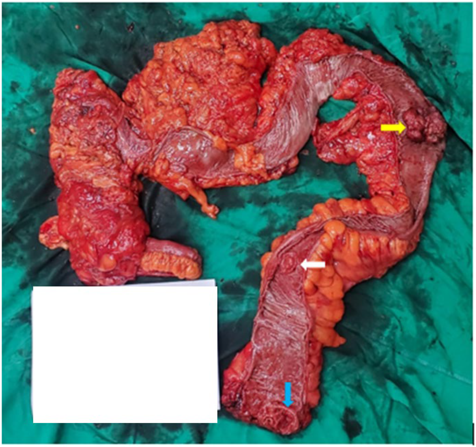
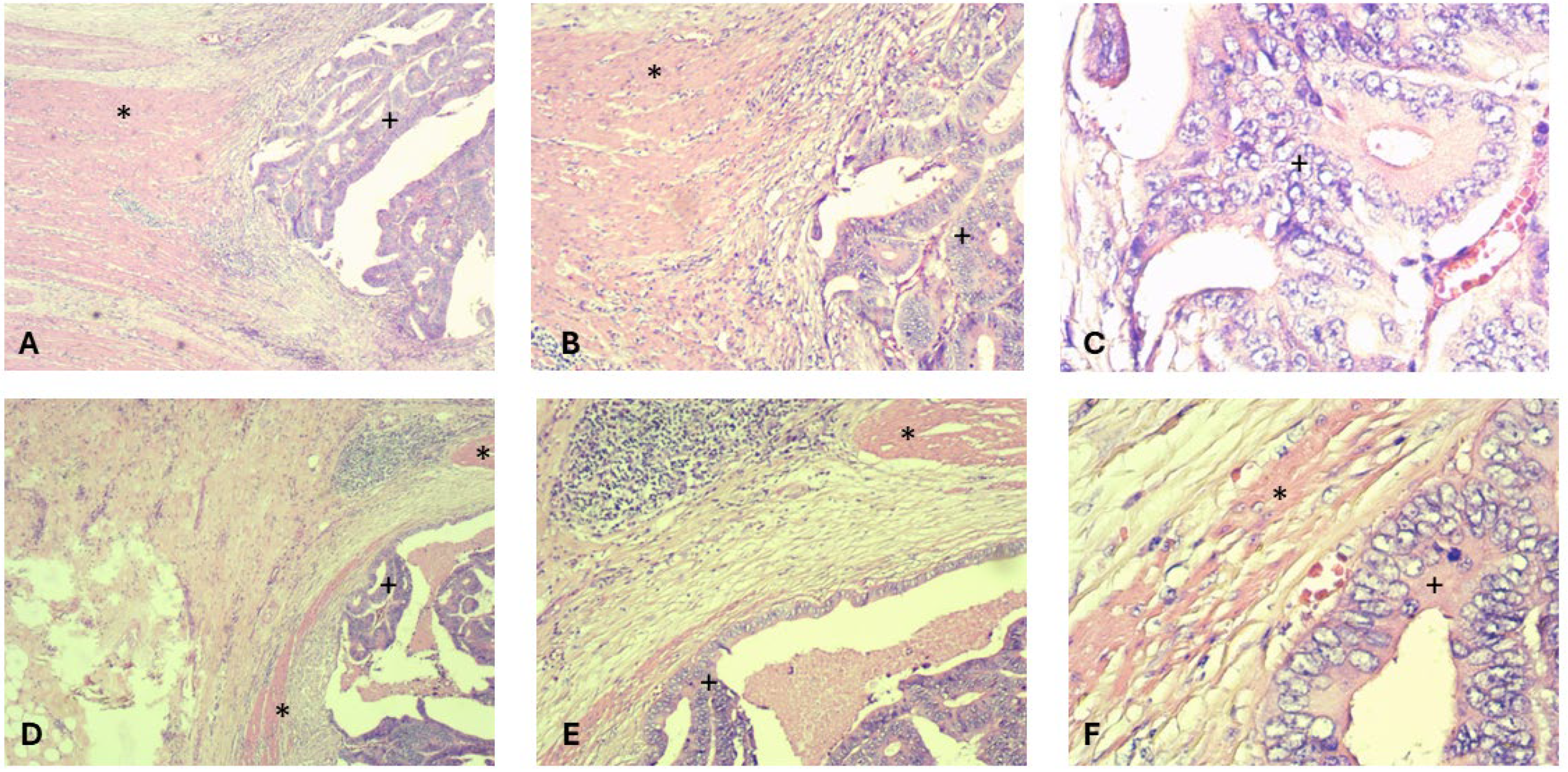
2. Case Presentation

A 65-year-old male patient presented to our hospital with a three-month history of abdominal discomfort, hematochezia, anorexia, and progressive weight loss, and no family history of malignancy. On physical examination, the patient showed tenderness in the right upper quadrant of the abdomen. Laboratory tests revealed a chronic anemia condition, with a hematocrit level of 24.9% and a hemoglobin concentration of 7.5 g/dL. An increased CA-19.9 level of 486.2 U/mL was noted, whereas other tumor markers, including alpha-fetoprotein (AFP), carcinoembryonic antigen (CEA), and prostate-specific antigen (PSA), were within normal ranges. Routine chest radiography and abdominal ultrasonography showed no abnormalities.
Abdominal contrast-enhanced computed tomography identified irregular colonic wall thickening and mesenteric infiltration at the splenic flexure and sigmoid portions, suggesting malignant lesions. A colonoscopy revealed a rectal tumor 10 cm from the anal verge, causing partial obstruction and limiting full examination. Histopathological examination of biopsy specimens suggested the presence of moderately differentiated adenocarcinoma. Other diagnostic imaging tools detected no abnormalities.
Given the diagnostic findings, an exploratory laparoscopy was performed. Intraoperatively, three distinct neoplastic lesions were identified: a 3 cm tumor in the rectum, a 7 cm tumor at the splenic flexure, and a 4 cm lesion in the sigmoid colon (Figure 1). No evidence of peritoneal or hepatic metastasis was observed. Suspiciously enlarged regional lymph nodes at preoperative palpation were present within the mesentery surrounding the splenic flexure. The operation was then converted to open surgery because of dense intra-abdominal adhesions. A total abdominal colectomy plus anterior resection was performed instead of a planned left extended hemicolectomy in conjunction with a low anterior resection because of the presence of an abnormal variant of mesenteric vessels. Total mesorectal excision was carried out in the patient as part of the surgical management.
The postoperative histopathological results confirmed three separate adenocarcinomas within the resected specimens, each showing distinct morphological characteristics. The tumors at the splenic flexure and sigmoid colon were moderately differentiated adenocarcinomas (Figure 2), while the rectal lesion was classified as poorly differentiated adenocarcinoma (Figure 3). Metastatic involvement was found in regional lymph nodes near the splenic flexure. The disease was staged as IIIc (pT3N2M0) according to the American Joint Committee on Cancer Tumor–Node–Metastasis classification. The postoperative course was uneventful, and the patient was initiated on adjuvant chemotherapy.

3. Discussion

This diagnosis of SCRC represents a complex clinical scenario rarely documented in the literature. The incidence of SCRC ranges from 1% to 8% among colorectal cancers [5,6,7]. Most SCRC cases involve two concurrent malignancies; nevertheless, rare instances with three or more tumors have been reported, with a recorded prevalence of up to 1.6% [8,9].
The risk factors for SCRC include age over 70, male sex, and a history of ulcerative colitis or adenomatous polyps [4,5]. However, this patient presented neither a history of gastrointestinal disease nor specific genetic syndromes. It is evident that colorectal cancer cannot be diagnosed merely with signs or symptoms. Determining the location of the lesions also poses more challenges given that the neoplasms are situated at various segments throughout the colon and rectum, as illustrated in our reported case. In this patient, the only significant symptoms observed are hematochezia and unintentional weight loss, which requires further investigation.
Determining the treatment strategy for SCRC presents another obstacle. While surgery is the priority, the procedure must be dependent upon the circumstances. The operative management of multiple cancers must be individualized, considering lesion location, invasion extent, and the patient’s overall condition [10,11]. In SCRC cases, therapy is more complicated, with decisions on the level of involvement of surgery or the initiation of neoadjuvant RCT strongly dependent on preoperative findings [12]. While some authors support laparoscopy, laparotomy is a preferred approach due to its reliability in confirming the diagnosis by palpating the probable lesions, especially in the early stages. Following the confirmation of the tumors, our initial strategy was to execute a left extended hemicolectomy in conjunction with a low anterior resection to guarantee a tumor-free resected margin. The decision was based on the patient’s preoperative performance state, indicating his fitness for surgery. However, abnormal mesenteric vessels complicated mobilization and made an anastomosis between the transverse colon and remnant rectum impossible. Consequently, we had to convert to a more extensive operation involving total abdominal colectomy plus low anterior resection with ileorectal anastomosis. Despite increased defecation frequency postoperatively, pathology revealed no malignancy in the resected margin, and the patient had a satisfactory recovery.
The postoperative pathological results confirmed the presence of three distinct adenocarcinomas. The lesions at the splenic flexure and sigmoid colon were moderately differentiated adenocarcinomas, and the tumor in the rectum was a poorly differentiated adenocarcinoma. The relatively homogeneous tumor characteristics helped to make planning adjuvant therapies more straightforward. The absence of lymphovascular or perineural invasion in the tumors was a positive prognostic indicator. Although the circumferential resection margin is essential for prognosis [11,12], we could not examine all specimens due to the heterogeneity in the locations and surrounding tissues of the three lesions.

4. Conclusions

In conclusion, our report presents a rare case of SCRC including triple synchronous malignancies with different histological types. Palpating the entire bowel during surgery is necessary to detect synchronous lesions. Further studies on the effective management of synchronous colorectal malignancies are essential.

Author Contributions

Conceptualization, P.V.L. (Phu Van La) and V.A.D.; methodology, P.V.L. (Phu Van La), D.N.N., D.M.T. and T.T.D.; validation, P.V.L. (Phu Van La), M.T.P.T., P.V.L. (Phuc Vinh La) and M.N.T.L.; formal analysis, P.V.L. (Phu Van La), C.P.D. and V.A.D.; investigation, T.T.D., M.T.P.T., P.V.L. (Phuc Vinh La) and M.N.T.L.; writing—original draft preparation, P.V.L. (Phu Van La) and C.P.D.; writing—review and editing, P.V.L. (Phu Van La) and C.P.D.; visualization, D.M.T., T.T.D. and M.T.P.T.; supervision, P.V.L. (Phu Van La) and V.A.D. All authors have read and agreed to the published version of the manuscript.

Funding

This research received no external funding.

Institutional Review Board Statement

Ethical review and approval were waived for this case report.

Informed Consent Statement

Informed consent for participation was obtained from all subjects involved in the study.

Data Availability Statement

The original contributions presented in the study are included in the article; further inquiries can be directed to the corresponding author.

Acknowledgments

We extend our sincere gratitude to Can Tho General Hospital for their kind approval. We also deeply appreciate the support provided by Can Tho University of Medicine and Pharmacy.

Conflicts of Interest

The authors declare no conflicts of interest.

References

  1. Flor, N.; Zanchetta, E.; Di Leo, G.; Mezzanzanica, M.; Greco, M.; Carrafiello, G.; Sardanelli, F. Synchronous colorectal cancer using CT colonography vs. other means: A systematic review and meta-analysis. Abdom. Radiol. 2018, 43, 3241–3249. [Google Scholar] [CrossRef] [PubMed]
  2. Welch, J.P. Multiple colorectal tumors: An appraisal of natural history and therapeutic options. Am. J. Surg. 1981, 142, 274–280. [Google Scholar] [CrossRef] [PubMed]
  3. Latournerie, M.; Jooste, V.; Cottet, V.; Lepage, C.; Faivre, J.; Bouvier, A. Epidemiology and prognosis of synchronous colorectal cancers. J. Br. Surg. 2008, 95, 1528–1533. [Google Scholar] [CrossRef] [PubMed]
  4. Yeh, C.-C.; Hsi, S.-C.; Chuu, C.-P.; Kao, Y.-H. Synchronous triple carcinoma of the colon and rectum. World J. Surg. Oncol. 2013, 11, 66. [Google Scholar] [CrossRef] [PubMed]
  5. Lam, A.K.-Y.; Chan, S.S.-Y.; Leung, M. Synchronous colorectal cancer: Clinical, pathological and molecular implications. World J. Gastroenterol. 2014, 20, 6815. [Google Scholar] [CrossRef] [PubMed]
  6. Box, J.C.; Rodriguez-Bigas, M.A.; Weber, T.K.; Petrelli, N.J. Clinical implications of multiple colorectal carcinomas in hereditary nonpolyposis colorectal carcinoma. Dis. Colon Rectum 1999, 42, 717–721. [Google Scholar] [CrossRef] [PubMed]
  7. Fante, R.; Roncucci, L.; Gregorio, C.D.; Tamassia, M.G.; Losi, L.; Benatti, P.; Pedroni, M.; Percesepe, A.; Pietri, S.D.; de Leon, M.P. Frequency and clinical features of multiple tumors of the large bowel in the general population and in patients with hereditary colorectal carcinoma. Cancer Interdiscip. Int. J. Am. Cancer Soc. 1996, 77, 2013–2021. [Google Scholar] [CrossRef]
  8. Bergeron, E.; Maniere, T.; Do, X.V.; Bensoussan, M.; De Broux, E. Three colonic cancers, two sites of complete occlusion, one patient: A case report. World J. Gastrointest. Surg. 2021, 13, 1095. [Google Scholar] [CrossRef] [PubMed]
  9. Kato, T.; Alonso, S.; Muto, Y.; Noda, H.; Miyakura, Y.; Suzuki, K.; Tsujinaka, S.; Saito, M.; Perucho, M.; Rikiyama, T. Clinical characteristics of synchronous colorectal cancers in Japan. World J. Surg. Oncol. 2016, 14, 272. [Google Scholar] [CrossRef] [PubMed]
  10. Michna, M.K.; Bichalska-Lach, M.; Rudzki, M.; Waniczek, D. A rare case of a trifocal synchronous colon cancer in a 65-year old patient. Pol. Ann. Med. 2023, 30, 140–143. [Google Scholar] [CrossRef]
  11. Van, T.N.; Van, N.P.; Van, D.M.; Van, H.N. Prone Position Laparoscopic Extra-Levator Abdomino-Perineal Excision (eLAPE) Improves Early Outcomes for Low Rectal Cancer. Surg. Gastroenterol. 2024, 29, 244–251. [Google Scholar] [CrossRef]
  12. Simu, P.; Jung, I.; Banias, L.; Kovacs, Z.; Fulop, Z.Z.; Bara, T.; Simu, I.; Gurzu, S. Synchronous Colorectal Cancer: Improving Accuracy of Detection and Analyzing Molecular Heterogeneity—The Main Keys for Optimal Approach. Diagnostics 2021, 11, 314. [Google Scholar] [CrossRef] [PubMed]
Figure 1. Macroscopic lesions: splenic flexure adenocarcinoma (yellow arrow), sigmoid colon adenocarcinoma (white arrow), and rectal adenocarcinoma (blue arrow). The white rectangle indicates individual notes for the patient during operation.
Gastrointestdisord 07 00036 g001
Figure 2. Histopathological examination of the lesion at the splenic flexure and sigmoid colon revealed a moderately differentiated adenocarcinoma (represented by the plus symbol) that invaded the muscularis propria (represented by the asterisk symbol). (AC) are images of the splenic flexure lesion with magnification at ×40; ×100; and ×400, respectively. (DF) are images of the sigmoid colon lesion with magnification at ×40; ×100; and ×400, respectively.
Gastrointestdisord 07 00036 g002
Figure 3. Histopathological examination of the rectal lesion reveals a poorly differentiated adenocarcinoma (represented by the plus symbol) that invaded the subserosal layer (represented by the asterisk symbol). (A) Magnification ×40; (B) magnification ×100; (C) magnification ×400.
Gastrointestdisord 07 00036 g003
Disclaimer/Publisher’s Note: The statements, opinions and data contained in all publications are solely those of the individual author(s) and contributor(s) and not of MDPI and/or the editor(s). MDPI and/or the editor(s) disclaim responsibility for any injury to people or property resulting from any ideas, methods, instructions or products referred to in the content.

