Article

Reliability of Kudo’s Glandular Pit Pattern in Predicting Colorectal Lesion Histology at Routine Colonoscopy with Digital Chromoendoscopy

by
Sabrina Gloria Giulia Testoni
1,†,
Pier Alberto Testoni
2,*,†,
Chiara Notaristefano
1,
Edi Viale
1 and
Giulia Martina Cavestro
2
1
Division of Gastroenterology and Gastrointestinal Endoscopy, Department of Experimental Oncology, IRCCS San Raffaele Scientific Institute, Via Olgettina 58, 20132 Milan, Italy
2
Division of Gastroenterology and Gastrointestinal Endoscopy, Department of Experimental Oncology, IRCCS San Raffaele Scientific Institute, Vita-Salute San Raffaele University, Via Olgettina 58, 20132 Milan, Italy
*
Author to whom correspondence should be addressed.
These authors contributed equally to this work.
Gastrointest. Disord. 2024, 6(3), 661-674; https://doi.org/10.3390/gidisord6030044
Submission received: 18 March 2024 / Revised: 24 June 2024 / Accepted: 3 July 2024 / Published: 10 July 2024
(This article belongs to the Special Issue Colorectal Cancer Screening (CRC) in the EU)

Abstract

:
Background. The large number of lesions detected via high-definition (HD) imaging during colonoscopy calls for the reliable real-time histological characterization of polyps, especially diminutive and small ones, to permit tailored management based on the neoplastic risk, such as a “resect-and-discard” or a “diagnose-and-leave” strategy for low-risk adenomas and hyperplastic polyps (HPs). The Kudo classification of glandular pit pattern is currently used for predicting polyp histology. Aim. The aim in this study was to assess whether Kudo’s glandular pit pattern, assessed via HD digital chromoendoscopy (i-Scan) without magnification and optical enhancement, reliably predicts polyp histology and differentiates neoplastic lesions (NLs) from non-neoplastic lesions (non-NLs) during routine colonoscopy. Methods. Consecutive colorectal lesions recorded in a database over 12 months, with Kudo’s glandular pit pattern classification, were retrospectively compared with histology. The diagnostic accuracy and negative predictive value (NPV) for adenomatous histology of Kudo’s pit patterns were assessed separately for diminutive (≤5 mm) and small (6–9 mm) polyps, accordingly to the American Society for Gastrointestinal Endoscopy (ASGE) Preservation and Incorporation of Valuable Endoscopic Innovations (PIVI), and in large (≥10 mm) lesions. Results. A total of 2230 lesions were recorded: 898 diminutive, 704 small, and 628 large. Kudo’s type II pit pattern was prevalent in diminutive polyps and recognized mostly in HPs (83.27%); it was also found in 38.8% of adenomas. In the right colon, Kudo’s type II pit pattern was prevalent in adenomas (70.04% vs. 20.74% in HPs); among the serrated lesions, it was evenly distributed between HPs and adenomas. Kudo’s type IIIL/IIIs/IV pit pattern was prevalent in NLs (61% vs. 8.37% of non-NLs) in diminutive polyps, evenly distributed between non-NLs and NLs in small polyps, and found only in NLs in large polyps. Kudo’s type Vi/Vn pit pattern correctly identified all but one adenocarcinoma. The NPV for adenomatous histology did not reach the recommended 90% PIVI threshold for differentiation between NLs and non-NLs in diminutive polyps showing Kudo’s type II pit pattern and in small polyps showing type IIIL/IIIs/IV pit pattern. Conclusions. Kudo’s pit pattern classification carried out with digital chromoendoscopy (i-Scan) during routine colonoscopy does not allow the reliable differentiation between non-NLs and NLs in diminutive and small polyps, so a “diagnose-and-leave” strategy for diminutive polyps may leave undetected adenomas, while a “resect-and-discard” strategy could miss lesions requiring closer follow-up.

1. Introduction

Colonoscopy is the most widely used screening method and has a protective role against colorectal cancer [1,2]. However, this role is far from complete, since neoplastic lesions (NLs) can be missed even during colonoscopy with good bowel preparation or adequate withdrawal time [3]; in fact, the reported adenoma miss rate varies from 2% to 25%, particularly for small and flat adenomas [4]. Advanced imaging technologies have improved the adenoma detection rate (ADR), but the large number of lesions detected calls for reliable real-time histological characterization, especially in diminutive and small lesions, to permit tailored management based on the neoplastic risk, such as a “resect-and-discard” or a “diagnose-and-leave” strategy for low-risk adenomas and hyperplastic polyps (HPs) [5]. These strategies have been shown to improve the cost-effectiveness of colorectal cancer screening, as documented in a study in which a Markov simulation model was used [6]. To adopt such a strategy, the American Society for Gastrointestinal Endoscopy (ASGE) Preservation and Incorporation of Valuable Endoscopic Innovations (PIVI) for the real-time endoscopic assessment of the histology of diminutive and small colorectal polyps recommends that a “diagnose-and-leave” strategy can be adopted for diminutive polyps and should have a >90% NPV value for adenomas, while a “resect-and-discard” strategy should require a >90% concordance between endoscopic characterization and histology in all diminutive and small polyps throughout the colon [5]. The “resect-and-discard” strategy for diminutive and small polyps based on the optical diagnosis was confirmed to offer substantial cost savings without adversely affecting the surveillance interval in the English Bowel Cancer Screening Programme (DISCARD 3 study) [7]. However, despite the widespread opinion that detecting and removing diminutive and small lesions in the colon may not have any significant clinical impact, some studies have found that some of these lesions can have unfavorable histology [8,9,10,11,12,13].
The Kudo classification of glandular pit pattern has been found useful for predicting lesion histology and distinguishing between neoplastic lesions (NLs) and non-neoplastic lesions (non-NLs) [14,15], and is currently used during routine colonoscopy with digital chromoendoscopy technologies, even if such a classification was established for surface characterization using magnification endoscopy under the aid of chromoendoscopy (dye staining). However, magnification is not commonly available. The glandular pit pattern is best assessed and classified with advanced imaging technologies applied with colonoscopy [16,17,18]. In the last years, artificial intelligence (AI) has been added to endoscopic imaging and has further improved the detection and characterization of colorectal lesions via the real-time computer-aided detection (CADe) of any protruding mucosal lesions. However, its performance is still suboptimal in the characterization of diminutive and small lesions, as well as in the presence of Kudo’s type II pit pattern [19]. Moreover, AI is expensive and not currently available in most endoscopy settings.
The aim of this retrospective study was, therefore, to assess the reliability of the Kudo glandular pit pattern classification in the real-time differentiation between NLs and non-NLs, in diminutive, small and large lesions, during colonoscopy with digital chromoendoscopy (Pentax i-Scan technology), without magnification and AI, as occurs in routine practice in most centers. Moreover, we aimed to assess its negative predictive value (NPV) for adenomatous histology, specifically according to the ASGE-PIVI for diminutive and small lesions [5].

2. Materials and Methods

2.1. Data Collection

Reports of consecutive routine colonoscopies performed with i-Scan imaging technology (PENTAX-HOYA Corporation, Tokyo, Japan) over a 12-month period were retrospectively analyzed. Only colonoscopies with at least one resected mucosal lesion with Kudo’s glandular pit pattern classification were considered for this study, and findings were compared with the histology. Data were collected from a computerized database in which colonoscopy reports were inserted prospectively, according to an observational protocol approved by the institutional Medical Ethics Committee (Register of diagnostic and therapeutic colonoscopies (HSR Reg/Colon). Data included patients’ main details and family history of colorectal cancer, indication for colonoscopy (screening, diagnosis, or surveillance), name of endoscopist, bowel preparation rating, whether the cecum was reached, and the reason for failure if not, time of withdrawal from cecum to rectum (minutes), complications during or immediately after the procedure, description of lesions with Paris classification for lesion morphology and Kudo’s classification for glandular pit pattern, and histological diagnosis of resected lesions. All patients gave informed consent for the procedures and use of data for scientific purposes.

2.2. Endoscopic Procedures and Lesion Characterization

Colonoscopies and Kudo’s pit pattern assessments were performed by four expert endoscopists, who had done 300–400 colonoscopies/year for at least 15 years and at least 300 procedures with i-Scan definition. These experts were previously trained in the glandular pit pattern evaluation reaching a moderate to good inter-observer agreement (k = 0.446) [20], similarly to that reported later in 2017 using the Simplified Identification Method for Polyp Labelling during Endoscopy (SIMPLE) (k = 0.46) [21].
All procedures were conducted using Pentax colonoscopes EC-3890FI (PENTAX-HOYA Corporation, Tokyo, Japan) and EC 3870FZK (PENTAX-HOYA Corporation, Japan) with high-definition (HD+) plus i-Scan and an EPKi processor (PENTAX-HOYA Corporation, Tokyo, Japan). The i-Scan imaging technology was a digital contrast method with a light filter that used different software algorithms, with real-time image mapping embedded in the EPKi processor. According to the current consensus [17,22], endoscopists routinely employed three predefined i-Scan settings in their clinical practice as follows: i-Scan 1 mode for lesion detection through surface enhancement (SE5) (PENTAX-HOYA Corporation, Tokyo, Japan), i-Scan 2 mode for lesion characterization combining SE and tone enhancement (SE4TEc) (PENTAX-HOYA Corporation, Tokyo, Japan), and i-Scan 3 mode for lesion demarcation adding contrast enhancement to the endoscopic image (SE4cCE2) (PENTAX-HOYA Corporation, Tokyo, Japan). By protocol, all colonoscopies started with i-Scan 1 setting; when a lesion was identified, i-Scan 2 and 3 settings were used to define its glandular pit pattern.
Lesions were grouped according to the size, Kudo’s glandular pit pattern classification, and site. The lesion size was routinely estimated from the width of standard fully open biopsy forceps (7 mm) and was classified as diminutive (≤5 mm), small (6–9 mm), or large (≥10 mm). The glandular pit pattern was classified as type I, II, IIIL/IIIs/IV, or Vi/Vn, according to Kudo’s classification (Figure 1) [14]. According to this classification, the assessment of pit patterns was made by comparison with the surrounding normal mucosa (type I): in type II, the pits are star-shaped and slightly larger than in type I, or open-shape and larger in the II-O subtype, histologically corresponding to HPs and serrated adenomas, respectively; in type III, the pits are elongated (IIIL) or round and smaller (IIIs) than those in type I, histologically corresponding to adenomas with low-grade to severe-grade focal dysplasia; in type IV, the pits are elongated and dendritic, reflecting villous elements within the lesion; in type V, the pits are various and irregularly distributed within the lesion (Vi), or deconstructed (Vn), histologically corresponding to high-grade dysplasia and cancer foci, respectively [14]. Lesions in the cecum, and ascending and transverse colon were defined as lesions of the “right colon”, whereas those in the descending and sigmoid colon and rectum as lesions of the “left colon”.
All the resected lesions were analyzed in the pathology unit of the same institute by highly experienced pathologists, with a standardized technique of preparation and histological analysis of the specimens.

2.3. Study Endpoints and Statistical Analysis

The endpoints of the study were (i) the diagnostic accuracy of Kudo’s glandular pit patterns, as assessed with an i-Scan, in the three groups of lesions (diminutive, small, and large), in differentiating between non-NLs (HPs) and NLs (conventional/serrated adenomas and adenocarcinomas) and (ii) the NPV for adenomatous histology, taking the histology as reference.
Assessment of data normality was made through the D’Agostino–Pearson test. Fisher’s exact or the chi-squared χ2 test was used to compare frequencies. Diagnostic accuracy and NPV with 95% confidence intervals (95% CIs) of Kudo’s glandular pit patterns were determined with a diagnostic test evaluation calculator and are expressed as percentages. The CIs for sensitivity, specificity and accuracy are “exact” Clopper–Pearson CIs; those for the predictive values are standard logit CI, as given by Mercaldo et al. [23], except when the predictive value was 0 or 100%, in which case Clopper–Pearson CIs are reported. Differences were considered significant at two-sided p < 0.05. Data were analyzed with MedCalc version 19.7 (MedCalc Statistical Software Ltd., Ostend, Belgium).

3. Results

3.1. Overview of Colorectal Lesions

This study included 2230 colorectal lesions, resected in 1358 consecutive colonoscopies, characterized with Kudo’s glandular pit pattern classification. Of these, 40.3% were diminutive, 31.6% small, and 28.1% large. Small and large lesions were predominant in the right colon (55.7% and 54.9% vs. 44.3% and 45.1% in the left colon), while diminutive lesions were evenly distributed in the right and left colon (52.1% and 47.9%). Kudo’s glandular pit pattern was recognized as type I in 1.5%, type II in 41.2%, type IIIL/IIIs/IV in 54%, and type Vi/Vn in 3.3% of lesions. Type I and II pit patterns were significantly recognized mostly among diminutive lesions (90.9% and 52.45%, respectively), while the type IIIL/IIIs/IV pit pattern was identified mostly among small lesions (35.2%). The type Vi/Vn pit pattern was detected in all large lesions (93.2%) but also in 6.85% of small lesions and absent in diminutive ones (Table 1).
At histological diagnosis, 13.2% of lesions were serrated HPs, 80.8% were adenomas (of these, 13.9% were traditional serrated adenomas (TSAs) or sessile serrated adenomas/polyps (SSA/ps) and 66.95% were conventional adenomas (Cas)), and 3.3% were adenocarcinomas (ADKs). The remaining specimens had normal mucosa. HPs and TSA-SSA/ps were found mostly among diminutive lesions (81.3% and 54.7%, respectively); CAs were significantly prevalent among small and large lesions (37.4% and 33.7%, respectively) (Table 2).
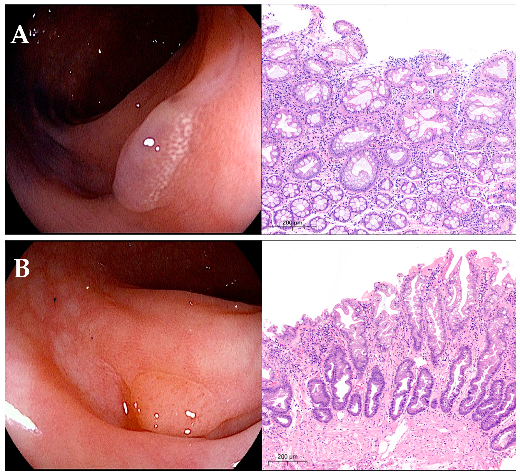
Kudo’s type I pit pattern was reported in 49.2% of cases with normal mucosa at histology. The type II pit pattern was significantly reported mostly among HPs (81.97%), in 35.85% of adenomas (serrated and conventional), and in one case of 6–9 mm ADK (1.37%) (p < 0.0001) (Figure 2). Among adenomas, the type II pit pattern was reported in 86.6% of serrated adenomas. In the right colon, it was prevalent in NLs (87.39%; p < 0.0001 vs. 12.61% of non-NLs), whereas in the left colon, it was evenly distributed between non-NLs and NLs (47.86% and 52.14%, respectively). The type IIIL/IIIs/IV pit pattern was prevalent in adenomas (95.6%%) but also seen in 4.39% of HPs (p < 0.0001). The rates of non-NLs and NLs recognized as type IIIL/IIIs/IV were similar between the right and left colon (4.46% and 4.32%, respectively, and 95.54%% and 95.67%, respectively). The type Vi/Vn pit pattern was recognized in all ADKs (98.6%) and in one 6–9 mm adenoma (1.37%) (p < 0.0001); it was found mostly in the right colon (63.01% vs. 36.99% in the left colon, p = 0.002).

3.2. Diagnostic Accuracy of Real-Time Characterization in Diminutive Polyps

Among the diminutive polyps, normal mucosa, HPs and adenomas (serrated and conventional) were found in 6.57%, 26.61%, and 66.82% of specimens, and neither high-grade dysplasia nor ADKs were found. HPs were found mostly in the left colon (42.3% vs. 12.2% of lesions in the right colon, p < 0.0001); adenomas were found mostly in the right colon (84.6% vs. 47.4% of lesions in the left colon, p < 0.0001) (Table 3).
The type I pit pattern was found in 9.7% and 0.3% of non-NLs (normal mucosa) and NLs (one serrated adenoma) (p < 0.0001). The type II pit pattern was found in 83.27% and 38.8% of non-NLs and NLs (p < 0.0001); in the right colon, it was prevalent in adenomas (70.04% vs. 20.74% in HPs, p < 0.0001); in the left colon, it was prevalent in HPs (65.66% vs. 25.66% in adenomas, p < 0.0001). The type IIIL/IIIs/IV pit pattern was found in 8.37% and 61% of non-NLs and NLs (p < 0.0001), being more prevalent in adenomas compared to HPs; this pit pattern was found evenly distributed in the right and left colon between NLs and non-NLs (95.04% and 4.96% vs. 94.4% vs. 5.55%, respectively) (Table 3).
The overall diagnostic accuracy of type II and type IIIL/IIIs/IV pit patterns in distinguishing non-NLs vs. NLs in diminutive lesions was 62.55% (95% CI: 58.9% to 66.1%) and 71.51% (95% CI: 58.9% to 66.1%), with an NPV for neoplastic histology of 84.52% (95% CI: 79.9% to 88.23%) and 92.13% (95% CI: 88.33% to 94.76%), respectively. In diminutive polyps, the NPV for neoplastic histology achieved the ASGE-PIVI-recommended 90% NPV threshold for differentiation between NLs and non-NLs only for lesions with the Kudo type IIIL/IIIs/IV glandular pattern.

3.3. Diagnostic Accuracy of Real-Time Characterization in Small Polyps

Among the small polyps, normal mucosa, HPs, adenomas (serrated and conventional), and adenocarcinomas were found in 0.3%, 7.7%, 91.3%, and 0.7% of specimens. HPs, adenomas, and adenocarcinomas represented 6.4% and 9.3%, 92.6% and 89.7%, and 0.8% and 0.6% of the lesions in the right and left colon, respectively (Table 4). High-grade dysplasia was reported in six adenomas (0.9%), all with a type IIIL/IIIs/IV pit pattern.
The type I pit pattern was found only in three cases (one normal mucosa, two serrated adenomas). The type II pit pattern was found in 84.6% and 41.5% of non-NLs and NLs (p < 0.0001), all HPs and adenomas, but with one case of normal mucosa in the right colon and one case of ADK in the left colon. This pattern was found in 45.4% and 30.13% of lesions in the right and left colon (p < 0.0001): HPs mostly in the left colon (4.49% vs. 1.79% in the right colon, p = 0.037) and adenomas (mostly conventional) mostly in the right colon (43.37% vs. 25.32% in the left colon, p < 0.0001). The type IIIL/IIIs/IV pit pattern was found in 58.93% and 60.34% of non-NLs and NLs (p = 0.836). The rates of non-NLs and NLs were similar in the right and left colon (p = 0.549) (Table 4).
The overall diagnostic accuracy of the type II and type IIIL/IIIs/IV pit patterns in distinguishing non-NLs vs. NLs was 60.67% (95% CI: 56.91% to 64.34%) and 69.51% (95% CI: 66.43% to 72.47%), with an NPV for neoplastic histology of 94.71% (95% CI: 92.94% to 96.06%) and 88.42% (95% CI: 88.47 to 91.47%), respectively. In contrast with diminutive polyps, in small polyps, the Kudo classification of the glandular pit pattern achieved the ASGE-PIVI-recommended 90% NPV threshold for the differentiation between NLs and non-NLs only for lesions showing a Kudo type II pit pattern but not for the type IIIL/IIIs/IV pit pattern.
The type Vi/Vn pit pattern was found mostly in ADKs (80%), and, in one case of adenoma, with a diagnostic accuracy of 99.7% (95% CI: 99.18% to 99.98%) and an NPV for neoplastic histology of 99.9% (95% CI: 98.97% to 99.97%).

3.4. Diagnostic Accuracy of Real-Time Characterization in Large Polyps

Among the large polyps, HPs, adenomas (serrated and conventional) and ADKs were found in 0.2%, 89% and 10.8% of specimens, while no normal mucosa was found. Adenomas were evenly distributed in the right and left colon (87.5% vs. 90.8%, respectively), as well as ADKs (12.5% in the right colon and 8.8% in the left colon) (Table 5). High-grade dysplasia was reported in 41 adenomas (7.3%).
The type II glandular pit pattern was found only in one HP in the left colon and in 26.16% of NLs (p < 0.0001), all adenomatous, and evenly distributed between the right and left colon (49.1% and 50.9%, respectively). The type IIIL/IIIs/IV pit pattern was found only in NLs (62.99%) and absent in non-NLs (p < 0.0001); it was evenly distributed in the right and left colon (64.06% and 61.48%, respectively) (Table 5).
The overall diagnostic accuracy of the type II and type IIIL/IIIs/IV pit patterns in distinguishing non-NLs vs. NLs was 70.69% (95% CI: 66.75% to 74.45%) and 100% (95% CI: 99.41% to 100%), with an NPV for neoplastic histology of 100% (95% CI: 99.07% to 100%) and 100% (95% CI: 98.43% to 100%), respectively.
The type Vi/Vn pit pattern was found only in ADKs, which were prevalent in the right colon (63.2%; p = 0.002), with a diagnostic accuracy and an NPV for neoplastic histology of 100% (95% CI: 99.22% to 100%) and 100% (95% CI: 99.10% to 100%), respectively.
The diagnostic accuracy in differentiation between non-NLs and NLs and the NPV for neoplastic histology of Kudo types II, IIIL/IIIs/IV, and Vi/Vn glandular pit patterns in diminutive, small, and large colorectal lesions are summarized in Table 6.

4. Discussion

A real-time classification system that distinguishes lesions as neoplastic or non-neoplastic, especially in presence of diminutive and small polyps, could offer a significant diagnostic advantage in clinical practice, enabling the endoscopist to decide which lesions need to be resected and sent for histopathological diagnosis, which ones could be resected and discarded, and which do not need to be resected at all.
However, up to 16% of adenomas ≤ 10 mm have been reported to have high-grade dysplasia, and 0.3% to 0.5% were ADKs [8,9]. Another study reported that small depressed colorectal lesions had up to a 40% chance of submucosal invasion [10]. Serrated adenomas are found mostly among small and diminutive lesions and harbor dysplasia in 3.7–42.9% of cases, accounting for up to 30% of colorectal cancer, as reported in systematic reviews [11,12]. Serrated adenomas proximal to the splenic flexure had the highest risk of cancer (OR: 12.42) [13]. Therefore, identifying and characterizing more lesions ≤ 10 mm may improve colorectal cancer prevention, because diminutive and small or flat lesions play a major role in the polyp miss rate and may cause interval cancers after screening colonoscopy.
The Kudo classification of glandular pit pattern makes it easier to distinguish NLs from non-NLs. A meta-analysis including 20 studies with a total of 5111 colorectal lesions showed a pooled sensitivity and specificity of 89.0% and 85.7% for the differentiation of NLs and non-NLs; the addition of chromoendoscopy yielded a sensitivity and specificity of 92.7% and 87.3% [24]. With unmagnified colonoscopy, Kudo’s pit pattern classification was reported to have a diagnostic accuracy of 82% in the differentiation between non-NLs and NLs [25]. However, defining the NPV for adenomatous histology according to PIVI recommendations can be difficult for diminutive and small polyps, especially in presence of = Kudo’s type II glandular pit pattern, which includes both serrated hyperplastic and adenomatous polyps, so that the differentiation between non-neoplastic and neoplastic histology remains challenging. In some studies, a review of polyps previously labeled as hyperplastic led to significant numbers being reclassified as adenomas [26,27], and in another study looking at long-term risk of colorectal cancer, 25% of serrated lesions could not be classified by pathology [28]. The Workgroup for Serrated Polyps and Polyposis (WASP) classification proposes validated endoscopic criteria for differentiating HPs and SSLs-TSAs; however, its accuracy in differentiating hyperplastic from serrated histology in diminutive lesions was not established [29]. The HiScope study proved i-Scan effective in the in vivo characterization of small polyps, fulfilling the ASGE performance thresholds for the assessment of diminutive colorectal polyps but only in a few lesions [30]. In a prospective multicenter study, i-Scan, associated with optical enhancement (OE) in diminutive polyps, showed an overall accuracy of 90% for histology prediction with sensitivity, specificity, positive, and negative predictive values, respectively, of 91%, 90%, 86%, and 94% [31]. Another study confirmed the high real-time diagnostic accuracy of i-Scan + OE for diminutive adenomas but not for serrated lesions [32]. The recent introduction of artificial intelligence further improved the characterization of lesions by implementing the ASGE-PIVI paradigm. However, most published data are based on small studies at tertiary care centers, with relatively small numbers of images used for the AI model’s training set, with possible biases in selection and randomization; moreover, CADe is currently unavailable in most endoscopic centers [11]. Convolutional neural network (CNN) training and validation achieved an overall accuracy of 94% for distinguishing adenomas and serrated polyps and a 97% NPV for adenomas in diminutive polyps [33]. However, again, this training method was adopted experimentally only in a single center.
We analyzed the diagnostic accuracy and the NPV for the neoplastic histology of Kudo’s glandular pit pattern, assessed with i-Scan digital chromoendoscopy, in diminutive, small, and large colorectal polyps during routine colonoscopy and outside of prospective studies. The present study very likely reflects the reliability of Kudo’s pit pattern in the differentiation between non-NLs and NLs in real life and covers the largest series of lesions so far on this topic.
Kudo’s type II pit pattern was prevalent in diminutive polyps and recognized mostly in HPs (83.27%); however, it was also found in about 33% and 41% of adenomas, in the left and right colon, respectively. Among the serrated lesions, it was evenly distributed between HPs and adenomas. In the right colon, this pit pattern was associated mainly with adenomas, while in the left colon, it was prevalent in HPs. In small polyps, this pit pattern was found in 84.6% and 41.5% of non-NLs and NLs, associated with adenomatous histology with similar frequencies in both the right and left colon, and evenly distributed between HPs and serrated adenomas (58.9% and 41.1%, respectively). In large polyps, it was found only in one HP and in about one-third of NLs, all adenomatous, and evenly distributed between the right and left colon: such a high rate of the type II pit pattern in NLs very likely accounts for serrated adenomas. The NPV for adenomatous histology was 84.5%, 94.7%, and 100% in diminutive, small, and large polyps, respectively.
Kudo’s type IIIL/IIIs/IV pit pattern was prevalent in NLs (61% vs. 8.7% of non-NLs) in diminutive polyps and evenly distributed with a similar rate in the right and left colon. In contrast, among small polyps, this pit pattern was found equally distributed between non-NLs and NLs (58.93% and 60.34%, respectively); these rates were maintained in both the right and left colon. In large polyps, the IIIL/IIIs/IV pit pattern was found only in NLs and absent in non-neoplastic ones; it was prevalent in the right colon. The NPV for adenomatous histology was 92.1%, 88.4%, and 100% in diminutive, small, and large polyps, respectively.
Kudo’s type Vi/Vn glandular pit pattern was correctly identified all but one ADK, independent of the size, with a diagnostic accuracy of 99.7% and 100% and an NPV for neoplastic histology of 99.9% and 100% in small and large lesions, respectively. This pit pattern was recognized in one small adenoma too.
Our data showed that Kudo’s glandular pit pattern classification assessed using digital chromoendoscopy without magnification did not allow the reliable differentiation between non-NLs and NLs in diminutive and small polyps. In fact, the NPV for adenomatous histology did not reach the recommended 90% threshold for differentiation between NLs and non-NLs in diminutive polyps with a Kudo type II pit pattern and in small polyps with a type IIIL/IIIs/IV pit pattern. Serrated lesions, which include both HPs and adenomas, may very likely account for the unsatisfactory NPV found in lesions smaller than 10 mm. In diminutive polyps with a type II pit pattern, an adenomatous histology was documented in about 8% of specimens, allowing a low-confidence optical diagnosis characterization. Our findings are confirmed by those of two other studies showing that the type II pit pattern is seen not only in serrated but also in CAs [34,35]. In these lesions, a “diagnose-and-leave” strategy could leave undetected adenomas in a high proportion of cases, even if the risk of malignant transformation appears to be negligible. If such strategy is adopted, a closer follow-up should be scheduled for these patients. Moreover, a “resect-and-discard” strategy, as suggested for diminutive polyps with high-confidence optical diagnosis characterization, appears to be difficult to adopt because of the challenging assessment of pit pattern in lesions ≤ 5 mm.
Among the small polyps showing a type II pit pattern, the level of confidence of optical diagnosis characterization was higher, but the method could not correctly characterize one case of ADK. In addition, in adenomas with a type IIIL/IIIs/IV pit pattern, high-grade dysplasia was reported in about 1% of lesions. Thus, in these polyps, the level of confidence of optical diagnosis was inadequate to identify advanced histology in some cases, so a “resect-and-discard” strategy might miss lesions, although rare, that require a closer follow-up in a short timeframe. If this strategy is adopted to reduce the work overload of the pathology units and, in general, the costs of a colorectal cancer screening, as documented in the DISCARD 3 study [7], closer monitoring should probably be considered, while in an individual colonoscopy setting, the cost-effectiveness benefit should be balanced against the risk of leaving unfollowed lesions with potential malignant evolution, which could play a role in the occurrence of interval cancer.
The limitation of this study is being retrospective conducted in a single tertiary care center, which potentially limit the broad applicability of the data. However, we analyzed a large series of polyps that were evaluated by expert endoscopists trained in lesion characterization and is one of the largest studies so far published on the diagnostic accuracy of Kudo’s glandular pit patterns for the real-time characterization of diminutive and small lesions in clinical practice during routine colonoscopy with digital chromoendoscopy.
In conclusion, based on our findings, all diminutive and small polyps should be removed, independent of Kudo pit pattern. A “resect and discard” strategy could be adopted for diminutive polyps of the left colon only within a colorectal cancer screening program but not in an individual colonoscopy setting, while diminutive polyps in the right colon, where adenomas are prevalent over hyperplastic polyps, and all small polyps should be sent to a pathologist for histological characterization in order to reduce the risk of leaving undetected adenomas.
However, further studies, prospective and multicenter, involving a larger number of expert endoscopists and of colorectal polyps, with a standardized expertise in optical diagnosis characterization, are required to validate our results. AI-assisted endoscopic technologies could give added value to these studies, although there is a gap between referral centers in which AI will be routinely adopted and the endoscopic reality in nonreferral centers, especially within the colorectal cancer screening programs proposed by the health national authorities.

Author Contributions

S.G.G.T. data curation, formal analysis, investigation, visualization, wrintng original draft; P.A.T. conceptualization, investigation, methodology, project administration, resources, supervision, writing original draft, review & editing; C.N. data curation, investigation, validation; E.V. investigation, validation; G.M.C. conceptualization, project administration, supervision, writing review & editing. All authors have read and agreed to the published version of the manuscript.

Funding

This study received no external funding.

Institutional Review Board Statement

The study was conducted according to the guidelines of the Declaration of Helsinki, and approved by the Ethics Committee of IRCCS San Raffaele Scientific Institute (Register of diagnostic and therapeutic colonoscopies-HSR Reg/Colon, approved 22 March 2011).

Informed Consent Statement

Informed consent for enrollment in the study was obtained from all subjects involved in the study. Written informed consent has been obtained from the patients to use their data for scientific purpose and publication.

Data Availability Statement

Data is contained within the article, further inquiries can be directed to the corresponding author.

Conflicts of Interest

The authors declare no conflict of interest.

References

  1. Qumseya, B.J.; Wallace, M.B. Advanced colorectal polyp detection techniques. Curr. Gastroenterol. Rep. 2012, 14, 414–420. [Google Scholar] [CrossRef]
  2. Brenner, H.; Stock, C.; Hoffmeister, M. Effect of screening sigmoidoscopy and screening colonoscopy on colorectal cancer incidence and mortality: Systematic review and meta-analysis of randomized controlled trials and observational studies. BMJ 2014, 348, 2467–2479. [Google Scholar] [CrossRef] [PubMed]
  3. Gomez, V.; Wallace, M.B. Advances in diagnostic and therapeutic colonoscopy. Curr. Opin. Gastroenterol. 2014, 30, 63–68. [Google Scholar] [CrossRef]
  4. van Rijn, J.C.; Reitsma, J.B.; Stoker, J.; Bossuyt, P.M.; van Deventer, S.J.; Dekker, E. Polyp miss rate determined by tandem colonoscopy: A systematic review. Am. J. Gastroenterol. 2006, 101, 343–350. [Google Scholar] [CrossRef]
  5. ASGE Technology Committee; Dayyeh, B.K.A.; Thosani, N.; Konda, V.; Wallace, M.B.; Rex, D.K.; Chauhan, S.S.; Hwang, J.H.; Komanduri, S.; Manfredi, M.; et al. ASGE Technology Committee systematic review and meta-analysis assessing the ASGE PIVI thresholds for adopting real-time endoscopic assessment of the histology of diminutive colorectal polyps. Gastrointest. Endosc. 2015, 81, 501–502. [Google Scholar]
  6. Hassan, C.; Pickhardt, P.J.; Rex, D.K. A resect and discard strategy would improve cost-effectiveness of colorectal cancer screening. Clin. Gastroenterol. Hepatol. 2010, 8, 865–869. [Google Scholar] [CrossRef]
  7. Orlovic, M.; Ahmad, A.; Saunders, B.P. Economic impact of implementing optical diagnosis with a “resect-and-discard” strategy within the English Bowel Cancer Screening Programme: Findings from the DISCARD3 study. Gastrointest. Endosc. 2023, 98, 73–81. [Google Scholar] [CrossRef]
  8. Kuwai, T.; Yamada, T.; Toyokawa, T.; Iwase, H.; Kudo, T.; Esaka, N.; Ohta, H.; Yamashita, H.; Hosoda, Y.; Watanabe, N.; et al. Local recurrence of diminutive colorectal polyps after cold forceps polypectomy with jumbo forceps followed by magnified narrowband imaging: A multicenter prospective study. Endoscopy 2019, 51, 253–260. [Google Scholar] [PubMed]
  9. Butterly, L.F.; Chase, M.P.; Pohl, H.; Fiarman, G.S. Prevalence of clinically important histology in small adenomas. Clin. Gastroenterol. Hepatol. 2006, 4, 343–348. [Google Scholar] [CrossRef]
  10. Gupta, N.; Bansal, A.; Rao, D.; Early, D.S.; Jonnalagadda, S.; Edmundowicz, S.A.; Sharma, P.; Rastogi, A. Accuracy of in vivo optical diagnosis of colon polyp histology by narrow-band imaging in predicting colonoscopy surveillance intervals. Gastrointest. Endosc. 2012, 75, 494–502. [Google Scholar] [CrossRef]
  11. Meester, R.G.; van Herk, M.M.; Lansdorp-Vogelaar, I.; Ladabaum, U. Prevalence and clinical features of sessile serrated polyps: A systematic review. Gastroenterology 2020, 159, 105–118. [Google Scholar] [CrossRef]
  12. East, J.E.; Atkin, W.S.; Bateman, A.C.; Clark, S.K.; Dolwani, S.; Ket, S.N.; Leedham, S.J.; Phull, P.S.; Rutter, M.D.; Shepherd, N.A.; et al. British Society of Gastroenterology position statement on serrated polyps in the colon and rectum. Gut 2017, 66, 1181–1196. [Google Scholar] [CrossRef]
  13. Erichsen, R.; Baron, J.A.; Hamilton-Dutoit, S.J.; Snover, D.C.; Torlakovic, E.E.; Pedersen, L.; Frøslev, T.; Vyberg, M.; Hamilton, S.R.; Sørensen, H.; et al. Increased risk of colorectal cancer development among patients with serrated polyps. Gastroenterology 2016, 150, 895–902. [Google Scholar] [CrossRef]
  14. Kudo, S.; Hirota, S.; Nakajima, T.; Hosobe, S.; Kusaka, H.; Kobayashi, T.; Himori, M.; Yagyuu, A. Colorectal tumors pit pattern. J. Clin. Pathol. 1994, 47, 880–885. [Google Scholar] [CrossRef]
  15. Tanaka, S.; Haruma, K.; Nagata, S.; Oka, S.; Chayama, K. Diagnosis of invasion depth in early colorectal carcinoma by pit pattern analysis with magnifying endoscopy. Dig. Endosc. 2001, 13, S2–S5. [Google Scholar] [CrossRef]
  16. Mason, S.E.; Poynter, L.; Takats, Z.; Darzi, A.; Kinross, J.M. Optical technologies for endoscopic real-time histologic assessment of colorectal polyps: A meta-analysis. Am. J. Gastroenterol. 2019, 114, 1219–1230. [Google Scholar] [CrossRef]
  17. Bhandari, P.; Thayalasekaran, S.; Keisslich, R.; Bisschops, R.; Hoffmann, A.; Haidry, R.; Esteban, J.; Viedma, B.L.; Godzhello, E.; Almadi, M.; et al. Detection and characterization of colorectal polyps using high-definition white light and i-Scan: Evidence-based consensus recommendations using a modified Delphi process. United Eur. Gastroenterol. J. 2018, 6, 748–754. [Google Scholar] [CrossRef]
  18. Glover, B.; Patel, N.; Ashrafian, H.; Teare, J. Diagnostic accuracy of i-scan image enhancement for real-time endoscopic diagnosis of small colorectal polyps: A meta-analysis. Ther. Adv. Gastroenterol. 2018, 11, 1756284818814948. [Google Scholar] [CrossRef]
  19. Parsa, N.; Byrne, M.F. Artificial intelligence for identification and characterization of colonic polyps. Ther. Adv. Gastrointest. Endosc. 2021, 14, 26317745211014698. [Google Scholar] [CrossRef]
  20. Masci, E.; Mangiavillano, B.; Crosta, C.; Fiori, G.; Trovato, C.; Viaggi, P.; Zambelli, A.; Buffoli, F.; Staiano, T.; Manfredi, G.; et al. Interobserver agreement among endoscopists on evaluation of polypoid colorectal lesions visualized with the Pentax i-Scan technique. Dig. Liver Dis. 2013, 45, 207–210. [Google Scholar] [CrossRef]
  21. Iacucci, M.; Trovato, C.; Daperno, M.; Akinola, O.; Greenwald, D.; Gross, S.A.; Hoffman, A.; Lee, J.; Lethebe, B.C.; Lowerison, M.; et al. SIMPLE classification investigator team. Development and validation of the SIMPLE endoscopic classification of diminutive and small colorectal polyps. Endoscopy 2018, 50, 779–789. [Google Scholar]
  22. Hoffman, A.; Kagel, C.; Goetz, M.; Tresch, A.; Mudter, J.; Biesterfeld, S.; Galle, P.R.; Neurath, M.F.; Kiesslich, R. Recognition and characterization of small colonic neoplasia with high-definition colonoscopy using i-Scan is as precise as chromoendoscopy. Dig. Liver Dis. 2010, 42, 45–50. [Google Scholar] [CrossRef]
  23. Mercaldo, N.D.; Lau, K.F.; Zhou, X.H. Confidence intervals for predictive values with an emphasis to case-control studies. Stat. Med. 2007, 26, 2170–2183. [Google Scholar] [CrossRef]
  24. Cocomazzi, F.; Gentile, M.; Perri, F.; Bossa, F.; Merla, A.; Ippolito, A.; Cubisino, R.; Carparelli, S.; Marra, A.; Mileti, A.; et al. Accuracy and inter-observer agreement of the NICE and Kudo classifications of superficial colonic lesions: A comparative study. Int. J. Color. Dis. 2021, 36, 1561–1568. [Google Scholar] [CrossRef]
  25. Li, M.; Ali, S.M.; Umm-a-OmarahGilani, S.; Liu, J.; Li, Y.Q.; Zuo, X.L. Kudo’s pit pattern classification for colorectal neoplasm: A meta-analysis. World J. Gastroenterol. 2014, 20, 12649–12656. [Google Scholar] [CrossRef]
  26. Kim, S.W.; Cha, J.M.; Lee, J.I.; Joo, K.R.; Shin, H.P.; Kim, G.Y.; Lim, S.J. A significant number of sessile serrated adenomas might not be accurately diagnosed in daily practice. Gut Liver 2010, 4, 498–502. [Google Scholar] [CrossRef]
  27. Khalid, O.; Radaideh, S.; Cummings, O.W.; O’brien, M.J.; Goldblum, J.R.; Rex, D.K. Reinterpretation of histology of proximal colon polyps called hyperplastic in 2001. World J. Gastroenterol. 2009, 15, 3767–3770. [Google Scholar] [CrossRef]
  28. Singh, H.; Bay, D.; Ip, S.; Bernstein, C.N.; Nugent, Z.; Gheorghe, R.; Wightman, R. Pathological reassessment of hyperplastic colon polyps in a city-wide pathology practice: Implications for polyp surveillance recommendations. Gastrointest. Endosc. 2012, 76, 1003–1008. [Google Scholar] [CrossRef]
  29. Ijspeert, J.E.G.; Bastiaansen, B.A.J.; E van Leerdam, M.; A Meijer, G.; van Eeden, S.; Sanduleanu, S.; Schoon, E.J.; Bisseling, T.M.; Spaander, M.C.; van Lelyveld, N.; et al. Development and validation of the WASP classification system for optical diagnosis of adenomas, hyperplastic polyps and sessile serrated adenomas/polyps. Dutch Workgroup serrAted polypS & Polyposis (WASP). Gut 2016, 65, 963–970. [Google Scholar]
  30. Basford, P.J.; Longcroft-Wheaton, G.; Higgins, B.; Bhandari, P. High-definition endoscopy with i-Scan for evaluation of small colon polyps: The HiSCOPE study. Gastrointest. Endosc. 2014, 79, 111–118. [Google Scholar] [CrossRef]
  31. Klenske, E.; Zopf, S.; Neufert, C.; Nägel, A.; Siebler, J.; Gschossmann, J.; Mühldorfer, S.; Pfeifer, L.; Fischer, S.; Vitali, F.; et al. I-scan optical enhancement for the in vivo prediction of diminutive colorectal polyp histology: Results from a prospective threephased multicentre trial. PLoS ONE 2018, 13, e0197520. [Google Scholar] [CrossRef] [PubMed]
  32. Soons, E.; Bisseling, T.M.; van der Post, R.S.; Nagtegaal, I.D.; Hazewinkel, Y.; van Kouwen, M.C.A.; Siersema, P.D. The Workgroup Serrated Polyps and Polyposis (WASP) classification for optical diagnosis of colorectal diminutive polyps with iScan and the impact of the revised World Health Organization (WHO) criteria. United Eur. Gastroenterol. J. 2021, 9, 819–828. [Google Scholar] [CrossRef] [PubMed]
  33. Zachariah, R.; Samarasena, J.; Luba, D.; Duh, E.; Dao, T.; Requa, J.; Ninh, A.; Karnes, W. Prediction of Polyp Pathology Using Convolutional Neural Networks Achieves “Resect and Discard” Thresholds. Am. J. Gastroenterol. 2020, 115, 138–144. [Google Scholar] [CrossRef] [PubMed]
  34. Kimura, T.; Yamamoto, E.; Yamano, H.-O.; Suzuki, H.; Kamimae, S.; Nojima, M.; Sawada, T.; Ashida, M.; Yoshikawa, K.; Takagi, R.; et al. A novel pit pattern identifies the precursor of colorectal cancer derived from sessile serrated adenoma. Am. J. Gastroenterol. 2012, 107, 460–469. [Google Scholar] [CrossRef]
  35. Nakao, Y.; Saito, S.; Ohya, T.; Aihara, H.; Arihiro, S.; Kato, T.; Ikegami, M.; Tajiri, H. Endoscopic features of colorectal serrated lesions using image-enhanced endoscopy with pathological analysis. Eur. J. Gastroenterol. Hepatol. 2013, 25, 981–988. [Google Scholar] [CrossRef]
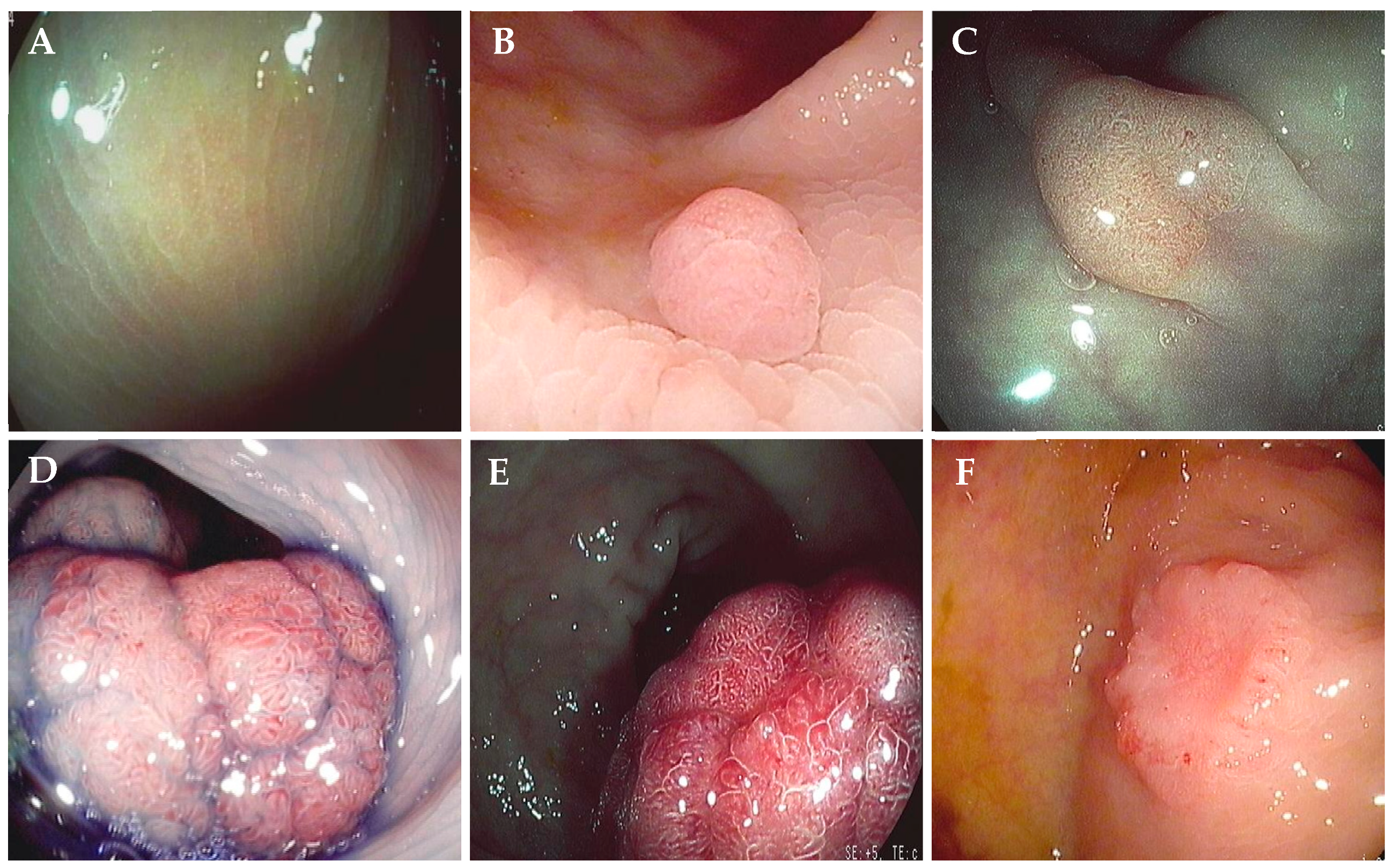
Figure 1. Kudo’s glandular pit pattern classification (from authors’ cases). (A) Type I pit pattern, with normal round pits (i-Scan 3 setting); (B) type II pit pattern, with asteroid, stellar or papillary pits (i-Scan 2 setting); (C) type IIIL/s pit pattern, with tubular or round pits (i-Scan 3 setting); (D) type IV, with dendritic or gyrus-like pits (i-Scan 2 setting); (E) type Vi pit pattern, with irregular arrangement (i-Scan 3 setting); (F) type Vn pit pattern, with loss of glandular structure (i-Scan 2 setting).
Gastrointestdisord 06 00044 g001
Figure 2. Endoscopic images of polyps classified at optical diagnosis as type II glandular pit pattern showing different histological diagnoses. (A) Hyperplastic polyp. (B) Sessile serrated adenoma.
Gastrointestdisord 06 00044 g002
Table 1. Colorectal lesions overview correlation between lesion size, site, and Kudo’s glandular pit pattern classification.
Site of LesionsSize of Lesionsp-Value
≤5 mm,
N. (%)
6–9 mm,
N. (%)
≥10 mm,
N. (%)
Total, N. (%)Left vs. Right Colon ≤ 5 mmLeft vs. Right Colon 6–9 mmLeft vs. Right Colon ≥ 10 mm
Right colon,
N. (%)
468
(52.12)
392
(55.68)
345
(54.94)
1205 (54.04)0.072<0.00010.0005
Left colon,
N. (%)
430
(47.88)
312
(44.32)
283
(45.06)
1025 (45.96)
Kudo glandular pit pattern Size of lesionsp-value
≤5 mm,
N. (%)
6–9 mm,
N. (%)
≥10 mm,
N. (%)
Total,
N. (%)
≤5 mm vs. 6–9 mm6–9 mm vs. ≥10 mm≤5 mm vs. ≥10 mm
Type I,
N. (%)
30
(90.9)
3
(9.09)
033
(1.48)
<0.00010.025<0.0001
Type II,
N. (%)
482 (52.45)272
(29.6)
165 (17.95)919
(41.21)
<0.0001<0.0001<0.0001
Type IIIs/IIIL/IV,
N. (%)
386 (32.03)424 (35.19)395 (32.78)1205 (54.04)<0.00010.00010.647
Type Vi/Vn,
N. (%)
05
(6.85)
68
(93.15)
73
(3.27)
0.016<0.0001<0.0001
Total, N. (%)898 (40.27)704 (31.57)628 (28.16)2230
Table 2. Histological diagnoses of colorectal polyps and correlation between histology and lesion size.
Histology of Lesions Size of Lesions p-Value
≤5 mm,
N. (%)
6–9 mm,
N. (%)
≥10 mm,
N. (%)
Total,
N. (%)
≤5 mm vs. 6–9 mm6–9 mm vs. ≥10 mm≤5 mm vs. ≥10 mm
Normal mucosa, N. (%)59 (96.72)2 (3.28)061 (2.74)<0.00010.083<0.0001
Serrated non-neoplastic (HPs),
N. (%)
239 (81.29)54 (18.37)1 (0.34)294 (13.18)<0.0001<0.0001<0.0001
Serrated adenomas (TSA-SSA/p),
N. (%)
169 (54.69)84 (27.18)56 (18.12)309 (13.86)<0.00010.0008<0.0001
Conventional adenomas (tubular, villous, tubulo-villous), N. (%)431 (28.87)559 (37.44)503 (33.69)1493 (66.95)<0.00010.0150.0009
Adenocarcinomas, N. (%)05 (6.85)68 (93.15)73 (3.27)0.003<0.0001<0.0001
Total, N. (%)898 (40.27)704 (31.57)628 (28.16)2.230
TSA: traditional serrated adenoma; SSA/p: sessile serrated adenomas/polyp.
Table 3. Correlation between lesion size, Kudo’s glandular pit pattern classification, and histology in the left and right colon in diminutive polyps (≤5 mm).
Size of LesionsSite of LesionsKudo Glandular Pit PatternNormal Mucosa, N. (%)HPs, N. (%)Adenomas (TSA-SSA/p, Conventional), N. (%)ADKs, N. (%)Total, N. (%)p-Value
Normal Mucosa vs. LesionsHPs vs. AdenomasHPs vs. Adenomas-ADKsAdenomas vs. ADKs
≤5 mmLeft colonType I, N. (%)21 (100)00021 (4.88)<0.0001
Type II, N. (%)23 (8.69)174 (65.66)68 (25.66)0265 (61.63)<0.0001<0.0001<0.0001<0.0001
Type IIIs/IIIL/IV, N. (%)08 (5.56)136 (94.44)0144 (33.49)<0.0001<0.0001<0.0001<0.0001
Type Vi/Vn, N. (%)00000
Total, N. (%)44 (10.23)182 (42.33)204 (47.44)0430 (47.88)
Right colonType I, N. (%)8 (88.89)01 (11.11)09 (1.92)0.0010.3170.3170.317
Type II, N. (%)7 (3.23)45 (20.74)165 (76.04)0217 (46.37)<0.0001<0.0001<0.0001<0.0001
Type IIIs/IIIL/IV, N. (%)012 (4.96)230 (95.04)0242 (51.71)<0.0001<0.0001<0.0001<0.0001
Type Vi/Vn, N. (%)00000
Total, N. (%)15 (3.21)57 (12.18)396 (84.62)0468 (52.12)
Grand Total, N. (%) 59 (6.57)239 (26.61)600 (66.82)0898
HPs: hyperplastic polyps; TSAs: traditional serrated adenomas; SSA/p: sessile serrated adenomas/polyp; ADKs: adenocarcinomas.
Table 4. Correlation between lesion size, Kudo’s glandular pit pattern classification, and histology in the left and right colon in small polyps (6–9 mm).
Size of LesionsSite of LesionsKudo Glandular Pit PatternNormal Mucosa, N. (%)HPs, N. (%)Adenomas (TSA-SSA/p, Conventional), N. (%)ADKs, N. (%)Total, N. (%)p-Value
Normal Mucosa vs. LesionsHPs vs. AdenomasHPs vs. Adenomas-ADKsAdenomas vs. ADKs
6–9 mmLeft colonType I, N. (%)1 (50)01 (50)02 (0.64)1.000.3170.3170.317
Type II, N. (%)014 (14.89)79 (84.04)1 (1.06)94 (30.13)<0.0001<0.0001<0.0001<0.0001
Type IIIs/IIIL/IV, N. (%)015 (7.01)199 (92.99)0214 (68.59)<0.0001<0.0001<0.0001<0.0001
Type Vi/Vn, N. (%)001 (50)1 (50)2 (0.64)0.0830.3170.0831000
Total, N. (%)1 (0.32)29 (9.29)280 (89.74)2 (0.64)312 (44.32)
Right colonType I, N. (%)001 (100)01 (0.26)0.3170.3170.3170.317
Type II, N. (%)1 (0.56)7 (3.93)170 (95.5)0178 (45.41)<0.0001<0.0001<0.0001<0.0001
Type IIIs/IIIL/IV, N. (%)018 (8.57)192 (91.43)0210 (53.57)<0.0001<0.0001<0.0001<0.0001
Type Vi/Vn, N. (%)0003 (100)3 (0.77)0.0250.0250.025
Total, N. (%)1 (0.26)25 (6.38)363 (92.6)3 (0.77)392 (55.68)
Grand Total, N. (%) 2 (0.28)54 (7.67)643 (91.34)5 (0.71)704
HPs: hyperplastic polyps; TSA: traditional serrated adenoma; SSA/p: sessile serrated adenomas/polyp; ADK: adenocarcinoma.
Table 5. Correlation between lesion size, Kudo’s glandular pit pattern classification, and histology, in the left and right colon, in large polyps (≥10 mm).
Size of LesionsSite of LesionsKudo Glandular Pit PatternNormal Mucosa, N. (%)HPs, N. (%)Adenomas (TSA-SSA/p, Conventional), N. (%)ADKs, N. (%)Total, N. (%)p-Value
Normal Mucosa vs. LesionsHPs vs. AdenomasHPs vs. Adenomas-ADKsAdenomas vs. ADKs
≥ 10 mmLeft colonType I, N. (%)00000
Type II, N. (%)01 (1.19)83 (98.81)084 (29.68)<0.0001<0.0001<0.0001<0.0001
Type IIIs/IIIL/IV, N. (%)00174 (100)0174 (61.48)<0.0001<0.0001<0.0001<0.0001
Type Vi/Vn, N. (%)00025 (100)25 (8.83)<0.0001<0.0001<0.0001
Total, N. (%)01 (0.35)257 (90.81)25 (8.83)283 (45.06)
Right colonType I, N. (%)00000
Type II, N. (%)0081 (100)081 (23.48)<0.0001<0.0001<0.0001<0.0001
Type IIIs/IIIL/IV, N. (%)00221 (100)0221 (64.06)<0.0001<0.0001<0.0001<0.0001
Type Vi/Vn, N. (%)00043 (100)43 (12.46)<0.0001<0.0001<0.0001
Total, N. (%)00302 (87.54)43 (12.46)345 (54.94)
Grand Total, N. (%) 01 (0.16)559 (89.01)68 (10.83)628
HP: hyperplastic polyp; TSA: traditional serrated adenoma; SSA/p: sessile serrated adenomas/polyp; ADK: adenocarcinoma.
Table 6. Diagnostic accuracy in differentiation between non-neoplastic and neoplastic histology and negative predictive value for neoplastic histology of Kudo type II, IIIs/IIIL/IV, and Vi/Vn glandular pit patterns, in diminutive (≤5 mm), small (6–9 mm), and large (≥10 mm) colorectal lesions.
Kudo’s Glandular Pit Pattern Size of Lesions
≤5 mm6–9 mm≥10 mm
Type IIDiagnostic accuracy (95% CI)62.55% (58.9 to 66.1)60.67% (56.91 to 64.34)70.69% (66.75 to 74.45)
NPV (95% CI) 84.52% (79.9 to 88.23)94.71% (92.94 to 96.06)100% (99.41 to 100)
Type IIL/IIIs/IVDiagnostic accuracy (95% CI)71.51% (58.9 to 66.1)69.51% (66.43 to 72.47)100% (98.43 to 100)
NPV (95% CI) 92.13% (88.33 to 94.76)88.42% (88.47 to 91.47)100% (99.41 to 100)
Type Vi/VnDiagnostic accuracy (95% CI)-99.7% (99.18 to 99.98)100% (99.1 to 100)
NPV (95% CI) -99.9% (98.97 to 99.97)100% (99.22 to 100)
NPV: negative predictive value.
Disclaimer/Publisher’s Note: The statements, opinions and data contained in all publications are solely those of the individual author(s) and contributor(s) and not of MDPI and/or the editor(s). MDPI and/or the editor(s) disclaim responsibility for any injury to people or property resulting from any ideas, methods, instructions or products referred to in the content.